Embed Size (px)
Citation preview

Oral and Maxillofacial Pathology Lecture 13Developmental disturbances of the Oral and Maxillofacial region ( Teeth, Soft tissue, Bone)

Disorders of development of
teeth

• The development of teeth is regulated
by genes, but the genetic program is
very sensitive to disturbances in the
environment such as infection, or toxic
chemicals.
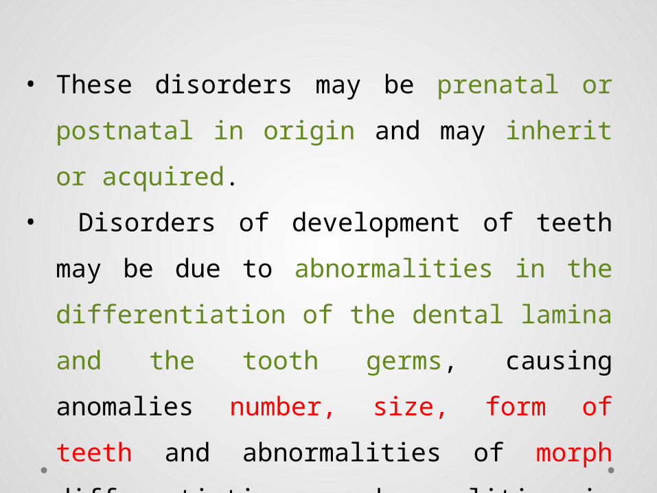
• These disorders may be prenatal or
postnatal in origin and may inherit or
acquired.
• Disorders of development of teeth may be
due to abnormalities in the differentiation
of the dental lamina and the tooth germs,
causing anomalies number, size, form of
teeth and abnormalities of morph
differentiation or abnormalities in the
formation of the dental hard tissue
resulting in disturbances in tooth
structure


Developmental Alterations in the Number of Teeth

Anodontia• Absence of teeth is known as anodontia.
• complete anodontia, when all teeth are missing
• partial anodontia or hypodontia, when one or
several teeth are missing.
• pseudoanodontia, when teeth are absent
clinically because of impaction or delayed eruption
• false anodontia, when teeth have been exfoliated
or extracted.

Complete anodontia is rare
but is often associated with a
syndrome known as
hereditary ectodermal
dysplasia
• Partial anodontia , few
teeth that are present are
usually conical.
• Hair, cutaneous
appendages, and nails are
also poorly developed in
this syndrome.

Hereditary ectodermal dysplasia resulting in lack of hair (including eyebrows and eyelashes) and poorly
developed sweat glands .

Anodontia of a permanent second premolar
with ankylosis of an erupted primary molar .

Supernumerary Teeth
• Extra teeth in the dentition
• result from continued proliferation of the
permanent or primary dental lamina to form a
third tooth germ.
• Most are isolated or familial
• others may be syndrome associated (Gardner’s
syndrome and cleidocranial dysplasia).
• The anterior midline of the maxilla is the most
common site, mesiodens.
• The maxillary molar area (fourth molar or
paramolar) is the second most common site.
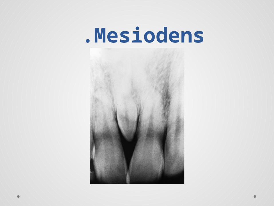
Mesiodens .

Mesiodens erupted .
• Teeth appearing at the time of birth are known as
natal teeth
• those appearing within 6 months following birth are
called neonatal teeth.
• Most of these teeth represent prematurely erupted
deciduous teeth, usually mandibular central incisors.
• A small percentage represent supernumer ary teeth.
Prematurely erupted primary teeth should be pre
served (provided they cause no injury to the infant or
the mother), and supernumeraries should be
extracted.


• Supernumerary teeth appearing after loss of
the perma nent teeth are known as
postpermanent dentition.
• This is generally regarded as a rare event.
• Most teeth appearing after extraction of the
permanent teeth are believed to arise from
eventual eruption of previously impacted
teeth.

Supernumerary premolar .

Developmental Alterations in the Size of Teeth

Microdontia • In generalized microdontia, all teeth in the
dentition appear smaller than normal. Teeth may actually be measurably smaller than normal, as in pituitary dwarfism, or they may be relatively small in comparison with a large mandible and maxilla.
• In focal, or localized, microdontia, a single tooth is smaller than normal. The shape of these microdonts is often altered with the reduced size. This phenomenon is most commonly seen with maxillary lateral incisors in which the tooth crown appears cone or peg shaped, prompting the designation peg lateral.
• Peg laterals are of no significance other than cosmetic appearance.
• The second most commonly seen microdont is the maxillary third molar, followed by super numerary teeth .


Microdont .

Macrodontia
• Generalized macrodontia is characterized by the
appear ance of enlarged teeth throughout the
dentition.
• it may be relative owing to a disproportionately
small maxilla and mandible.
• Focal, or localized, macrodontia is characterized by
an abnormally large tooth or group of teeth. This
relatively uncommon condition usually with
mandibular third molars.
• In the rare condition known as hemifacial hypertro
phy, teeth on the affected side are abnormally large
compared with the unaffected side.

Localized Disturbances in EruptionPrimary Impaction: Impaction of teeth is a common event that most often affects the mandibular third molars and maxillary canines. Less commonly, premolars, mandibular canines, and second molars are involved. • Impaction occurs because of obstruction from
crowding or from some other physical barrier. • Occa sionally, it may be due to an abnormal eruption
path, pre sumably caused by unusual orientation of the tooth germ.


Ankylosis, the fusion of a tooth to
surrounding bone, is another cause of
impaction. This usually occurs in associa tion
with erupted primary molars.
• The reason for ankylosis is unknown, but it is
believed to be related to periapical inflam
mation and subsequent bone repair.
• With focal loss of the periodontal ligament,
bone and cementum become mixed, causing
fusion of the tooth to alveolar bone.

Ankylosis:Eruption continues after the emergence of the teeth to
compensate for masticatory wear and the growth of the jaws.
The cessation of eruption after emergence is termed ankylosis
and occurs from an anatomic fusion of tooth cementum or dentin
with the alveolar bone.

Developmental Alterations in the
Shape of Teeth
• Disturbances in tooth form may involve the
crown, the roots or both.
• The most frequent variations of the crowns
of the teeth affect maxillary permanent
lateral incisors which may be peg-shaped.
• Premolars and molars with an increased or
decreased number of cusps are also seen.

Double teeth:- a descriptive term used to describe a
developmental anomaly where two teeth appear joined together.
It occurs mostly in primary than permanent dentition especially
anterior teeth.
1-Gemination
It is the fusion of two teeth from a single enamel organ. The typical
result is partial cleavage, with the appearance of two crowns that
share the same root canal.
Complete cleavage, or twinning, occasionally occurs, resulting in
two teeth from one tooth germ.
Although trauma has been suggested as a possible cause, the
cause of gemina tion is unknown.
These teeth may be cosmetically unaccept able and may cause
crowding..


Gemination .

2-Fusion is the joining of two developing tooth germs, resulting in
a single large tooth structure. The fusion process may
involve the entire length of the teeth, or it may involve
the roots only, in which case cementum and dentin are
shared.
Root canals may also be separate or shared.
It may be impossible to differentiate fusion of normal and
supernumerary teeth from gemination. The cause of this
condition is unknown, although trauma has been
suggested.
The etiology of double teeth remains unclear.

Fusion .
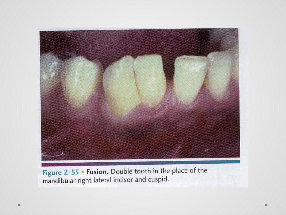

Fusion .

Concrescence:
• Concrescence is an acquired union form in which
adjacent, already formed teeth are joined by
cementum only.
• This may take place before or after eruption of
teeth and is believed to be related to trauma or
overcrowding. Mostly seen in permanent than
primary dentition.
• Concres cence is most commonly seen in
association with the maxil lary second and third
molars.


Concrescence

ACCESSORY CUSPS
The cuspal morphology of teeth exhibits
minor variations among different
populations.
(1) Cusp of Carabelli.
(2) Talon cusp.
When an accessory cusp is present, the
other permanent teeth often exhibit a
slightly increased tooth size.

Clinical and Radiographic Features
1-The cusp of Carabelli is an accessory cusp located on the
palatal surface of the mesio lingual cusp of a maxillary
molar.
The cusp is most pronounced on the first molar.
2-Talon cusp A talon cusp (dens evaginatus of anterior
tooth) is a well-delineated additional cusp that is located on
the surface of an anterior tooth and extends at least half
the distance from the cementoenamel junction to the incisal
edge.


Odontoma like lesions:
1-Dens Invaginatus : Also known as dens in dente or tooth
within a tooth arises as a result of invagination of a portion of
enamel organ into the dental papilla at an early stage in
odontogenesis before the formation of calcified tissue.
• This defect ranges in severity from superficial, in which
only the crown is affected, to deep, in which both the
crown and the root are involved.
• The permanent maxillary lateral incisors are most
commonly involved.
• The cause of this developmental condition is unknown.

Dens invaginatus of lateral incisors .
2-Dens Evaginatus :- is a relatively common
developmental con dition affecting predominantly
premolar teeth .
The defect, which is often bilateral, is an
anomalous tubercle, or cusp, located at the
center of the occlusal surface.
Because of occlusal abrasion, the tubercle wears
relatively quickly causing early exposure of an
accessory pulp horn that extends into the
tubercle.

Morphology of dens invaginatus and dens
evaginatus

Dens evaginatus of mandibular second
premolars .

Dens evaginatus with associated periapical lesions .

Dens evaginatus. Ground section showing pulpal extension
through dentin to the surface of a worn occlusal cusp .

3-Enamel Pearls (enamelonoma): Droplets of ectopic enamel,
or so-called enamel pearls, may occasionally be found on the
roots of teeth.
• Maxillary molars are more commonly affected than
mandibular molars.
• These deposits are occasionally sup ported by dentin and
rarely may have a pulp horn extending into them.
• This developmental disturbance of enamel forma tion may be
detected on radiographic examination.
• It gener ally is of little significance except when located in an
area of periodontal disease. because a periodontal ligament
attachment would not be expected and hygiene would be more
difficult.

Enamel pearl .

Taurodontism
Taurodontism is a variation in tooth form in which
teeth have elongated crowns or apically displaced
furcations, resulting in pulp chambers that have
increased apical-occlusal height.
• taurodontism is of little clinical significance
unless the tooth becomes nonvital, in which case
it becomes a challenging endodontic problem.
No treatment is required.

Taurodontism

Dilaceration
A term used to describe a deformity in which the crown is
displaced from its normal alignment with the root, so that
the tooth is severly bent along its long axis. The cause of
this condition has been related to trauma during root
development.
• Eruption generally continues without problems.
However, extraction may be difficult.

Dilaceration .

Supernumerary Roots
Accessory roots are most commonly seen in
mandibular canines, premolars, and molars
(especially third molars). They are rarely found in
upper anterior teeth and mandibu lar incisors.
Radiographic recognition of an extraordinary
number of roots becomes important when
extractions or root canal fillings are necessary.


Environmental alteration of teeth

Amelogenesis Imperfecta
• It is a clinically and genetically het erogeneous group of
disorders of enamel formation that affect both dentitions.
• Most cases of amelogen esis imperfecta fall into one of two
clinical types: hypoplastic or hypocalcified. A third type,
known as hypomaturation, has been added to the list.

• Several genes that are involved in enamel formation (amelogenin,
enamelin, kallikrein 4, MMP20, others) are mutated in various
forms of this condition.
• Most cases of amelogenesis imperfecta are inherited as an
autosomal-dominant trait, with variable clinical manifestations.
• Affected males may have a very thin, smooth enamel layer,
whereas females may have thicker enamel with vertical grooves

• hypoplastic type of amelogenesis imperfecta, teeth erupt with
insufficient amounts of enamel, ranging from pits and grooves
in one patient to complete absence (aplasia) in another. Because
of reduced enamel thickness in some cases, abnormal contour
and absent interproximal contact points may be evident.
• hypocalcified type, the quantity of enamel is normal, but it is
soft and friable, so that it fractures and wears readily.


• Radiographically, enamel appears reduced in bulk, often
showing a thin layer over occlusal and interproximal surfaces.
Dentin and pulp cham bers appear normal. Although the enamel
is soft and irregu lar, teeth are not caries prone.
• Treatment focuses on esthetics and protection of tooth tissue.
Restorative dental procedures at an early age not only preserve
teeth but have a significant effect on the patient’s self-esteem.

DEFECTS OF DENTIN
Dentinogenesis Imperfecta• It is an autosomal-dominant trait with variable expressivity.
• Muta tions in the dentin sialo phospho protein gene have
been described.
• It typically affects the dentin of both primary and
permanent dentitions.
• Because of the clinical discoloration of teeth, this condition
has also been known as (hereditary) opalescent dentin.

Dentinogenesis imperfecta has been divided into three types:
1. type I or syndrome-associated, in which the dentin abnormality
occurs in patients with osteogenesis imperfecta,
primary teeth are more severely affected than permanent teeth.
2. type II, patients have only dentin abnor malities and no bone
disease.
3. type III, or the (Brandywine type) ,only dental defects occur.
This type is similar to type II, but has some clinical and
radiographic variations.
• Features of type III that are not seen in types I and II include
multiple pulp exposures, periapical radiolucencies, and a variable
radiographic appearance.

Dentinogenesis imperfecta has an autosomal dominant
pattern of inheritance.
• Dentinogenesis imperfecta type I is caused by muta
tions in the genes that encode collagen type I.
• Dentinogen esis imperfecta types II and III, on the
other hand, have been shown to be related to
mutations in a gene known as dentin sialo phospho
protein that encodes non collagen proteins of dentin.

Clinically
all three types share numerous features.
• In both dentitions, the teeth exhibit an unusual translucent, opales cent
appearance, with color variation from yellow-brown to gray.
• The entire crown appears discolored because of the abnormal underlying
dentin. Although the enamel is struc turally and chemically normal, it fractures
easily, resulting in rapid wear. The enamel fracturing is believed to be due to
the poor support provided by abnormal dentin
• Overall tooth morphology is unusual for its excessive constriction at the
cementoenamel junction, Roots are shortened and blunted.
• The teeth do not exhibit any greater susceptibility to caries, and they may in
fact show some resistance because of the rapid wear and absence of interdental
contacts.


Radiographically
types I and II exhibit identical changes.
• Opacification of dental pulps occurs as the result of
contin ued deposition of abnormal dentin.
• The short roots and the bell-shaped crowns are also
obvious on radiographic exami nation.
In type III, the dentin appears thin and the pulp chambers
and root canals extremely large, giving the appear ance of
thin dentin shells—hence the previous designation of
shell teeth.


Microscopically
• the dentin of teeth in dentinogenesis imperfecta
contains fewer, but larger and irregular, dentinal
tubules.
• The pulpal space is nearly completely replaced
over time by irregular dentin. Enamel appears
normal, but the dentino enamel junction is
smooth instead of scalloped.

Treatment
is directed toward protecting tooth tissue from wear and toward,
thereby improving the esthetic appearance of the teeth.
• Generally, fitting with full crowns at an early age is the
treatment of choice. Despite the qualitatively poor dentin,
support for the crowns is adequate.
• These teeth should not be used as abutments because the roots
are prone to fracture under stress.

Dentin Dysplasia
• subdivided into types I and II, is another autosomal-dominant
condition that affects dentin.
• The incidence of this rare disorder is approximately 10 times
less than that of dentinogenesis imperfecta.
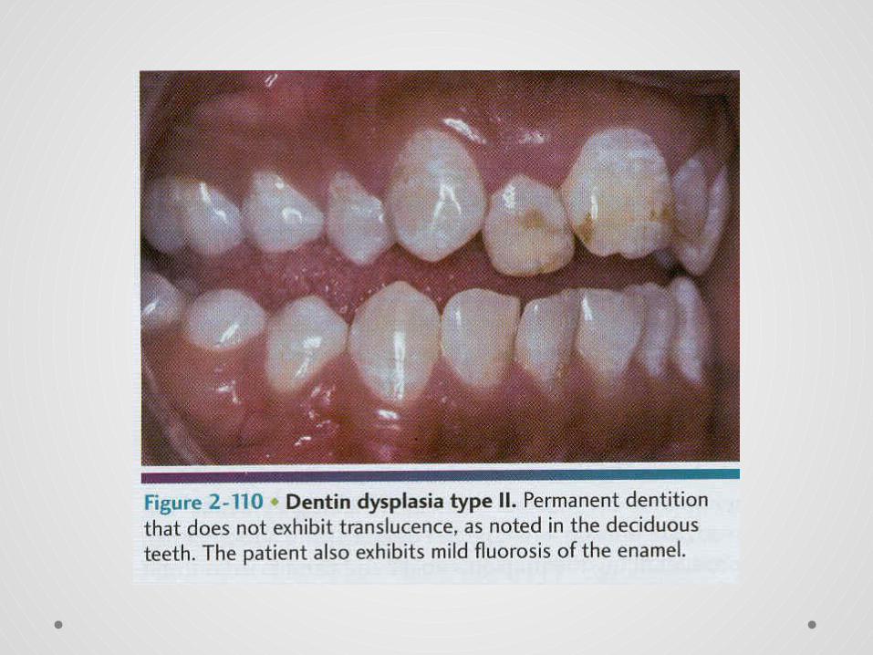
• In dentin dysplasia type II, the color of the primary denti tion is
opalescent and the permanent dentition is normal.
• in type I, both dentitions are of normal color. The coronal pulps
in type II are usually large and are filled with globules of
abnormal dentin.
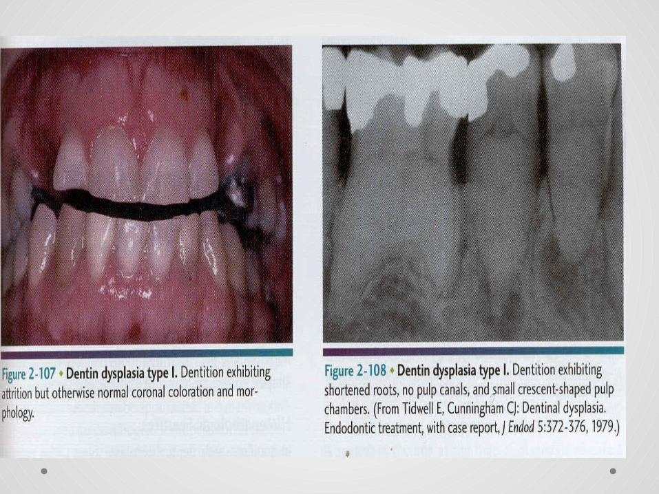
Clinically
• the crowns in dentin dysplasia type I appear to be normal in
color and shape.
• Premature tooth loss may occur because of short roots or
periapical inflammatory lesions. Teeth show greater resistance
to caries when com pared with normal teeth.


Radiographically
• in dentin dysplasia type I, roots appear extremely short and
pulps are almost completely obliterated.
• Residual fragments of pulp tissue appear typically as hori zontal
lucencies (chevrons).
• Periapical lucencies are typically seen; they represent chronic
abscesses, granulomas, or cysts.
• In dentin dysplasia type II, deciduous teeth are similar in
radiographic appearance to those in type I, but permanent teeth
exhibit enlarged pulp chambers that have been described as
thistle tube in appearance.


Microscopically
• the enamel and the immediately subja cent dentin
appear normal.
• Deeper layers of dentin show atypical tubular
patterns, with amorphous, atubular areas and
irregular organization.
• On the pulpal side of the normal-appearing mantle
of dentin, globular or nodular masses of abnormal
dentin are seen.
Treatment
is directed toward retention of teeth for as long as
possible. However, because of the short roots and
periapi cal lesions, the prognosis for prolonged retention
is poor.

DEFECTS OF ENAMEL AND DENTIN
Regional Odontodysplasia
• Regional odontodysplasia is a dental abnormality that involves the hard
tissues derived from both epithelial (enamel) and mesenchymal (dentin
and cementum) compo nents of the tooth-forming apparatus.
• The teeth in a region or quadrant of the maxilla or mandible are affected
to the extent that they exhibit short roots, open apical foramina, and
enlarged pulp chambers.

• The thinness and poor mineralization quality of the
enamel and dentin layers have given rise to the
term ghost teeth.
• The permanent teeth are more affected than the
primary teeth
• the maxillary anterior teeth are more affected than
other teeth.
• Eruption of the affected teeth is delayed


ENVIRONMENTAL EFFECTS ON TOOTH STRUCTURE
DEVELOPMENT
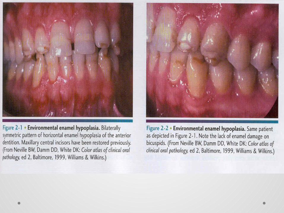
DEFECTS OF ENAMEL Environmental Defects of Enamel
During enamel formation, ameloblasts are susceptible to various external factors
that may be reflected in erupted teeth. Metabolic injury, if severe enough and
long enough, can cause defects in the quantity and shape of enamel or in the
quality and color of enamel.
• Quantitatively defective enamel, when of normal hardness, is known as
enamel hypoplasia.
• Qualitatively defective enamel, in which normal amounts of enamel are
produced but are hypomineralized, is known as enamel hypocalcifica tion. In
this defect, the enamel is softer than normal.


The extent of the enamel defect is dependent on three conditions:
(1) the intensity of the causative factor.
(2) the duration of the factor’s presence.
(3) the time at which the factor occurs during crown development.
• Affected teeth may have areas of coronal discol oration, or they may have actual
pits and irregularities. This is most commonly seen in permanent teeth in which
the overlying deciduous tooth becomes abscessed or is physi cally forced into the
enamel organ of the permanent tooth. The resulting hypoplastic or hypocalcified
permanent tooth is sometimes known as Turner’s tooth (Turners hypoplasia).


Hypoplasia caused by antineoplastic therapy.
• As modern medicine increases its prevalence of successful
therapy against childhood cancer. it has become evident that
a number of developmental alterations arise secondary to use
of therapeutic radiation or chemotherapy.
• The degree and severity of the developmental alterations are
related to the patient's age at treatment. form of therapy. and
the dose and field of radiation

Dental fluorosis. The ingestion of excess amounts of
fluoride also can result in significant enamel defects known
as dental fluorosis. .


Syphilitic hypoplasia.
Congenital syphilis results in a pattern
of enamel hypoplasia that is well
known but currently so rare .
• Anterior teeth altered by syphilis
are termed Hutchinson's incisors
and exhibit crowns that are shaped
like straight-edge screw drivers.
• Altered posterior teeth are termed
mulberry molars .
Post developmental Loss of Tooth
Attrition, Abrasion, Erosion and Abfraction
Attrition is the physiologic wearing of teeth as a
result of mastication. It is an age-related process


Abrasion is the pathologic wearing of teeth caused
by an abnormal habit or abnormal use of abrasive
substances orally.
Pipe smoking, tobacco chewing, aggressive tooth
brushing, and use of abrasive dentifrices are among
the more common causes.


Erosion is the loss of tooth structure through a nonbacte rial chemical
process. Most commonly, acids are involved in the dissolution process from an
external or an internal source.
Externally, acid may be found in the work environment (e.g., battery
manufacturing) or in the diet (e.g., citrus fruits, acid-containing soft drinks).
• The internal source of acid may be seen in any disorder of which chronic
vomiting is a part.
• The pattern of erosion associated with vomit ing is usually generalized tooth
loss on the lingual surfaces of maxillary teeth. However, all surfaces may be
affected.


Abfraction loss of tooth from occlusal stress that create
repeated tooth flexure with failure of enamel and dentin
at a location away from the point of locking, it appears
as wedge-shaped defects limited to the cervical area of
the teeth and may closely resemble cervical abrasion or
erosion.


Internal and External Resorption
Internal ResorptionResorption of the dentin of the pulpal walls may be seen as
part of an inflammatory response to pulpal injury. The
resorption occurs as a result of activation of osteoclasts or
dentinoclasts on internal surfaces of the root or crown.

teeth may appear pink because of the proximity of
pulp tissue to the tooth surface.
The treatment of choice is root canal therapy before
per foration. Once communication between pulp and
periodon tal ligament occurs, the prognosis for
saving the tooth is very poor.


External Resorption
This change may be the result of an adjacent pathologic process,
such as
(1) chronic inflammatory lesions.
(2) cysts.
(3) benign tumors.
(4) malignant neoplasms.
External resorption of teeth may also be seen in association with
(1) trauma.
(2) Re implantation or transplan tation of teeth.
(3) impaction.


Endogenous Stains
Discoloration of teeth resulting from deposits of systemically circulating
substances during tooth development is defined as endogenous or intrinsic
staining.
Systemic ingestion of tetracycline during tooth develop ment is a well-known
cause of endogenous staining of teeth.
• Tetracycline binds calcium and therefore is deposited in developing teeth and
bones. The bright yellow color of the drug is reflected in subsequently erupted
teeth.
• Because tetracycline can cross the placenta, it may stain primary teeth if taken
during pregnancy.
• If it is administered between birth and age 6 or 7 years, permanent teeth may be
affected.


Environmental Discoloration of Teeth
Exogenous Stains
Stains on the surfaces of teeth that can be removed with
abrasives are known as exogenous or extrinsic stains. The color
change may be caused by pigments in dietary sub stances (e.g.,
coffee, “betel” areca nut, tobacco) or by the colored by-products
of chromogenic bacteria in dental plaque.