Embed Size (px)
Citation preview
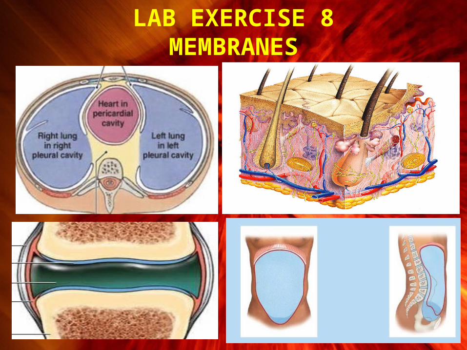
LAB EXERCISE 8MEMBRANES

Membranes • Membranes
– Line or cover body surfaces– Consist of epithelium supported by
connective tissue
• Four Types of Membranes
1. Cutaneous membrane
2. Mucous membranes
3. Synovial membranes
4. Serous membranes

• Cutaneous membrane– Covers surface of body– Skin– Relatively thick, waterproof, and dry comparatively– Tissues Include
» Stratified squamous epithelium» Dense irregular connective tissue » Loose areolar CT
Membranes

Membranes
• Mucous membranes– Line organs that communicate to the outside– Located in digestive, respiratory, urinary, and reproductive tracts
– Epithelial surfaces must be moist
• To reduce friction
• To facilitate absorption and excretion » Lubricated by mucus or bodily fluids
– Supported by areolar connective tissue of the lamina propria

• Synovial membranes
– Line moving, articulating joint cavities
– Produce synovial fluid (lubricant)
– Protect the ends of bones
– Lack a true epithelium
Membranes

• Serous membranes– Line cavities that do not open to the outside– Mesothelium supported by areolar connective tissue
• Are thin but strong
– Have a liquid fluid called transudate to reduce friction and
allows the viscera to slide somewhat during movements.
Membranes

• Serous membranes
– Have a parietal portion covering the cavity
– Have a visceral portion (serosa) covering the organs
– Three subdivisions of ventral body cavity• Pleura
– Line pleural cavity and cover the lungs• Peritoneum
– Line peritoneal cavity and cover visceral organs• Pericardium
– Line pericardial cavity and cover the heart
Membranes

Serous Membranes
Pericardium Pleura

Serous MembranePeritoneum
• Peritoneum– Largest serous membrane
of the body– Visceral layer
• Covers organs– Parietal layer
• Lines the walls of body cavity
• Peritoneal cavity– Slim space containing a
bit of serous fluid between the parietal & visceral layers

Abdominal Organs
• Retroperitoneal organs– These organs are posterior to the
peritoneum and lose their mesentery• Kidneys, Pancreas and parts
of the large intestine
• Intraperitoneal organs– Digestive organs that remain in
the peritoneal cavity and maintain their mesentery• The rest

MEDICAL IMAGING
• A specialized branch of anatomy and physiology that is essential for the diagnosis of many disorders is medical imaging, one division of which is radiography, which includes the use of x-rays.
• Medical imaging techniques allow physicians to peer inside the body to provide clues to abnormal anatomy and deviations from normal physiology in order to help diagnose disease.

Conventional Radiography• A single burst of
xrays• Produces 2-D
image on film• Known as
radiography or xray• Poor resolution of
soft tissues• Major use is
osteology

Computed Tomography (CT Scan)
• Moving x-ray beam• Image produced on a
video monitor of a cross-section through body
• Computer generated image reveals more soft tissue detail– kidney & gallstones
• Multiple scans used to build 3D views

Digital Subtraction Angiography(DSA)
• Radiopaque material injected into blood vessels
• Before and after images compared with a computer program
• Image of blood vessel is shown on a monitor

Ultrasound (US)• High-frequency sound
waves emitted by hand-held device
• Safe, noninvasive & painless
• Image or sonogram is displayed on video monitor
• Used for fetal ultrasound and examination of pelvic & abdominal organs, heart and blood flow through blood vessels

Magnetic Resonance Imaging (MRI)
• Body exposed to high-energy magnetic field
• Protons align themselves relative to magnetic field
• Pulse of radiowaves used to generate an image on video monitor
• Can not use on patient with metal in their body
• Reveals fine detail within soft tissues

Positron Emission Tomography(PET)
• Substance that emits positively charged particles is injected into body
• Collision with negatively charged electrons in tissues releases gamma rays
• Camera detects gamma rays & computer generates image displayed on monitor