Embed Size (px)
Citation preview
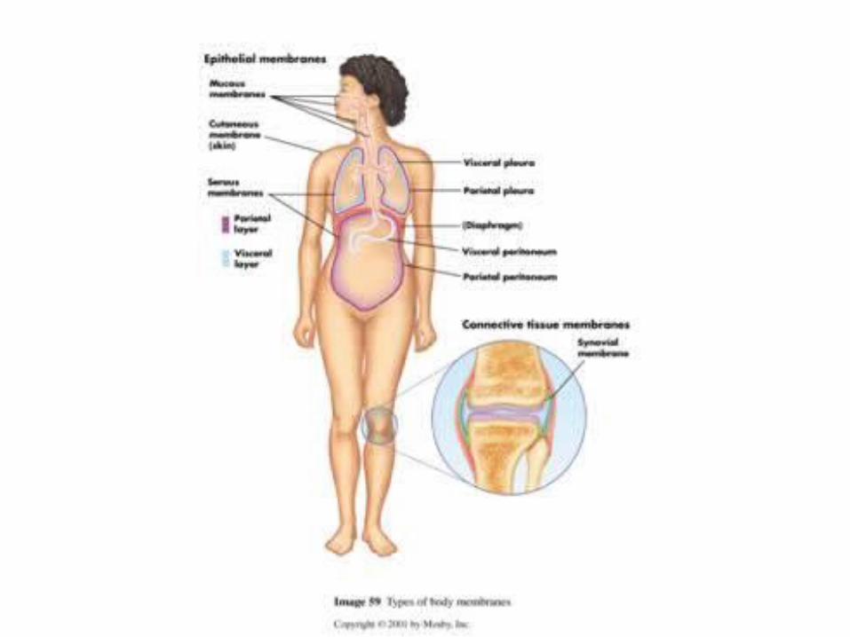
I. Membranes Classified by tissue types.
1. Epithelial Membranes (layer of epithelia and connective tissue) = Simple organ
a. Cutaneous Membrane (skin) composed of stratified squamous and dense connective
tissue. Unique because it is primarily a “dry” membrane.
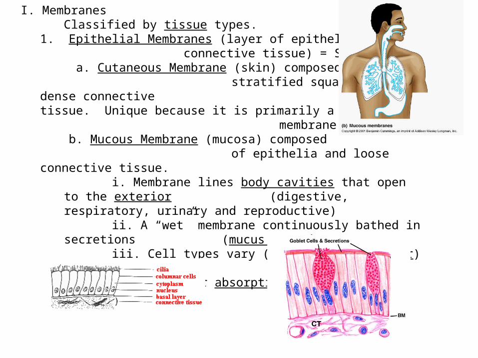
b. Mucous Membrane (mucosa) composed of epithelia and loose connective tissue.
i. Membrane lines body cavities that open to the exterior (digestive, respiratory, urinary and reproductive)
ii. A “wet” membrane continuously bathed in secretions (mucus or urine).
iii. Cell types vary (squamous or columnar) typically specialized for secretion (mucus) or absorption
(digestive tract).

c. Serous Membranes (serosa) composed of simple squamous and aerolar connective tissue.
i. Line body cavities that are closed to the exterior (except dorsal cavity and joints)ii. Occurs in pairs: 1. Layer that lines the body
cavity is known as the parietal layer 2. Layer that lines the body
organ is the visceral layer 3. Fluid between layers

iii. Function of membrane is to decrease friction between organs. Ex. Heart and lungs, stomach and intestine.
iv. Location of membrane can dictate name. Ex. Pleura around lungs; pericardium around heart, peritoneum in
abdominal cavity.

2. Connective Tissue Membranes/Synovial Membranes (composed of soft aerolar tissue)
a. Lines:i.
fibrous capsules of your joints,
ii. bursa (sacs of connective tissue at some joints),
iii. tendon sheaths
b. Protects against friction between moving parts of the body.

• Friction is the force resisting the relative motion of solid surfaces, fluid layers, and material elements sliding against each other.

Cool Skin Facts
• Your skin weighs around 9 pounds
• You lose 30-40 thousand skin cells each minute (around a billion in a day)
• These mites live in your
Eyelash follicles!
Hi! I live in your
eyelashes!

II The Integument System Structure (skin and its derivatives)A. Epidermis – top most superficial layer 1. Composed of stratified squamous epithelial
2. Avascular and not innervated (no nerves)3. Different cell types:
a. Keratinocytes – produce keratin helps with waterproofing of the skin; these cells are lost through friction.
b. Melanocytes – produce melanin (primary skin pigment), found in the lowest level of epidermis.
Function is to protect the DNA in the nucleus from the UV radiation of the sun.
Amount and kind of melanin determine skin color.
c. Immune system cells in lower layers for early detection (called Langerhans cells)


4. Layers of epidermisa. Stratum corneum – most superficial layer, numerous cell layers (20 to 30); cells are dead (cornified) and flake off easily. Major protective layer.b. Stratum Lucidum – Only present on palms and soles of your feet. Cells are clear and dead (too far to receive nutrients and oxygen)c. Stratum Granulosum d. Stratum Spinosume. Stratum Basale/Germinativum – layer of rapid cell division
How tatoos work.
"Come, Let's Get Sun Burned":· From superficial to deep:CorneumLucidumGranulosumSpinosumBasale [Germinativum]
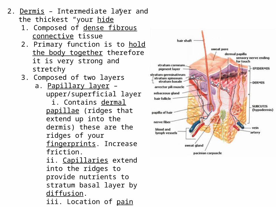
2. Dermis – Intermediate layer and the thickest “your hide”1. Composed of dense fibrous connective
tissue2. Primary function is to hold the body
together therefore it is very strong and stretchy
3. Composed of two layersa. Papillary layer – upper/superficial
layer i. Contains dermal papillae (ridges
that extend up into the dermis) these are the ridges of your fingerprints. Increase friction.ii. Capillaries extend into the ridges to provide nutrients to stratum basal layer by diffusion.iii. Location of pain receptors
(Meissner’s corpuscles)

b. Reticular layer – thicker layer of the dermisi. Contains blood vessels, sweat and oil glands, pressure receptors (Pacinian corpuscles), phagocytes (immune cells) and collagen and elastic fibers. a. Collagen – help keep skin hydratedb. Elastic – help with skin “stretchyness”
c. Hypodermis – Deepest layer of skin 1. Composed of adipose tissue2. Anchors the two other layers to underlying organs also insulate and act as a shock absorber.

Skin Layer Review
"Come, Let's Get Sun Burned":· From superficial to deep:CorneumLucidumGranulosumSpinosumBasale [Germinativum]

III Appendages of the Skin – Arise from the epidermis (stratum basal)A. Cutaneous Exocrine (have a duct) glands
1. Sebaceous Gland / Oil Glands
a. Found everywhere except palms of hands and feet.
b. Duct empties into a hair follicle.
c. Sebum is the product excreted composed of oils, chemicals that kill bacteria and fragmented cells.
d. Function to lubricate the skin and hair and protection against microbial invasion.
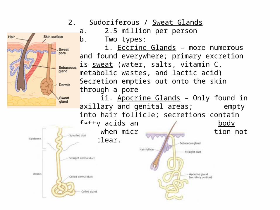
2. Sudoriferous / Sweat Glandsa. 2.5 million per person b. Two types: i. Eccrine Glands – more numerous and found everywhere; primary excretion is sweat (water, salts, vitamin C, metabolic wastes, and lactic acid) Secretion empties out onto the skin through a pore ii. Apocrine Glands – Only found in axillary and genital areas; empty into hair follicle; secretions contain fatty acids and proteins (causes body odor when microbes digest); function not yet clear.

B. Hair and Hair Follicle1. Layers of hair – cuticle (outer most, looks like shingles on a roof), cortex (area of color) and medulla (core, different pattern in each species)2. Types of hair:
a. Vellus hair – “peach fuzz” covers the entire body.
b. Terminal hair – darker hair on head, eye lashes, eye brows, pubic area, etc…
The lovely mites that live in our eyelash follicles!
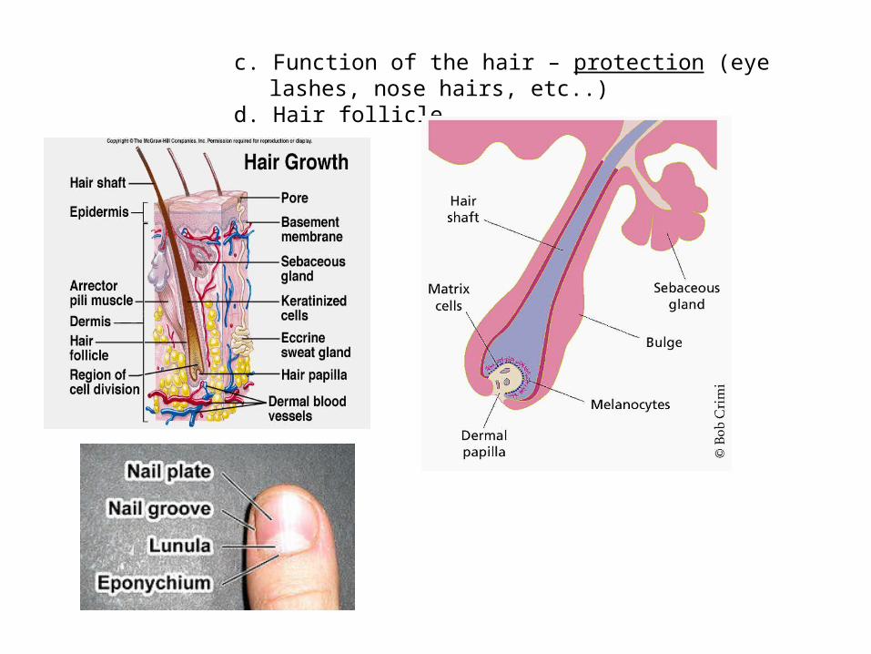
c. Function of the hair – protection (eye lashes, nose hairs, etc..)
d. Hair follicle

IV Function of the integument (the best coat you’ll ever wear)A. Thermoregulation – maintaining proper body temperature
1. Capillaries in the dermis can dilate (get bigger) releasing heat from the skin (red and swollen) causing a flushed appearance; or become restricted and redirect blood flow
away from skin to more vital parts of the body (why your skin feels cold)
2. Sweat glands -release of water from sweat results in evaporative cooling. B. Physical and Chemical Damage - bumps, cuts, scrapes, burns.
a. Keratinized cells in upper epidermis
b. Nerve receptors that alert central nervous system to a problem

C. Microbe damage – bacteria 1. “acid mantle” secretions of epidermis slow/prevent bacterial growth2. Phagocytes in dermis that engulf pathogen (disease causing microbes)
D. Desiccation – drying out1. Keratinized epidermal cells- waterproof
E. Excretion 1. Sweat – uric acid, heat, other toxins and chemicals.2. Pheromones – through apocrine sweat glands3. Oil – through sebaceous glands
F. Synthesis 1. Vitamin D (modified cholesterol and sunlight) helps with calcium absorption in the digestive tract. 2. Proteins necessary for immunity.
G. UV radiation1. Melanin protecting DNA in nucleus from mutations.

Fingerprints
• Arch
• Whorl Double loop

Eccrine vs. Apocrine Glands
Use your notes to explain their location, secretion, functions

Question of the Week Why do we have fingernails?
• “We have fingernails because we're primates," said John Hawks, a biological anthropologist at the University of Wisconsin-Madison.
• “Fingernails distinguish primates, they are essentially flattened forms of claws to grab onto things, to climb things, to scratch things, and to dig holes."
• While claws would have provided excellent grip as our mammalian ancestors clambered up large tree trunks, they would have been a nuisance for larger-bodied primates trying to grasp smaller branches while scrambling across tree canopies for fruits. Rather, primates developed broader fingertips made for grasping.
• Nails serve as a visual advertisement of a person's health, he said. For instance, malnutrition can change the coloring of nails.
• small pits in fingernails can signal the skin condition psoriasis.• Spoon-shaped--- anemia• A healthy nail has a specific shape—slightly raised in the middle, then curving down a bit at the tip.• Dry and brittle it’s possible for an under-active thyroid to cause both dry skin and brittle nails• Yellow-When all of the nails turn yellow it can be a sign of lung disease or diabetes,” • White spots- nail injury or can be ZINC defeciency, or air spaces in nail plate
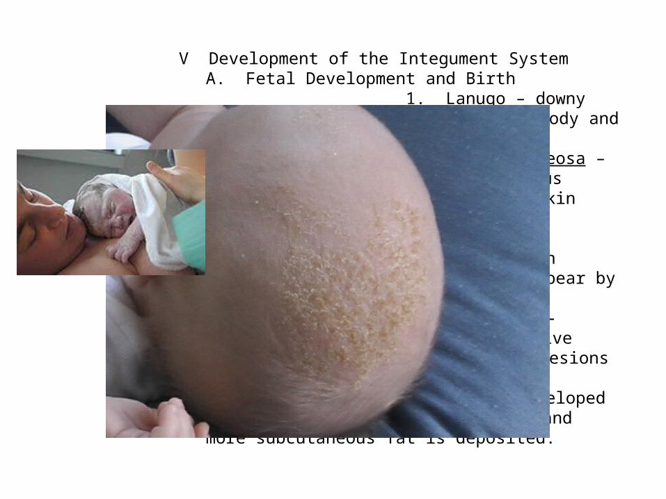
V Development of the Integument SystemA. Fetal Development and Birth
1. Lanugo – downy hair that covers the fetus’s body and is typically shed by birth.
2. Vernix caseosa – white secretion by the sebaceous glands which protects the baby’s skin in the uterus.
3. Milia – accumulation of sebaceous glands on nose and forehead, typically disappear by 3rd week.
4. Seborrhea – “cradle cap” formed from overactive sebaceous glands forming raised lesions that crust over and slough off.
5. As baby developed skin becomes thicker and moist and more subcutaneous fat is deposited.
B. Adolescence1. Sebaceous glands increase their activity and acne (infection of
sebaceous gland) can appear. a. White head – sebaceous gland blocked
by sebumb. Black head – when accumulated
material oxidizes and driesc. 20’s and 30’s your skin is the best it will
ever look!C. Old Age
1. Subcutaneous fat deposits decrease (intolerance of cold due to lack of insulation and thinness of skin can lead to increase in bruising)2. Decrease oil production and fewer collagen fibers contribute to
overall decrease in moisture level of skin. 3. Sagging is caused by the loss of elasticity of the elastic fibers in the skin.4. Overexposure to sun can lead to increase incidence of skin cancer
(accumulation of mutations over the years)5. Hair follicle numbers decrease by 1/3 by the age of 50 resulting in
hair loss (alopecia) and melanocytes can stop producing melanin in the follicle and the hair appears “gray”
(due to a delayed action gene)

D. How can we reverse this aging trend?1. Stay out of the sun /use sunscreen2. Good balanced diet (antiaging foods- blueberries, nuts, brocolli, green leafy veggies, garlic, tomatoes, soy beans, prunes, etc.)3. Plenty of water4. Cleanliness / good hygiene

VI Homeostatic Imbalances of the Skin (there are over 1000)A. Changes in color
1. Redness /Erythema – dilation of capillaries in the skin
2. Blanching/Pallor – pale skin can be indicative of anemia, emotional stress, low blood pressure.
3. Jaundice – yellowing of skin from a liver disorder (not breaking down bile properly and the pigments are circulated through the blood)
4. Bruises – blood has escaped the vessels and clotted in the tissues (hematoma)
In Raynaud's disease, smaller arteries that supply blood to your skin narrow, limiting blood circulation to affected areas.
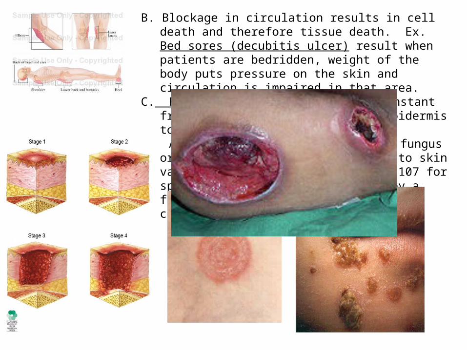
B. Blockage in circulation results in cell death and therefore tissue death. Ex. Bed sores (decubitis ulcer) result when patients are bedridden, weight of the body puts pressure on the skin and circulation is impaired in that area.
C. Blisters - skin is exposed to constant friction causing the dermis and epidermis to separate
D. Attack by bacteria, virus and/or fungus or allergy. Symptoms and effects to skin vary with cause. (read over pg. 107 for specific types) Ringworm, caused by a fungus in middle picture and impetigo caused by bacteria in third picture.
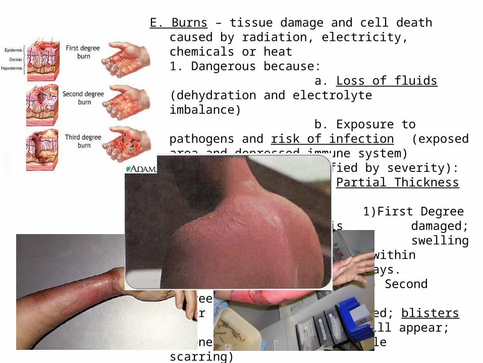
E. Burns – tissue damage and cell death caused by radiation, electricity, chemicals or heat1. Dangerous because:
a. Loss of fluids (dehydration and electrolyte imbalance)
b. Exposure to pathogens and risk of infection (exposed area and depressed immune system)2. Three types (classified by severity):
a. Partial Thickness Burns1)First Degree Burn
– Only epidermis is damaged; symptoms are redness and swelling in the area. Heals usually within 2 to 3 days.
2) Second Degree Burn – Epidermis and upper layer of dermis affected; blisters will appear; regeneration can occur (little scarring)
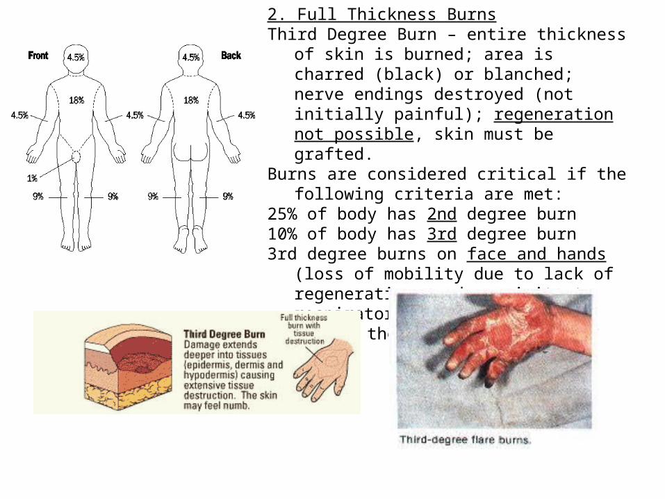
2. Full Thickness BurnsThird Degree Burn – entire thickness of skin is
burned; area is charred (black) or blanched; nerve endings destroyed (not initially painful); regeneration not possible, skin must be grafted.
Burns are considered critical if the following criteria are met:
25% of body has 2nd degree burn10% of body has 3rd degree burn3rd degree burns on face and hands (loss of
mobility due to lack of regeneration and proximity to respiratory system)
Look over the Rule of 9’s on page 108.

F. Skin Cancer1.Cells divide uncontrollably and the cancer can metastasize (move to other areas of the body); growth can also be benign such as warts.
2. Factors that can predispose a person to skin cancer: exposure to UV radiation (sun), frequent irritations, genetics.
3.Types:Basal Cell Carcinoma – least malignant; most common; cells stop producing keratin and invade the dermis. Relatively slow growing and 99% full cure if lesions removed surgically. Sun induced!Squamous Cell Carcinoma – Arise from stratum spinosum layer; lesion is scaly in appearance; appears on scalp, ears, tops of hands and lower lips; grows more rapidly and can metastize. Malignant melanoma – Cancer of melanocytes; often deadly metastases quickly; survival 50% with early detection.

» Rule for detection – ABCD Rule» Asymmetry of the lesion» Border irregularity (not smooth)» Color changes and different colors in same area» Diameter is larger than 6mm (pencil eraser
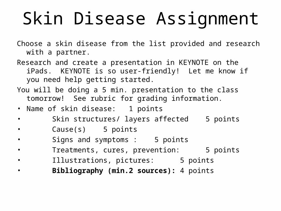
Skin Disease AssignmentChoose a skin disease from the list provided and research with a partner.
Research and create a presentation in KEYNOTE on the iPads. KEYNOTE is so user-friendly! Let me know if you need help getting started.
You will be doing a 5 min. presentation to the class tomorrow! See rubric for grading information.
• Name of skin disease: 1 points• Skin structures/ layers affected 5 points
• Cause(s) 5 points
• Signs and symptoms : 5 points
• Treatments, cures, prevention: 5 points• Illustrations, pictures: 5 points• Bibliography (min.2 sources): 4 points