Embed Size (px)
Citation preview
I’ve
Lab 4: Gel Electrophoresis MiniOne
Updated: June 2021
Project Guide

LAB 4: GEL ELECTROPHORESIS 2
Table of Contents
Page Contents
3 Introduction
4 Key Elements for Gel Electrophoresis
5 How to Read a Gel
6 How to Interpret Gel Electrophoresis Results
Gel Electrophoresis Lab
7 Pre-Lab Questions
8 Getting Started
9 Visual Equipment and Reagents Checklist
11 Gel Electrophoresis Protocol: MiniOne
13 Results
14 Post-Lab Questions
15 Troubleshooting
17 Database Checklist
18 Glossary
Content is made available under the Creative Commons Attribution-NonCommercial-No Derivatives International License. Contact ([email protected]) if you would like to make adaptations for distribution beyond the classroom. The Wolbachia Project: Discover the Microbes Within! was developed by a collaboration of scientists, educators, and outreach specialists. It is directed by the Bordenstein Lab at Vanderbilt University. https://www.vanderbilt.edu/wolbachiaproject
Some figures created with BioRender.com
LAB 4: GEL ELECTROPHORESIS 3
Introduction
This lab will determine the presence or absence of amplified DNA in your samples by visualization on an agarose gel. Arthropod and Wolbachia DNA, if present, will be distinguishable based on the size, or base pair (bp) length, of the DNA molecule.
Gel electrophoresis
Gel electrophoresis is a method of separating DNA fragments by movement through a Jello-like substance called agarose. Derived from a seaweed polysaccharide, agarose gels form small pores that act as sieves to separate DNA based on size; whereby smaller DNA molecules move through the pores faster and easier than larger molecules. Loading wells are oriented at the top of the gel to allow for precise insertion of PCR products into the gel. An electrical current is applied to move negatively charged DNA molecules away from a negative electrode (-) and toward a positive electrode (+). DNA migrates through the gel in a single, vertical lane. Three factors influence the speed of movement: (i) the voltage of the electrical field, (ii) the concentration of agarose, and (iii) most importantly, the size of the DNA molecule.
DNA Visualization DNA itself is not visible within an agarose gel. Therefore, a fluorescent stain is added to the gel that binds DNA and fluoresces under UV or blue light. DNA will appear as a horizontal line, or band, on the agarose gel.
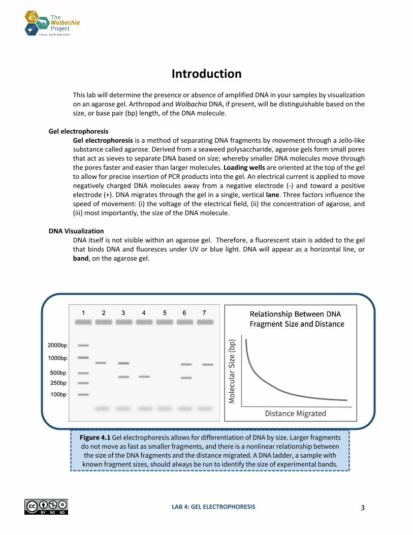
Figure 4.1 Gel electrophoresis allows for differentiation of DNA by size. Larger fragments do not move as fast as smaller fragments, and there is a nonlinear relationship between
the size of the DNA fragments and the distance migrated. A DNA ladder, a sample with known fragment sizes, should always be run to identify the size of experimental bands.

LAB 4: GEL ELECTROPHORESIS 4
Key Elements for Gel Electrophoresis PCR Products (DNA)
The purpose of this lab is to visualize the PCR products, or amplified DNA, from your arthropod samples.
DNA Ladder A DNA ladder is a cocktail of DNA fragments with pre-determined sizes. The ladder, also called a DNA marker, is loaded alongside experimental samples as a reference tool for estimating band size.
Agarose Gel MiniOne GelCups contain solidified chunks of agarose that are melted and re-solidified in a casting tray to form the agarose gel. DNA will migrate through the gel and form separate bands based on size (correlating to length in bp).
DNA Stain A DNA stain is added to the agarose gel to visualize DNA under a UV or blue light. It has already been pre-mixed into the MiniOne GreenGel GelCups.
Running Buffer Running buffer is a conductive liquid that allows the DNA to migrate through the agarose gel. It is important that the agarose gel be made using the same buffer.
Loading Dye Loading dye has two primary components: (i) a visible dye indicates how far the DNA has run on the gel and (ii) glycerol, which is denser than the buffer, ensures that samples fall into the loading wells rather than float back out. Some Taq Master Mixes (e.g., Promega GoTaq) already contain a pre-mixed loading dye.
Electrophoresis System Running buffer and the agarose gel will be placed into the chamber of an electrophoresis system. After loading the samples, an electric current is applied to move the negatively charged DNA towards the positive end of the system. Without this electric field, the DNA will not migrate through the agarose gel. If the electric field is reversed, the DNA will run in the opposite direction, towards the top of the gel, and eventually exit the gel.
Figure 4.2. Pictograph of all necessary components for gel electrophoresis.
LAB 4: GEL ELECTROPHORESIS 5
How to Read a Gel Lanes DNA that was loaded into each well will migrate in a single, vertical lane towards the (+) charge. Bands
When DNA becomes separated by size due to gel electrophoresis, they appear as bands in the gel. These are clearly defined, bright lines in the gel.
DNA Ladder The DNA ladder will contain multiple bands in one lane. Each band represents a pre-determined length of DNA and can be used as a reference tool to estimate DNA size for each of the experimental samples. Refer to the product information for specific band sizes.
Primer Dimers PCR reactions are set up with an excess of primers. In addition, some primers bind to each other instead of binding to the DNA, creating primer dimers. Primers are ~25bp long, so excess primers appear as fuzzy bands on the bottom of the gel ~25-50bp. This is normal and to be expected.
Reading a Single PCR vs. Duplex PCR Gel A single PCR gel will contain only one amplified PCR product, either Wolbachia or arthropod, in each lane. A separate gel will need to be run for each DNA type. A duplex PCR means that both the arthropod barcoding gene and the 16S rRNA fragment from Wolbachia were amplified in the same PCR reaction. When visualizing this PCR reaction, two bands should appear in the same lane if Wolbachia is present, and only one band will appear if the arthropod is uninfected.
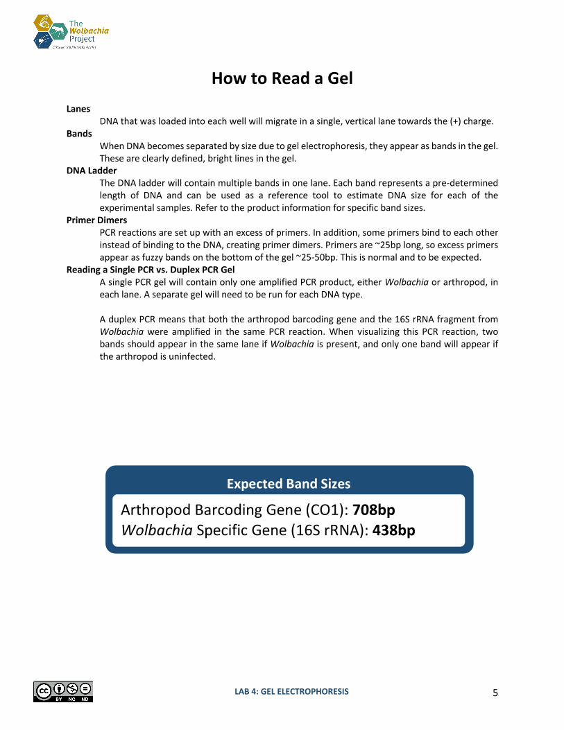
Arthropod Barcoding Gene (CO1): 708bp Wolbachia Specific Gene (16S rRNA): 438bp
Expected Band Sizes
LAB 4: GEL ELECTROPHORESIS 6
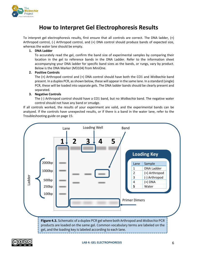
How to Interpret Gel Electrophoresis Results To interpret gel electrophoresis results, first ensure that all controls are correct. The DNA ladder, (+) Arthropod control, (-) Arthropod control, and (+) DNA control should produce bands of expected size, whereas the water lane should be empty.
1. DNA Ladder To accurately read the gel, confirm the band size of experimental samples by comparing their location in the gel to reference bands in the DNA Ladder. Refer to the information sheet accompanying your DNA ladder for specific band sizes as the bands, or rungs, vary by product. Below is the DNA Marker (M3104) from MiniOne.
2. Positive Controls The (+) Arthropod control and (+) DNA control should have both the CO1 and Wolbachia band present. In a duplex PCR, as shown below, these will appear in the same lane. In a standard (single) PCR, these will be loaded into separate gels. The DNA ladder bands should be clearly present and separated.
3. Negative Controls The (-) Arthropod control should have a CO1 band, but no Wolbachia band. The negative water control should not have any band or smudge.
If all controls worked, the results of your experiment are valid, and the experimental bands can be analyzed. If the controls have unexpected results, or if there is a band in the water lane, refer to the Troubleshooting guide on page 15.
Figure 4.3. Schematic of a duplex PCR gel where both Arthropod and Wolbachia PCR products are loaded on the same gel. Common vocabulary terms are labeled on the gel, and the loading key is labeled according to each lane.
1000bp
500bp
2000bp
250bp
100bp
Lane
4
Lane Sample 1 DNA Ladder 2 (+) Arthropod 3 (-) Arthropod 4 (+) DNA 5 Water
Loading Key
Primer Dimers
Loading Well
Ladd
er
3 2 5 1
Band

LAB 4: GEL ELECTROPHORESIS 7
Pre-Lab Questions Read through the entire lab activity and answer the questions below.
1. Assume that single PCR reactions were loaded into two separate gels for arthropod and Wolbachia DNA analysis. Fill in the expected bands for lanes 2-7 using the table below.
2. This experiment included five controls. In the table below, list the following for each lab activity: • (+) for positive control • (-) for negative control • N/A for not applicable
Control DNA Extraction PCR Gel Electrophoresis
DNA Ladder
(+) Arthropod Control
(-) Arthropod Control
(+) DNA Control
Water
Lane Sample 1 DNA Ladder (bands already shown) 2 An arthropod sample positive for Wolbachia 3 An arthropod sample negative for Wolbachia 4 (+) Arthropod Control 5 (-) Arthropod Control 6 (+) DNA Control 7 Water
Arthropod (COI) gel Wolbachia (16S rRNA) gel

LAB 4: GEL ELECTROPHORESIS 8
Getting Started Introduction
In this activity, you will be using agarose gel electrophoresis to determine the presence and size of two different gene fragments (arthropod COI, and Wolbachia 16S rRNA) previously amplified by PCR. If you ran two separate PCR reactions, arthropod and Wolbachia, you should prepare and run two gels. If you set up a duplex reaction (both primer sets in one PCR tube), you will only need one gel.
MiniOne Gel Electrophoresis System
This protocol uses the MiniOne electrophoresis system (https://theminione.com/) and GreenGel cups (MiniOne #M3102TBE). The unit contains a built-in transilluminator (blue light) and photo hood. DNA can be visualized as it migrates through the gel by turning on the blue light. Be careful to not overuse the light feature as the stain is photosensitive and will bleach out with prolonged exposure to light. If you wish to make your own gels, you will need a DNA stain that is compatible with blue light, such as GelGreen or SYBR Safe. The MiniOne casting trays hold about 11 mL of molten agarose and the electrophoresis system will need about 135 mL of running buffer. A cell phone or other mobile device is required to take pictures.
Pre-Lab Preparation
If using the GreenGel cups, little to no pre-lab preparation is required. It is recommended to prepare a working solution of the TBE running buffer prior to class. Prepare electrophoresis buffer by adding 1 part TBE concentrate to 19 parts deionized or distilled water. Distilled water from the grocery store is suitable. Do NOT use tap water.
MiniOne Resource Center
MiniOne Benchtop Guide MiniOne User Manual MiniOne Instructional Video
LAB 4: GEL ELECTROPHORESIS 9
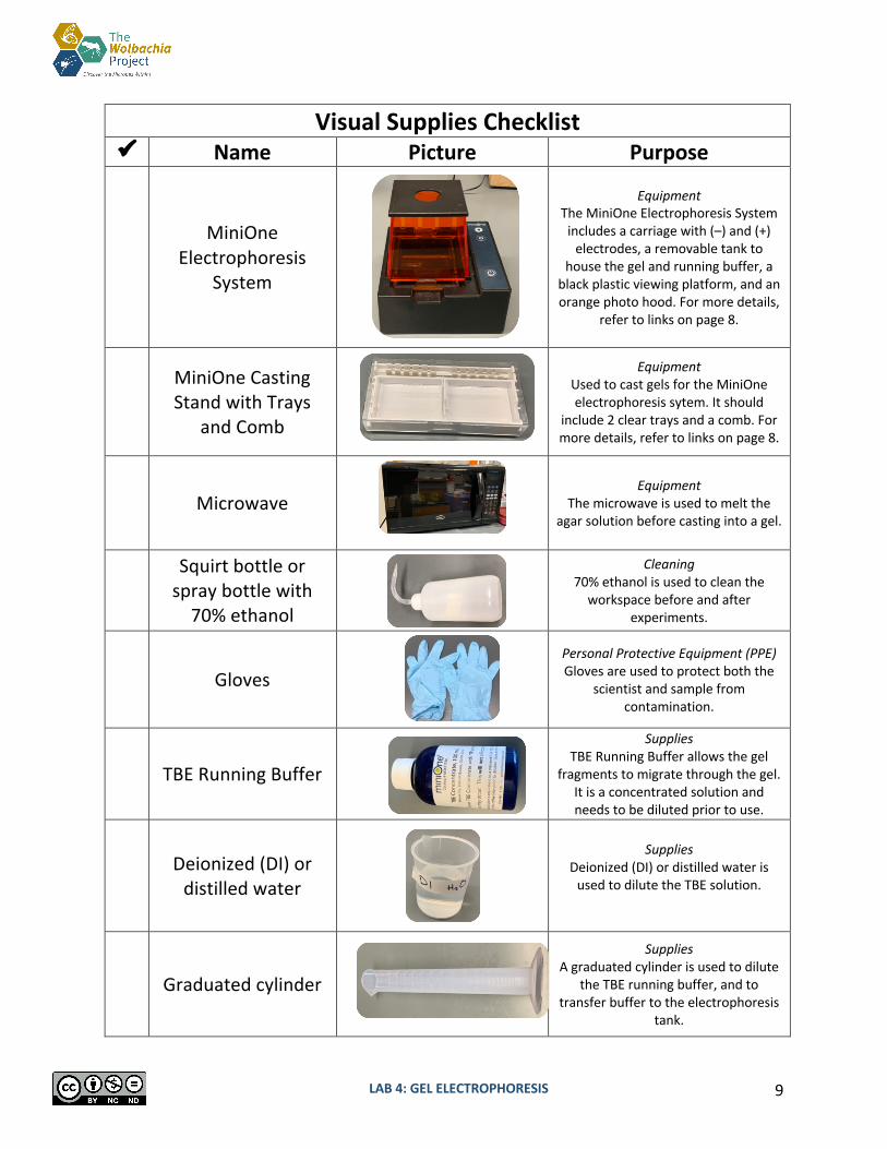
Visual Supplies Checklist ü Name Picture Purpose
MiniOne Electrophoresis
System
Equipment The MiniOne Electrophoresis System
includes a carriage with (–) and (+) electrodes, a removable tank to
house the gel and running buffer, a black plastic viewing platform, and an orange photo hood. For more details,
refer to links on page 8.
MiniOne Casting Stand with Trays
and Comb
Equipment Used to cast gels for the MiniOne electrophoresis sytem. It should
include 2 clear trays and a comb. For more details, refer to links on page 8.
Microwave
Equipment The microwave is used to melt the
agar solution before casting into a gel.
Squirt bottle or spray bottle with
70% ethanol
Cleaning 70% ethanol is used to clean the
workspace before and after experiments.
Gloves
Personal Protective Equipment (PPE) Gloves are used to protect both the
scientist and sample from contamination.
TBE Running Buffer
Supplies TBE Running Buffer allows the gel
fragments to migrate through the gel. It is a concentrated solution and needs to be diluted prior to use.
Deionized (DI) or
distilled water
Supplies
Deionized (DI) or distilled water is used to dilute the TBE solution.
Graduated cylinder
Supplies A graduated cylinder is used to dilute
the TBE running buffer, and to transfer buffer to the electrophoresis
tank.
LAB 4: GEL ELECTROPHORESIS 10
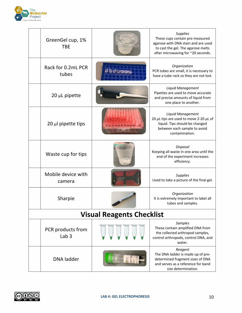
GreenGel cup, 1%
TBE
Supplies These cups contain pre-measured
agarose with DNA stain and are used to cast the gel. The agarose melts
after microwaving for ~20 seconds.
Rack for 0.2mL PCR
tubes
Organization
PCR tubes are small, it is necessary to have a tube rack so they are not lost.
20 μL pipette
Liquid Management Pipettes are used to move accurate and precise amounts of liquid from
one place to another.
20 μl pipette tips
Liquid Management 20 μL tips are used to move 2-20 μL of
liquid. Tips should be changed between each sample to avoid
contamination.
Waste cup for tips
Disposal
Keeping all waste in one area until the end of the experiment increases
efficiency.
Mobile device with camera
Supplies
Used to take a picture of the final gel.
Sharpie
Organization It is extremely important to label all
tubes and samples.
Visual Reagents Checklist
PCR products from Lab 3
Samples These contain amplified DNA from the collected arthropod samples,
control arthropods, control DNA, and water.
DNA ladder
Reagent The DNA ladder is made up of pre-determined fragment sizes of DNA and serves as a reference for band
size determination.

LAB 4: GEL ELECTROPHORESIS 11
Gel Electrophoresis Protocol MiniOne
Class Preparation: TBE Buffer Working Solution 1. Together with your class, prepare a working solution of electrophoresis running buffer by
adding 1 part TBE concentrate to 19 parts deionized or distilled water. This may have already been prepared in advance.
Prepare Lab Space
2. Remove all unnecessary items from your lab station. 3. Put on nitrile gloves and clean all surfaces by wiping down with 70% Ethanol.
Prepare the Gel(s)
4. Place the MiniOne casting stand on a level surface and insert two gel trays. 5. Select the 9-tooth comb (in order to accommodate the DNA ladder and 6 PCR products) and
insert into the top ridges of the casting tray. 6. Partially peel the film of a GreenGel cup to vent and place in the microwave for 20 seconds.
Make sure it is completely melted- look for chunks or shadows. Do not microwave more than five cups at a time.
If you are running two gels in one class period, they can be heated and prepared at the same time.
7. Allow the cup to cool for 15 seconds. Carefully handle the cup to prevent bubbles from forming in the agarose solution.
8. Carefully pour the hot gel into one gel tray and let it sit for at least 10 minutes. Do not move the tray. Moving the tray or not waiting until the gel is fully set may affect your results.
9. While you wait for the gel to solidify, use a graduated cylinder to measure out 135 mL of the diluted TBE running buffer and fill out the Loading Key on the next page.
10. Once the gel is solidified, carefully and slowly remove the comb. Lift the gel tray from the casting tray and wipe off excess agarose from the bottom of the gel tray. Do not remove the gel from the tray.
Prepare the Electrophoresis System
11. Insert the black plastic viewing platform into the middle of the tank and place the solidified gel (along with gel tray) on top of the platform. The wells should be aligned with the (-) end of the tank. The white “Do NOT pour buffer into this cartridge” sticker should be covered and not visible.
12. Carefully place the tank into the carriage so it is level, and the - /+ ends are properly aligned. The electrodes should be touching the rivets.
13. Pour the diluted TBE running buffer into one side of the tank and allow it to flow to the other side. Ensure that air bubbles are not trapped under the tray.
14. Plug the power supply into the wall. Load the Gel
15. Press the smaller light button to turn on the low intensity blue light. This will help to visualize wells while loading samples.
LAB 4: GEL ELECTROPHORESIS 12
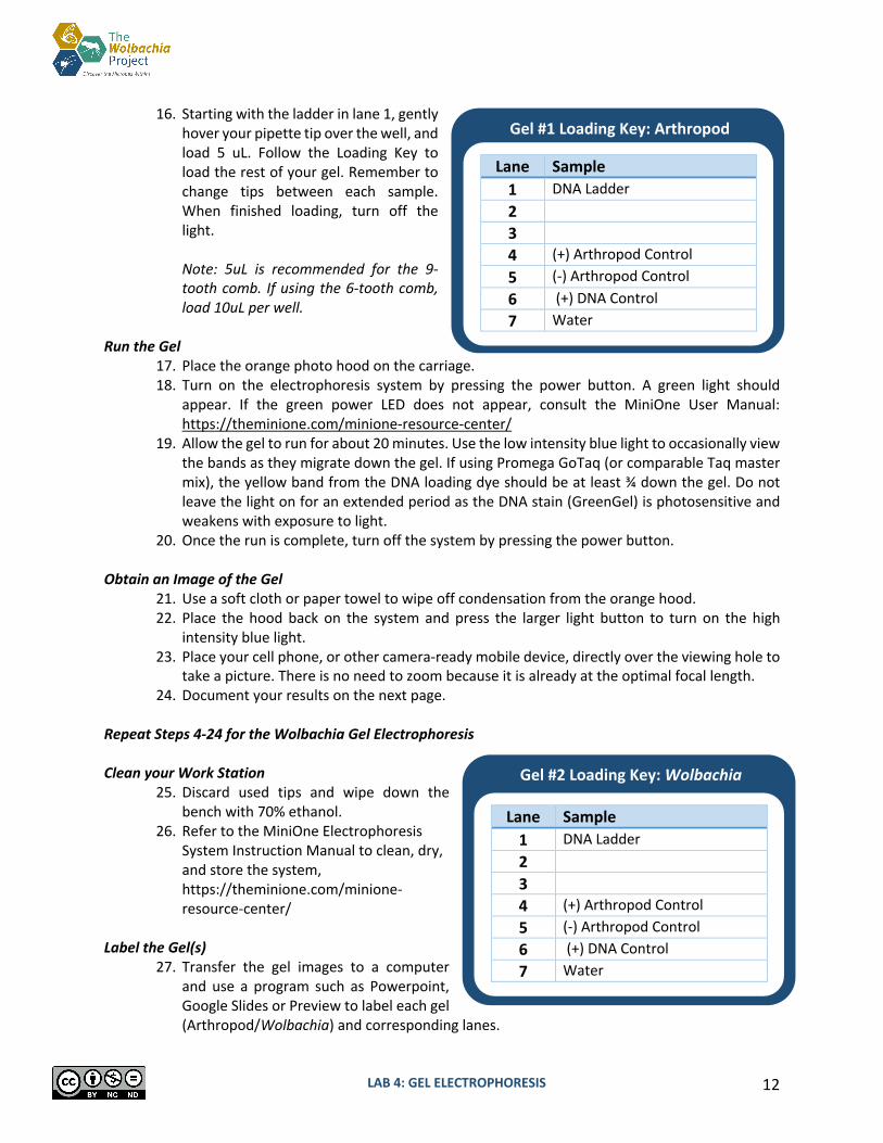
16. Starting with the ladder in lane 1, gently hover your pipette tip over the well, and load 5 uL. Follow the Loading Key to load the rest of your gel. Remember to change tips between each sample. When finished loading, turn off the light.
Note: 5uL is recommended for the 9-tooth comb. If using the 6-tooth comb, load 10uL per well.
Run the Gel
17. Place the orange photo hood on the carriage. 18. Turn on the electrophoresis system by pressing the power button. A green light should
appear. If the green power LED does not appear, consult the MiniOne User Manual: https://theminione.com/minione-resource-center/
19. Allow the gel to run for about 20 minutes. Use the low intensity blue light to occasionally view the bands as they migrate down the gel. If using Promega GoTaq (or comparable Taq master mix), the yellow band from the DNA loading dye should be at least ¾ down the gel. Do not leave the light on for an extended period as the DNA stain (GreenGel) is photosensitive and weakens with exposure to light.
20. Once the run is complete, turn off the system by pressing the power button. Obtain an Image of the Gel
21. Use a soft cloth or paper towel to wipe off condensation from the orange hood. 22. Place the hood back on the system and press the larger light button to turn on the high
intensity blue light. 23. Place your cell phone, or other camera-ready mobile device, directly over the viewing hole to
take a picture. There is no need to zoom because it is already at the optimal focal length. 24. Document your results on the next page.
Repeat Steps 4-24 for the Wolbachia Gel Electrophoresis Clean your Work Station
25. Discard used tips and wipe down the bench with 70% ethanol.
26. Refer to the MiniOne Electrophoresis System Instruction Manual to clean, dry, and store the system, https://theminione.com/minione-resource-center/
Label the Gel(s)
27. Transfer the gel images to a computer and use a program such as Powerpoint, Google Slides or Preview to label each gel (Arthropod/Wolbachia) and corresponding lanes.
Gel #1 Loading Key: Arthropod
Lane Sample 1 DNA Ladder 2 3 4 (+) Arthropod Control 5 (-) Arthropod Control 6 (+) DNA Control 7 Water
Gel #2 Loading Key: Wolbachia
Lane Sample 1 DNA Ladder 2 3 4 (+) Arthropod Control 5 (-) Arthropod Control 6 (+) DNA Control 7 Water

LAB 4: GEL ELECTROPHORESIS 13
Results Use the table below to record presence (+) or absence (-) of bands.
Do your control bands match the expected results you drew in Pre-Lab Question 1? Based on your answer, how confident are you that the experimental results are valid? Why? Complete the table below:
Tube label Arthropod ID Wolbachia-infected? (Yes, No, Unknown) Confidence (High, Low)
Lane DNA Source Arthropod CO1 Band?
Wolbachia 16S Band?
1 DNA Ladder N/A N/A 2 3
4 (+) Arthropod Control 5 (-) Arthropod Control
6 (+) DNA Control
7 Water
LAB 4: GEL ELECTROPHORESIS 14
Post-Lab Questions
1. In gel electrophoresis, pore size depends on agarose content. Higher % gels have smaller pores whereas lower % gels have larger pores. In this lab, you used a 1% agarose gel. What would happen to the DNA if you used a 2% agarose gel, but ran it for the same amount of time?
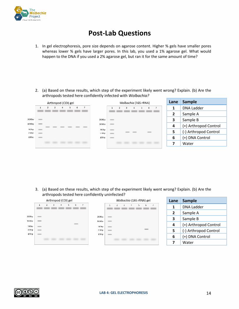
2. (a) Based on these results, which step of the experiment likely went wrong? Explain. (b) Are the arthropods tested here confidently infected with Wolbachia?
3. (a) Based on these results, which step of the experiment likely went wrong? Explain. (b) Are the arthropods tested here confidently uninfected?
Lane Sample 1 DNA Ladder 2 Sample A 3 Sample B 4 (+) Arthropod Control 5 (-) Arthropod Control 6 (+) DNA Control 7 Water
Lane Sample 1 DNA Ladder 2 Sample A 3 Sample B 4 (+) Arthropod Control 5 (-) Arthropod Control 6 (+) DNA Control 7 Water
Note 3.1: You will use 6 tubes because previously purified samples of Wolbachia DNA has been included as a (+) control and water as a (-) control.
LAB 4: GEL ELECTROPHORESIS 15
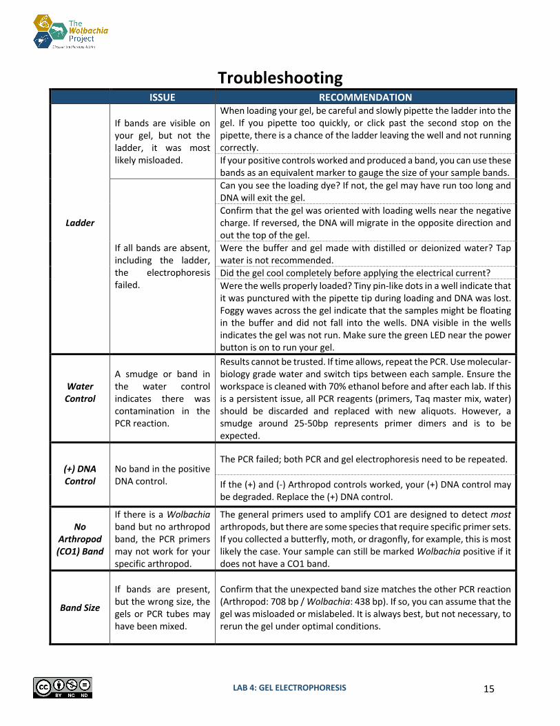
Troubleshooting ISSUE RECOMMENDATION
Ladder
If bands are visible on your gel, but not the ladder, it was most likely misloaded.
When loading your gel, be careful and slowly pipette the ladder into the gel. If you pipette too quickly, or click past the second stop on the pipette, there is a chance of the ladder leaving the well and not running correctly. If your positive controls worked and produced a band, you can use these bands as an equivalent marker to gauge the size of your sample bands.
If all bands are absent, including the ladder, the electrophoresis failed.
Can you see the loading dye? If not, the gel may have run too long and DNA will exit the gel. Confirm that the gel was oriented with loading wells near the negative charge. If reversed, the DNA will migrate in the opposite direction and out the top of the gel. Were the buffer and gel made with distilled or deionized water? Tap water is not recommended. Did the gel cool completely before applying the electrical current? Were the wells properly loaded? Tiny pin-like dots in a well indicate that it was punctured with the pipette tip during loading and DNA was lost. Foggy waves across the gel indicate that the samples might be floating in the buffer and did not fall into the wells. DNA visible in the wells indicates the gel was not run. Make sure the green LED near the power button is on to run your gel.
Water Control
A smudge or band in the water control indicates there was contamination in the PCR reaction.
Results cannot be trusted. If time allows, repeat the PCR. Use molecular-biology grade water and switch tips between each sample. Ensure the workspace is cleaned with 70% ethanol before and after each lab. If this is a persistent issue, all PCR reagents (primers, Taq master mix, water) should be discarded and replaced with new aliquots. However, a smudge around 25-50bp represents primer dimers and is to be expected.
(+) DNA Control
No band in the positive DNA control.
The PCR failed; both PCR and gel electrophoresis need to be repeated.
If the (+) and (-) Arthropod controls worked, your (+) DNA control may be degraded. Replace the (+) DNA control.
No Arthropod (CO1) Band
If there is a Wolbachia band but no arthropod band, the PCR primers may not work for your specific arthropod.
The general primers used to amplify CO1 are designed to detect most arthropods, but there are some species that require specific primer sets. If you collected a butterfly, moth, or dragonfly, for example, this is most likely the case. Your sample can still be marked Wolbachia positive if it does not have a CO1 band.
Band Size
If bands are present, but the wrong size, the gels or PCR tubes may have been mixed.
Confirm that the unexpected band size matches the other PCR reaction (Arthropod: 708 bp / Wolbachia: 438 bp). If so, you can assume that the gel was misloaded or mislabeled. It is always best, but not necessary, to rerun the gel under optimal conditions.
Note 3.1: You will use 6 tubes because previously purified samples of Wolbachia DNA has been included as a (+) control and water as a (-) control.
LAB 4: GEL ELECTROPHORESIS 16
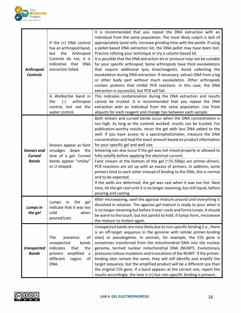
Arthropod Controls
If the (+) DNA control has an arthropod band, but the Arthropod Controls do not, it is indicative that DNA extraction failed.
It is recommended that you repeat the DNA extraction with an individual from the same population. The most likely culprit is lack of appropriately lysed cells. Increase grinding time with the pestle. If using a pellet-based DNA extraction kit, the DNA pellet may have been lost. Practice refining your technique or try a column-based kit. It is possible that the DNA extraction kit or protocol may not be suitable for your specific arthropod. Some arthropods have thick exoskeletons that require additional lysis time/reagents. Avoid collecting the exoskeleton during DNA extraction. If necessary, extract DNA from a leg or other body part without much exoskeleton. Other arthropods contain proteins that inhibit PCR reactions. In this case, the DNA extraction is successful, but PCR will fail.
A Wolbachia band in the (-) arthropod control, but not the water control.
This indicates contamination during the DNA extraction and results cannot be trusted. It is recommended that you repeat the DNA extraction with an individual from the same population. Use fresh aliquots for each reagent and change tips between each sample.
Smears and Curved Bands
Smears appear as faint smudges down the lane of a gel. Curved bands appear “smiley” or U-shaped.
Both smears and curved bands occur when the DNA concentration is too high. As long as the controls worked, results can be trusted. For publication-worthy results, rerun the gel with less DNA added to the well. If you have access to a spectrophotometer, measure the DNA concentration and load the exact amount based on product information for your specific gel and well size. Smearing can also occur if the gel was not mixed properly or allowed to fully solidify before applying the electrical current. Faint smears at the bottom of the gel (~25-50bp) are primer-dimers. PCR reactions are set up with an excess of primers. In addition, some primers bind to each other instead of binding to the DNA, this is normal and to be expected. If the wells are deformed, the gel was cast when it was too hot. Next time, let the gel cool until it is no longer steaming, but still liquid, before pouring and casting.
Lumps in the gel
Lumps in the gel indicate that it was too cold when poured/cast.
After microwaving, swirl the agarose mixture around until everything is dissolved in solution. The agarose gel mixture is ready to pour when it is no longer steaming but before it over-cools and forms lumps. It should be warm to the touch, but not painful to hold. If lumps form, microwave the mixture to molten again.
Unexpected Bands
The presence of unexpected bands indicates that the primers amplified a different region of DNA.
Unexpected bands are most likely due to non-specific binding (i.e., there is an off-target sequence in the genome with similar primer-binding sites) or pseudogenes. In animals, for example, the COI gene is sometimes transferred from the mitochondrial DNA into the nuclear genome, termed nuclear mitochondrial DNA (NUMT). Evolutionary pressures induce mutations and truncations of the NUMT. If the primer-binding sites remain the same, they will still identify and amplify the target sequence, but the amplified product will be a different size than the original COI gene. If a band appears at the correct size, report the results accordingly: the lane is (+) but non-specific binding is present.

LAB 4: GEL ELECTROPHORESIS 17
Database Entry
After completing the Gel Electrophoresis Lab, open your entries in The Wolbachia Project Database and record Methods and Results. A comprehensive guide is located under the Resources tab.
https://wolbachiaprojectdb.org/
Observations Methods
ü DNA extraction kit ü DNA extraction location ü Taq polymerase used ü Single/duplex PCR reaction q Upload gel image q Gel electrophoresis system q Buffer q DNA stain q Update protocol notes
Results q Wolbachia positive? q Confidence level q Explain confidence level
Database Fields to Complete
LAB 4: GEL ELECTROPHORESIS 18
Glossary
Agarose: A polysaccharide purified from seaweed. When dry agarose is boiled in a buffer solution, it will harden into a flexible, gelatin-like slab when it cools.
Band: A clearly visible and defined mark on a gel, indicating DNA presence. The location of the band in relation to reference bands in the ladder indicates the size of the DNA product amplified in PCR.
DNA Stain: A dye that binds to the DNA, making it visible in UV or blue light. Electrophoresis: A method of separating substances based on the rate of movement while under
the influence of an electric field. Ladder: A mix of pre-cut DNA of defined sizes. Each ladder will have different “rungs” of pre-
determined sizes; check the product information to find the sizes of DNA for your specific ladder.
Lane: The vertical path of DNA migration below each loading well. Loading Dye: Loading dye is added to aid in loading of an agarose gel. It contains colored dye and
glycerol. Loading Well: An indentation in the agarose gel in which samples are loaded. Negative Control: Ensures the process and samples are not contaminated, it is designed to
produce a negative result. Positive Control: A well-understood variable; should result in an expected positive result. Primer Dimers: Primers that bind to each other instead of the target DNA during PCR. This is the
“haze” seen at the bottom of the gel, ~25-50bp. This area may also include excess primers that were not used during PCR.
Running Buffer: A conductive liquid that allows the DNA to migrate through the agarose gel.