Embed Size (px)
Citation preview
Clinical Practice Guidelines
DAVID S. LOGERSTEDT, PT, PhD • DAVID A. SCALZITTI, PT, PhD • KIM L. BENNELL, PT, PhD • RANA S. HINMAN, PT, PhDHOLLY SILVERS-GRANELLI, PT, PhD • JAY EBERT, PhD • KAREN HAMBLY, PT, PhD • JAMES L. CAREY, MD, MPH
LYNN SNYDER-MACKLER, PT, ScD, FAPTA • MICHAEL J. AXE, MD • CHRISTINE M. MCDONOUGH, PT, PhD
Knee Pain and Mobility Impairments: Meniscal and Articular Cartilage Lesions
Revision 2018Clinical Practice Guidelines Linked to the
International Classification of Functioning, Disability and Health From the Orthopaedic Section
of the American Physical Therapy AssociationJ Orthop Sports Phys Ther. 2018;48(2):A1-A50. doi:10.2519/jospt.2018.0301
REVIEWERS: Paul Beattie, PT, PhD • John DeWitt, DPT • Amanda Ferland, DPT • Jennifer S. Howard, ATC, PhD Sandra Kaplan, PT, PhD • David Killoran, PhD • Laura Schmitt, PT, PhD • Jonas Bloch Thorlund, PhD • Leslie Torburn, DPT
For author, coordinator, contributor, and reviewer affiliations, see end of text. ©2018 Orthopaedic Section, American Physical Therapy Association (APTA), Inc, and the Journal of Orthopaedic & Sports Physical Therapy. The Orthopaedic Section, APTA, Inc, and the Journal of Orthopaedic & Sports Physical Therapy consent to the reproduction and distribution of this guideline for educational purposes. Address correspondence to Brenda Johnson, ICF-Based Clinical Practice Guidelines Coordinator, Orthopaedic Section, APTA, Inc, 2920 East Avenue South, Suite 200, La Crosse, WI 54601. E-mail: [email protected]
SUMMARY OF RECOMMENDATIONS . . . . . . . . . . . . . . . . . . . . . . . . . . . . . . A2
INTRODUCTION. . . . . . . . . . . . . . . . . . . . . . . . . . . . . . . . . . . . . . . . . . . . . . . . . . . . . . . . . . . . A3
METHODS . . . . . . . . . . . . . . . . . . . . . . . . . . . . . . . . . . . . . . . . . . . . . . . . . . . . . . . . . . . . . . . . . . . A4
CLINICAL GUIDELINES: Impairment/Function-Based Diagnosis . . . . . . . . . . . . . . . . . . A7
CLINICAL GUIDELINES: Examination . . . . . . . . . . . . . . . . . . . . . . . . . . . . . . . . . . . . . . . . . . . . . . . . . . . . . . . . . . A20
CLINICAL GUIDELINES: Interventions . . . . . . . . . . . . . . . . . . . . . . . . . . . . . . . . . . . . . . . . . . . . . . . . . . . . . . . . . . . A23
AUTHOR/REVIEWER AFFILIATIONS AND CONTACTS . . . . . . A27
REFERENCES . . . . . . . . . . . . . . . . . . . . . . . . . . . . . . . . . . . . . . . . . . . . . . . . . . . . . . . . . . . . . A28
Jour
nal o
f Orth
opae
dic
& S
ports
Phy
sica
l The
rapy
®
Dow
nloa
ded
from
ww
w.jo
spt.o
rg a
t on
Febr
uary
8, 2
018.
For
per
sona
l use
onl
y. N
o ot
her u
ses w
ithou
t per
mis
sion
. C
opyr
ight
© 2
018
Jour
nal o
f Orth
opae
dic
& S
ports
Phy
sica
l The
rapy
®. A
ll rig
hts r
eser
ved.
Knee Pain and Mobility Impairments: Clinical Practice Guidelines Revision 2018
a2 | february 2018 | volume 48 | number 2 | journal of orthopaedic & sports physical therapy
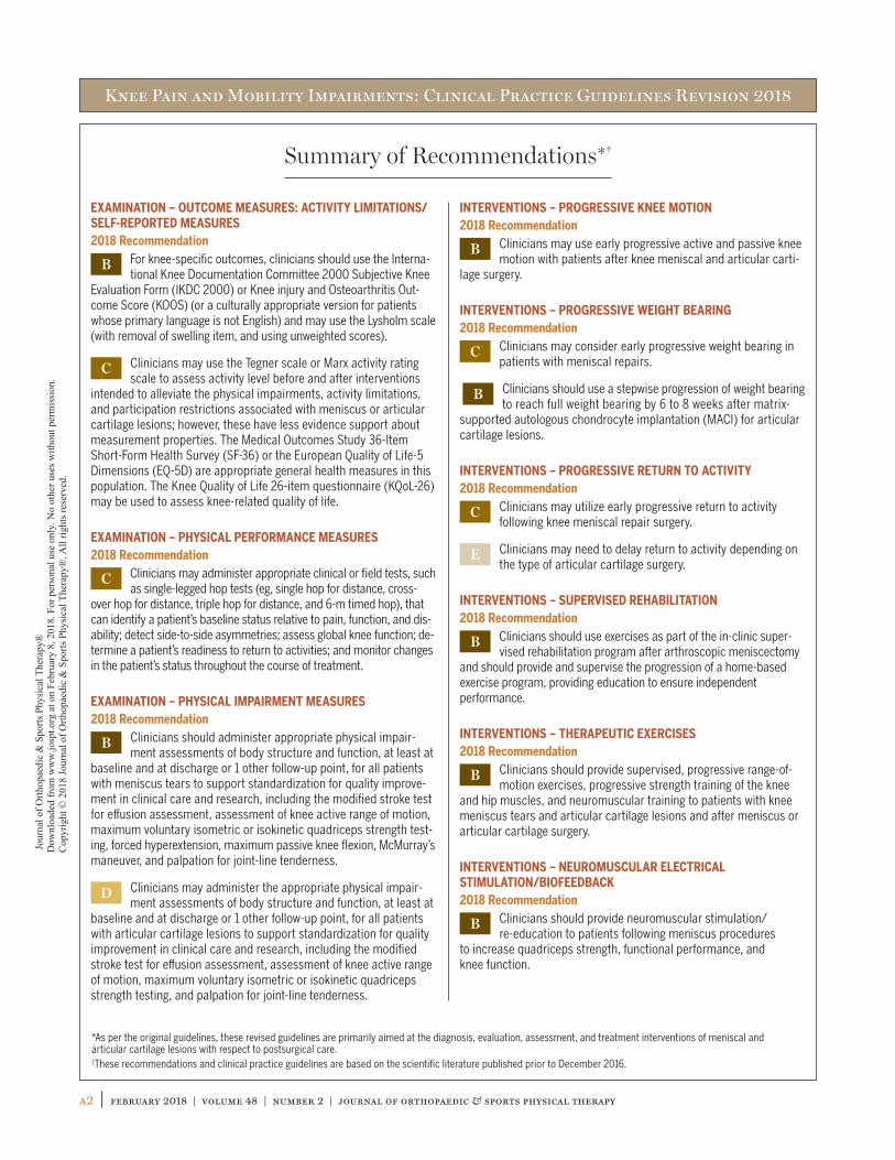
EXAMINATION – OUTCOME MEASURES: ACTIVITY LIMITATIONS/SELF-REPORTED MEASURES2018 Recommendation
B For knee-specific outcomes, clinicians should use the Interna-tional Knee Documentation Committee 2000 Subjective Knee
Evaluation Form (IKDC 2000) or Knee injury and Osteoarthritis Out-come Score (KOOS) (or a culturally appropriate version for patients whose primary language is not English) and may use the Lysholm scale (with removal of swelling item, and using unweighted scores).
C Clinicians may use the Tegner scale or Marx activity rating scale to assess activity level before and after interventions
intended to alleviate the physical im pairments, activity limitations, and participation restrictions associated with meniscus or articular cartilage lesions; however, these have less evidence support about measurement properties. The Medical Outcomes Study 36-Item Short-Form Health Survey (SF-36) or the European Quality of Life-5 Dimensions (EQ-5D) are appropriate general health measures in this population. The Knee Quality of Life 26-item questionnaire (KQoL-26) may be used to assess knee-related quality of life.
EXAMINATION – PHYSICAL PERFORMANCE MEASURES2018 Recommendation
C Clinicians may administer appropriate clinical or field tests, such as single-legged hop tests (eg, single hop for distance, cross-
over hop for distance, triple hop for distance, and 6-m timed hop), that can identify a patient’s baseline status relative to pain, function, and dis-ability; detect side-to-side asymmetries; assess global knee function; de-termine a patient’s readiness to return to activities; and monitor changes in the patient’s status throughout the course of treatment.
EXAMINATION – PHYSICAL IMPAIRMENT MEASURES2018 Recommendation
B Clinicians should administer appropriate physical impair-ment assessments of body structure and function, at least at
baseline and at discharge or 1 other follow-up point, for all patients with meniscus tears to support standardization for quality improve-ment in clinical care and research, including the modified stroke test for effusion assessment, assessment of knee active range of motion, maximum voluntary isometric or isokinetic quadriceps strength test-ing, forced hyperextension, maximum passive knee flexion, McMurray’s maneuver, and palpation for joint-line tenderness.
D Clinicians may administer the appropriate physical impair-ment assessments of body structure and function, at least at
baseline and at discharge or 1 other follow-up point, for all patients with articular cartilage lesions to support standardization for quality improvement in clinical care and research, including the modified stroke test for effusion assessment, assessment of knee active range of motion, maximum voluntary isometric or isokinetic quadriceps strength testing, and palpation for joint-line tenderness.
INTERVENTIONS – PROGRESSIVE KNEE MOTION2018 Recommendation
B Clinicians may use early progressive active and passive knee mo tion with patients after knee meniscal and articular carti-
lage surgery.
INTERVENTIONS – PROGRESSIVE WEIGHT BEARING2018 Recommendation
C Clinicians may consider early progressive weight bearing in patients with meniscal repairs.
B Clinicians should use a stepwise progression of weight bearing to reach full weight bearing by 6 to 8 weeks after matrix-
supported autologous chondrocyte implantation (MACI) for articular cartilage lesions.
INTERVENTIONS – PROGRESSIVE RETURN TO ACTIVITY2018 Recommendation
C Clinicians may utilize early progressive return to activity following knee meniscal repair surgery.
E Clinicians may need to delay return to activity depending on the type of articular cartilage surgery.
INTERVENTIONS – SUPERVISED REHABILITATION2018 Recommendation
B Clinicians should use exercises as part of the in-clinic super-vised rehabilitation program after arthroscopic meniscectomy
and should provide and supervise the progression of a home-based exercise program, providing education to ensure independent performance.
INTERVENTIONS – THERAPEUTIC EXERCISES2018 Recommendation
B Clinicians should provide supervised, progressive range-of-motion exercises, progressive strength training of the knee
and hip muscles, and neuromuscular training to patients with knee meniscus tears and articular cartilage lesions and after meniscus or articular cartilage surgery.
INTERVENTIONS – NEUROMUSCULAR ELECTRICAL STIMULATION/BIOFEEDBACK2018 Recommendation
B Clinicians should provide neuromuscular stimulation/ re-education to patients following meniscus procedures
to increase quadriceps strength, functional performance, and knee function.
Summary of Recommendations*†
*As per the original guidelines, these revised guidelines are primarily aimed at the diagnosis, evaluation, assessment, and treatment interventions of meniscal and articular cartilage lesions with respect to postsurgical care.†These recommendations and clinical practice guidelines are based on the scientific literature published prior to December 2016.
Jour
nal o
f Orth
opae
dic
& S
ports
Phy
sica
l The
rapy
®
Dow
nloa
ded
from
ww
w.jo
spt.o
rg a
t on
Febr
uary
8, 2
018.
For
per
sona
l use
onl
y. N
o ot
her u
ses w
ithou
t per
mis
sion
. C
opyr
ight
© 2
018
Jour
nal o
f Orth
opae
dic
& S
ports
Phy
sica
l The
rapy
®. A
ll rig
hts r
eser
ved.
Knee Pain and Mobility Impairments: Clinical Practice Guidelines Revision 2018
journal of orthopaedic & sports physical therapy | volume 48 | number 2 | february 2018 | a3
List of Abbreviations
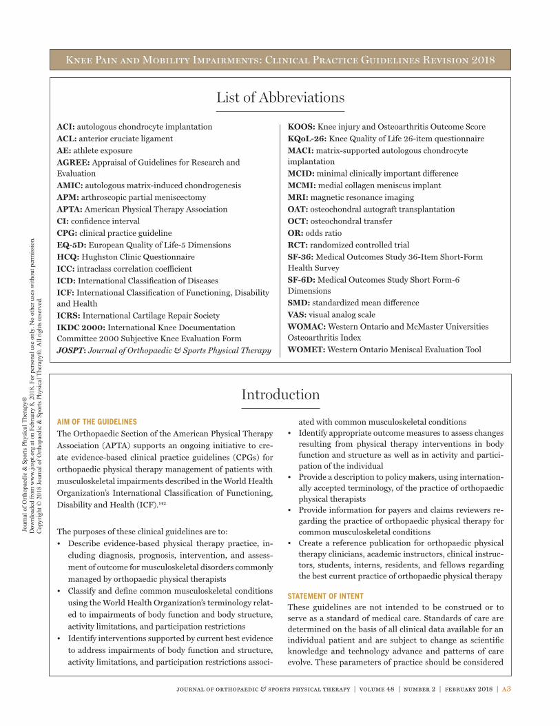
ACI: autologous chondrocyte implantationACL: anterior cruciate ligamentAE: athlete exposureAGREE: Appraisal of Guidelines for Research and EvaluationAMIC: autologous matrix-induced chondrogenesisAPM: arthroscopic partial meniscectomyAPTA: American Physical Therapy AssociationCI: confidence intervalCPG: clinical practice guidelineEQ-5D: European Quality of Life-5 DimensionsHCQ: Hughston Clinic QuestionnaireICC: intraclass correlation coefficientICD: International Classification of DiseasesICF: International Classification of Functioning, Disability and HealthICRS: International Cartilage Repair SocietyIKDC 2000: International Knee Documentation Committee 2000 Subjective Knee Evaluation FormJOSPT: Journal of Orthopaedic & Sports Physical Therapy
KOOS: Knee injury and Osteoarthritis Outcome ScoreKQoL-26: Knee Quality of Life 26-item questionnaireMACI: matrix-supported autologous chondrocyte implantationMCID: minimal clinically important differenceMCMI: medial collagen meniscus implantMRI: magnetic resonance imagingOAT: osteochondral autograft transplantationOCT: osteochondral transferOR: odds ratioRCT: randomized controlled trialSF-36: Medical Outcomes Study 36-Item Short-Form Health SurveySF-6D: Medical Outcomes Study Short Form-6 DimensionsSMD: standardized mean differenceVAS: visual analog scaleWOMAC: Western Ontario and McMaster Universities Osteoarthritis IndexWOMET: Western Ontario Meniscal Evaluation Tool
AIM OF THE GUIDELINES
The Orthopaedic Section of the American Physical Therapy Association (APTA) supports an ongoing initiative to cre-ate evidence-based clinical practice guidelines (CPGs) for orthopaedic physical therapy management of patients with musculoskeletal impairments described in the World Health Organization’s International Classification of Functioning, Disability and Health (ICF).142
The purposes of these clinical guidelines are to:• Describe evidence-based physical therapy practice, in-
cluding diagnosis, prognosis, intervention, and assess-ment of outcome for musculoskeletal disorders commonly managed by orthopaedic physical therapists
• Classify and define common musculoskeletal conditions using the World Health Organization’s terminology relat-ed to impairments of body function and body structure, activity limitations, and participation restrictions
• Identify interventions supported by current best evidence to address impairments of body function and structure, activity limitations, and participation restrictions associ-
ated with common musculoskeletal conditions• Identify appropriate outcome measures to assess changes
resulting from physical therapy interventions in body function and structure as well as in activity and partici-pation of the individual
• Provide a description to policy makers, using internation-ally accepted terminology, of the practice of orthopaedic physical therapists
• Provide information for payers and claims reviewers re-garding the practice of orthopaedic physical therapy for common musculoskeletal conditions
• Create a reference publication for orthopaedic physical therapy clinicians, academic instructors, clinical instruc-tors, students, interns, residents, and fellows regarding the best current practice of orthopaedic physical therapy
STATEMENT OF INTENTThese guidelines are not intended to be construed or to serve as a standard of medical care. Standards of care are determined on the basis of all clinical data available for an individual patient and are subject to change as scientific knowledge and technology advance and patterns of care evolve. These parameters of practice should be considered
Introduction
Jour
nal o
f Orth
opae
dic
& S
ports
Phy
sica
l The
rapy
®
Dow
nloa
ded
from
ww
w.jo
spt.o
rg a
t on
Febr
uary
8, 2
018.
For
per
sona
l use
onl
y. N
o ot
her u
ses w
ithou
t per
mis
sion
. C
opyr
ight
© 2
018
Jour
nal o
f Orth
opae
dic
& S
ports
Phy
sica
l The
rapy
®. A
ll rig
hts r
eser
ved.

Knee Pain and Mobility Impairments: Clinical Practice Guidelines Revision 2018
a4 | february 2018 | volume 48 | number 2 | journal of orthopaedic & sports physical therapy
guidelines only. Adherence to them will not ensure a suc-cessful outcome in every patient, nor should they be con-strued as including all proper methods of care or excluding other acceptable methods of care aimed at the same results. The ultimate judgment regarding a particular clinical pro-cedure or treatment plan must be made based on clinician experience and expertise in light of the clinical presentation of the patient, the available evidence, available diagnostic and treatment options, and the patient’s values, expecta-tions, and preferences. However, we suggest that significant departures from accepted guidelines should be documented in the patient’s medical records at the time the relevant clin-ical decision is made.
SCOPEThe aims of the revision were to provide a concise summary of the evidence since publication of the original guideline in 2010 and to develop new recommendations or revise previ-ously published recommendations to support evidence-based practice. The original guidelines were primarily aimed at the diagnosis, evaluation, assessment, and treatment interven-tions of meniscus and articular cartilage lesions with respect to postsurgical care, and this revision builds on the original guidelines. The state of the literature in the nonoperative management of meniscus and articular cartilage lesions is rapidly evolving and will be explored and presented in the next iteration of this CPG.
Introduction (continued)
Content experts with relevant physical therapy, medical, and surgical expertise were appointed by the Orthopaedic Section, APTA, Inc to conduct a review of the literature and to develop an updated Knee Pain and Mobility Impairments Meniscal and Articular Cartilage Lesions CPG as indicated by the current state of the evidence in the field. Four au-thors of this guideline revision completed the Appraisal of Guidelines for Research and Evaluation (AGREE) II tool to assess the quality and reporting of the CPG published in 2010, and to identify areas for improvement. The authors of this guideline revision worked with the CPG Editors and medical librarians for methodological guidance. The re-search librarians were chosen for their expertise in system-atic review rehabilitation literature search, and to perform systematic searches for concepts associated with meniscus and articular cartilage injuries of the knee in articles pub-lished from 2008 related to classification, examination, and intervention strategies consistent with previous guideline development methods related to ICF classification.91 Briefly, the following databases were searched from 2008 to De-cember 31, 2016: MEDLINE (PubMed, 2008 to date), Sco-pus (Elsevier BV, 2008 to date), CINAHL (EBSCO, 2008 to date), SPORTDiscus (EBSCO, 2008 to date), and Cochrane Library (Wiley, 2008 to date). (See APPENDIX A for full search strategies and APPENDIX B for search dates and results, avail-able at www.orthopt.org.)
The authors declared relationships and developed a conflict management plan that included submitting a Conflict of In-terest form to the Orthopaedic Section, APTA, Inc. Articles that were authored by a reviewer were assigned to an alter-
nate reviewer. Funding was provided to the CPG develop-ment team for travel and expenses for CPG development training by the Orthopaedic Section, APTA, Inc. The CPG development team maintained editorial independence.
Articles contributing to recommendations were reviewed based on specified inclusion and exclusion criteria with the goal of identifying evidence relevant to physical therapist clinical decision making for adult persons with knee pain and mobility impairments/knee meniscal/articular cartilage lesions. The title and abstract of each article were reviewed independently by 2 members of the CPG development team for inclusion. (See APPENDIX C for inclusion and exclusion criteria, available at www.orthopt.org.) Full-text review was then similarly conducted to obtain the final set of articles for contribution to recommendations. The team leader (D.S.L.) provided the final decision for discrepancies that were not re-solved by the review team. (See APPENDIX D for a flow chart of articles and APPENDIX E for articles included in recommenda-tions by topic, available at www.orthopt.org.) For selected rel-evant topics that were not appropriate for the development of recommendations, such as incidence and imaging, articles were not subject to the systematic review process and were not included in the flow chart. Evidence tables for this CPG are available on the Clinical Practice Guidelines page of the Orthopaedic Section of the APTA website: www.orthopt.org.
This guideline was issued in 2018 based on the published literature up to December 2016, and will be considered for review in 2022, or sooner if new evidence becomes available that may change the recommendations. Any updates to the
Methods
Jour
nal o
f Orth
opae
dic
& S
ports
Phy
sica
l The
rapy
®
Dow
nloa
ded
from
ww
w.jo
spt.o
rg a
t on
Febr
uary
8, 2
018.
For
per
sona
l use
onl
y. N
o ot
her u
ses w
ithou
t per
mis
sion
. C
opyr
ight
© 2
018
Jour
nal o
f Orth
opae
dic
& S
ports
Phy
sica
l The
rapy
®. A
ll rig
hts r
eser
ved.
Knee Pain and Mobility Impairments: Clinical Practice Guidelines Revision 2018
journal of orthopaedic & sports physical therapy | volume 48 | number 2 | february 2018 | a5
guideline in the interim period will be noted on the Ortho-paedic Section of the APTA website: www.orthopt.org.
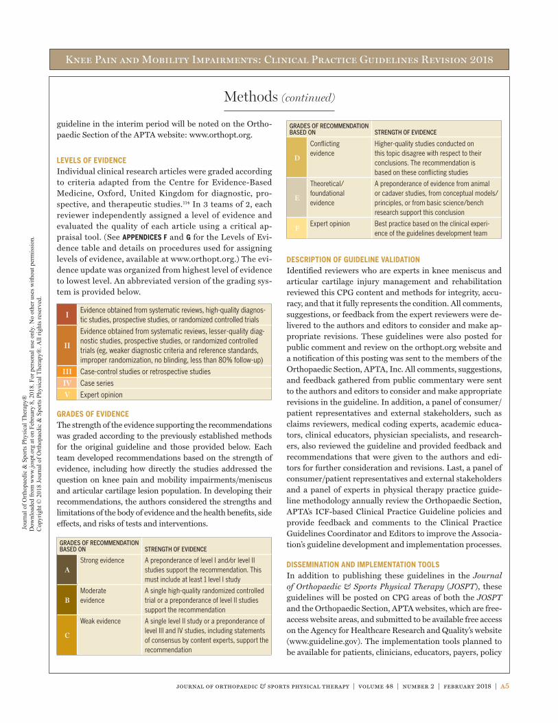
LEVELS OF EVIDENCEIndividual clinical research articles were graded according to criteria adapted from the Centre for Evidence-Based Medicine, Oxford, United Kingdom for diagnostic, pro-spective, and therapeutic studies.114 In 3 teams of 2, each reviewer independently assigned a level of evidence and evaluated the quality of each article using a critical ap-praisal tool. (See APPENDICES F and G for the Levels of Evi-dence table and details on procedures used for assigning levels of evidence, available at www.orthopt.org.) The evi-dence update was organized from highest level of evidence to lowest level. An abbreviated version of the grading sys-tem is provided below.
IEvidence obtained from systematic reviews, high-quality diagnos-tic studies, prospective studies, or randomized controlled trials
II
Evidence obtained from systematic reviews, lesser-quality diag-nostic studies, prospective studies, or randomized controlled trials (eg, weaker diagnostic criteria and reference standards, improper randomization, no blinding, less than 80% follow-up)
III Case-control studies or retrospective studies
IV Case series
V Expert opinion
GRADES OF EVIDENCEThe strength of the evidence supporting the recommendations was graded according to the previously established methods for the original guideline and those provided below. Each team developed recommendations based on the strength of evidence, including how directly the studies addressed the question on knee pain and mobility impairments/meniscus and articular cartilage lesion population. In developing their recommendations, the authors considered the strengths and limitations of the body of evidence and the health benefits, side effects, and risks of tests and interventions.
GRADES OF RECOMMENDATION BASED ON STRENGTH OF EVIDENCE
AStrong evidence A preponderance of level I and/or level II
studies support the recommendation. This must include at least 1 level I study
BModerate evidence
A single high-quality randomized controlled trial or a preponderance of level II studies support the recommendation
C
Weak evidence A single level II study or a preponderance of level III and IV studies, including statements of consensus by content experts, support the recommendation
GRADES OF RECOMMENDATION BASED ON STRENGTH OF EVIDENCE
D
Conflicting evidence
Higher-quality studies conducted on this topic disagree with respect to their conclusions. The recommendation is based on these conflicting studies
E
Theoretical/ foundational evidence
A preponderance of evidence from animal or cadaver studies, from conceptual models/principles, or from basic science/bench research support this conclusion
FExpert opinion Best practice based on the clinical experi-
ence of the guidelines development team
DESCRIPTION OF GUIDELINE VALIDATIONIdentified reviewers who are experts in knee meniscus and articular cartilage injury management and rehabilitation reviewed this CPG content and methods for integrity, accu-racy, and that it fully represents the condition. All comments, suggestions, or feedback from the expert reviewers were de-livered to the authors and editors to consider and make ap-propriate revisions. These guidelines were also posted for public comment and review on the orthopt.org website and a notification of this posting was sent to the members of the Orthopaedic Section, APTA, Inc. All comments, suggestions, and feedback gathered from public commentary were sent to the authors and editors to consider and make appropriate revisions in the guideline. In addition, a panel of consumer/patient representatives and external stakeholders, such as claims reviewers, medical coding experts, academic educa-tors, clinical educators, physician specialists, and research-ers, also reviewed the guideline and provided feedback and recommendations that were given to the authors and edi-tors for further consideration and revisions. Last, a panel of consumer/patient representatives and external stakeholders and a panel of experts in physical therapy practice guide-line methodology annually review the Orthopaedic Section, APTA’s ICF-based Clinical Practice Guideline policies and provide feedback and comments to the Clinical Practice Guidelines Coordinator and Editors to improve the Associa-tion’s guideline development and implementation processes.
DISSEMINATION AND IMPLEMENTATION TOOLSIn addition to publishing these guidelines in the Journal of Orthopaedic & Sports Physical Therapy (JOSPT), these guidelines will be posted on CPG areas of both the JOSPT and the Orthopaedic Section, APTA websites, which are free-access website areas, and submitted to be available free access on the Agency for Healthcare Research and Quality’s website (www.guideline.gov). The implementation tools planned to be available for patients, clinicians, educators, payers, policy
Methods (continued)
Jour
nal o
f Orth
opae
dic
& S
ports
Phy
sica
l The
rapy
®
Dow
nloa
ded
from
ww
w.jo
spt.o
rg a
t on
Febr
uary
8, 2
018.
For
per
sona
l use
onl
y. N
o ot
her u
ses w
ithou
t per
mis
sion
. C
opyr
ight
© 2
018
Jour
nal o
f Orth
opae
dic
& S
ports
Phy
sica
l The
rapy
®. A
ll rig
hts r
eser
ved.
Knee Pain and Mobility Impairments: Clinical Practice Guidelines Revision 2018
a6 | february 2018 | volume 48 | number 2 | journal of orthopaedic & sports physical therapy
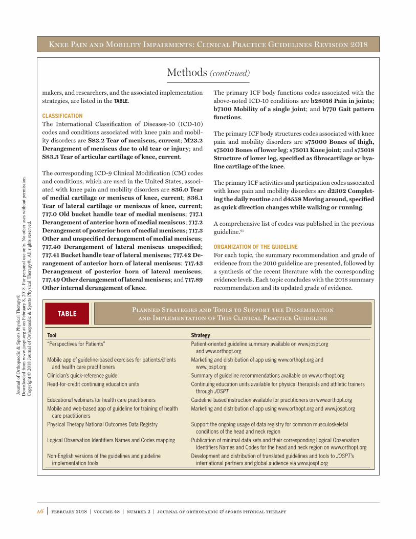
makers, and researchers, and the associated implementation strategies, are listed in the TABLE.
CLASSIFICATIONThe International Classification of Diseases-10 (ICD-10) codes and conditions associated with knee pain and mobil-ity disorders are S83.2 Tear of meniscus, current; M23.2 Derangement of meniscus due to old tear or injury; and S83.3 Tear of articular cartilage of knee, current.
The corresponding ICD-9 Clinical Modification (CM) codes and conditions, which are used in the United States, associ-ated with knee pain and mobility disorders are 836.0 Tear of medial cartilage or meniscus of knee, current; 836.1 Tear of lateral cartilage or meniscus of knee, current; 717.0 Old bucket handle tear of medial meniscus; 717.1 Derangement of anterior horn of medial meniscus; 717.2 Derangement of posterior horn of medial meniscus; 717.3 Other and unspecified derangement of medial meniscus; 717.40 Derangement of lateral meniscus unspecified; 717.41 Bucket handle tear of lateral meniscus; 717.42 De-rangement of anterior horn of lateral meniscus; 717.43 Derangement of posterior horn of lateral meniscus; 717.49 Other derangement of lateral meniscus; and 717.89 Other internal derangement of knee.
The primary ICF body functions codes associated with the above-noted ICD-10 conditions are b28016 Pain in joints; b7100 Mobility of a single joint; and b770 Gait pattern functions.
The primary ICF body structures codes associated with knee pain and mobility disorders are s75000 Bones of thigh, s75010 Bones of lower leg; s75011 Knee joint; and s75018 Structure of low er leg, specified as fibrocartilage or hya-line cartilage of the knee.
The primary ICF activities and participation codes associated with knee pain and mobility disorders are d2302 Complet-ing the daily routine and d4558 Moving around, specified as quick direction changes while walking or running.
A comprehensive list of codes was published in the previous guideline.91
ORGANIZATION OF THE GUIDELINEFor each topic, the summary recommendation and grade of evidence from the 2010 guideline are presented, followed by a synthesis of the recent literature with the corresponding evidence levels. Each topic concludes with the 2018 summary recommendation and its updated grade of evidence.
Methods (continued)
TABLEPlanned Strategies and Tools to Support the Dissemination
and Implementation of This Clinical Practice Guideline
Tool Strategy
“Perspectives for Patients” Patient-oriented guideline summary available on www.jospt.org and www.orthopt.org
Mobile app of guideline-based exercises for patients/clients and health care practitioners
Marketing and distribution of app using www.orthopt.org and www.jospt.org
Clinician’s quick-reference guide Summary of guideline recommendations available on www.orthopt.org
Read-for-credit continuing education units Continuing education units available for physical therapists and athletic trainers through JOSPT
Educational webinars for health care practitioners Guideline-based instruction available for practitioners on www.orthopt.org
Mobile and web-based app of guideline for training of health care practitioners
Marketing and distribution of app using www.orthopt.org and www.jospt.org
Physical Therapy National Outcomes Data Registry Support the ongoing usage of data registry for common musculoskeletal conditions of the head and neck region
Logical Observation Identifiers Names and Codes mapping Publication of minimal data sets and their corresponding Logical Observation Identifiers Names and Codes for the head and neck region on www.orthopt.org
Non-English versions of the guidelines and guideline implementation tools
Development and distribution of translated guidelines and tools to JOSPT’s international partners and global audience via www.jospt.org
Jour
nal o
f Orth
opae
dic
& S
ports
Phy
sica
l The
rapy
®
Dow
nloa
ded
from
ww
w.jo
spt.o
rg a
t on
Febr
uary
8, 2
018.
For
per
sona
l use
onl
y. N
o ot
her u
ses w
ithou
t per
mis
sion
. C
opyr
ight
© 2
018
Jour
nal o
f Orth
opae
dic
& S
ports
Phy
sica
l The
rapy
®. A
ll rig
hts r
eser
ved.
Knee Pain and Mobility Impairments: Clinical Practice Guidelines Revision 2018
journal of orthopaedic & sports physical therapy | volume 48 | number 2 | february 2018 | a7
INCIDENCE2010 SummaryMeniscusInjuries to the menisci are the second most common injury to the knee, with a prevalence of 12% to 14% and an incidence of 61 cases per 100 000 persons.96,128 A high incidence of menis-cal tears occur with injury to the anterior cruciate ligament (ACL), ranging from 22% to 86%.105 In the United States, 10% to 20% of all orthopaedic surgeries consist of surgery to the meniscus on an estimated 850 000 patients each year.117
Articular CartilageBased on studies of knee arthroscopies, the prevalence of ar-ticular cartilage pathologies is reported to be between 60% and 70%.8,69 The incidence of isolated articular cartilage le-sions (30%) is lower than that of nonisolated cartilage le-sions.139 Thirty-two percent to 58% of all articular cartilage lesions are the result of a traumatic, noncontact mechanism of injury.74,139 Sixty-four percent of all chondral lesions were less than 1 cm2.139 Thirty-three percent to 60% of articu-lar cartilage lesions are greater than grade 3 lesions on the International Cartilage Repair Society (ICRS) grading sys-tem.36,130 The ICRS cartilage injury classification consists of 5 grading levels, from grade 0 (normal cartilage without no-table defects) to grade 4 (severely abnormal, full-thickness osteochondral injury).21 The most frequent localizations of cartilage lesions were to the medial femoral condyle and the patellar articular surface.139 Medial meniscal tears (37%) and ACL ruptures (36%) were the most common injuries con-comitant with articular cartilage injuries.
Evidence UpdateMeniscusTear patterns of the knee meniscus can be classified as either traumatic tears or degenerative tears.46 Younger active par-ticipants are more likely to sustain traumatic meniscus inju-ries, such as longitudinal or radial tears. Older individuals are more likely to have degenerative tears, such as horizontal cleavages, flap or complex tears, or meniscal maceration or destruction.46
IIIn active-duty US military service personnel, Jones et al75 reported an unadjusted incidence rate of 8.27 per 1000 person-years (95% CI: 8.22, 8.32) for
acute meniscal injury. For men, the adjusted rate per 1000 person-years was 7.08 and for women was 6.02. Oldest ser-vice personnel (older than 40 years of age) had more than 4 times (4.25) the adjusted rate of meniscus tears compared to youngest (less than 20 years of age) service personnel.
IIIYeh et al146 identified 129 isolated meniscus tears over a 21-season span in 1797 professional basket-ball players. One hundred eleven injuries (86.7%)
were the result of a single incident. Lateral meniscus tears were involved in 59.2% and medial meniscus tears were in-volved in 40.8% of cases. Isolated tears accounted for 87.8% of cases, whereas 12.2% of cases were concomitant with a ligamentous injury. They reported an overall clinical inci-dence of 8.2 meniscus tears per 100 athletes. Lateral menis-cus tears were more likely to occur in younger athletes (younger than or equal to 30 years of age), whereas medial meniscus tears were more prevalent in athletes older than 30 years of age.
IVIn an injury surveillance study of high school ath-letes, the meniscus was involved in 23.0% of all knee injuries in all reported sports, corresponding
to 0.51 injuries per 10 000 athlete exposures (AEs).129 In sex-comparable sports, boys had 0.22 injuries per 10 000 AEs and girls had 0.42 injuries per 10 000 AEs, resulting in girls having a higher rate of meniscus injuries compared to boys (rate ratio = 1.88; 95% CI: 1.48, 2.40).
IVIn a claims analysis study, Abrams et al1 reported that from 2005 to 2011, 387 833 meniscectomies and 23 640 meniscus repairs were performed in the
United States. The majority of meniscectomies performed were in the 45-to-54-year-old and 55-to-64-year-old age groups (32.9% and 32.2%, respectively, in 2011), whereas the majority of meniscal repairs were performed in the under-25-year-old and 25-to-34-year-old age groups (55.2% and 19.5%, respectively, in 2011). The authors reported only a small increase in the number of yearly meniscectomies from 2005 to 2011 (4.7%), but there was a larger increase (11.4%) in the number of yearly meniscus repairs. The overall inci-dence of meniscectomies went from 0.21% per year to 0.24% per year, whereas the incidence of meniscal repairs went from 0.01% per year to 0.02% per year.
CLINICAL GUIDELINES
Impairment/Function-Based Diagnosis
Jour
nal o
f Orth
opae
dic
& S
ports
Phy
sica
l The
rapy
®
Dow
nloa
ded
from
ww
w.jo
spt.o
rg a
t on
Febr
uary
8, 2
018.
For
per
sona
l use
onl
y. N
o ot
her u
ses w
ithou
t per
mis
sion
. C
opyr
ight
© 2
018
Jour
nal o
f Orth
opae
dic
& S
ports
Phy
sica
l The
rapy
®. A
ll rig
hts r
eser
ved.
Knee Pain and Mobility Impairments: Clinical Practice Guidelines Revision 2018
a8 | february 2018 | volume 48 | number 2 | journal of orthopaedic & sports physical therapy
IVSimilarly, in Denmark from 2000 to 2011, the num-ber of yearly meniscus procedures doubled from 8750 to 17 368.134 The largest increases in incidence
rate in the same time period were seen in patients older than 55 years (3-fold increase) and in patients between 35 and 55 years of age (2-fold increase).
Articular Cartilage
IIA systematic review of 11 studies (931 participants) looking at the prevalence of chondral lesions in ath-letes’ knees identified by arthroscopy or magnetic
resonance imaging (MRI) found that the overall prevalence of full-thickness focal chondral lesions was 36% (range, 2.4%-75%).51 Thirty-five percent of lesions were located in the femoral condyles, 37% in the patella and trochlea, and 25% in the tibial plateaus. The prevalence of full-thickness focal chondral lesions in asymptomatic individuals was 14%, but was substantially higher in basketball players and endur-ance runners (59%; range, 18%-63%).
IIBrophy et al22 examined 725 participants with revi-sion ACL reconstructions to determine the pres-ence of chondral lesions and their relationship with
prior meniscus surgery. After adjusting for patient age, knees with prior partial meniscectomy were more likely to have car-tilage deterioration compared to knees with prior meniscus repair or no previous history of meniscus surgery.
IVNepple et al103 identified 432 articular cartilage ab-normalities in 704 knee MRI scans from 594 par-ticipants from the National Football League
Scouting Combine. Full-thickness lesions were present in 17% of knees, with the lateral compartment being the most common site. Previous surgery to the knee was significantly associated with full-thickness articular cartilage lesions.
IVIn a retrospective review, Ralles et al115 reported that a delay in ACL reconstruction (greater than 12 months from the index injury) was associated with
an increased incidence of medial meniscus lesions and carti-lage lesions. Additionally, less active patients (based on Marx activity rating scale less than 7) were more likely to have car-tilage lesions and medial meniscus tears compared to those who were more active.
Meniscus and Articular Cartilage
IIIWyatt et al144 investigated the prevalence of menis-cus and cartilage lesions in a sample of 261 patients who had primary and subsequent revision ACL re-
construction. The prevalence of cartilage injuries was twice as common among those undergoing revision ACL recon-struction (31.8%) compared to those undergoing primary ACL reconstruction (14.9%). There was a higher prevalence
of meniscus tears at primary ACL reconstruction (54.8%) compared to revision ACL reconstruction (43.7%). There was a higher prevalence of lateral meniscus tears at primary ACL reconstruction (37.2%) compared to revision ACL recon-struction (18.4%), but no difference in prevalence of medial meniscus tears between primary (32.6%) and revision recon-struction (32.6%).
IVKuikka et al87 reported on population-based inci-dence in young military men. They reported an inci-dence of 3.1 per 1000 person-years (95% CI: 2.7, 3.4)
for old meniscus tears, 2.2 per 1000 person-years (95% CI: 1.9, 2.5) for new meniscus tears, and 0.2 per 1000 person-years (95% CI: 0.1, 0.3) for fresh chondral lesions. Twenty-seven percent of individuals were hospitalized for old meniscus tears, 19.9% for new meniscus tears, and 1.7% for chondral lesions. They reported that one third of service class changes were the result of meniscal tears and new chondral lesions.
2018 SummaryMeniscus lesions account for almost one quarter of all knee injuries. In high school athletes, girls may have higher inci-dence of meniscus tears than boys. Older individuals have a higher rate of meniscus tears compared to younger indi-viduals. Lateral meniscus tears are more likely to occur in younger athletes, and medial meniscus tears are more likely to occur in older people. A high prevalence of meniscus tears are present in individuals undergoing primary and revision ACL reconstruction. Individuals older than 45 years of age are more likely to have meniscectomy, whereas individuals younger than 35 years of age are more likely to have meniscus repair. The incidence rate of meniscus procedures (partial meniscectomies and meniscus repairs) has substantially in-creased over the past decade.
The prevalence of articular cartilage lesions in athletes’ knees ranges from 17% to 59%, some of those athletes being asymp-tomatic. The incidence rate of articular cartilage lesions is high after partial meniscectomy or second ACL injury.
PATHOANATOMICAL FEATURES2010 SummaryMeniscusThe medial and lateral menisci cover the superior aspect of the tibia.20 Each meniscus is composed of fibrocartilage and is wedge shaped. The lateral meniscus is more circular, whereas the medial meniscus is more crescent shaped. The lateral me-niscus is more mobile than the medial meniscus. The menisci function to distribute stress across the knee during weight bearing, provide shock absorption, serve as secondary joint stabilizers, provide articular cartilage nutrition and lubrication, facilitate joint gliding, prevent hyperextension, and protect the
Jour
nal o
f Orth
opae
dic
& S
ports
Phy
sica
l The
rapy
®
Dow
nloa
ded
from
ww
w.jo
spt.o
rg a
t on
Febr
uary
8, 2
018.
For
per
sona
l use
onl
y. N
o ot
her u
ses w
ithou
t per
mis
sion
. C
opyr
ight
© 2
018
Jour
nal o
f Orth
opae
dic
& S
ports
Phy
sica
l The
rapy
®. A
ll rig
hts r
eser
ved.
Knee Pain and Mobility Impairments: Clinical Practice Guidelines Revision 2018
journal of orthopaedic & sports physical therapy | volume 48 | number 2 | february 2018 | a9
surgery compared to other treatments for pain that was then absent at 1 to 2 years.135 Furthermore, harms, such as symptomatic deep venous thrombosis, pulmonary embo-lism, infection, and death, are associated with knee arthroscopy.135
IIn a randomized controlled trial (RCT), Frobell et al52 reported that the number of meniscus surgeries over a 5-year period after ACL injury was similar in
those who had early ACL reconstruction (n = 29) and those who had initial rehabilitation with the option of later recon-struction (n = 32). However, the frequency of repeated me-niscus surgery was lower in those who had early ACL reconstruction compared to those who had initial rehabilita-tion with the option of later reconstruction.
IKatz et al78 randomized 351 patients with a menis-cus tear and mild to moderate knee osteoarthritis into either APM and rehabilitation or rehabilita-
tion only. Patients were followed up at 6 and 12 months, and results were similar for the 2 groups. In the intention-to-treat analysis (adjusted for study site), at 6 months, the mean Western Ontario and McMaster Universities Osteoarthritis Index (WOMAC) physical function score improved by 20.9 points for the surgical group and 18.5 points for the rehabili-tation group. At 12 months, the mean scores improved by 23.5 and 22.8 points for the surgical and rehabilitation groups, respectively. Similar improvements in both groups were reported in Knee injury and Osteoarthritis Outcome Score (KOOS) pain subscale scores at both time points. At 6 months, 30% of the patients assigned to the rehabilitation group crossed over to the surgery group, whereas 5% of pa-tients assigned to the surgery group chose not to undergo surgery.
IIA systematic review of 367 participants from 7 studies (1 RCT and 6 retrospective observational trials) evaluated outcomes comparing meniscal re-
pair to meniscectomy.145 Patients post meniscus repair re-ported similar long-term International Knee Documentation Committee 2000 Subjective Knee Evaluation Form (IKDC 2000) scores, higher Lysholm scores (mean difference, 5.24), and less change in Tegner scores (median difference, –0.81) compared to patients post meniscectomy. Patients post me-niscus repair had better self-reported knee function and less activity loss compared to those post meniscectomy. However, the length of follow-up after surgery and type of study design may have influenced the outcomes.
IIHall et al61 performed a systematic review on knee extensor muscle strength in patients older than 29 years undergoing APM, reporting on 11 studies in-
volving 596 individuals. Before APM surgery, patients with
joint margins.20 Individuals who sustain a meniscal tear report a similar history as an individual with an ACL tear, such as feel-ing a “pop” while suddenly changing direction with or without contact.20 The rate of medial meniscal tears increases over time, whereas lateral meniscal tears do not.76,105,130 Prolonged delays in ACL reconstruction are related to increased occurrence of meniscus injuries.105
Articular CartilageThe articular cartilage that covers the gliding surfaces of the knee joint is hyaline in nature.16,88 Hyaline cartilage decreases the friction between gliding surfaces, withstands compres-sion by acting as a shock absorber, and resists wear during normal situations.16,24 Injuries to the articular cartilage can be the result of acute trauma or repetitive minor trauma.16,74,139 Some individuals who sustain articular surface injury do not seek treatment. Many lesions are nonprogressive and remain asymptomatic, while some experts believe that even small asymptomatic lesions may increase in size and eventually become painful if left untreated.55 Four methods of opera-tive care that are most widely used are arthroscopic lavage and debridement, microfracture, autologous chondrocyte implantation (ACI), and osteochondral autograft transplan-tation (OAT).88
Evidence UpdateNone.
2018 SummaryPartial meniscectomy is the primary surgical procedure used to treat meniscus tears. Microfracture procedures for articu-lar cartilage lesions are largely used for young patients, are associated with good outcomes, and have been combined with an extrinsic matrix known as autologous matrix-induced chrondrogenesis (AMIC).
CLINICAL COURSE2010 Recommendation
CKnee pain and mobility impairments associated with meniscal and articular cartilage tears can be the result of a contact or noncontact incident,
which can result in damage to one or more structures. Clini-cians should assess for impairments in range of motion, mo-tor control, strength, and endurance of the limb associated with the identified meniscal or articular cartilage pathology or following meniscal or chondral surgery.
Evidence UpdateMeniscus
IA systematic review of arthroscopy surgery for de-generative meniscus tears reported minimal short-term improvement favoring arthroscopy
Jour
nal o
f Orth
opae
dic
& S
ports
Phy
sica
l The
rapy
®
Dow
nloa
ded
from
ww
w.jo
spt.o
rg a
t on
Febr
uary
8, 2
018.
For
per
sona
l use
onl
y. N
o ot
her u
ses w
ithou
t per
mis
sion
. C
opyr
ight
© 2
018
Jour
nal o
f Orth
opae
dic
& S
ports
Phy
sica
l The
rapy
®. A
ll rig
hts r
eser
ved.
Knee Pain and Mobility Impairments: Clinical Practice Guidelines Revision 2018
a10 | february 2018 | volume 48 | number 2 | journal of orthopaedic & sports physical therapy
(85%) who underwent APM and completed 3-month follow-up assessment, a large effect size (1.0) was observed for im-provement in body pain and a moderate effect size (0.70) for the physical component summary of the SF-36.
IIFabricant et al48 studied factors related to patient recovery 12 months following APM. There were 141 patients included at baseline (tested 2-6 weeks
prior to surgery) and 126 (89%) completed the study. Pain and knee function were rated by the surgeon. Variables as-sessed to predict recovery rate included osteoarthritis sever-ity (modified Outerbridge score), meniscal excision depth, involvement of both menisci, extent of tear, sex, age, body mass index, and time (preoperative and 1, 3, 8, 16, 24, and 48 weeks post surgery). Of the variables assessed, female sex and greater osteoarthritis severity were associated with slower rate of short- to intermediate-term pain recovery, functional recovery, and overall knee status.
IIIn this 10-year study, Zaffagnini et al147 compared clinical and structural outcomes in patients receiv-ing a medial collagen meniscus implant (MCMI)
compared to patients undergoing APM. Thirty-three of the 36 patients returned for reassessment (92%), and results showed that on average, patients receiving MCMI (n = 17) compared to the APM group (n = 16) had similar pain (VAS, 1.2 versus 1.8), higher physical activity levels (Tegner activity scale, 7.5 versus 5.0), and less joint space narrowing (radio-graphs, 0.48 mm versus 2.13 mm).
IIKijowski et al81 evaluated whether preoperative MRI features were associated with clinical out-comes 1 year later. In 100 patients undergoing
APM, clinical outcomes were assessed using the IKDC 2000 and structural integrity was assessed using the Boston Leads Osteoarthritis Knee scoring system. Poorer clinical outcome after surgery was associated with greater severity of cartilage loss and bone edema, specific to the compartment of the meniscal tear. Meniscal root tears were associated with an increased risk for limited improvement in middle-aged and older patients following APM.
IIThorlund et al132 assessed knee muscle strength, including maximal isometric knee extension and flexion, 1-leg hop for distance, and maximum num-
ber of 1-leg hops in 30 seconds, and found no difference in change in knee muscle strength from 2 years post APM to 4 years post APM in patients who had undergone APM com-pared to healthy controls. The KOOS quality of life subscale was lower in patients 4 years after APM (mean SD, 78.7 3.6) compared to healthy controls (90.0 2.7; Cohen d = 3.6), with no differences in the other 4 KOOS subscale scores be-tween patients and controls.
meniscus tear had lower knee extensor strength compared to healthy controls or their noninjured limb, with a standard-ized mean difference (SMD) of –0.58 (95% CI: –1.13, –0.04,). After surgery, the lower knee extensor muscle strength per-sisted for up to 4 years (1 week after surgery: SMD, –2.42; 95% CI: –3.36, –1.48; 3-4 weeks after surgery: SMD, –0.47; 95% CI: –1.06, 0.12; 12 weeks after surgery: SMD, –0.47; 95% CI: –0.91, 0.02; 6 months after surgery: SMD, –0.56; 95% CI: –1.05, –0.07; 2 years after surgery: SMD, –0.01; 95% CI: –0.36, 0.35; and 4 years after surgery: SMD, –0.56; 95% CI: –1.20, 0.08). They reported that the involved limb was 11% to 12% weaker than controls before APM and up to 4 years after APM (except for the 2-year time point after APM).
IIA systematic review of 4 studies (prospective and cross-sectional) assessing quadriceps strength after APM reported large quadriceps strength deficits
less than 1 month after surgery (Cohen’s d = –1.01 to –1.62), small to large deficits 1 to 3 months after surgery (d = –0.40 to –8.04), small to large deficits 3 to 6 months after surgery (d = –0.40 to –5.11), and small deficits (d = –0.30 to –0.37) more than 6 months after surgery.97
IIIn patients with degenerative meniscus lesions, Østerås et al109 randomized 17 patients to either specialized exercise therapy or APM. The exercise
therapy group had similar to better adjusted differences in change from baseline to 3 months’ follow-up compared to the APM group for visual analog scale (VAS) pain scores (exercise therapy, –1.1; APM, –1.1), total KOOS scores (exercise thera-py, –10.7; APM, –8.9), Hospital Anxiety and Depression Scale scores (exercise therapy, –1.7; APM, –0.7), and quadriceps muscle strength with maximal external load using 5 repeti-tions (exercise therapy, 10.5; APM, 4.1).
IIAl-Dadah et al3 investigated proprioception and self-reported knee function preoperatively (base-line) and 3 months later (follow-up) in patients
undergoing knee arthroscopy. At baseline, the group scheduled for APM (n = 50) had impaired proprioception compared to healthy controls and the contralateral unin-jured knee. At follow-up, despite improvements in per-ceived knee function according to Lysholm, Cincinnati, and IKDC 2000 scores compared to preoperative scores, the APM leg continued to demonstrate impaired proprio-ception compared to the normal contralateral knee and to healthy controls.
IIBusija et al26 assessed the change in Medical Out-comes Study 36-Item Short-Form Health Survey (SF-36) scores in patients undergoing 4 types of
orthopaedic surgeries (APM, ACL reconstruction, total hip arthroplasty, and total knee arthroplasty). In 63 patients
Jour
nal o
f Orth
opae
dic
& S
ports
Phy
sica
l The
rapy
®
Dow
nloa
ded
from
ww
w.jo
spt.o
rg a
t on
Febr
uary
8, 2
018.
For
per
sona
l use
onl
y. N
o ot
her u
ses w
ithou
t per
mis
sion
. C
opyr
ight
© 2
018
Jour
nal o
f Orth
opae
dic
& S
ports
Phy
sica
l The
rapy
®. A
ll rig
hts r
eser
ved.
Knee Pain and Mobility Impairments: Clinical Practice Guidelines Revision 2018
journal of orthopaedic & sports physical therapy | volume 48 | number 2 | february 2018 | a11
following surgery, while those older than 30 years returned to sports later, on average 89 days following surgery. Patients with medial meniscus tears had a longer return-to-sport time (79 days) than those with lateral meniscus tears (61 days). Elite and competitive athletes had shorter return-to-sport time (53-54 days) than recreational athletes (88 days). There-fore, age, level of physical activity, and which meniscus is torn may influence time to return to sport.
Articular Cartilage
IGoyal et al58 performed a systematic review of level I and II studies on microfracture surgery, reporting on 6 studies with long-term follow-up and 9 with
short-term follow-up. Patients with small articular cartilage lesions (less than 5 cm2) treated with microfracture surgery who returned to low-load activities postoperatively had good short-term outcomes. Patients with small lesions who re-turned to higher-demand activities had an increased progres-sive failure rate. For large lesions (greater than 4 cm2), self-reported outcomes improved up to 5 years after micro-fracture surgery. The authors of the review reported that younger patients, regardless of lesion size, had better out-comes than older patients.
IGoyal et al57 performed a systematic review of level I and II studies on osteochondral transfer (OCT) procedures, compared to other articular cartilage
repair procedures. They reported that high-demand athletes with OCT had superior clinical and self-reported outcome measures compared to athletes with microfracture surgery. Additionally, 93% of athletes with OCT returned to sports, compared to 52% after microfracture. At 10-year follow-up, 75% of athletes with OCT maintained their same level of sports, compared to 37% after microfracture.
IIIn a systematic review, Campbell et al27 reported 20 studies involving 970 individuals on return to prein-jury sport level, with 78% among athletic popula-
tions returning after articular cartilage surgeries. In patients after specific articular cartilage repair procedures, 75% re-turned after microfracture surgery, 84% to 86% after ACI sur-geries, and 88% to 89% after OCT surgeries. The average time to return to sports was 11.2 months after articular cartilage surgical procedures. The average time to return to sports after microfracture was 8.6 months, after ACI was 16.0 months, and after OCT surgeries was 7.1 to 9.6 months. The majority of total patients (72%) returned to sports at their preinjury level, with 69% returning after microfracture, 71% to 76% after ACI, and 70% to 79% after OCT surgeries.
IIIn a systematic review, Filardo et al50 reported on failure rates after ACI surgeries (5-12 years post surgery) in 193 patients. They reported that failure
IIA series of publications from a 2-year longitudinal cohort study assessed 82 patients 3 months post APM of the medial meniscus (baseline), with 66
(80%) who returned 2 years later for reassessment (follow-up).62-64,133 Thirty-eight healthy controls were assessed at base-line and 23 (61%) returned for reassessment 2 years later. At baseline, the operated leg had a lower maximum loading rate during early stance phase of walking compared to healthy con-trols. The peak vertical force during stance increased (relative to baseline) in the operated leg compared to healthy controls over time.63 Knee muscle weakness in the operated leg report-ed at 3 months following surgery compared to controls had recovered 2 years later, such that no differences were observed at follow-up between groups.64 Higher peak knee adduction moment and knee adduction moment impulse (indicators of knee joint loading) during walking were found in patients 3 months following surgery compared to healthy controls. Knee muscle weakness 3 months following APM was not associated with change in the knee adduction moment over the subse-quent 2 years.62 At baseline, in a subgroup of these patients (n = 66), greater varus, valgus, and total knee joint angular laxity were found compared to healthy controls. No differences were observed in change in stiffness over the 2-year period between the operated legs and controls.133
IIIStein et al126 investigated clinical and radiographic outcomes in patients with an isolated traumatic medial meniscal tear who had undergone a menis-
cal repair (n = 42) or partial meniscectomy (n = 39). At long-term follow-up (5-8 years after surgery), 56% of the cohort (meniscal repair, 62%; partial meniscectomy, 51%) returned for follow-up, and osteoarthritis progression was greater in the meniscectomy group (40%) compared to the meniscal repair group (20%). There was no difference between groups in knee function using the Lysholm score (meniscal repair, 91.5; partial meniscectomy, 88.4). Following rehabilitation, 95% of the repair group returned to preinjury activity levels based upon Tegner activity scale measures, compared to 50% in the meniscectomy group.
IIIScanzello et al122 investigated whether synovitis in patients undergoing APM (n = 33) predicted post-operative symptoms. Synovitis and hyperplasia were
assessed via surgical biopsies. In patients with inflammation, Lysholm scores and the physical component summary of the Medical Outcomes Study 12-Item Short-Form Health Survey were worse preoperatively. However, there was no association between synovial inflammation and self-reported symptoms at 16 weeks, 1 year, and 2 years postoperatively.
IIIKim et al82 evaluated return to sport after surgery in 56 athletes undergoing APM. Athletes younger than 30 years returned to sport on average 54 days
Jour
nal o
f Orth
opae
dic
& S
ports
Phy
sica
l The
rapy
®
Dow
nloa
ded
from
ww
w.jo
spt.o
rg a
t on
Febr
uary
8, 2
018.
For
per
sona
l use
onl
y. N
o ot
her u
ses w
ithou
t per
mis
sion
. C
opyr
ight
© 2
018
Jour
nal o
f Orth
opae
dic
& S
ports
Phy
sica
l The
rapy
®. A
ll rig
hts r
eser
ved.
Knee Pain and Mobility Impairments: Clinical Practice Guidelines Revision 2018
a12 | february 2018 | volume 48 | number 2 | journal of orthopaedic & sports physical therapy
91% 2% after OAT surgeries. The time to return to sports varied from 7 to 18 months, depending on the surgical pro-cedure. Time to return to sports after microfracture was 8
1 months, after ACI was 18 4 months, and after OAT was 7 2 months. The majority of patients (68% 4%) returned to sports at their preinjury level, with 68% 5% returning after microfracture, 71% 12% after ACI, and 70% 3% after OAT.
2018 SummaryThe clinical course for most patients after meniscus injury managed with or without surgery is satisfactory, though these patients will report lower knee function compared to the general population. Patients who have nonoperative management for meniscus tear have similar to better out-comes in terms of strength and perceived knee function in the short term and intermediate term compared to those who had APM.
Impairments in proprioception and muscle strength and poor patient-reported outcomes are present early after meniscal injury and in the short-term time period (less than 6 months) after APM. Most of these impairments and limi-tations in patient-reported outcomes may resolve within 2 years after APM. However, perceived knee function and quality of life are lower than for healthy controls as much as 4 years after APM. Demographics, meniscus tear location, physical impairments, and functional levels as determined by performance-based tests and patient-reported outcomes can influence return-to-sport rates after APM.
Young patients who have meniscus repair have similar to better perceived knee function, less activity loss, and higher rates of return to activity compared to those who have APM. Elite and competitive athletes or athletes younger than 30 years are likely to return to sport less than 2 months after APM, and athletes older than 30 years are likely to return by 3 months after APM.
Athletes with OAT procedures have a higher rate of self-reported knee function, return to sports, and mainte-nance of level of activity compared to athletes with ACI or microfracture.
Return to activity after ACI procedures is high, but patients are delayed in their return to sport. Failure rates and reopera-tion for complications after ACI procedures are high.
Microfracture procedures are most appropriate with good outcomes for small articular cartilage lesions and those re-turning to low-demand sports. Those with small lesions re-turning to high-demand sports have a progressively higher failure rate.
rates varied based on the definition criteria: (1) surgical: the percentage of patients needing revision surgery (10.4% fail-ure rate), (2) clinical improvement based on minimally clini-cally important difference (MCID) on the IKDC 2000 (21.2% failure rate), (3) absolute IKDC 2000 scores less than 60 (24.4% failure rate), or (4) IKDC clinical knee scores that were “severely abnormal” (3.6% failure rate). When all crite-ria were combined, the failure rate was 33.7% at a mean follow-up of 8.5 years.
IIHarris et al65 performed a systematic review of fail-ures and reoperation rates after ACI procedures, reporting on 82 studies involving 5276 patients.
They reported that the overall failure rate was 5.8%; with first-generation ACI, the failure rate was 1.5% to 7.7%, and with second-generation ACI, the failure rate was 0.83% to 3.3%. Thirty-three percent (33.3%) required a reoperation after primary ACI surgery, with a mean time to reoperation of 21.6 months.
IIChalmers et al30 performed a systematic review of patient-reported outcomes after microfracture, osteochondral autograft, and ACI procedures
from preoperation to 2 years after surgery. They reported that patients with ACI had better 1-year Tegner (4.6 versus 3.0) and 2-year IKDC 2000 (82.6 versus 72.6) scores com-pared to those with microfracture, whereas those with mi-crofracture had better 1-year Lysholm (82.5 versus 73.7) scores compared to those with ACI. They reported that pa-tients with osteochondral autograft had better 1-year Tegner (5.0 versus 3.0) scores, 2-year Marx activity rating scale (7.3 versus 3.7) scores, and 2-year SF-36 (53.5 versus 47.3) scores compared to those with microfracture, whereas those with microfracture had better 1-year Lysholm (82.5 versus 68.3) scores compared to those with osteochondral autograft.
IIHoward et al70 evaluated patient-reported out-comes in 48 (60% men) patients prior to and 3, 6, and 12 months after ACI surgery. When comparing
scores prior to surgery to 6 and 12 months after surgery, mean SD IKDC 2000 scores improved from 38.4 12.50 to 51.1 18.3 and 56.2 20.6, respectively; Lysholm scores improved from 47 18 to 61 23 and 65 24, respectively; and mean WOMAC scores improved from 33 17 to 22 19 and 20 19, respectively.
IIMithoefer et al,99 in a systematic review, reported on 20 studies involving 1363 patients after articu-lar cartilage repair, with a mean SD of 73% 5%
of patients returning to sports. In patients after specific ar-ticular cartilage repair procedures, 66% 6% returned after microfracture surgery, 67% 17% after ACI surgeries, and
Jour
nal o
f Orth
opae
dic
& S
ports
Phy
sica
l The
rapy
®
Dow
nloa
ded
from
ww
w.jo
spt.o
rg a
t on
Febr
uary
8, 2
018.
For
per
sona
l use
onl
y. N
o ot
her u
ses w
ithou
t per
mis
sion
. C
opyr
ight
© 2
018
Jour
nal o
f Orth
opae
dic
& S
ports
Phy
sica
l The
rapy
®. A
ll rig
hts r
eser
ved.
Knee Pain and Mobility Impairments: Clinical Practice Guidelines Revision 2018
journal of orthopaedic & sports physical therapy | volume 48 | number 2 | february 2018 | a13
IIRosenberger et al118 found that women had poorer knee function on the Lysholm scale than men until 48 weeks post APM. Among women, previous knee
injury or impairment and lower preoperative fitness level were risk factors for slower postoperative recovery following partial meniscectomy for patients with meniscus tear.
IIIn a study of all meniscal repairs and any concomi-tant procedures from a New York statewide data-base, risk factors for meniscectomy after meniscal
repairs were identified.94 Older age (older than 40 years of age) (hazard ratio = 0.53), lateral meniscus injury (hazard ratio = 0.71), and surgeon characteristics (high annual vol-ume of meniscus repairs) (hazard ratio = 0.37) were associ-ated with lower likelihood of subsequent meniscectomy after an initial isolated meniscus repair.
IIIBrambilla et al19 retrospectively examined the prev-alence of associated meniscus and cartilage lesions in ACL reconstruction. They reported an increase
of an average of 0.6% of associated lesion for each month of delay of ACL reconstruction. A delay of 12 months for ACL reconstruction increased the odds of developing a medial me-niscus tear (OR = 1.81; 95% CI: 1.32, 2.48), and developing a cartilage lesion on the medial femoral condyle (OR = 2.35; 95% CI: 1.50, 3.68) and on the medial tibial plateau (OR = 5.57; 95% CI: 1.91, 16.26). Male sex increased the odds for developing lateral meniscal tears (OR = 2.29; 95% CI: 1.60, 3.28) and medial meniscal tears (OR = 1.75; 95% CI: 1.28, 2.40).
IIIIn a retrospective analysis, Hwang et al71 investi-gated the risk factors associated with medial menis-cus posterior root tears. Patients with medial
meniscus posterior root tears were older, more likely to be female, and had a higher body mass index (greater than 30 kg/m2), greater varus mechanical axis angle, lower sports ac-tivity level, and higher Kellgren-Lawrence grade than pa-tients with other types of meniscus tears.
IIIIn a case-control study, Englund et al47 reported that any history of meniscus tear (either traumatic or degenerative), independent of meniscectomy
and adjusted for patient demographics, physical activity, and mechanical alignment, as compared to no meniscus tear, is highly predictive (OR = 5.7) of the development of radio-graphic tibiofemoral osteoarthritis.
IIIIn a retrospective analysis of 1252 patients in the Kaiser Permanente Anterior Cruciate Ligament Re-construction Registry, time from injury to ACL re-
construction of greater than 12 months increased the risk of medial meniscus injury at the time of ACL reconstruction. At
RISK FACTORS2010 Recommendation
CClinicians should consider age and greater time from injury as predisposing factors for having a meniscal injury. Patients who participated in high-
level sports or had increased knee laxity after an ACL injury are more likely to have late meniscal surgery.
Clinicians should consider the patients’ age and presence of a meniscal tear for the odds of having a chondral lesion sub-sequent to having an ACL injury. The greater a patient’s age and longer time from initial ACL injury are predictive factors of the severity of chondral lesions, and time from initial ACL injury is significantly associated with the number of chondral lesions.
Evidence UpdateMeniscus
IIA systematic review of 11 studies of risk factors for meniscus tears found strong evidence that older age (greater than 60 years) (odds ratio [OR] =
2.32), male sex (OR = 2.98), work-related kneeling and squatting (OR = 2.69), and climbing more than 30 flights of stairs per day (OR = 2.28) were associated with the occur-rence of degenerative meniscus tears.124 Playing soccer (OR = 3.58) and rugby (OR = 2.84) were strong risk factors for acute meniscus tears. Additionally, delayed ACL reconstruc-tion (OR = 3.50) was a strong risk factor for future medial meniscus tears.
IIPapalia et al110 performed a systematic review of 32 studies to identify risk factors of knee osteoarthritis after meniscectomy. The overall mean prevalence
of knee osteoarthritis was 53.5% (range, 16%-92.9%). They found strong evidence that medial and lateral meniscectomy and duration of preoperative symptoms were associated with knee osteoarthritis. Consistent evidence was found that the extent of meniscectomy was associated with knee osteoar-thritis. Incidence of knee osteoarthritis was reported higher after meniscectomy in those with degenerative meniscus tears compared to those with traumatic tears. Age at surgery, sex, duration of follow-up, cartilage status, body mass index, functional results, and impairments were inconsistent in their association with knee osteoarthritis.
IIA systematic review of 5 studies with a minimum of 8-year follow-up on factors associated with knee osteoarthritis after partial meniscectomy found
normal or nearly normal clinical results based on clinician grading scores, such as IKDC grading or Fairbanks grading, in 80% to 100% of patients.113 Radiographic evidence of joint degeneration after partial meniscectomy was present in up to 60% of patients.
Jour
nal o
f Orth
opae
dic
& S
ports
Phy
sica
l The
rapy
®
Dow
nloa
ded
from
ww
w.jo
spt.o
rg a
t on
Febr
uary
8, 2
018.
For
per
sona
l use
onl
y. N
o ot
her u
ses w
ithou
t per
mis
sion
. C
opyr
ight
© 2
018
Jour
nal o
f Orth
opae
dic
& S
ports
Phy
sica
l The
rapy
®. A
ll rig
hts r
eser
ved.
Knee Pain and Mobility Impairments: Clinical Practice Guidelines Revision 2018
a14 | february 2018 | volume 48 | number 2 | journal of orthopaedic & sports physical therapy
erative pain and function, smoking, and follow-up time were predictive of lower IKDC 2000 scores. Lower preoperative pain and function, smoking, and patellofemoral lesions were related to higher probability of reoperation.
IJungmann et al, 77 in a study of 88 patients, report-ed that women (OR = 1.7) and having previous mul-tiple knee surgeries (OR = 4.0), previous bone
marrow stimulation procedures (OR = 1.9), and periosteum patch-covered ACI (OR = 2.0-2.4) were associated with sig-nificantly higher risk of surgical revision of the index knee.
IIEbert et al42 performed a retrospective analysis of 104 patients (62 men; mean SD age, 37.9 11.6 years). They reported that higher preoperative SF-
36 mental and physical component summary scores, and shorter duration of symptoms, were associated with more favorable KOOS sports/recreation scores 5 years after MACI. Younger age, higher SF-36 mental component scores, shorter duration of symptoms, fewer previous knee procedures, and smaller graft size predicted better 5-year MRI scores. Earlier return to full weight bearing was associated with higher 5-year patient satisfaction scores.
IIIIn a case-control study of 122 patients, people with a higher body mass index prior to ACI procedure were more likely to have poorer knee function as
reported by the modified Cincinnati scores 24 months after surgery, independent of other demographic and lesion characteristics.73
Meniscus and Articular Cartilage
IIn a prospective, longitudinal observational study of 152 women older than 40 years of age, Crema et al34 reported that cartilage loss in the medial tibia
(total medial tibia and external medial tibia regions) was positively associated with complex medial meniscus tears or medial meniscus maceration. However, cartilage loss in the medial femoral condyle was not associated with single medial meniscus tears.
IIIKluczynski et al,84 in a prospective case-control study of 541 patients, reported that male sex was positively associated with overall lateral meniscus
tears in patients undergoing ACL reconstruction, while male sex and delayed surgery up to 6 weeks were associated with lateral meniscus tear surgical management. Male sex, obesity, sports injuries, and a greater number of instability episodes were identified as risk factors for medial meniscus tears in patients undergoing ACL reconstruction and medial menis-cus tear surgical management. Older age, obesity, and de-layed surgery up to 12 weeks were associated with chondral lesions in patients undergoing ACL reconstruction.
the time of ACL reconstruction, women had a lower risk of lateral meniscus injury as compared to men.31 Increasing age and greater delay in time to ACL reconstruction increased the risk for cartilage injury at the time of ACL reconstruction. A decrease in the rate of medial meniscus repairs relative to medial meniscus injury was associated with delayed time to ACL reconstruction and increasing age.
IIIIn a cross-sectional analysis of 2131 knees from the Multicenter Osteoarthritis Study,35 the risk of me-niscus extrusion (meniscal margin extending be-
yond the tibial margin) from meniscus tears in the medial compartment had an OR of 6.3 and tears in the lateral com-partment had an OR of 10.3. Varus and valgus malalignment, and cartilage damage in the medial and lateral compart-ments, respectively, were also associated with meniscus extrusion.
IVIn a retrospective analysis of 210 patients with hori-zontal or radial meniscus tears by Wu et al,143 the prevalence of radial tears in the posterior horn of
the medial meniscus was 25.3% and of horizontal tears in the posterior horn was 26.3%. Higher static varus angle of the knee (OR = 12.58; 95% CI: 2.83, 55.90), older age (OR = 0.88; 95% CI: 0.78, 0.94), and higher Outerbridge grade were risk factors for radial tears in the posterior horn of the medial meniscus.
IVIn a retrospective analysis of 129 patients with ACL reconstruction, delay in ACL reconstruction of greater than 24 weeks was identified as a risk factor
of medial, lateral, or both meniscus tears at time of surgery.72
Articular Cartilage
IPestka et al112 evaluated clinical outcomes after MACI using the IKDC 2000 questionnaire. They reported that patients with IKDC 2000 scores
greater than 80 at 6 (100% probability), 12 (91% probability), and 24 months (89% probability) after surgery were more likely to have IKDC 2000 scores greater than 80 at 36 months, whereas patients with IKDC 2000 scores less than 65 at 12 (61% probability) and 24 months (81% probability) after surgery were more likely to show no improvement (IKDC 2000 score greater than 65) by 36 months.
IIn a retrospective analysis of 454 patients, Salz-mann et al121 found that absence of previous knee trauma, longer symptom duration, female sex, and
previous surgery to the index knee predicted lower IKDC 2000 scores in all patients undergoing microfracture surgery. In patients who failed microfracture surgery, absence of pre-vious knee trauma, longer symptom duration, lower preop-
Jour
nal o
f Orth
opae
dic
& S
ports
Phy
sica
l The
rapy
®
Dow
nloa
ded
from
ww
w.jo
spt.o
rg a
t on
Febr
uary
8, 2
018.
For
per
sona
l use
onl
y. N
o ot
her u
ses w
ithou
t per
mis
sion
. C
opyr
ight
© 2
018
Jour
nal o
f Orth
opae
dic
& S
ports
Phy
sica
l The
rapy
®. A
ll rig
hts r
eser
ved.
Knee Pain and Mobility Impairments: Clinical Practice Guidelines Revision 2018
journal of orthopaedic & sports physical therapy | volume 48 | number 2 | february 2018 | a15
- Sensitivity, 76% (95% CI: 73%, 80%)• Medial meniscus, 83% (95% CI: 71%, 90%)• Lateral meniscus, 68% (95% CI: 46%, 85%)
- Specificity, 77% (95% CI: 64%, 87%)• Medial meniscus, 76% (95% CI: 55%, 89%)• Lateral meniscus, 97% (95% CI: 89%, 99%)
• Discomfort or a sense of locking or catching in the knee over either the medial or lateral joint line during the Thessaly test when performed at 20° of knee flexion- Sensitivity
• Medial meniscus, 59% to 89%• Lateral meniscus, 67% to 92%
- Specificity• Medial meniscus, 83% to 97%• Lateral meniscus, 95% to 96%
• Meniscal Pathology Composite Score: the combination of history of “catching” or “locking,” pain with forced hyper-extension, pain with maximum passive knee flexion, joint-line tenderness, and pain or audible click with McMurray’s maneuver- Greater than 5 positive findings
• Sensitivity, 11.2%• Specificity, 99.0%
- Greater than 3 positive findings• Sensitivity, 30.8%• Specificity, 90.2%
- Greater than 1 positive finding• Sensitivity, 76.6%• Specificity, 43.1%
- Zero positive findings• Sensitivity, 23.4%• Specificity, 56.9%
The ICD diagnosis of an articular cartilage defect and the associated ICF diagnosis of joint pain and mobility impair-ments are made with a low level of certainty when the patient presents with the following clinical findings23:• Acute trauma with hemarthrosis (0-2 hours) (associated
with osteochondral fracture)• Insidious onset aggravated by repetitive impact• Intermittent pain and swelling• History of “catching” or “locking”• Joint-line tenderness
Evidence UpdateNone.
2018 Summary for Diagnosing Meniscal LesionsClinical findings of knee pain, history of twisting knee mechanism injury, history of “catching” or “locking,” de-layed onset of effusion, and a Meniscal Pathology Composite Score greater than 3 positive findings may be used to clas-sify patients with knee pain and mobility disorders into the
IVAmong 103 patients (range, 14-85 years of age) pro-spectively followed, individuals with isolated root and radial/flap meniscus tears had greater articular
cartilage degeneration on the medial femoral condyle.68 Those with isolated root and complex meniscus tears had more articular cartilage degeneration on the medial tibial plateau, whereas those with isolated radial/flap meniscus tears had more articular cartilage degeneration on the lateral tibial plateau. An increase in age and body mass index de-creased the Noyes lateral compartment score for a bucket handle/vertical meniscus tear, and an increase in age de-creased the Noyes medial compartment score for a bucket handle/vertical meniscus tear.
IVIn a case series of 97 patients, symptoms lasting more than 6 months after initial injury (OR = 4.98) and a wedge-shaped (asymmetrical) discoid lateral
meniscus (OR = 5.36) were associated with the number of articular cartilage lesions as observed on arthroscopy.40
2018 SummaryCutting and pivoting sports are risk factors for acute menis-cus tears. Increased age and delayed ACL reconstruction are risk factors for future medial and lateral meniscus tears. Fe-male sex, older age, higher body mass index, lower physical activity, and delayed ACL reconstruction are risk factors for medial meniscus tears. Female sex, older age, higher body mass index, longer symptom duration, previous procedures and surgeries, and lower self-reported knee function are as-sociated with higher failures with articular cartilage repair surgical procedures.
DIAGNOSIS/CLASSIFICATION2010 SummaryThe ICD diagnosis of a meniscal tear and the associated ICF diagnosis of joint pain and mobil ity impairments are made with a fair level of certainty when the patient presents with the following clinical findings9,14,21,67,93,98,119:• Twisting injury• Tearing sensation at time of injury• Delayed effusion (6-24 hours post injury)• History of “catching” or “locking”• Pain with forced hyperextension• Pain with maximum passive knee flexion• Pain or audible click with McMurray’s maneuver
- Sensitivity, 55% (95% CI: 50%, 60%)• Medial meniscus, 50% (95% CI: 38%, 62%)• Lateral meniscus, 21% (95% CI: 9%, 43%)
- Specificity, 77% (95% CI: 62%, 87%)• Medial meniscus, 77% (95% CI: 57%, 90%)• Lateral meniscus, 94% (95% CI: 85%, 98%)
• Joint-line tenderness
Jour
nal o
f Orth
opae
dic
& S
ports
Phy
sica
l The
rapy
®
Dow
nloa
ded
from
ww
w.jo
spt.o
rg a
t on
Febr
uary
8, 2
018.
For
per
sona
l use
onl
y. N
o ot
her u
ses w
ithou
t per
mis
sion
. C
opyr
ight
© 2
018
Jour
nal o
f Orth
opae
dic
& S
ports
Phy
sica
l The
rapy
®. A
ll rig
hts r
eser
ved.
Knee Pain and Mobility Impairments: Clinical Practice Guidelines Revision 2018
a16 | february 2018 | volume 48 | number 2 | journal of orthopaedic & sports physical therapy
the therapist to employ specific patient education strategies to optimize patient outcomes from physical therapy inter-ventions and potentially provide indications for referring the patient for consultation with another medical or mental health practitioner.15
Component 2Differential evaluation of musculoskeletal clinical findings is to determine the most relevant physical impairments as sociated with the patient’s reported activity limitations and medical diagnosis.79 Clusters of these clinical find-ings are described as impairment patterns in the physical therapy literature, and are labeled according to the key impairment(s) of body function associ ated with that cluster. The ICD-10 and pri mary and secondary ICF codes associ-ated with meniscus/articular cartilage lesions are provided in the 2010 ICF-based meniscus/articular cartilage lesions CPG.91 These impairment patterns impact the selection of interventions, which focus on normalizing the key impair-ments of body function, which in turn improves the move-ment and function of the patient and lessens or alleviates the activity limitations commonly reported by the patients who meet the diagnostic criteria of that specific pattern. The FIGURE lists the key clinical findings used to rule in or rule out the common impairment patterns, and their as-sociated medical conditions. Impairment-based classifica-tion is critical for matching the intervention strategy that is most likely to provide the optimal outcome for a patient’s clinical findings.79 However, it is important for clinicians to understand that the impairment pattern, the most relevant impairments of body function, and the associated interven-tion strategies often change during the patient’s episode of care. Thus, continual re-eval uation of the patient’s response to treatment and the patient’s emerging clinical findings are important for providing op timal interventions throughout the patient’s episode of care.17
Component 3Irritability is a term used by rehabilitation practitioners to reflect the tissue’s ability to handle physical stress,101 and is presumably related to physical status and the extent of injury and inflammatory activity that is present. There are cases where the irritability level and the duration of symptoms do not match, requiring clinicians to make judgments when ap-plying time-based research results to individual patients.17 Diagnosis of tissue irritability is important for guiding the clinical decisions regarding treatment frequency, intensity, duration, and type, with the goal of matching the optimal dosage of treatment to the status of the tissue being treat-ed.17,79 There are other biopsychosocial elements that may relate to staging of the condition, including, but not limited to, the level of disability reported by the patient and activity avoidance.32
ICD category of tear of the meniscus and the associated ICF impairment-based categories of knee pain (b28016 Pain in joint) and mobility impairments (b7100 Mobility of a single joint).
2018 Summary for Diagnosing Articular Cartilage LesionsThe clinical findings of intermittent knee pain, history of acute trauma to the knee, history of “catching” or “locking,” effusion, and joint-line tenderness may classify patients with knee pain and mobility disorders into the ICD category of tear of the articular cartilage and the associated ICF impairment-based categories of knee pain (b28016 Pain in joint) and mobility impairments (b7100 Mobility of a single joint).
Decision Tree ModelA pathoanatomical/medical diagnosis of meniscus/articular cartilage lesion can provide valuable information in describ-ing tissue pathology and may assist in nonoperative or pre-operative planning and predicting prognosis. The proposed model for examination, diagnosis, and treatment planning for patients with knee pain and mobility impairments associ-ated with knee meniscus/articular cartilage lesions uses the following components: (1) medical screening; (2) classify the condition through evaluation of clinical findings suggestive of musculoskeletal impairments of body functioning (ICF) and associated tissue pathology/disease (ICD); (3) determi-nation of irritability stage; (4) determination of evaluative outcome measure instruments; and (5) intervention strat-egies for patients with meniscus/articular cartilage lesions with respect to postsurgical care. This model is depicted in the FIGURE.
Component 1Medical screening incorporates the findings of the history and physical examination to determine whether the pa-tient’s symptoms originate from a condition that requires referral to another health care provider. The Ottawa knee rules are one example of tools that may be helpful in this decision-making process. In addition to those conditions that require a provider referral, clinicians should screen for the presence of psychosocial issues that may affect progno-sis and rehabilitation treatment decision making. Psycho-logical stress negatively influences recovery. Fear of reinjury is a frequently cited reason that athletes do not return to sport or reduce their level of physical activity.5,6 Low inter-nal health locus of control (the belief in one’s ability to con-trol one’s life), lower self-efficacy, and depressive symptoms prior to surgery result in worse outcomes after ACL recon-struction.53,131 Athletes who did not return to sport after ACL reconstruction had significantly lower preoperative motiva-tion and more negative psychological response than those who did return.7 Accordingly, identifying cognitive behav-ioral tendencies during the patient’s evaluation can direct
Jour
nal o
f Orth
opae
dic
& S
ports
Phy
sica
l The
rapy
®
Dow
nloa
ded
from
ww
w.jo
spt.o
rg a
t on
Febr
uary
8, 2
018.
For
per
sona
l use
onl
y. N
o ot
her u
ses w
ithou
t per
mis
sion
. C
opyr
ight
© 2
018
Jour
nal o
f Orth
opae
dic
& S
ports
Phy
sica
l The
rapy
®. A
ll rig
hts r
eser
ved.
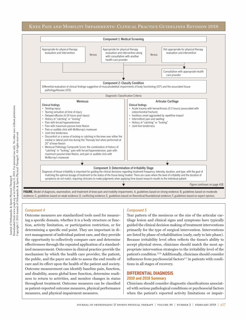
Knee Pain and Mobility Impairments: Clinical Practice Guidelines Revision 2018
journal of orthopaedic & sports physical therapy | volume 48 | number 2 | february 2018 | a17
Appropriate for physical therapy evaluation and intervention
Appropriate for physical therapy evaluation and intervention along with consultation with another health care provider
Not appropriate for physical therapy evaluation and intervention
Consultation with appropriate health care provider
Component 1: Medical Screening
Component 2: Classify ConditionDifferential evaluation of clinical findings suggestive of musculoskeletal impairments of body functioning (ICF) and the associated tissue
pathology/disease (ICD)
Component 3: Determination of Irritability StageDiagnosis of tissue irritability is important for guiding the clinical decisions regarding treatment frequency, intensity, duration, and type, with the goal of
matching the optimal dosage of treatment to the status of the tissue being treated. There are cases where the level of irritability and the duration of symptoms do not match, requiring clinicians to make judgments when applying time-based research results to the individual patient.
MeniscusClinical findings• Twisting injury• Tearing sensation at time of injury• Delayed effusion (6-24 hours post injury)• History of “catching” or “locking”• Pain with forced hyperextension• Pain with maximum passive knee flexion• Pain or audible click with McMurray’s maneuver• Joint-line tenderness• Discomfort or a sense of locking or catching in the knee over either the
medial or lateral joint line during the Thessaly test when performed at 20° of knee flexion
• Meniscal Pathology Composite Score: the combination of history of “catching” or “locking,” pain with forced hyperextension, pain with maximum passive knee flexion, and pain or audible click with McMurray’s maneuver
Articular CartilageClinical findings• Acute trauma with hemarthrosis (0-2 hours) (associated with
osteochondral fracture)• Insidious onset aggravated by repetitive impact• Intermittent pain and swelling• History of “catching” or “locking”• Joint-line tenderness
Versus Versus
Diagnostic Classification Criteria
FIGURE. Model of diagnosis, examination, and treatment of knee pain and mobility impairments. A, guidelines based on strong evidence; B, guidelines based on moderate evidence; C, guidelines based on weak evidence; D, conflicting evidence; E, guidelines based on theoretical/foundational evidence; F, guidelines based on expert opinion.
Figure continues on page A18.
Component 4Outcome measures are standardized tools used for measur-ing a specific domain, whether it is a body structure or func-tion, activity limitation, or participation restriction, or for determining a specific end point. They are important in di-rect management of individual patient care, and they provide the opportunity to collectively compare care and determine effectiveness through the repeated application of a standard-ized measurement. Outcomes in clinical practice provide the mechanism by which the health care provider, the patient, the public, and the payer are able to assess the end results of care and its effect upon the health of the patient and society. Outcome measurement can identify baseline pain, function, and disability, assess global knee function, determine readi-ness to return to activities, and monitor changes in status throughout treatment. Outcome measures can be classified as patient-reported outcome measures, physical performance measures, and physical impairment measures.
Component 5Tear pattern of the meniscus or the size of the articular car-tilage lesion and clinical signs and symptoms have typically guided the clinical decision making of treatment interventions primarily for the type of surgical intervention. Interventions are listed by phase of rehabilitation (early, early to late phase). Because irritability level often reflects the tissue’s ability to accept physical stress, clinicians should match the most ap-propriate intervention strategies to the irritability level of the patient’s condition.17,79 Additionally, clinicians should consider influences from psychosocial factors5-7 in patients with condi-tions in all stages of recovery.
DIFFERENTIAL DIAGNOSIS2010 and 2018 SummaryClinicians should consider diagnostic classifications associat-ed with serious pathological conditions or psychosocial factors when the patient’s reported activity limitations or impair-
Jour
nal o
f Orth
opae
dic
& S
ports
Phy
sica
l The
rapy
®
Dow
nloa
ded
from
ww
w.jo
spt.o
rg a
t on
Febr
uary
8, 2
018.
For
per
sona
l use
onl
y. N
o ot
her u
ses w
ithou
t per
mis
sion
. C
opyr
ight
© 2
018
Jour
nal o
f Orth
opae
dic
& S
ports
Phy
sica
l The
rapy
®. A
ll rig
hts r
eser
ved.
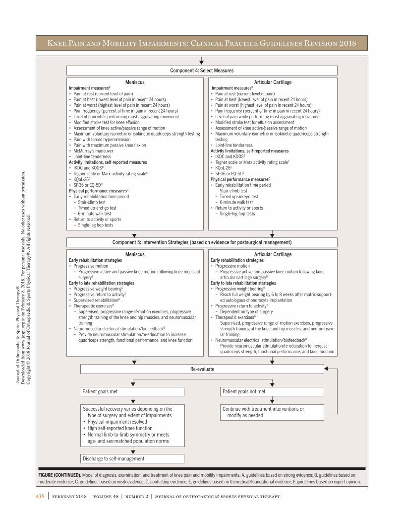
Knee Pain and Mobility Impairments: Clinical Practice Guidelines Revision 2018
a18 | february 2018 | volume 48 | number 2 | journal of orthopaedic & sports physical therapy
Component 5: Intervention Strategies (based on evidence for postsurgical management)
Component 4: Select Measures
MeniscusImpairment measuresB
• Pain at rest (current level of pain)• Pain at best (lowest level of pain in recent 24 hours)• Pain at worst (highest level of pain in recent 24 hours)• Pain frequency (percent of time in pain in recent 24 hours)• Level of pain while performing most aggravating movement• Modified stroke test for knee effusion• Assessment of knee active/passive range of motion• Maximum voluntary isometric or isokinetic quadriceps strength testing• Pain with forced hyperextension• Pain with maximum passive knee flexion• McMurray’s maneuver• Joint-line tendernessActivity limitations, self-reported measures• IKDC and KOOSB
• Tegner scale or Marx activity rating scaleC
• KQoL-26C
• SF-36 or EQ-5DC
Physical performance measuresC
• Early rehabilitation time period – Stair-climb test – Timed up-and-go test – 6-minute walk test• Return to activity or sports – Single-leg hop tests
Articular Cartilage Impairment measuresD
• Pain at rest (current level of pain)• Pain at best (lowest level of pain in recent 24 hours)• Pain at worst (highest level of pain in recent 24 hours)• Pain frequency (percent of time in pain in recent 24 hours)• Level of pain while performing most aggravating movement• Modified stroke test for effusion assessment • Assessment of knee active/passive range of motion• Maximum voluntary isometric or isokinetic quadriceps strength
testing• Joint-line tendernessActivity limitations, self-reported measures• IKDC and KOOSB
• Tegner scale or Marx activity rating scaleC
• KQoL-26C
• SF-36 or EQ-5DC
Physical performance measuresC
• Early rehabilitation time period – Stair-climb test – Timed up-and-go test – 6-minute walk test• Return to activity or sports – Single-leg hop tests
MeniscusEarly rehabilitation strategies• Progressive motion – Progressive active and passive knee motion following knee meniscal
surgeryB
Early to late rehabilitation strategies• Progressive weight bearingC
• Progressive return to activityC
• Supervised rehabilitationB
• Therapeutic exercisesB
– Supervised, progressive range-of-motion exercises, progressive strength training of the knee and hip muscles, and neuromuscular training
• Neuromuscular electrical stimulation/biofeedbackB
– Provide neuromuscular stimulation/re-education to increase quadriceps strength, functional performance, and knee function
Articular CartilageEarly rehabilitation strategies• Progressive motion – Progressive active and passive knee motion following knee
articular cartilage surgeryB
Early to late rehabilitation strategies• Progressive weight bearingB
– Reach full weight bearing by 6 to 8 weeks after matrix-support-ed autologous chondrocyte implantation
• Progressive return to activityE
– Dependent on type of surgery• Therapeutic exercisesB
– Supervised, progressive range-of-motion exercises, progressive strength training of the knee and hip muscles, and neuromuscu-lar training
• Neuromuscular electrical stimulation/biofeedbackB
– Provide neuromuscular stimulation/re-education to increase quadriceps strength, functional performance, and knee function
Re-evaluate
Patient goals met
Discharge to self-management
Patient goals not met
Continue with treatment interventions or modify as needed
Successful recovery varies depending on the type of surgery and extent of impairments
• Physical impairment resolved• High self-reported knee function• Normal limb-to-limb symmetry or meets
age- and sex-matched population norms
FIGURE (CONTINUED). Model of diagnosis, examination, and treatment of knee pain and mobility impairments. A, guidelines based on strong evidence; B, guidelines based on moderate evidence; C, guidelines based on weak evidence; D, conflicting evidence; E, guidelines based on theoretical/foundational evidence; F, guidelines based on expert opinion.
Jour
nal o
f Orth
opae
dic
& S
ports
Phy
sica
l The
rapy
®
Dow
nloa
ded
from
ww
w.jo
spt.o
rg a
t on
Febr
uary
8, 2
018.
For
per
sona
l use
onl
y. N
o ot
her u
ses w
ithou
t per
mis
sion
. C
opyr
ight
© 2
018
Jour
nal o
f Orth
opae
dic
& S
ports
Phy
sica
l The
rapy
®. A
ll rig
hts r
eser
ved.
Knee Pain and Mobility Impairments: Clinical Practice Guidelines Revision 2018
journal of orthopaedic & sports physical therapy | volume 48 | number 2 | february 2018 | a19
• Isolated tenderness of patella (no bone tenderness of knee other than patella)
• Tenderness of head of the fibula• Inability to flex knee to 90°• Inability to bear weight both immediately and in the emer-
gency department for 4 steps regardless of limping
Clinical examination by well-trained clinicians appears to be as accurate as MRI in regard to the diagnosis of meniscal le-sions.10,85,95 A lower threshold of suspicion of a meniscal tear is warranted in middle-aged and elderly patients.59,95 Magnetic resonance imaging may be reserved for more complicated or confusing cases85 and may assist an orthopaedic surgeon in preoperative planning and prognosis.85,95 Imaging may be used to monitor the status of meniscus repair or articular cartilage repair or restoration procedures.25,104
ments of body function and structure are inconsistent with those presented in the diagnosis/classification section of this guideline, or when the patient’s symptoms are not resolving with appropriate interventions.
IMAGING STUDIES2010 and 2018 Summary (unchanged from 2010)When a patient reports a history of knee trauma, the thera-pist needs to be alert for the presence of a fracture in associ-ated lower extremity bones. The Ottawa knee rule has been developed and validated to assist clinicians in determining when to order radiographs in individuals with acute knee in-jury.12,127 The Ottawa knee rule has a sensitivity of 0.99 and specificity of 0.49.12 A knee radiograph series is required in patients with any of the following criteria:• Aged 55 years or older
Jour
nal o
f Orth
opae
dic
& S
ports
Phy
sica
l The
rapy
®
Dow
nloa
ded
from
ww
w.jo
spt.o
rg a
t on
Febr
uary
8, 2
018.
For
per
sona
l use
onl
y. N
o ot
her u
ses w
ithou
t per
mis
sion
. C
opyr
ight
© 2
018
Jour
nal o
f Orth
opae
dic
& S
ports
Phy
sica
l The
rapy
®. A
ll rig
hts r
eser
ved.
Knee Pain and Mobility Impairments: Clinical Practice Guidelines Revision 2018
a20 | february 2018 | volume 48 | number 2 | journal of orthopaedic & sports physical therapy
OUTCOME MEASURES – ACTIVITY LIMITATIONS/SELF-REPORTED MEASURES2010 Recommendation
BClinicians should use a validated patient-reported outcome measure, a general health questionnaire, and a validated activity scale for patients with knee
pain and mobility impairments. These tools are useful for identifying a patient’s baseline status relative to pain, func-tion, and disability and for monitoring changes in the pa-tient’s status throughout the course of treatment.
Evidence Update
IIThe KOOS has been evaluated for its reliability and validity in people with articular cartilage lesions.45 Using qualitative methodology, content validity of
the KOOS was demonstrated in people who had undergone, or were candidates for, articular cartilage repair. In the quan-titative analysis, KOOS subscales showed test-retest reliabil-ity (all intraclass correlation coefficients [ICCs] greater than 0.70), and construct validity was demonstrated against the SF-36, although correlation between the KOOS quality of life subscale and SF-36 general health was nonsignificant. The KOOS showed sensitivity to change from baseline to 12 months after baseline, with standardized response means from 0.8 to 1.2 and minimal detectable change estimates ranging between 7.4 and 12.1.
IIThe psychometric properties (internal consistency, convergent validity, sensitivity to change, and floor and ceiling effects) of the generic European Quality
of Life-5 Dimensions (EQ-5D) and Medical Outcomes Study Short Form-6 Dimensions (SF-6D) were compared to the knee-specific Hughston Clinic Questionnaire (HCQ) in 84 patients on average 5 days, 6 weeks, and 6 months following APM.56 The EQ-5D was more consistently responsive to change over time, was better at distinguishing differences between groups, and better reflected the results of the joint-specific HCQ than the SF-6D. Thus, in this patient popula-tion, the EQ-5D is preferable to the SF-6D when used alongside a knee-specific instrument such as the HCQ.
IIThe Knee Quality of Life 26-item questionnaire (KQoL-26) for patients with a suspected ligamen-tous or meniscal injury contains 26 items with 3
subscales of knee-related quality of life: physical functioning, activity limitations, and emotional functioning.54 The KQoL-26 was found to have evidence for internal reliability (Cron-
bach = .91-.94), test-retest reliability (estimates of 0.80-0.93), construct validity (correlations with other knee scales including Lysholm knee scale: r = 0.58-0.76 with the 3 KQoL-26 subscales; EQ-5D questionnaire: r = 0.21-0.54 with the 3 KQoL-26 subscales; SF-36: r = 0.39-0.64 with the 3 KQoL-26 subscales; and knee symptom questions), respon-siveness (effect size: KQoL-26, 0.86-1.13; EQ-5D, 0.46; SF-36, 0.03-0.65 and responsiveness index: KQoL-26, 1.50-2.13; EQ-5D, 0.51; SF-36, 0.03-1.12).
IIIThe KOOS has been cross-culturally adapted for use in both the Persian and Arabic languages. In patients from Iran with ACL, meniscus, and com-
bined meniscus and ACL injuries, the Persian version had test-retest reliability (ICCs) on all subscales greater than 0.70, except the KOOS sports/recreation subscale (ICC = 0.61), and the Persian KOOS had good construct validity against the SF-36.120 The Arabic version showed test-retest reliability (ICCs) for all subscales above 0.70, as well as con-struct validity against subscales of the RAND-36 (Arabic ver-sion of SF-36) (r = 0.61-0.78) scores of pain in people from Egypt with ACL, meniscus, and combined knee injuries.4
IIIThe measurement properties of the Dutch-language versions of the IKDC 2000, KOOS, and WOMAC were compared in patients with meniscal tears.136
The Cronbach alpha for the IKDC 2000 was .90, for KOOS was .97, for KOOS domains was .72 to .95, for WOMAC was .96, and for WOMAC domains was .84 to .95. Test-retest reli-ability for the IKDC 2000 was 0.93 (95% CI: 0.89, 0.96), for KOOS was 0.93 (95% CI: 0.89, 0.96), and for WOMAC was 0.89 (95% CI: 0.83, 0.93). The standard error of the mea-surement for the IKDC 2000 was 5.3, for KOOS was 5.4, and for WOMAC was 7.2. The IKDC 2000, KOOS, and WOMAC demonstrated little to no floor or ceiling effects. The KOOS and WOMAC domains performed suboptimally with respect to internal consistency, measurement error, ability to mea-sure true change, and content validity.
IIIIn a study of 53 individuals obtained from a sports injury database and electronic medical records sys-tem, Balain et al13 investigated response shift in 3
self-report measures: Lysholm scale, VAS for worst pain, and the modified IKDC 2000 scale. When patients were asked to retrospectively rate their preoperative knee function 6 months following microfracture, retrospective ratings were lower on all 3 scales than ratings completed preoperatively,
CLINICAL GUIDELINES
Examination
Jour
nal o
f Orth
opae
dic
& S
ports
Phy
sica
l The
rapy
®
Dow
nloa
ded
from
ww
w.jo
spt.o
rg a
t on
Febr
uary
8, 2
018.
For
per
sona
l use
onl
y. N
o ot
her u
ses w
ithou
t per
mis
sion
. C
opyr
ight
© 2
018
Jour
nal o
f Orth
opae
dic
& S
ports
Phy
sica
l The
rapy
®. A
ll rig
hts r
eser
ved.
Knee Pain and Mobility Impairments: Clinical Practice Guidelines Revision 2018
journal of orthopaedic & sports physical therapy | volume 48 | number 2 | february 2018 | a21
suggesting that preoperative disability may have been greater than patients realized prior to surgery. However, adjusting for this response shift did not affect the clinical interpretation of the modified IKDC 2000 scales or the Lysholm scale.
IIIA Rasch model was used to assess the internal con-struct validity of the Lysholm knee scale in 157 pa-tients with chondral pathology.123 Fit to the Rasch
model with 7 remaining items was achieved after removal of the swelling item. There was a high degree of agreement be-tween the patient and health professional scoring (ICC = 0.90). By removing the swelling item and using unweighted scores, a modified version of the Lysholm knee scale can be used as an outcome measure for knee chondral damage.
IIIA study translated and culturally adapted the Western Ontario Meniscal Evaluation Tool (WOMET) into Turkish and evaluated the reliabil-
ity and validity of the translated tool in 96 patients with meniscal pathology.29 Validity of the tool was compared against the Lysholm knee scale and the SF-36. The WOM-ET had a Cronbach alpha of .89. Test-retest reliability of the Turkish version of the WOMET was r = 0.80 to 0.87, and had correlations with the Lysholm knee scale (r = 0.49) and SF-36 physical component and physical scores (r = 0.39-0.63). Lower correlations were observed with several SF-36 domains, predominantly mental component and emotional role scores (r = 0.03-0.11).
IIIA cross-cultural adaptation of the KOOS into Spanish was evaluated in 20 patients who underwent arthroscopic surgery for knee cartilage
defects with a microfracture technique.137 Validity was assessed against the SF-36. The Spanish KOOS demonstrated adequate test-retest reliability, with ICCs exceeding 0.8 for all domains. Agreement between the Spanish-version KOOS and the SF-36 domains of physical function (r = 0.54-0.81) and pain was observed.
2018 Recommendation
BFor knee-specific outcomes, clinicians should use the IKDC 2000 or KOOS (or a culturally appropri-ate version for patients whose primary language is
not English) and may use the Lysholm scale (with removal of the swelling item, and using unweighted scores).
CClinicians may use the Tegner scale or Marx activity rating scale to assess activity level before and after interventions intended to alleviate the physical im-
pairments, activity limitations, and participation restrictions associated with meniscus or articular cartilage lesions; how-ever, these have less evidence support about measurement properties. The SF-36 or the EQ-5D are appropriate general
health measures in this population. The KQoL-26 may be used to assess knee-related quality of life.
PHYSICAL PERFORMANCE MEASURESRefer to the 2010 Knee Pain and Mobility Impairments CPG for a list of activity limitation measures and their measure-ment properties.91
2010 Recommendation
CClinicians should utilize easily reproducible physi-cal performance measures, such as single-limb hop tests, 6-minute walk test, or timed up-and-go test,
to assess activity limitations and participation restrictions associated with their patient’s knee pain or mobility impair-ment and to assess the changes in the patient’s level of func-tion over the episode of care.
Evidence UpdateNone.
2018 Recommendation
CClinicians may administer appropriate clinical or field tests, such as single-legged hop tests (eg, single hop for distance, crossover hop for distance, triple
hop for distance, and 6-m timed hop), that can identify a patient’s baseline status relative to pain, function, and dis-ability; detect side-to-side asymmetries; assess global knee function; determine a patient’s readiness to return to activi-ties; and monitor changes in the patient’s status throughout the course of treatment.
PHYSICAL IMPAIRMENT MEASURESRefer to the 2010 Knee Pain and Mobility Impairments CPG for a list of physical impairment measures and their measure-ment properties.91
Evidence Update
IIA systematic review of 4 articles examined the va-lidity and reliability of tests to assess meniscus tears.37 They reported that the Thessaly test had fair
reliability ( = 0.54) based on 1 study of moderate quality. The McMurray and joint-line-tenderness tests had poor reliabil-ity ( 0.38) based on 3 studies of low to moderate quality.
IIIn a large diagnostic study of 292 patients with knee pathology and 75 healthy controls, Blyth et al18 examined the diagnostic accuracy of several menis-
cal tear clinical tests compared to MRI in primary care clini-cians. McMurray’s test had poor to fair diagnostic accuracy, with sensitivity of 0.58 (95% CI: 0.49, 0.67), specificity of 0.56 (95% CI: 0.45, 0.66), and OR of 1.79 (95% CI: 1.04,
Jour
nal o
f Orth
opae
dic
& S
ports
Phy
sica
l The
rapy
®
Dow
nloa
ded
from
ww
w.jo
spt.o
rg a
t on
Febr
uary
8, 2
018.
For
per
sona
l use
onl
y. N
o ot
her u
ses w
ithou
t per
mis
sion
. C
opyr
ight
© 2
018
Jour
nal o
f Orth
opae
dic
& S
ports
Phy
sica
l The
rapy
®. A
ll rig
hts r
eser
ved.
Knee Pain and Mobility Impairments: Clinical Practice Guidelines Revision 2018
a22 | february 2018 | volume 48 | number 2 | journal of orthopaedic & sports physical therapy
to support standardization for quality improvement in clinical care and research, including the modified stroke test for effu-sion assessment, assessment of knee active range of motion, maximum voluntary isometric or isokinetic quadriceps strength testing, and joint-line tenderness to palpation.
BEST-PRACTICE POINTEssential Data ElementsClinicians should document the following measures, at least at baseline and discharge or at 1 other follow-up point, for all patients with meniscus tears to support standardization for quality improvement in clinical care and research:Activity Limitation – Self-report Measures• IKDC 2000 and KOOSActivity Limitation – Physical Performance Measures• Early rehabilitation time period
- 30-second chair-stand test- Stair-climb test- Timed up-and-go test- 6-minute walk test
• Return to activity or sports- Single-leg hop tests
Physical Impairment Measures• Modified stroke test for effusion assessment• Assessment of knee active range of motion• Maximum voluntary isometric or isokinetic quadriceps
strength testing• Forced hyperextension• Maximum passive knee flexion• McMurray’s maneuver• Joint-line tenderness
Clinicians should document the following measures, at least at baseline and discharge or at 1 other follow-up point, for all patients with articular cartilage lesions to support standard-ization for quality improvement in clinical care and research:Activity Limitation – Self-report Measures• IKDC 2000 and KOOSActivity Limitation – Physical Performance Measures• Early rehabilitation time period
- 30-second chair-stand test- Stair-climb test- Timed up-and-go test- 6-minute walk test
• Return to activity or sports- Single-leg hop tests
Physical Impairment Measures• Modified stroke test for effusion assessment• Assessment of knee active range of motion• Maximum voluntary isometric or isokinetic quadriceps
strength testing• Joint-line tenderness
3.09) compared to MRI. The Thessaly test had sensitivity of 0.66 (95% CI: 0.57, 0.74), specificity of 0.39 (95% CI: 0.29, 0.50), and OR of 1.24 (95% CI: 0.71, 2.18) compared to MRI. Apley’s test had sensitivity of 0.53 (95% CI: 0.44, 0.62), spec-ificity of 0.53 (95% CI: 0.42, 0.63), and OR of 1.24 (95% CI: 0.73, 2.12) compared to MRI. The joint-line-tenderness test had sensitivity of 0.77 (95% CI: 0.68, 0.84), specificity of 0.26 (95% CI: 0.18, 0.36), and OR of 1.16 (95% CI: 0.63, 2.13) compared to MRI.
IIIHaviv et al66 investigated the accuracy of joint-line tenderness of meniscus tears in 134 men and 61 women. Joint-line tenderness for medial and lateral
meniscus tears in men had sensitivity of 0.50 to 0.58, speci-ficity of 0.74 to 1.00, and diagnostic accuracy of 0.63 to 0.86. Joint-line tenderness for medial and lateral meniscus tears in women had sensitivity of 0.40 to 0.49, specificity of 0.71 to 0.98, and diagnostic accuracy of 0.57 to 0.93.
IIISnoeker et al125 investigated the reliability and di-agnostic accuracy of deep squat, Thessaly test, and the joint-line-tenderness test. The Thessaly test
had a kappa of 0.54, sensitivity of 0.52 to 0.67, specificity of 0.38 to 0.44, positive likelihood ratio of 0.91 to 1.07, and negative likelihood ratio of 0.88 to 1.12. The deep squat test had a kappa of 0.46, sensitivity of 0.75 to 0.77, specificity of 0.36 to 0.42, positive likelihood ratio of 1.20 to 1.29, and negative likelihood ratio of 0.60 to 0.64. The joint-line-tenderness test had a kappa of 0.17.
IVCampbell et al28 examined the association between patients’ pain symptom location and arthroscopy findings in patients with meniscus tear. They re-
ported that pain symptom location was not correlated with the location of the meniscus tear.
2018 Recommendation
BClinicians should administer appropriate physical impairment assessments of body structure and function, at least at baseline and at discharge or 1
other follow-up point, for all patients with meniscus tears to support standardization for quality improvement in clinical care and research, including the modified stroke test for ef-fusion assessment, assessment of knee active range of mo-tion, maximum voluntary isometric or isokinetic quadriceps strength testing, forced hyperextension, maximum passive knee flexion, McMurray’s maneuver, and joint-line tender-ness to palpation.
DClinicians may administer the appropriate physical impairment assessments of body structure and func-tion, at least at baseline and at discharge or 1 other
follow-up point, for all patients with articular cartilage lesions
Jour
nal o
f Orth
opae
dic
& S
ports
Phy
sica
l The
rapy
®
Dow
nloa
ded
from
ww
w.jo
spt.o
rg a
t on
Febr
uary
8, 2
018.
For
per
sona
l use
onl
y. N
o ot
her u
ses w
ithou
t per
mis
sion
. C
opyr
ight
© 2
018
Jour
nal o
f Orth
opae
dic
& S
ports
Phy
sica
l The
rapy
®. A
ll rig
hts r
eser
ved.
Knee Pain and Mobility Impairments: Clinical Practice Guidelines Revision 2018
journal of orthopaedic & sports physical therapy | volume 48 | number 2 | february 2018 | a23
PROGRESSIVE KNEE MOTION2010 Recommendation
CClinicians may utilize early progressive knee mo-tion following knee meniscal and articular cartilage surgery.
Evidence Update
IIIn a randomized controlled trial, patients random-ized to the supervised active-range-of-motion group (n = 14) using an adjustable pedal arm sta-
tionary cycle ergometer had significantly better gait measures (presence or absence of antalgic gait and limp during gait) early after partial meniscectomy compared to the control group (n = 14) who did not have supervised therapy.80 No differences were reported between the groups over time in range of motion, effusion, or IKDC 2000 scores.
IIA systematic review of 4 level III studies on clinical effectiveness of continuous passive motion after ar-ticular lesion surgery did not find improved histo-
logical outcomes on second-look arthroscopic biopsies or improved radiographic findings greater than 1 year after sur-gery.49 Mixed results in clinical outcomes were reported be-tween the continuous passive motion groups and the active-range-of-motion groups.
2018 Recommendation
BClinicians may use early progressive active and pas-sive knee mo tion with patients after knee meniscal and articular cartilage surgery.
PROGRESSIVE WEIGHT BEARING2010 Recommendation
DThere are conflicting opinions regarding the best use of progressive weight bearing in patients with meniscal repairs or chondral lesions.
Evidence Update
IEbert et al41 randomized 62 patients after MACI to an accelerated weight-bearing group (stepwise pro-gression in weight bearing, with full weight bearing
by 8 weeks) or to a standard of care weight-bearing group (5 weeks of 20% partial weight bearing followed by stepwise progression in weight bearing, with full weight bearing by week 11). Three months after MACI, patients in the acceler-ated group had better KOOS scores compared to those in the
standard of care group (range for KOOS subscales: 11.84 to 83.32 versus 6.82 to 78.55). Both groups demonstrated pro-gressive graft tissue healing over time, with no difference between groups at any time period (no complete graft de-lamination).
ITwenty-eight consecutive patients after MACI were randomized to an accelerated weight-bearing group (stepwise progression in weight bearing, with full
weight bearing by 6 weeks) (n = 14) or to a standard of care weight-bearing group (stepwise progression in weight bear-ing, with full weight bearing by 8 weeks) (n = 14).43 Six and 12 months after MACI, patients in the accelerated group had better KOOS quality of life scores compared to those in the standard of care group (6 months, 62 versus 50; 12 months, 77 versus 58). Both groups demonstrated progressive graft tissue healing over time, with no difference between groups at any time period.
IThirty-one patients after ACI were randomized to an accelerated weight-bearing group (stepwise progression in weight bearing, with full weight
bearing after 6 weeks) or to a standard of care weight-bearing group (stepwise progression in weight bearing, with full weight bearing after 8 weeks).141 Both groups showed improvement in clinical scores (IKDC 2000 and Tegner scale) and MRI scores over 2 years, but no signifi-cant differences between groups were noted at 1 year and 2 years after ACI.
IILind et al90 randomized 60 patients after isolated meniscal repair to receive either free rehabilitation (restricted range of motion and toe-touch weight
bearing and no brace for 2 weeks with unrestricted activity and free range of motion afterward) or restricted rehabilita-tion (braced toe-touch weight bearing and progressive re-stricted range of motion for 6 weeks). Patients were followed at 3 months and 1 and 2 years on KOOS and Tegner mea-sures. Patients who underwent repeat arthroscopy demon-strated little to partial healing in approximately one third of patients in each group (n = 19). The KOOS and Tegner scores were similar in both groups at 1 and 2 years.
IVA retrospective analysis of 34 patients with degen-erative medial meniscus tear and knee osteoarthri-tis using a foot-worn biomechanical device during
activities of daily living was assessed before use and 3 months
CLINICAL GUIDELINES
Interventions
Jour
nal o
f Orth
opae
dic
& S
ports
Phy
sica
l The
rapy
®
Dow
nloa
ded
from
ww
w.jo
spt.o
rg a
t on
Febr
uary
8, 2
018.
For
per
sona
l use
onl
y. N
o ot
her u
ses w
ithou
t per
mis
sion
. C
opyr
ight
© 2
018
Jour
nal o
f Orth
opae
dic
& S
ports
Phy
sica
l The
rapy
®. A
ll rig
hts r
eser
ved.
Knee Pain and Mobility Impairments: Clinical Practice Guidelines Revision 2018
a24 | february 2018 | volume 48 | number 2 | journal of orthopaedic & sports physical therapy
and 12 months after wearing the device.44 Using a gait mat, patients had significant improvement in gait velocity, step length, and single-limb support of the involved knee and im-proved limb symmetry 3 months after device use. These re-sults were maintained 12 months after device use.
2018 Recommendation
CClinicians may consider early progressive weight bearing in patients with meniscal repairs.
BClinicians should use a stepwise progression of weight bearing to reach full bearing by 6 to 8 weeks after MACI for articular cartilage lesions.
PROGRESSIVE RETURN TO ACTIVITY2010 and 2018 Recommendation
CClinicians may utilize early progressive return to activity following knee meniscal repair surgery.
EClinicians may need to delay return to activity de-pending on the type of articular cartilage surgery.
SUPERVISED REHABILITATION2010 Recommendation
DThere are conflicting opinions regarding the best use of clinic-based programs for patients following meniscectomy to increase quadriceps strength and
functional performance.
Evidence Update
IIA systematic review of 18 RCTs and meta-analysis of 6 RCTs conducted by Dias et al39 supports the utili-zation of outpatient physical therapy with a home
exercise program compared to a home exercise program alone to improve knee range of motion and self-reported knee func-tion and reduce knee joint effusion in patients after APM. However, the studies were of moderate to high risk of bias.
IIIn a systematic review of 12 articles conducted by Reid et al,116 supervised clinic-based rehabilitation or a well-structured home exercise program dem-
onstrated improvements in knee muscle performance and knee function early after partial meniscectomy. However, the evidence is limited on the use of exercise to prevent the de-velopment of osteoarthritis or total knee joint arthroplasty.
IIIn a systematic review by Coppola and Collins,33 5 RCTs were identified comparing outcomes of home-based versus supervised outpatient rehabilitation
after meniscectomy. In early and intermediate follow-ups, there was no difference between groups in patient-reported outcomes at 3 weeks and 1 year after meniscectomy. However, the mean scores for these groups were lower than the popula-tion norm, which may suggest that patients in both groups were not fully rehabilitated. Two studies100,138 reported on higher vertical jump height and single hop distances in the supervised rehabilitation group (vertical jump, 22.5 cm; single hop, 113.8 cm) compared to the home-based group (vertical jump, 20.1 cm; single hop distance, 94.7 cm), though both studies had short follow-ups (less than 4 weeks).
IIPapalia et al,111 in a systematic review, evaluated the same 5 RCTs as Coppola and Collins,33 com-paring outcomes between home-based versus su-
pervised outpatient rehabilitation after meniscectomy. They reached similar conclusions that differences were demon-strated in performance-based outcomes (vertical jump height, single hop distance, and knee extensor strength), but not in patient-reported outcomes (Lysholm scale, Teg-ner score, Hughston questionnaire).
2018 Recommendation
BClinicians should use exercises as part of the in-clinic supervised rehabilitation program after ar-throscopic meniscectomy and should provide and
supervise the progression of a home-based exercise pro-gram, providing education to ensure independent performance.
THERAPEUTIC EXERCISES2010 Recommendation
BClinicians should consider strength training and functional exercise to increase quadriceps and hamstrings strength, quadriceps endurance, and
functional performance following meniscectomy.
Evidence Update
IØsterås107 randomized 42 participants after degen-erative meniscectomy to receive either 12 weeks of specialized exercise therapy (n = 22) or no exercise
therapy (n = 20). Four participants (2 in each group) were lost to follow-up. Improvements in pain (VAS, 1.9), muscle strength (quadriceps peak torque, 38.1 Nm), and KOOS scores (18.0 points) were significantly higher in the specialized exercise therapy group compared to the no-exercise-therapy group (VAS, 0.6; quadriceps peak torque, 10.4 Nm; KOOS, 6.5) after the intervention period and 12 months later.
IIn a similar study, Østerås et al108 randomized 75 participants with degenerative meniscus tear to receive either 12 weeks of specialized exercise
Jour
nal o
f Orth
opae
dic
& S
ports
Phy
sica
l The
rapy
®
Dow
nloa
ded
from
ww
w.jo
spt.o
rg a
t on
Febr
uary
8, 2
018.
For
per
sona
l use
onl
y. N
o ot
her u
ses w
ithou
t per
mis
sion
. C
opyr
ight
© 2
018
Jour
nal o
f Orth
opae
dic
& S
ports
Phy
sica
l The
rapy
®. A
ll rig
hts r
eser
ved.
Knee Pain and Mobility Impairments: Clinical Practice Guidelines Revision 2018
journal of orthopaedic & sports physical therapy | volume 48 | number 2 | february 2018 | a25
IIKoutras and colleagues86 randomized 20 male pa-tients after APM to either receive standard reha-bilitation augmented with progressive isokinetic
muscle strength training or progressive isotonic muscle strength training. Both groups demonstrated a significant improvement in knee extensor and flexor isokinetic strength and single-legged hop limb-to-limb symmetry (knee extensor at 60°/s, 17% improvement; knee flexor at 60°/s, 12% im-provement; single hop: 14% improvement; triple hop: 17% improvement; vertical hop: 18% improvement) and in Lysholm scores (17% improvement) over time, but no signifi-cant differences were noted between groups.
IILind et al90 randomized 60 patients after isolated meniscal repair to receive either free rehabilitation (restricted range of motion and toe-touch weight
bearing and no brace for 2 weeks with unrestricted activity and free range of motion afterward) or restricted rehabilita-tion (braced toe-touch weight bearing and progressive re-stricted range of motion for 6 weeks). Patients were followed at 3 months and 1 and 2 years on KOOS and Tegner mea-sures. Patients who underwent repeat arthroscopy demon-strated little to partial healing in approximately one third of patients in each group (n = 19). The KOOS and Tegner scores were similar in both groups at 1 and 2 years.
IIIDella Villa et al38 evaluated an intensive rehabilita-tion program in 31 highly competitive male athletes after an ACI procedure compared to a standard
program in 34 nonathletic participants after the same ACI procedure. They reported that at 1 year post surgery, the ath-letic cohort had higher IKDC 2000 scores than the nonath-letic cohort (mean SD, 84.7 11.7 versus 71.3 16.9), and at 5 years (90.7 11.7 versus 75.7 22.4). Both groups had a decrease in Tegner scores from preinjury to 5 years follow-up (athletic cohort: preinjury, 8.3 1.2; 5 years, 7.3 1.6 and nonathletic cohort: preinjury, 5.9 1.3; 5 years, 4.3 2.1). No severe adverse events were reported in either cohort.
IVIn a retrospective study, 30 patients with nontrau-matic posterior root tear of the medial meniscus had supervised physical therapy, focusing on knee
range of motion and knee muscle strength for at least 8 weeks, and were prescribed nonsteroidal anti-inflammatory drugs for 8 to 12 weeks.89 Patients demonstrated significant and clinically meaningful improvements in pain levels (4-point improvement on VAS) and self-reported knee func-tion (13-point improvement in Lysholm scores).
IVNeogi et al102 reported benefit in symptoms and function with 12-week rehabilitation and analgesics (up to 6 weeks) in 37 patients with degenerative
therapy (n = 38) or no physical therapy (n = 37). Eleven participants (5 in the exercise group, 6 in the no-therapy group) were lost to follow-up. Improvements in pain, mus-cle strength, and patient-reported measures were signifi-cantly higher in the exercise therapy group compared to the no-therapy group after the intervention period and 12 months later.
IAssche et al11 implemented the same standardized rehabilitation protocol to patients who were ini-tially randomized into an ACI surgery group (n =
57) or a microfracture surgery group (n = 61). Both groups received the same rehabilitation program consisting of pro-gressive, stepwise weight bearing, joint mobilization exer-cises, progressive strength training to the knee muscles, neuromuscular training, and return-to-sports integration. The authors reported no differences in recovery between the 2 groups at 2-year follow-up. When assessing patient recov-ery, activities that were repetitive movements in low-load conditions (range of motion, non–weight-bearing strength-ening exercises, proprioceptive exercises) were considered low-load modalities. Patients who had low levels of activity (less than 12 minutes per day of activity) in these low-load modalities had poorer outcomes in quadriceps strength and single-legged hop performance than patients who had high levels of activity (greater than 12 minutes per day of activity) in low-load modalities.
IHall et al60 performed an RCT to investigate the effects of a neuromuscular training program on knee kinetics, cartilage quality, and physical func-
tion during walking and single-legged sit-to-stand after APM. Groups were randomly assigned to the neuromuscu-lar training group or a control group receiving no interven-tions. The authors reported no differences in peak knee adduction moment, cartilage quality, and physical function. The neuromuscular group was more likely to demonstrate improvements in physical function and overall improve-ment compared to the control group.
IKise et al83 randomized 140 participants into 2 treatment groups: exercise therapy (n = 70) or APM. Thirteen (19%) of 70 participants crossed
over to the APM group and were analyzed in the “as treated group.” The authors reported no clinically relevant differ-ences in KOOS change scores from baseline to 2-year fol-low-up between groups (0.9 points; 95% CI: –4.3, 6.1). Both groups demonstrated similar improvements from baseline to 2-year follow-up (exercise group, 25.3 points; 95% CI: 21.6, 29.0 and APM group, 24.4 points; 95% CI: 20.7, 28.0). The exercise group had greater improvement in muscle strength at 3 and 12 months (P<.03).
Jour
nal o
f Orth
opae
dic
& S
ports
Phy
sica
l The
rapy
®
Dow
nloa
ded
from
ww
w.jo
spt.o
rg a
t on
Febr
uary
8, 2
018.
For
per
sona
l use
onl
y. N
o ot
her u
ses w
ithou
t per
mis
sion
. C
opyr
ight
© 2
018
Jour
nal o
f Orth
opae
dic
& S
ports
Phy
sica
l The
rapy
®. A
ll rig
hts r
eser
ved.
Knee Pain and Mobility Impairments: Clinical Practice Guidelines Revision 2018
a26 | february 2018 | volume 48 | number 2 | journal of orthopaedic & sports physical therapy
meniscus. Patients demonstrated improvements in Lysholm scores from pretreatment to final follow-up (56 to 79), Tegner scores (2 to 4), and VAS of pain at rest (2 to 0). Despite the improvement, the number of participants with radiographic osteoarthritis had increased by the final follow-up from 24 knees with Kellgren-Lawrence classifications at grades 0 and 1 and 9 knees at stage 2 or greater at pretreatment to 12 knees with grade 0 and 1 and 21 knees at stage 2 or greater at final follow-up.
IVForty-eight patients with full-thickness articular cartilage lesions with poor knee function partici-pated in a 3-month rehabilitation program consist-
ing of cardiovascular training, progressive strength training of the knee and hip muscles, and neuromuscular training.140 Primary outcome measures were KOOS and IKDC 2000 scores, and isokinetic muscle strength and hop test scores. The authors reported an 83% adherence rate to the rehabili-tation program. They reported clinically significant increases in KOOS sports/recreation and KOOS quality of life sub-scales. Patients also had large positive effects in standardized response means for muscle strength (0.99 to 1.22) and hop performance (0.53 to 0.75). Four (8.3%) patients showed in-creases in pain and effusion.
2018 Recommendation
BClinicians should provide supervised, progressive range-of-motion exercises, progressive strength training of the knee and hip muscles, and neuro-
muscular training to patients with knee meniscus tears and articular cartilage lesions and after meniscus or articular car-tilage surgery.
NEUROMUSCULAR ELECTRICAL STIMULATION/BIOFEEDBACK2010 Recommendation
BNeuromuscular electrical stimulation can be used with patients following meniscal or chondral inju-ries to increase quadriceps muscle strength.
Evidence Update
IIAkkaya et al2 conducted a 3-arm RCT in 45 patients after APM comparing (1) a home exercise program (without any biofeedback or electrical stimulation),
(2) electromyographic biofeedback to the quadriceps plus a home exercise program, and (3) electrical stimulation to the quadriceps plus a home exercise program. All 3 groups had similar gait measures and muscle performance values (no statistical differences between groups) 2 and 6 weeks after surgery. All groups had significant improvement in pain dur-ing walking and Lysholm scores early after partial meniscectomy.
IIIn an RCT, 64 participants were randomized to re-ceive either electromyographic biofeedback (n = 33) or usual care (n = 31) early after meniscal re-
pair.106 Electromyographic values and KOOS sport/recreation scores were significantly better in the biofeedback group (electromyographic, 16% to 25% higher; KOOS sport/recre-ation, 6% higher) compared to the usual care group 8 weeks after meniscal repair. However, these differences may not be clinically meaningful.
2018 Recommendation
BClinicians should provide neuromuscular stimula-tion/re-education to patients following meniscus procedures to increase quadriceps strength, func-
tional performance, and knee function.
Jour
nal o
f Orth
opae
dic
& S
ports
Phy
sica
l The
rapy
®
Dow
nloa
ded
from
ww
w.jo
spt.o
rg a
t on
Febr
uary
8, 2
018.
For
per
sona
l use
onl
y. N
o ot
her u
ses w
ithou
t per
mis
sion
. C
opyr
ight
© 2
018
Jour
nal o
f Orth
opae
dic
& S
ports
Phy
sica
l The
rapy
®. A
ll rig
hts r
eser
ved.

Knee Pain and Mobility Impairments: Clinical Practice Guidelines Revision 2018
journal of orthopaedic & sports physical therapy | volume 48 | number 2 | february 2018 | a27
AUTHORSDavid S. Logerstedt, PT, PhDAssistant ProfessorDepartment of Physical TherapyUniversity of the SciencesPhiladelphia, [email protected] ProfessorDepartment of Health Policy and
Public HealthUniversity of the SciencesPhiladelphia, PA
David A. Scalzitti, PT, PhDAssistant ProfessorDepartment of Physical TherapyGeorge Washington UniversityWashington, [email protected]
Kim L. Bennell, PT, PhDProfessorDepartment of PhysiotherapyUniversity of MelbourneParkville, Victoria, [email protected]
Rana S. Hinman, PT, PhDAssociate ProfessorDepartment of PhysiotherapyUniversity of MelbourneParkville, Victoria, [email protected]
Holly Silvers-Granelli, PT, PhDPhysical TherapistVelocity Physical TherapySanta Monica, [email protected]
Jay Ebert, PhDLecturerSchool of Human Sciences (Exercise
and Sport Science)The University of Western AustraliaCrawley, Western Australia, [email protected]
Karen Hambly, PT, PhDSenior LecturerMedway BuildingUniversity of KentChatham Maritime, Kent, United
James L. Carey, MD, MPHAssistant ProfessorDepartment of Orthopaedic
SurgeryDirector, Penn Center for Cartilage
Repair and Osteochondritis Dissecans Treatment
Hospital of the University of Pennsylvania
Philadelphia, [email protected]
Lynn Snyder-Mackler, PT, ScD, FAPTAAlumni Distinguished ProfessorDepartment of Physical TherapyFaculty Athletics RepresentativeUniversity of DelawareNewark, [email protected]
Michael J. Axe, MDFirst State OrthopaedicsNewark, [email protected]
Christine M. McDonough, PT, PhDAssistant ProfessorPhysical TherapySchool of Health and Rehabilitation
SciencesUniversity of PittsburghPittsburgh, PA [email protected] Clinical Practice Guidelines
EditorOrthopaedic Section, APTA, IncLa Crosse, WI
REVIEWERSPaul Beattie, PT, PhDClinical ProfessorDoctoral Program in Physical TherapyDepartment of Exercise Science, Arnold
School of Public HealthUniversity of South CarolinaColumbia, [email protected]
John DeWitt, DPTDirector of Physical Therapy Sports &
Orthopaedic ResidenciesThe Ohio State UniversityColumbus, [email protected]
Amanda Ferland, DPTClinical FacultyTongji University/USC Division of
Biokinesiology and Physical TherapyOrthopaedic Physical Therapy
Residency and Spine Rehabilitation Fellowship
Shanghai, [email protected]
Jennifer S. Howard, ATC, PhDAssistant Professor, Athletic TrainingDepartment of Health and Exercise
ScienceBeaver College of Health SciencesAppalachian State UniversityBoone, [email protected]
Sandra Kaplan, PT, PhDClinical Practice Guidelines CoordinatorAcademy of Pediatric Physical Therapy,
APTA, IncandProfessor, Doctoral Programs in
Physical TherapyRutgers UniversityNewark, [email protected]
David Killoran, PhDPatient/Consumer Representative
for the ICF-Based Clinical Practice Guidelines
Orthopaedic Section, APTA, IncLa Crosse, WIandProfessor EmeritusLoyola Marymount UniversityLos Angeles, [email protected]
Laura Schmitt, PT, PhDAssistant ProfessorSchool of Health and Rehabilitation
SciencesThe Ohio State UniversityColumbus, [email protected]
Jonas Bloch Thorlund, PhDAssociate ProfessorUniversity of Southern DenmarkDepartment of Sports Science and
Clinical BiomechanicsResearch Unit for Musculoskeletal
Function and PhysiotherapyOdense M, [email protected]
Leslie Torburn, DPTPrincipal and ConsultantSilhouette Consulting, IncSacramento, [email protected] Clinical Practice Guidelines
Editor
GUIDELINES EDITORSChristine M. McDonough, PT, PhDAssistant ProfessorPhysical TherapySchool of Health and Rehabilitation
SciencesUniversity of PittsburghPittsburgh, PA [email protected] Clinical Practice Guidelines
EditorOrthopaedic Section, APTA, IncLa Crosse, WI
Guy G. Simoneau, PT, PhD, FAPTAICF-Based Clinical Practice Guidelines
EditorOrthopaedic Section, APTA, IncLa Crosse, WIandProfessorPhysical Therapy DepartmentMarquette UniversityMilwaukee, [email protected]
Joseph J. Godges, DPT, MAAdjunct Associate Professor of Clinical
Physical TherapyDivision of Biokinesiology and Physical
TherapyUniversity of Southern CaliforniaLos Angeles, [email protected] Clinical Practice Guidelines
EditorOrthopaedic Section, APTA, IncLa Crosse, WI
AFFILIATIONS AND CONTACTS
ACKNOWLEDGMENTS: The authors would like to acknowledge the contributions of George Washington University Himmelfarb Health Sciences librarian Tom Harrod for his guidance and assistance in the design and implementation of the literature search. The authors would also like to acknowledge the assistance in screening articles provided by Nicholas Ienni, Doctor of Physical Therapy student. The authors would like to acknowledge the assistance in the writing of the evidence tables provided by Gong Chen, Doctor of Physical Therapy student.
Jour
nal o
f Orth
opae
dic
& S
ports
Phy
sica
l The
rapy
®
Dow
nloa
ded
from
ww
w.jo
spt.o
rg a
t on
Febr
uary
8, 2
018.
For
per
sona
l use
onl
y. N
o ot
her u
ses w
ithou
t per
mis
sion
. C
opyr
ight
© 2
018
Jour
nal o
f Orth
opae
dic
& S
ports
Phy
sica
l The
rapy
®. A
ll rig
hts r
eser
ved.
Knee Pain and Mobility Impairments: Clinical Practice Guidelines Revision 2018
a28 | february 2018 | volume 48 | number 2 | journal of orthopaedic & sports physical therapy
1. Abrams GD, Frank RM, Gupta AK, Harris JD, McCormick FM, Cole BJ. Trends in meniscus repair and meniscectomy in the United States, 2005-2011. Am J Sports Med. 2013;41:2333-2339. https://doi.org/10.1177/0363546513495641
2. Akkaya N, Ardic F, Ozgen M, Akkaya S, Sahin F, Kilic A. Efficacy of electro-myographic biofeedback and electrical stimulation following arthroscopic partial meniscectomy: a randomized controlled trial. Clin Rehabil. 2012;26:224-236. https://doi.org/10.1177/0269215511419382
3. Al-Dadah O, Shepstone L, Donell ST. Proprioception following partial meniscectomy in stable knees. Knee Surg Sports Traumatol Arthrosc. 2011;19:207-213. https://doi.org/10.1007/s00167-010-1237-7
4. Almangoush A, Herrington L, Attia I, et al. Cross-cultural adaptation, reliability, internal consistency and validation of the Arabic version of the Knee injury and Osteoarthritis Outcome Score (KOOS) for Egyptian people with knee injuries. Osteoarthritis Cartilage. 2013;21:1855-1864. https://doi.org/10.1016/j.joca.2013.09.010
5. Ardern CL, Österberg A, Tagesson S, Gauffin H, Webster KE, Kvist J. The impact of psychological readiness to return to sport and recreational activities after anterior cruciate ligament reconstruction. Br J Sports Med. 2014;48:1613-1619. https://doi.org/10.1136/bjsports-2014-093842
6. Ardern CL, Taylor NF, Feller JA, Webster KE. Fear of re-injury in people who have returned to sport following anterior cruciate ligament reconstruction surgery. J Sci Med Sport. 2012;15:488-495. https://doi.org/10.1016/j.jsams.2012.03.015
7. Ardern CL, Taylor NF, Feller JA, Whitehead TS, Webster KE. Psychological responses matter in returning to preinjury level of sport after anterior cruciate ligament reconstruction surgery. Am J Sports Med. 2013;41:1549-1558. https://doi.org/10.1177/0363546513489284
8. Årøen A, Løken S, Heir S, et al. Articular cartilage lesions in 993 consecu-tive knee arthroscopies. Am J Sports Med. 2004;32:211-215. https://doi.org/10.1177/0363546503259345
9. Arroll B, Robb G, Sutich E, et al. The Diagnosis and Management of Soft Tissue Knee Injuries: Internal Derangements. Wellington, New Zealand: New Zealand Guidelines Group; 2003.
10. Ashraf S, Zahoor A. Magnetic resonance imaging of articular cartilage. JBJS Rev. 2016;4:1. https://doi.org/10.2106/JBJS.RVW.15.00093
11. Assche DV, Caspel DV, Staes F, et al. Implementing one standardized rehabilitation protocol following autologous chondrocyte implantation or microfracture in the knee results in comparable physical therapy management. Physiother Theory Pract. 2011;27:125-136. https://doi.org/10.3109/09593981003681046
12. Bachmann LM, Haberzeth S, Steurer J, ter Riet G. The accuracy of the Ottawa knee rule to rule out knee fractures: a systematic review. Ann Intern Med. 2004;140:121-124. https://doi.org/10.7326/0003-4819-140-5-200403020-00013
13. Balain B, Ennis O, Kanes G, et al. Response shift in self-reported func-tional scores after knee microfracture for full thickness cartilage lesions. Osteoarthritis Cartilage. 2009;17:1009-1013. https://doi.org/10.1016/j.joca.2009.02.007
14. Bansal P, Deehan DJ, Gregory RJ. Diagnosing the acutely locked knee. In-jury. 2002;33:495-498. https://doi.org/10.1016/S0020-1383(02)00081-5
15. Bergbom S, Boersma K, Overmeer T, Linton SJ. Relationship among pain catastrophizing, depressed mood, and outcomes across physical therapy treatments. Phys Ther. 2011;91:754-764. https://doi.org/10.2522/ptj.20100136
16. Bhosale AM, Richardson JB. Articular cartilage: structure, injuries and review of management. Br Med Bull. 2008;87:77-95. https://doi.org/10.1093/bmb/ldn025
17. Blanpied PR, Gross AR, Elliott JM, et al. Neck pain: revision 2017. J Orthop Sports Phys Ther. 2017;47:A1-A83. https://doi.org/10.2519/jospt.2017.0302
18. Blyth M, Anthony I, Francq B, et al. Diagnostic accuracy of the Thessaly test, standardised clinical history and other clinical examination tests (Ap-ley’s, McMurray’s and joint line tenderness) for meniscal tears in compari-son with magnetic resonance imaging diagnosis. Health Technol Assess. 2015;19:1-62. https://doi.org/10.3310/hta19620
19. Brambilla L, Pulici L, Carimati G, et al. Prevalence of associated lesions in anterior cruciate ligament reconstruction: correlation with surgical timing and with patient age, sex, and body mass index. Am J Sports Med. 2015;43:2966-2973. https://doi.org/10.1177/0363546515608483
20. Brindle T, Nyland J, Johnson DL. The meniscus: review of basic prin-ciples with application to surgery and rehabilitation. J Athl Train. 2001;36:160-169.
21. Brittberg M, Winalski CS. Evaluation of cartilage injuries and repair. J Bone Joint Surg Am. 2003;85-A suppl 2:58-69.
22. Brophy RH, Wright RW, David TS, et al. Association between previous meniscal surgery and the incidence of chondral lesions at revision anterior cruciate ligament reconstruction. Am J Sports Med. 2012;40:808-814. https://doi.org/10.1177/0363546512437722
23. Brukner P, Khan K. Clinical Sports Medicine. 3rd ed. Sydney, Australia: McGraw-Hill; 2007.
24. Buckwalter JA, Mankin HJ. Articular cartilage: tissue design and chondro-cyte-matrix interactions. Instr Course Lect. 1998;47:477-486.
25. Burstein D. Tracking longitudinal changes in knee degeneration and repair. J Bone Joint Surg Am. 2009;91 suppl 1:51-53.
26. Busija L, Osborne RH, Nilsdotter A, Buchbinder R, Roos EM. Magni-tude and meaningfulness of change in SF-36 scores in four types of orthopedic surgery. Health Qual Life Outcomes. 2008;6:55. https://doi.org/10.1186/1477-7525-6-55
27. Campbell AB, Pineda M, Harris JD, Flanigan DC. Return to sport after ar-ticular cartilage repair in athletes’ knees: a systematic review. Arthroscopy. 2016;32:651-668.e1. https://doi.org/10.1016/j.arthro.2015.08.028
28. Campbell J, Harte A, Kerr DP, Murray P. The location of knee pain and pathology in patients with a presumed meniscus tear: preoperative symp-toms compared to arthroscopic findings. Ir J Med Sci. 2014;183:23-31. https://doi.org/10.1007/s11845-013-0965-3
29. Celik D, Demirel M, Ku G, Erdil M, Özdinçler AR. Translation, cross-cultur-al adaptation, reliability and validity of the Turkish version of the Western Ontario Meniscal Evaluation Tool (WOMET). Knee Surg Sports Traumatol Arthrosc. 2015;23:816-825. https://doi.org/10.1007/s00167-013-2753-z
30. Chalmers PN, Vigneswaran H, Harris JD, Cole BJ. Activity-related out-comes of articular cartilage surgery: a systematic review. Cartilage. 2013;4:193-203. https://doi.org/10.1177/1947603513481603
31. Chhadia AM, Inacio MC, Maletis GB, Csintalan RP, Davis BR, Funahashi TT. Are meniscus and cartilage injuries related to time to anterior cruciate ligament reconstruction? Am J Sports Med. 2011;39:1894-1899. https://doi.org/10.1177/0363546511410380
32. Cook CE. Orthopedic Manual Therapy: An Evidence-Based Approach. 2nd ed. Upper Saddle River, NJ: Pearson Education; 2012.
33. Coppola SM, Collins SM. Is physical therapy more beneficial than un-supervised home exercise in treatment of post surgical knee disorders? A systematic review. Knee. 2009;16:171-175. https://doi.org/10.1016/j.knee.2008.09.001
34. Crema MD, Guermazi A, Li L, et al. The association of prevalent medial meniscal pathology with cartilage loss in the medial tibiofemoral com-partment over a 2-year period. Osteoarthritis Cartilage. 2010;18:336-343. https://doi.org/10.1016/j.joca.2009.11.003
REFERENCES
Jour
nal o
f Orth
opae
dic
& S
ports
Phy
sica
l The
rapy
®
Dow
nloa
ded
from
ww
w.jo
spt.o
rg a
t on
Febr
uary
8, 2
018.
For
per
sona
l use
onl
y. N
o ot
her u
ses w
ithou
t per
mis
sion
. C
opyr
ight
© 2
018
Jour
nal o
f Orth
opae
dic
& S
ports
Phy
sica
l The
rapy
®. A
ll rig
hts r
eser
ved.
Knee Pain and Mobility Impairments: Clinical Practice Guidelines Revision 2018
journal of orthopaedic & sports physical therapy | volume 48 | number 2 | february 2018 | a29
35. Crema MD, Roemer FW, Felson DT, et al. Factors associated with meniscal extrusion in knees with or at risk for osteoarthritis: the Multicenter Os-teoarthritis study. Radiology. 2012;264:494-503. https://doi.org/10.1148/radiol.12110986
36. Curl WW, Krome J, Gordon ES, Rushing J, Smith BP, Poehling GG. Cartilage injuries: a review of 31,516 knee arthroscopies. Arthroscopy. 1997;13:456-460. https://doi.org/10.1016/S0749-8063(97)90124-9
37. Décary S, Ouellet P, Vendittoli PA, Desmeules F. Reliability of physical examination tests for the diagnosis of knee disorders: evidence from a systematic review. Man Ther. 2016;26:172-182. https://doi.org/10.1016/j.math.2016.09.007
38. Della Villa S, Kon E, Filardo G, et al. Does intensive rehabilitation per-mit early return to sport without compromising the clinical outcome after arthroscopic autologous chondrocyte implantation in highly competitive athletes? Am J Sports Med. 2010;38:68-77. https://doi.org/10.1177/0363546509348490
39. Dias JM, Mazuquin BF, Mostagi FQ, et al. The effectiveness of postop-erative physical therapy treatment in patients who have undergone arthroscopic partial meniscectomy: systematic review with meta-analysis. J Orthop Sports Phys Ther. 2013;43:560-576. https://doi.org/10.2519/jospt.2013.4255
40. Ding J, Zhao J, He Y, Huangfu X, Zeng B. Risk factors for articular car-tilage lesions in symptomatic discoid lateral meniscus. Arthroscopy. 2009;25:1423-1426. https://doi.org/10.1016/j.arthro.2009.06.024
41. Ebert JR, Robertson WB, Lloyd DG, Zheng MH, Wood DJ, Ackland T. Tradi-tional vs accelerated approaches to post-operative rehabilitation following matrix-induced autologous chondrocyte implantation (MACI): comparison of clinical, biomechanical and radiographic outcomes. Osteoarthritis Car-tilage. 2008;16:1131-1140. https://doi.org/10.1016/j.joca.2008.03.010
42. Ebert JR, Smith A, Edwards PK, Hambly K, Wood DJ, Ackland TR. Fac-tors predictive of outcome 5 years after matrix-induced autologous chondrocyte implantation in the tibiofemoral joint. Am J Sports Med. 2013;41:1245-1254. https://doi.org/10.1177/0363546513484696
43. Edwards PK, Ackland TR, Ebert JR. Accelerated weightbearing rehabilita-tion after matrix-induced autologous chondrocyte implantation in the tibiofemoral joint: early clinical and radiological outcomes. Am J Sports Med. 2013;41:2314-2324. https://doi.org/10.1177/0363546513495637
44. Elbaz A, Beer Y, Rath E, et al. A unique foot-worn device for patients with degenerative meniscal tear. Knee Surg Sports Traumatol Arthrosc. 2013;21:380-387. https://doi.org/10.1007/s00167-012-2026-2
45. Engelhart L, Nelson L, Lewis S, et al. Validation of the Knee Injury and Os-teoarthritis Outcome Score subscales for patients with articular cartilage lesions of the knee. Am J Sports Med. 2012;40:2264-2272. https://doi.org/10.1177/0363546512457646
46. Englund M, Guermazi A, Lohmander SL. The role of the meniscus in knee osteoarthritis: a cause or consequence? Radiol Clin North Am. 2009;47:703-712. https://doi.org/10.1016/j.rcl.2009.03.003
47. Englund M, Guermazi A, Roemer FW, et al. Meniscal tear in knees with-out surgery and the development of radiographic osteoarthritis among middle-aged and elderly persons: the Multicenter Osteoarthritis Study. Arthritis Rheum. 2009;60:831-839. https://doi.org/10.1002/art.24383
48. Fabricant PD, Rosenberger PH, Jokl P, Ickovics JR. Predictors of short-term recovery differ from those of long-term outcome after arthroscopic partial meniscectomy. Arthroscopy. 2008;24:769-778. https://doi.org/10.1016/j.arthro.2008.02.015
49. Fazalare JA, Griesser MJ, Siston RA, Flanigan DC. The use of continuous passive motion following knee cartilage defect sur-gery: a systematic review. Orthopedics. 2010;33:878. https://doi.org/10.3928/01477447-20101021-16
50. Filardo G, Andriolo L, Balboni F, Marcacci M, Kon E. Cartilage failures. Sys-tematic literature review, critical survey analysis, and definition. Knee Surg Sports Traumatol Arthrosc. 2015;23:3660-3669. https://doi.org/10.1007/s00167-014-3272-2
51. Flanigan DC, Harris JD, Trinh TQ, Siston RA, Brophy RH. Preva-lence of chondral defects in athletes’ knees: a systematic review. Med Sci Sports Exerc. 2010;42:1795-1801. https://doi.org/10.1249/MSS.0b013e3181d9eea0
52. Frobell RB, Roos HP, Roos EM, Roemer FW, Ranstam J, Lohmander LS. Treatment for acute anterior cruciate ligament tear: five year outcome of randomised trial. BMJ. 2013;346:f232. https://doi.org/10.1136/bmj.f232
53. Garcia GH, Wu HH, Park MJ, et al. Depression symptomatology and anteri-or cruciate ligament injury: incidence and effect on functional outcome—a prospective cohort study. Am J Sports Med. 2016;44:572-579. https://doi.org/10.1177/0363546515612466
54. Garratt AM, Brealey S, Robling M, et al. Development of the Knee Quality of Life (KQoL-26) 26-item questionnaire: data quality, reliability, validity and responsiveness. Health Qual Life Outcomes. 2008;6:48. https://doi.org/10.1186/1477-7525-6-48
55. Gobbi A, Nunag P, Malinowski K. Treatment of full thickness chondral lesions of the knee with microfracture in a group of athletes. Knee Surg Sports Traumatol Arthrosc. 2005;13:213-221. https://doi.org/10.1007/s00167-004-0499-3
56. Goodwin PC, Ratcliffe J, Morris J, Morrissey MC. Using the knee-specific Hughston Clinic Questionnaire, EQ-5D and SF-6D following arthroscopic partial meniscectomy surgery: a comparison of psychometric prop-erties. Qual Life Res. 2011;20:1437-1446. https://doi.org/10.1007/s11136-011-9880-0
57. Goyal D, Keyhani S, Goyal A, Lee EH, Hui JH, Vaziri AS. Evidence-based status of osteochondral cylinder transfer techniques: a systematic re-view of level I and II studies. Arthroscopy. 2014;30:497-505. https://doi.org/10.1016/j.arthro.2013.12.023
58. Goyal D, Keyhani S, Lee EH, Hui JH. Evidence-based status of microfrac-ture technique: a systematic review of level I and II studies. Arthroscopy. 2013;29:1579-1588. https://doi.org/10.1016/j.arthro.2013.05.027
59. Habata T, Uematsu K, Hattori K, Takakura Y, Fujisawa Y. Clinical features of the posterior horn tear in the medial meniscus. Arch Orthop Trauma Surg. 2004;124:642-645. https://doi.org/10.1007/s00402-004-0659-4
60. Hall M, Hinman RS, Wrigley TV, et al. Neuromuscular exercise post partial medial meniscectomy: randomized controlled trial. Med Sci Sports Exerc. 2015;47:1557-1566. https://doi.org/10.1249/MSS.0000000000000596
61. Hall M, Juhl CB, Lund H, Thorlund JB. Knee extensor muscle strength in middle-aged and older individuals undergoing arthroscopic partial meniscectomy: a systematic review and meta-analysis. Arthritis Care Res (Hoboken). 2015;67:1289-1296. https://doi.org/10.1002/acr.22581
62. Hall M, Wrigley TV, Metcalf BR, et al. Knee muscle strength after recent partial meniscectomy does not relate to 2-year change in knee adduc-tion moment. Clin Orthop Relat Res. 2014;472:3114-3120. https://doi.org/10.1007/s11999-014-3737-0
63. Hall M, Wrigley TV, Metcalf BR, et al. A longitudinal study of impact and early stance loads during gait following arthroscopic partial meniscectomy. J Bio-mech. 2014;47:2852-2857. https://doi.org/10.1016/j.jbiomech.2014.07.029
64. Hall M, Wrigley TV, Metcalf BR, et al. A longitudinal study of strength and gait after arthroscopic partial meniscectomy. Med Sci Sports Exerc. 2013;45:2036-2043. https://doi.org/10.1249/MSS.0b013e318299982a
65. Harris JD, Siston RA, Brophy RH, Lattermann C, Carey JL, Flanigan DC. Failures, re-operations, and complications after autologous chondrocyte implantation – a systematic review. Osteoarthritis Cartilage. 2011;19:779-791. https://doi.org/10.1016/j.joca.2011.02.010
Jour
nal o
f Orth
opae
dic
& S
ports
Phy
sica
l The
rapy
®
Dow
nloa
ded
from
ww
w.jo
spt.o
rg a
t on
Febr
uary
8, 2
018.
For
per
sona
l use
onl
y. N
o ot
her u
ses w
ithou
t per
mis
sion
. C
opyr
ight
© 2
018
Jour
nal o
f Orth
opae
dic
& S
ports
Phy
sica
l The
rapy
®. A
ll rig
hts r
eser
ved.
Knee Pain and Mobility Impairments: Clinical Practice Guidelines Revision 2018
a30 | february 2018 | volume 48 | number 2 | journal of orthopaedic & sports physical therapy
66. Haviv B, Bronak S, Kosashvili Y, Thein R. Gender differences in the accu-racy of joint line tenderness for arthroscopically confirmed meniscal tears. Arch Orthop Trauma Surg. 2015;135:1567-1570. https://doi.org/10.1007/s00402-015-2305-8
67. Hegedus EJ, Cook C, Hasselblad V, Goode A, McCrory DC. Physical exami-nation tests for assessing a torn meniscus in the knee: a systematic review with meta-analysis. J Orthop Sports Phys Ther. 2007;37:541-550. https://doi.org/10.2519/jospt.2007.2560
68. Henry S, Mascarenhas R, Kowalchuk D, Forsythe B, Irrgang JJ, Harner CD. Medial meniscus tear morphology and chondral degeneration of the knee: is there a relationship? Arthroscopy. 2012;28:1124-1134.e2. https://doi.org/10.1016/j.arthro.2011.12.020
69. Hjelle K, Solheim E, Strand T, Muri R, Brittberg M. Articular cartilage de-fects in 1,000 knee arthroscopies. Arthroscopy. 2002;18:730-734. https://doi.org/10.1053/jars.2002.32839
70. Howard JS, Mattacola CG, Mullineaux DR, English RA, Lattermann C. Patient-oriented and performance-based outcomes after knee autologous chondrocyte implantation: a timeline for the first year of recovery. J Sport Rehabil. 2014;23:223-234. https://doi.org/10.1123/jsr.2013-0094
71. Hwang BY, Kim SJ, Lee SW, et al. Risk factors for medial meniscus posterior root tear. Am J Sports Med. 2012;40:1606-1610. https://doi.org/10.1177/0363546512447792
72. Jacob KM, Oommen AT. A retrospective analysis of risk factors for menis-cal co-morbidities in anterior cruciate ligament injuries. Indian J Orthop. 2012;46:566-569. https://doi.org/10.4103/0019-5413.101038
73. Jaiswal PK, Bentley G, Carrington RW, Skinner JA, Briggs TW. The adverse effect of elevated body mass index on outcome after autologous chondro-cyte implantation. J Bone Joint Surg Br. 2012;94:1377-1381. https://doi.org/10.1302/0301-620X.94B10.29388
74. Johnson-Nurse C, Dandy DJ. Fracture-separation of articular cartilage in the adult knee. J Bone Joint Surg Br. 1985;67:42-43.
75. Jones JC, Burks R, Owens BD, Sturdivant RX, Svoboda SJ, Cameron KL. Incidence and risk factors associated with meniscal injuries among active-duty US military service members. J Athl Train. 2012;47:67-73.
76. Joseph C, Pathak SS, Aravinda M, Rajan D. Is ACL reconstruction only for athletes? A study of the incidence of meniscal and cartilage injuries in an ACL-deficient athlete and non-athlete population – an Indian experience. Int Orthop. 2008;32:57-61. https://doi.org/10.1007/s00264-006-0273-x
77. Jungmann PM, Salzmann GM, Schmal H, Pestka JM, Südkamp NP, Nie-meyer P. Autologous chondrocyte implantation for treatment of cartilage defects of the knee: what predicts the need for reintervention? Am J Sports Med. 2012;40:58-67. https://doi.org/10.1177/0363546511423522
78. Katz JN, Brophy RH, Chaisson CE, et al. Surgery versus physical therapy for a meniscal tear and osteoarthritis. N Engl J Med. 2013;368:1675-1684. https://doi.org/10.1056/NEJMoa1301408
79. Kelley MJ, Shaffer MA, Kuhn JE, et al. Shoulder pain and mobility deficits: adhesive capsulitis. J Orthop Sports Phys Ther. 2013;43:A1-A31. https://doi.org/10.2519/jospt.2013.0302
80. Kelln BM, Ingersoll CD, Saliba S, Miller MD, Hertel J. Effect of early active range of motion rehabilitation on outcome measures after partial menis-cectomy. Knee Surg Sports Traumatol Arthrosc. 2009;17:607-616. https://doi.org/10.1007/s00167-009-0723-2
81. Kijowski R, Woods MA, McGuine TA, Wilson JJ, Graf BK, De Smet AA. Arthroscopic partial meniscectomy: MR imaging for prediction of outcome in middle-aged and elderly patients. Radiology. 2011;259:203-212. https://doi.org/10.1148/radiol.11101392
82. Kim SG, Nagao M, Kamata K, Maeda K, Nozawa M. Return to sport after arthroscopic meniscectomy on stable knees. BMC Sports Sci Med Reha-
bil. 2013;5:23. https://doi.org/10.1186/2052-1847-5-23
83. Kise NJ, Risberg MA, Stensrud S, Ranstam J, Engebretsen L, Roos EM. Ex-ercise therapy versus arthroscopic partial meniscectomy for degenerative meniscal tear in middle aged patients: randomised controlled trial with two year follow-up. BMJ. 2016;354:i3740. https://doi.org/10.1136/bmj.i3740
84. Kluczynski MA, Marzo JM, Bisson LJ. Factors associated with menis-cal tears and chondral lesions in patients undergoing anterior cruci-ate ligament reconstruction: a prospective study. Am J Sports Med. 2013;41:2759-2765. https://doi.org/10.1177/0363546513503448
85. Kocabey Y, Tetik O, Isbell WM, Atay ÖA, Johnson DL. The value of clini-cal examination versus magnetic resonance imaging in the diagnosis of meniscal tears and anterior cruciate ligament rupture. Arthroscopy. 2004;20:696-700. https://doi.org/10.1016/j.arthro.2004.06.008
86. Koutras G, Letsi M, Papadopoulos P, Gigis I, Pappas E. A randomized trial of isokinetic versus isotonic rehabilitation program after arthroscopic meniscectomy. Int J Sports Phys Ther. 2012;7:31-38.
87. Kuikka PI, Pihlajamäki HK, Mattila VM. Knee injuries related to sports in young adult males during military service – incidence and risk factors. Scand J Med Sci Sports. 2013;23:281-287. https://doi.org/10.1111/j.1600-0838.2011.01397.x
88. Lewis PB, McCarty LP, 3rd, Kang RW, Cole BJ. Basic science and treat-ment options for articular cartilage injuries. J Orthop Sports Phys Ther. 2006;36:717-727. https://doi.org/10.2519/jospt.2006.2175
89. Lim HC, Bae JH, Wang JH, Seok CW, Kim MK. Non-operative treatment of degenerative posterior root tear of the medial meniscus. Knee Surg Sports Traumatol Arthrosc. 2010;18:535-539. https://doi.org/10.1007/s00167-009-0891-0
90. Lind M, Nielsen T, Faunø P, Lund B, Christiansen SE. Free rehabilitation is safe after isolated meniscus repair: a prospective randomized trial comparing free with restricted rehabilitation regimens. Am J Sports Med. 2013;41:2753-2758. https://doi.org/10.1177/0363546513505079
91. Logerstedt DS, Snyder-Mackler L, Ritter RC, Axe MJ. Knee pain and mobil-ity impairments: meniscal and articular cartilage lesions. J Orthop Sports Phys Ther. 2010;40:A1-A35. https://doi.org/10.2519/jospt.2010.0304
92. Logerstedt DS, Snyder-Mackler L, Ritter RC, Axe MJ, Godges JJ. Knee stability and movement coordination impairments: knee ligament sprain. J Orthop Sports Phys Ther. 2010;40:A1-A37. https://doi.org/10.2519/jospt.2010.0303
93. Lowery DJ, Farley TD, Wing DW, Sterett WI, Steadman JR. A clinical composite score accurately detects meniscal pathology. Arthroscopy. 2006;22:1174-1179. https://doi.org/10.1016/j.arthro.2006.06.014
94. Lyman S, Hidaka C, Valdez AS, et al. Risk factors for meniscectomy after meniscal repair. Am J Sports Med. 2013;41:2772-2778. https://doi.org/10.1177/0363546513503444
95. Madhusudhan TR, Kumar TM, Bastawrous SS, Sinha A. Clinical ex-amination, MRI and arthroscopy in meniscal and ligamentous knee Injuries – a prospective study. J Orthop Surg Res. 2008;3:19. https://doi.org/10.1186/1749-799X-3-19
96. Majewski M, Habelt S, Steinbrück K. Epidemiology of athletic knee inju-ries: a 10-year study. Knee. 2006;13:184-188. https://doi.org/10.1016/j.knee.2006.01.005
97. McLeod MM, Gribble P, Pfile KR, Pietrosimone BG. Effects of arthroscopic partial meniscectomy on quadriceps strength: a systematic review. J Sport Rehabil. 2012;21:285-295. https://doi.org/10.1123/jsr.21.3.285
98. Meserve BB, Cleland JA, Boucher TR. A meta-analysis examining clinical test utilities for assessing meniscal injury. Clin Rehabil. 2008;22:143-161. https://doi.org/10.1177/0269215507080130
Jour
nal o
f Orth
opae
dic
& S
ports
Phy
sica
l The
rapy
®
Dow
nloa
ded
from
ww
w.jo
spt.o
rg a
t on
Febr
uary
8, 2
018.
For
per
sona
l use
onl
y. N
o ot
her u
ses w
ithou
t per
mis
sion
. C
opyr
ight
© 2
018
Jour
nal o
f Orth
opae
dic
& S
ports
Phy
sica
l The
rapy
®. A
ll rig
hts r
eser
ved.
Knee Pain and Mobility Impairments: Clinical Practice Guidelines Revision 2018
journal of orthopaedic & sports physical therapy | volume 48 | number 2 | february 2018 | a31
99. Mithoefer K, Hambly K, Della Villa S, Silvers H, Mandelbaum BR. Return to sports participation after articular cartilage repair in the knee: scientific evidence. Am J Sports Med. 2009;37 suppl 1:167S-176S. https://doi.org/10.1177/0363546509351650
100. Moffet H, Richards CL, Malouin F, Bravo G, Paradis G. Early and intensive physiotherapy accelerates recovery postarthroscopic meniscectomy: results of a randomized controlled study. Arch Phys Med Rehabil. 1994;75:415-426.
101. Mueller MJ, Maluf KS. Tissue adaptation to physical stress: a proposed “Physical Stress Theory” to guide physical therapist practice, education, and research. Phys Ther. 2002;82:383-403.
102. Neogi DS, Kumar A, Rijal L, Yadav CS, Jaiman A, Nag HL. Role of nonoperative treatment in managing degenerative tears of the medial meniscus posterior root. J Orthop Traumatol. 2013;14:193-199. https://doi.org/10.1007/s10195-013-0234-2
103. Nepple JJ, Wright RW, Matava MJ, Brophy RH. Full-thickness knee ar-ticular cartilage defects in National Football League combine athletes undergoing magnetic resonance imaging: prevalence, location, and asso-ciation with previous surgery. Arthroscopy. 2012;28:798-806. https://doi.org/10.1016/j.arthro.2011.11.010
104. Nieminen MT, Nissi MJ, Mattila L, Kiviranta I. Evaluation of chondral repair using quantitative MRI. J Magn Reson Imaging. 2012;36:1287-1299. https://doi.org/10.1002/jmri.23644
105. O’Connor DP, Laughlin MS, Woods GW. Factors related to additional knee injuries after anterior cruciate ligament injury. Arthroscopy. 2005;21:431-438. https://doi.org/10.1016/j.arthro.2004.12.004
106. Oravitan M, Avram C. The effectiveness of electromyographic biofeedback as part of a meniscal repair rehabilitation programme. J Sports Sci Med. 2013;12:526-532.
107. Østerås H. A 12-week medical exercise therapy program leads to signifi-cant improvement in knee function after degenerative meniscectomy: a randomized controlled trial with one year follow-up. J Bodyw Mov Ther. 2014;18:374-382. https://doi.org/10.1016/j.jbmt.2013.11.015
108. Østerås H, Østerås B, Torstensen TA. Is postoperative exercise therapy necessary in patients with degenerative meniscus? A randomized con-trolled trial with one year follow-up. Knee Surg Sports Traumatol Arthrosc. 2014;22:200-206. https://doi.org/10.1007/s00167-012-2354-2
109. Østerås H, Østerås B, Torstensen TA. Medical exercise therapy, and not arthroscopic surgery, resulted in decreased depression and anxiety in pa-tients with degenerative meniscus injury. J Bodyw Mov Ther. 2012;16:456-463. https://doi.org/10.1016/j.jbmt.2012.04.003
110. Papalia R, Del Buono A, Osti L, Denaro V, Maffulli N. Meniscectomy as a risk factor for knee osteoarthritis: a systematic review. Br Med Bull. 2011;99:89-106. https://doi.org/10.1093/bmb/ldq043
111. Papalia R, Vasta S, Tecame A, D’Adamio S, Maffulli N, Denaro V. Home-based vs supervised rehabilitation programs following knee surgery: a systematic review. Br Med Bull. 2013;108:55-72. https://doi.org/10.1093/bmb/ldt014
112. Pestka JM, Bode G, Salzmann G, et al. Clinical outcomes after cell-seeded autologous chondrocyte implantation of the knee: when can success or failure be predicted? Am J Sports Med. 2014;42:208-215. https://doi.org/10.1177/0363546513507768
113. Petty CA, Lubowitz JH. Does arthroscopic partial meniscectomy result in knee osteoarthritis? A systematic review with a minimum of 8 years’ follow-up. Arthroscopy. 2011;27:419-424. https://doi.org/10.1016/j.arthro.2010.08.016
114. Phillips B, Ball C, Sackett D, et al. Oxford Centre for Evidence-based Medi-cine - Levels of Evidence (March 2009). Available at: http://www.cebm.net/index.aspx?o=1025. Accessed August 4, 2009.
115. Ralles S, Agel J, Obermeier M, Tompkins M. Incidence of second-ary intra-articular injuries with time to anterior cruciate ligament reconstruction. Am J Sports Med. 2015;43:1373-1379. https://doi.org/10.1177/0363546515574061
116. Reid D, Rydwanski J, Hing W, White S. The effectiveness of post-operative rehabilitation following partial meniscectomy of the knee. Phys Ther Rev. 2012;17:45-54. https://doi.org/10.1179/1743288X11Y.0000000046
117. Renström P, Johnson RJ. Anatomy and biomechanics of the menisci. Clin Sports Med. 1990;9:523-538.
118. Rosenberger PH, Dhabhar FS, Epel E, Jokl P, Ickovics JR. Sex differ-ences in factors influencing recovery from arthroscopic knee surgery. Clin Orthop Relat Res. 2010;468:3399-3405. https://doi.org/10.1007/s11999-010-1562-7
119. Ryzewicz M, Peterson B, Siparsky PN, Bartz RL. The diagnosis of menis-cus tears: the role of MRI and clinical examination. Clin Orthop Relat Res. 2007;455:123-133.
120. Salavati M, Mazaheri M, Negahban H, et al. Validation of a Persian-version of Knee injury and Osteoarthritis Outcome Score (KOOS) in Iranians with knee injuries. Osteoarthritis Cartilage. 2008;16:1178-1182. https://doi.org/10.1016/j.joca.2008.03.004
121. Salzmann GM, Sah B, Südkamp NP, Niemeyer P. Reoperative character-istics after microfracture of knee cartilage lesions in 454 patients. Knee Surg Sports Traumatol Arthrosc. 2013;21:365-371. https://doi.org/10.1007/s00167-012-1973-y
122. Scanzello CR, Albert AS, DiCarlo E, et al. The influence of synovial inflam-mation and hyperplasia on symptomatic outcomes up to 2 years post-operatively in patients undergoing partial meniscectomy. Osteoarthritis Cartilage. 2013;21:1392-1399. https://doi.org/10.1016/j.joca.2013.05.011
123. Smith HJ, Richardson JB, Tennant A. Modification and validation of the Lysholm Knee Scale to assess articular cartilage damage. Osteoarthritis Cartilage. 2009;17:53-58. https://doi.org/10.1016/j.joca.2008.05.002
124. Snoeker BA, Bakker EW, Kegel CA, Lucas C. Risk factors for meniscal tears: a systematic review including meta-analysis. J Orthop Sports Phys Ther. 2013;43:352-367. https://doi.org/10.2519/jospt.2013.4295
125. Snoeker BA, Lindeboom R, Zwinderman AH, Vincken PW, Jansen JA, Lucas C. Detecting meniscal tears in primary care: reproducibility and accuracy of 2 weight-bearing tests and 1 non–weight-bearing test. J Orthop Sports Phys Ther. 2015;45:693-702. https://doi.org/10.2519/jospt.2015.5712
126. Stein T, Mehling AP, Welsch F, von Eisenhart-Rothe R, Jäger A. Long-term outcome after arthroscopic meniscal repair versus arthroscopic partial meniscectomy for traumatic meniscal tears. Am J Sports Med. 2010;38:1542-1548. https://doi.org/10.1177/0363546510364052
127. Stiell IG, Greenberg GH, Wells GA, et al. Derivation of a decision rule for the use of radiography in acute knee injuries. Ann Emerg Med. 1995;26:405-413. https://doi.org/10.1016/S0196-0644(95)70106-0
128. Stocker BD, Nyland JA, Caborn DN, Sternes R, Ray JM. Results of the Kentucky high school football knee injury survey. J Ky Med Assoc. 1997;95:458-464.
129. Swenson DM, Collins CL, Best TM, Flanigan DC, Fields SK, Comstock RD. Epidemiology of knee injuries among U.S. high school athletes, 2005/2006–2010/2011. Med Sci Sports Exerc. 2013;45:462-469. https://doi.org/10.1249/MSS.0b013e318277acca
130. Tandogan RN, Ta er Ö, Kayaalp A, et al. Analysis of meniscal and chondral lesions accompanying anterior cruciate ligament tears: relationship with age, time from injury, and level of sport. Knee Surg Sports Traumatol Arthrosc. 2004;12:262-270. https://doi.org/10.1007/s00167-003-0398-z
131. te Wierike SC, van der Sluis A, van den Akker-Scheek I, Elferink-Gemser
Jour
nal o
f Orth
opae
dic
& S
ports
Phy
sica
l The
rapy
®
Dow
nloa
ded
from
ww
w.jo
spt.o
rg a
t on
Febr
uary
8, 2
018.
For
per
sona
l use
onl
y. N
o ot
her u
ses w
ithou
t per
mis
sion
. C
opyr
ight
© 2
018
Jour
nal o
f Orth
opae
dic
& S
ports
Phy
sica
l The
rapy
®. A
ll rig
hts r
eser
ved.
Knee Pain and Mobility Impairments: Clinical Practice Guidelines Revision 2018
a32 | february 2018 | volume 48 | number 2 | journal of orthopaedic & sports physical therapy
MT, Visscher C. Psychosocial factors influencing the recovery of athletes with anterior cruciate ligament injury: a systematic review. Scand J Med Sci Sports. 2013;23:527-540. https://doi.org/10.1111/sms.12010
132. Thorlund JB, Aagaard P, Roos EM. Muscle strength and functional perfor-mance in patients at high risk of knee osteoarthritis: a follow-up study. Knee Surg Sports Traumatol Arthrosc. 2012;20:1110-1117. https://doi.org/10.1007/s00167-011-1719-2
133. Thorlund JB, Creaby MW, Wrigley TV, Metcalf BR, Bennell KL. Knee joint laxity and passive stiffness in meniscectomized patients compared with healthy controls. Knee. 2014;21:886-890. https://doi.org/10.1016/j.knee.2014.06.001
134. Thorlund JB, Hare KB, Lohmander LS. Large increase in arthroscopic me-niscus surgery in the middle-aged and older population in Denmark from 2000 to 2011. Acta Orthop. 2014;85:287-292. https://doi.org/10.3109/17453674.2014.919558
135. Thorlund JB, Juhl CB, Roos EM, Lohmander LS. Arthroscopic surgery for degenerative knee: systematic review and meta-analysis of benefits and harms. Br J Sports Med. 2015;49:1229-1235. https://doi.org/10.1136/bjsports-2015-h2747rep
136. van de Graaf VA, Wolterbeek N, Scholtes VA, Mutsaerts EL, Poolman RW. Reliability and validity of the IKDC, KOOS, and WOMAC for patients with meniscal injuries. Am J Sports Med. 2014;42:1408-1416. https://doi.org/10.1177/0363546514524698
137. Vaquero J, Longo UG, Forriol F, Martinelli N, Vethencourt R, Denaro V. Reliability, validity and responsiveness of the Spanish version of the Knee Injury and Osteoarthritis Outcome Score (KOOS) in patients with chondral lesion of the knee. Knee Surg Sports Traumatol Arthrosc. 2014;22:104-108. https://doi.org/10.1007/s00167-012-2290-1
138. Vervest AM, Maurer CA, Schambergen TG, de Bie RA, Bulstra SK. Effec-tiveness of physiotherapy after meniscectomy. Knee Surg Sports Trauma-tol Arthrosc. 1999;7:360-364. https://doi.org/10.1007/s001670050181
139. Widuchowski W, Widuchowski J, Trzaska T. Articular cartilage defects: study of 25,124 knee arthroscopies. Knee. 2007;14:177-182. https://doi.org/10.1016/j.knee.2007.02.001
140. Wondrasch B, Årøen A, Røtterud JH, Høysveen T, Bølstad K, Risberg MA.
The feasibility of a 3-month active rehabilitation program for patients with knee full-thickness articular cartilage lesions: the Oslo Cartilage Active Rehabilitation and Education Study. J Orthop Sports Phys Ther. 2013;43:310-324. https://doi.org/10.2519/jospt.2013.4354
141. Wondrasch B, Zak L, Welsch GH, Marlovits S. Effect of accelerated weight-bearing after matrix-associated autologous chondrocyte implantation on the femoral condyle on radiographic and clinical outcome after 2 years: a prospective, randomized controlled pilot study. Am J Sports Med. 2009;37 suppl 1:88S-96S. https://doi.org/10.1177/0363546509351272
142. World Health Organization. International Classification of Functioning, Disabil-ity and Health: ICF. Geneva, Switzerland: World Health Organization; 2009.
143. Wu J, Huang JM, Zhao B, Cao JG, Chen X. Risk factors comparison for radial and horizontal tears. J Knee Surg. 2016;29:679-683. https://doi.org/10.1055/s-0036-1572415
144. Wyatt RW, Inacio MC, Liddle KD, Maletis GB. Prevalence and in-cidence of cartilage injuries and meniscus tears in patients who underwent both primary and revision anterior cruciate ligament reconstructions. Am J Sports Med. 2014;42:1841-1846. https://doi.org/10.1177/0363546514536020
145. Xu C, Zhao J. A meta-analysis comparing meniscal repair with meniscec-tomy in the treatment of meniscal tears: the more meniscus, the better outcome? Knee Surg Sports Traumatol Arthrosc. 2015;23:164-170. https://doi.org/10.1007/s00167-013-2528-6
146. Yeh PC, Starkey C, Lombardo S, Vitti G, Kharrazi FD. Epidemiology of isolated meniscal injury and its effect on performance in athletes from the National Basketball Association. Am J Sports Med. 2012;40:589-594. https://doi.org/10.1177/0363546511428601
147. Zaffagnini S, Marcheggiani Muccioli GM, Lopomo N, et al. Prospective long-term outcomes of the medial collagen meniscus implant versus partial medial meniscectomy: a minimum 10-year follow-up study. Am J Sports Med. 2011;39:977-985. https://doi.org/10.1177/0363546510391179
@ MORE INFORMATIONWWW.JOSPT.ORG
Jour
nal o
f Orth
opae
dic
& S
ports
Phy
sica
l The
rapy
®
Dow
nloa
ded
from
ww
w.jo
spt.o
rg a
t on
Febr
uary
8, 2
018.
For
per
sona
l use
onl
y. N
o ot
her u
ses w
ithou
t per
mis
sion
. C
opyr
ight
© 2
018
Jour
nal o
f Orth
opae
dic
& S
ports
Phy
sica
l The
rapy
®. A
ll rig
hts r
eser
ved.
Knee Pain and Mobility Impairments: Clinical Practice Guidelines Revision 2018
journal of orthopaedic & sports physical therapy | volume 48 | number 2 | february 2018 | a33
APPENDIX A
SEARCH STRATEGIES FOR ALL DATABASES SEARCHEDMEDLINE((“Menisci, Tibial” [MH]) OR (knee joint [MH] AND (menisc* [TW] OR “articular cartilage” [TW] OR chondral [TW]))) AND (classif* [TW])
((“Menisci, Tibial”[MH]) OR (knee joint[MH] AND (menisc*[TW] OR “articular cartilage”[TW] OR chondral[TW]))) AND (sensitiv*[Title/Abstract] OR sensitivity and specificity[MeSH Terms] OR diagnos*[Title/Abstract] OR diagnosis[MeSH:noexp] OR diagnostic[MeSH:noexp] OR diagnosis, differential[MeSH:noexp] OR diagnosis[Subheading:noexp] OR questionnaires[Mesh] OR “disability evaluation”[mesh:noexp] OR questionnaire[tiab] OR questionnaires[tiab] OR instrument[tiab] OR instruments[tiab] OR scale[tiab] OR scales[tiab] OR measurement[tiab] OR measurements[tiab] OR index[tiab] OR indices[tiab] OR score[tiab] OR scores[tiab])
((“Menisci, Tibial” [MH]) OR (knee joint [MH] AND (menisc* [TW] OR “articular cartilage” [TW] OR chondral [TW]))) AND (physical therapy modalities [MH] OR recovery of function [MH] OR rehabilita-tion [MH] OR therapeutics [MH] OR “physical therapy” [TW] OR physiother* [TW] OR recovery [TW] OR restoration [TW] OR re-ed-ucation [TW] OR early ambulation [MH] OR strengthening [TW] OR resistance training [MH] OR “resistance methods” [TW] OR exercise therapy [MH] OR biofeedback, psychology [MH] OR “neuromuscular electrical stimulation” [TW] OR pain management [MH] OR pain measurement [MH] OR mobilization* [TW] OR “continuous passive motion” [TW] OR manipulation, spinal [MH] OR ultrasonography [TW] OR ultrasound [TW] OR acupuncture [TW] OR laser* [TW] OR patient education as topic [MH] OR electrical stimulation [MH] OR electrical stimulation therapy [MH] OR Transcutaneous electric nerve stimulation [MH] OR taping [TW] OR bracing [TW] OR orthotic* [TW] OR weight-bearing [MH] OR Range of motion [MH] OR Treat-ment Outcome [MH] OR Exercise [MH] OR “physical therapy treat-ments” [TW] OR “training program” [TW])
((“Menisci, Tibial” [MH]) OR (knee joint [MH] AND (menisc* [TW] OR “articular cartilage” [TW] OR chondral [TW]))) AND (prognos* [tw] OR return to work [tw] OR return to work [MH] OR return to sport [tw])
((“Menisci, Tibial” [MH]) OR (knee joint [MH] AND (menisc* [TW] OR “articular cartilage” [TW] OR chondral [TW]))) AND (preval* [tw] OR incidenc* [tw] OR epidem* [tw])
((“Menisci, Tibial” [MH]) OR (knee joint [MH] AND (menisc* [TW] OR “articular cartilage” [TW] OR chondral [TW]))) AND (associat* [tw] OR risk* [tw] OR probabil* [tw] OR odds* [tw] OR relat* [tw] OR prevalen* [tw] OR predict* [tw] OR caus* [tw] OR etiol* [tw] OR interact* [tw])
Scopus((TITLE-ABS-KEY (“menisc*”) AND (TITLE-ABS-KEY (tibial) OR TITLE-ABS-KEY (medial) OR TITLE-ABS-KEY (lateral))) OR (TITLE-ABS-KEY (semilunar) AND TITLE-ABS-KEY (cartilage*)) OR (TITLE-ABS-KEY (“knee joint”) AND (TITLE-ABS-KEY (menisc*) OR TITLE-ABS-KEY (“articular cartilage”) OR TITLE-ABS-KEY (chondral)))) AND (TITLE-ABS-KEY (classif*))
((TITLE-ABS-KEY (“menisc*”) AND (TITLE-ABS-KEY (tibial) OR TITLE-ABS-KEY (medial) OR TITLE-ABS-KEY (lateral))) OR (TITLE-ABS-KEY (semilunar) AND TITLE-ABS-KEY (cartilage*)) OR (TITLE-ABS-KEY (“knee joint”) AND (TITLE-ABS-KEY (menisc*) OR TITLE-ABS-KEY (“articular cartilage”) OR TITLE-ABS-KEY (chondral)))) AND (TITLE-ABS-KEY (sensitiv*) OR TITLE-ABS-KEY (sensitivity and specificity) OR TITLE-ABS-KEY (diagnos*) OR TITLE-ABS-KEY (questionnaires) OR TITLE-ABS-KEY (“disability evaluation”) OR TITLE-ABS-KEY (ques-tionnaire) OR TITLE-ABS-KEY (questionnaires) OR TITLE-ABS-KEY (instrument) OR TITLE-ABS-KEY (instruments) OR TITLE-ABS-KEY (scale) OR TITLE-ABS-KEY (scales) OR TITLE-ABS-KEY (measure-ment) OR TITLE-ABS-KEY (measurements) OR TITLE-ABS-KEY (index) OR TITLE-ABS-KEY (indices) OR TITLE-ABS-KEY (score) OR TITLE-ABS-KEY (scores))
((TITLE-ABS-KEY (“menisc*”) AND (TITLE-ABS-KEY (tibial) OR TITLE-ABS-KEY (medial) OR TITLE-ABS-KEY (lateral))) OR (TITLE-ABS-KEY (semilunar) AND TITLE-ABS-KEY (cartilage*)) OR (TITLE-ABS-KEY (“knee joint”) AND (TITLE-ABS-KEY (menisc*) OR TITLE-ABS-KEY (“articular cartilage”) OR TITLE-ABS-KEY (chondral)))) AND (TITLE-ABS-KEY (“physical therapy modalities”) OR TITLE-ABS-KEY (“recovery of function”) OR TITLE-ABS-KEY (rehabilitation) OR TITLE-ABS-KEY (therapeutics) OR TITLE-ABS-KEY (“physical therapy”) OR TITLE-ABS-KEY (physiother*) OR TITLE-ABS-KEY (recovery) OR TITLE-ABS-KEY (restoration) OR TITLE-ABS-KEY (re-education) OR TITLE-ABS-KEY (“early ambulation”) OR TITLE-ABS-KEY (strengthen-ing) OR TITLE-ABS-KEY (“resistance training”) OR TITLE-ABS-KEY (“resistance methods”) OR TITLE-ABS-KEY (“exercise therapy”) OR TITLE-ABS-KEY (biofeedback) OR TITLE-ABS-KEY (“neuromuscular electrical stimulation”) OR TITLE-ABS-KEY (“pain management”) OR TITLE-ABS-KEY (“pain measurement”) OR TITLE-ABS-KEY (mobilization*) OR TITLE-ABS-KEY (“continuous passive motion”) OR TITLE-ABS-KEY (“spinal manipulation”) OR TITLE-ABS-KEY (ul-trasonography) OR TITLE-ABS-KEY (ultrasound) OR TITLE-ABS-KEY (acupuncture) OR TITLE-ABS-KEY (laser*) OR TITLE-ABS-KEY (“pa-tient education”) OR TITLE-ABS-KEY (“electrical stimulation”) OR TITLE-ABS-KEY (“electrical stimulation therapy”) OR TITLE-ABS-KEY (“Transcutaneous electric nerve stimulation”) OR TITLE-ABS-KEY (taping) OR TITLE-ABS-KEY (bracing) OR TITLE-ABS-KEY (orthotic*) OR TITLE-ABS-KEY (weight-bearing) OR TITLE-ABS-KEY (“Range of motion”) OR TITLE-ABS-KEY (“Treatment Outcome”) OR TITLE-ABS-KEY (Exercise) OR TITLE-ABS-KEY (“physical therapy treatments”) OR TITLE-ABS-KEY (“training program”))
((TITLE-ABS-KEY (“menisc*”) AND (TITLE-ABS-KEY (tibial) OR TITLE-
Jour
nal o
f Orth
opae
dic
& S
ports
Phy
sica
l The
rapy
®
Dow
nloa
ded
from
ww
w.jo
spt.o
rg a
t on
Febr
uary
8, 2
018.
For
per
sona
l use
onl
y. N
o ot
her u
ses w
ithou
t per
mis
sion
. C
opyr
ight
© 2
018
Jour
nal o
f Orth
opae
dic
& S
ports
Phy
sica
l The
rapy
®. A
ll rig
hts r
eser
ved.
Knee Pain and Mobility Impairments: Clinical Practice Guidelines Revision 2018
a34 | february 2018 | volume 48 | number 2 | journal of orthopaedic & sports physical therapy
APPENDIX A
ABS-KEY (medial) OR TITLE-ABS-KEY (lateral))) OR (TITLE-ABS-KEY (semilunar) AND TITLE-ABS-KEY (cartilage*)) OR (TITLE-ABS-KEY (“knee joint”) AND (TITLE-ABS-KEY (menisc*) OR TITLE-ABS-KEY (“articular cartilage”) OR TITLE-ABS-KEY (chondral)))) AND (TITLE-ABS-KEY (prognos*) OR TITLE-ABS-KEY (return to work) OR TITLE-ABS-KEY (return to sport))
((TITLE-ABS-KEY (“menisc*”) AND (TITLE-ABS-KEY (tibial) OR TITLE-ABS-KEY (medial) OR TITLE-ABS-KEY (lateral))) OR (TITLE-ABS-KEY (semilunar) AND TITLE-ABS-KEY (cartilage*)) OR (TITLE-ABS-KEY (“knee joint”) AND (TITLE-ABS-KEY (menisc*) OR TITLE-ABS-KEY (“articular cartilage”) OR TITLE-ABS-KEY (chondral)))) AND ((TITLE (prevalence) OR KEY (prevalence)) OR (TITLE (incidence) OR KEY (incidence)) OR (TITLE (epidemiology) OR KEY (epidemiology)))
((TITLE-ABS-KEY (“menisc*”) AND (TITLE-ABS-KEY (tibial) OR TITLE-ABS-KEY (medial) OR TITLE-ABS-KEY (lateral))) OR (TITLE-ABS-KEY (semilunar) AND TITLE-ABS-KEY (cartilage*)) OR (TITLE-ABS-KEY (“knee joint”) AND (TITLE-ABS-KEY (menisc*) OR TITLE-ABS-KEY (“articular cartilage”) OR TITLE-ABS-KEY (chondral)))) AND (TITLE-ABS-KEY (associat*) OR TITLE-ABS-KEY (risk*) OR TITLE-ABS-KEY (probabil*) OR TITLE-ABS-KEY (odds*) OR TITLE-ABS-KEY (relat*) OR TITLE-ABS-KEY (prevalen*) OR TITLE-ABS-KEY (predict*) OR TITLE-ABS-KEY (caus*) OR TITLE-ABS-KEY (etiol*) OR TITLE-ABS-KEY (interact*))
CINAHL((TX (“menisc*”) AND (TX (tibial) OR TX (medial) OR TX (lateral))) OR (TX (semilunar) AND TX (cartilage*)) OR (TX (“knee joint”) AND (TX (menisc*) OR TX (“articular cartilage”) OR TX (chondral)))) AND (TX (classif*))
((TX (“menisc*”) AND (TX (tibial) OR TX (medial) OR TX (lateral))) OR (TX (semilunar) AND TX (cartilage*)) OR (TX (“knee joint”) AND (TX (menisc*) OR TX (“articular cartilage”) OR TX (chondral)))) AND (TX (sensitiv*) OR TX (sensitivity and specificity) OR TX (diagnos*) OR TX (questionnaires) OR TX (“disability evaluation”) OR TX (ques-tionnaire) OR TX (questionnaires) OR TX (instrument) OR TX (instru-ments) OR TX (scale) OR TX (scales) OR TX (measurement) OR TX (measurements) OR TX (index) OR TX (indices) OR TX (score) OR TX (scores))
((TX (“menisc*”) AND (TX (tibial) OR TX (medial) OR TX (lateral))) OR (TX (semilunar) AND TX (cartilage*)) OR (TX (“knee joint”) AND (TX (menisc*) OR TX (“articular cartilage”) OR TX (chondral)))) AND (TX (“physical therapy modalities”) OR TX (“recovery of func-tion”) OR TX (rehabilitation) OR TX (therapeutics) OR TX (“physical therapy”) OR TX (physiother*) OR TX (recovery) OR TX (restoration) OR TX (re-education) OR TX (“early ambulation”) OR TX (strengthen-ing) OR TX (“resistance training”) OR TX (“resistance methods”) OR TX (“exercise therapy”) OR TX (biofeedback) OR TX (“neuromuscular electrical stimulation”) OR TX (“pain management”) OR TX (“pain
measurement”) OR TX (mobilization*) OR TX (“continuous passive motion”) OR TX (“spinal manipulation”) OR TX (ultrasonography) OR TX (ultrasound) OR TX (acupuncture) OR TX (laser*) OR TX (“patient education”) OR TX (“electrical stimulation”) OR TX (“electrical stimu-lation therapy”) OR TX (“Transcutaneous electric nerve stimulation”) OR TX (taping) OR TX (bracing) OR TX (orthotic*) OR TX (weight-bearing) OR TX (“Range of motion”) OR TX (“Treatment Outcome”) OR TX (Exercise) OR TX (“physical therapy treatments”) OR TX (“training program”))
((TX (“menisc*”) AND (TX (tibial) OR TX (medial) OR TX (lateral))) OR (TX (semilunar) AND TX (cartilage*)) OR (TX (“knee joint”) AND (TX (menisc*) OR TX (“articular cartilage”) OR TX (chondral)))) AND (TX (prognos*) OR TX (return to work) OR TX (return to sport))
((TX (“menisc*”) AND (TX (tibial) OR TX (medial) OR TX (lateral))) OR (TX (semilunar) AND TX (cartilage*)) OR (TX (“knee joint”) AND (TX (menisc*) OR TX (“articular cartilage”) OR TX (chondral)))) AND ((TI (prevalence) OR SU (prevalence)) OR (TI (incidence) OR SU (in-cidence)) OR (TI (epidemiology) OR SU (epidemiology)))
((TX (“menisc*”) AND (TX (tibial) OR TX (medial) OR TX (lateral))) OR (TX (semilunar) AND TX (cartilage*)) OR (TX (“knee joint”) AND (TX (menisc*) OR TX (“articular cartilage”) OR TX (chondral)))) AND (TX (associat*) OR TX (risk*) OR TX (probabil*) OR TX (odds*) OR TX (relat*) OR TX (prevalen*) OR TX (predict*) OR TX (caus*) OR TX (etiol* ) OR TX (interact*))
SPORTDiscus((TX (“menisc*”) AND (TX (tibial) OR TX (medial) OR TX (lateral))) OR (TX (semilunar) AND TX (cartilage*)) OR (TX (“knee joint”) AND (TX (menisc*) OR TX (“articular cartilage”) OR TX (chondral)))) AND (TX (classif*))
((TX (“menisc*”) AND (TX (tibial) OR TX (medial) OR TX (lateral))) OR (TX (semilunar) AND TX (cartilage*)) OR (TX (“knee joint”) AND (TX (menisc*) OR TX (“articular cartilage”) OR TX (chondral)))) AND (TX (sensitiv*) OR TX (sensitivity and specificity) OR TX (diagnos*) OR TX (questionnaires) OR TX (“disability evaluation”) OR TX (ques-tionnaire) OR TX (questionnaires) OR TX (instrument) OR TX (instru-ments) OR TX (scale) OR TX (scales) OR TX (measurement) OR TX (measurements) OR TX (index) OR TX (indices) OR TX (score) OR TX (scores))
((TX (“menisc*”) AND (TX (tibial) OR TX (medial) OR TX (lateral))) OR (TX (semilunar) AND TX (cartilage*)) OR (TX (“knee joint”) AND (TX (menisc*) OR TX (“articular cartilage”) OR TX (chondral)))) AND (TX (“physical therapy modalities”) OR TX (“recovery of func-tion”) OR TX (rehabilitation) OR TX (therapeutics) OR TX (“physical therapy”) OR TX (physiother*) OR TX (recovery) OR TX (restoration) OR TX (re-education) OR TX (“early ambulation”) OR TX (strengthen-ing) OR TX (“resistance training”) OR TX (“resistance methods”) OR
Jour
nal o
f Orth
opae
dic
& S
ports
Phy
sica
l The
rapy
®
Dow
nloa
ded
from
ww
w.jo
spt.o
rg a
t on
Febr
uary
8, 2
018.
For
per
sona
l use
onl
y. N
o ot
her u
ses w
ithou
t per
mis
sion
. C
opyr
ight
© 2
018
Jour
nal o
f Orth
opae
dic
& S
ports
Phy
sica
l The
rapy
®. A
ll rig
hts r
eser
ved.
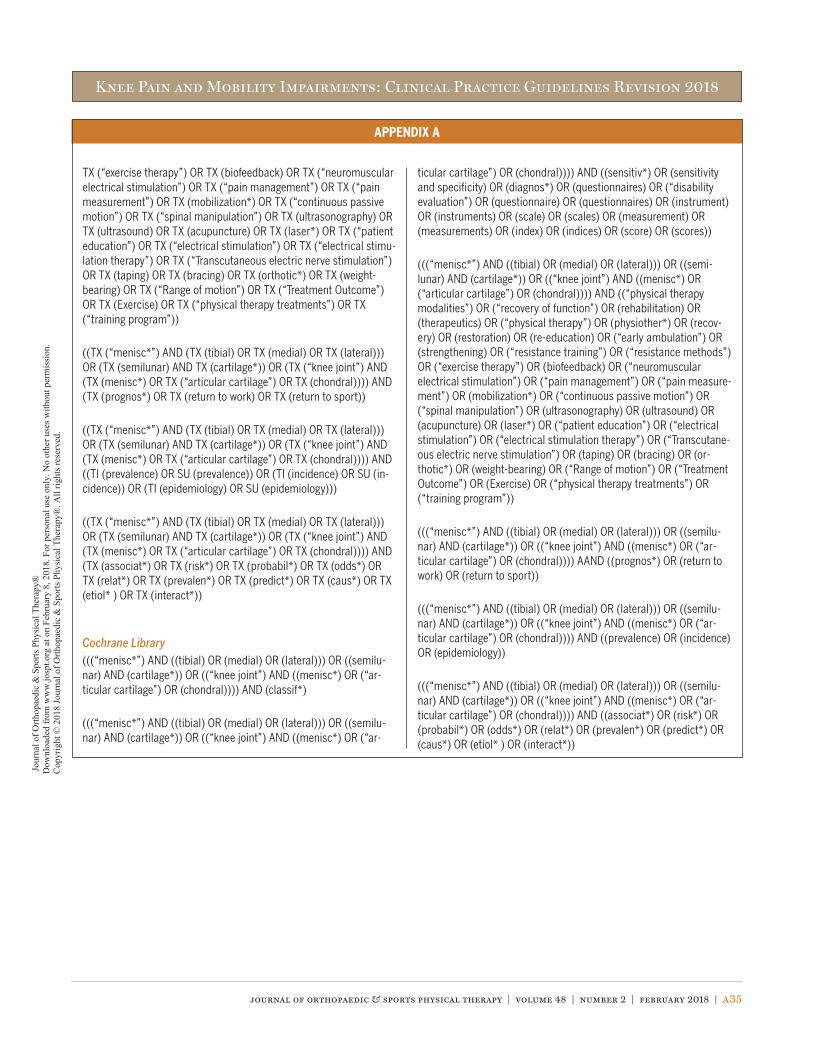
Knee Pain and Mobility Impairments: Clinical Practice Guidelines Revision 2018
journal of orthopaedic & sports physical therapy | volume 48 | number 2 | february 2018 | a35
APPENDIX A
TX (“exercise therapy”) OR TX (biofeedback) OR TX (“neuromuscular electrical stimulation”) OR TX (“pain management”) OR TX (“pain measurement”) OR TX (mobilization*) OR TX (“continuous passive motion”) OR TX (“spinal manipulation”) OR TX (ultrasonography) OR TX (ultrasound) OR TX (acupuncture) OR TX (laser*) OR TX (“patient education”) OR TX (“electrical stimulation”) OR TX (“electrical stimu-lation therapy”) OR TX (“Transcutaneous electric nerve stimulation”) OR TX (taping) OR TX (bracing) OR TX (orthotic*) OR TX (weight-bearing) OR TX (“Range of motion”) OR TX (“Treatment Outcome”) OR TX (Exercise) OR TX (“physical therapy treatments”) OR TX (“training program”))
((TX (“menisc*”) AND (TX (tibial) OR TX (medial) OR TX (lateral))) OR (TX (semilunar) AND TX (cartilage*)) OR (TX (“knee joint”) AND (TX (menisc*) OR TX (“articular cartilage”) OR TX (chondral)))) AND (TX (prognos*) OR TX (return to work) OR TX (return to sport))
((TX (“menisc*”) AND (TX (tibial) OR TX (medial) OR TX (lateral))) OR (TX (semilunar) AND TX (cartilage*)) OR (TX (“knee joint”) AND (TX (menisc*) OR TX (“articular cartilage”) OR TX (chondral)))) AND ((TI (prevalence) OR SU (prevalence)) OR (TI (incidence) OR SU (in-cidence)) OR (TI (epidemiology) OR SU (epidemiology)))
((TX (“menisc*”) AND (TX (tibial) OR TX (medial) OR TX (lateral))) OR (TX (semilunar) AND TX (cartilage*)) OR (TX (“knee joint”) AND (TX (menisc*) OR TX (“articular cartilage”) OR TX (chondral)))) AND (TX (associat*) OR TX (risk*) OR TX (probabil*) OR TX (odds*) OR TX (relat*) OR TX (prevalen*) OR TX (predict*) OR TX (caus*) OR TX (etiol* ) OR TX (interact*))
Cochrane Library(((“menisc*”) AND ((tibial) OR (medial) OR (lateral))) OR ((semilu-nar) AND (cartilage*)) OR ((“knee joint”) AND ((menisc*) OR (“ar-ticular cartilage”) OR (chondral)))) AND (classif*)
(((“menisc*”) AND ((tibial) OR (medial) OR (lateral))) OR ((semilu-nar) AND (cartilage*)) OR ((“knee joint”) AND ((menisc*) OR (“ar-
ticular cartilage”) OR (chondral)))) AND ((sensitiv*) OR (sensitivity and specificity) OR (diagnos*) OR (questionnaires) OR (“disability evaluation”) OR (questionnaire) OR (questionnaires) OR (instrument) OR (instruments) OR (scale) OR (scales) OR (measurement) OR (measurements) OR (index) OR (indices) OR (score) OR (scores))
(((“menisc*”) AND ((tibial) OR (medial) OR (lateral))) OR ((semi-lunar) AND (cartilage*)) OR ((“knee joint”) AND ((menisc*) OR (“articular cartilage”) OR (chondral)))) AND ((“physical therapy modalities”) OR (“recovery of function”) OR (rehabilitation) OR (therapeutics) OR (“physical therapy”) OR (physiother*) OR (recov-ery) OR (restoration) OR (re-education) OR (“early ambulation”) OR (strengthening) OR (“resistance training”) OR (“resistance methods”) OR (“exercise therapy”) OR (biofeedback) OR (“neuromuscular electrical stimulation”) OR (“pain management”) OR (“pain measure-ment”) OR (mobilization*) OR (“continuous passive motion”) OR (“spinal manipulation”) OR (ultrasonography) OR (ultrasound) OR (acupuncture) OR (laser*) OR (“patient education”) OR (“electrical stimulation”) OR (“electrical stimulation therapy”) OR (“Transcutane-ous electric nerve stimulation”) OR (taping) OR (bracing) OR (or-thotic*) OR (weight-bearing) OR (“Range of motion”) OR (“Treatment Outcome”) OR (Exercise) OR (“physical therapy treatments”) OR (“training program”))
(((“menisc*”) AND ((tibial) OR (medial) OR (lateral))) OR ((semilu-nar) AND (cartilage*)) OR ((“knee joint”) AND ((menisc*) OR (“ar-ticular cartilage”) OR (chondral)))) AAND ((prognos*) OR (return to work) OR (return to sport))
(((“menisc*”) AND ((tibial) OR (medial) OR (lateral))) OR ((semilu-nar) AND (cartilage*)) OR ((“knee joint”) AND ((menisc*) OR (“ar-ticular cartilage”) OR (chondral)))) AND ((prevalence) OR (incidence) OR (epidemiology))
(((“menisc*”) AND ((tibial) OR (medial) OR (lateral))) OR ((semilu-nar) AND (cartilage*)) OR ((“knee joint”) AND ((menisc*) OR (“ar-ticular cartilage”) OR (chondral)))) AND ((associat*) OR (risk*) OR (probabil*) OR (odds*) OR (relat*) OR (prevalen*) OR (predict*) OR (caus*) OR (etiol* ) OR (interact*))
Jour
nal o
f Orth
opae
dic
& S
ports
Phy
sica
l The
rapy
®
Dow
nloa
ded
from
ww
w.jo
spt.o
rg a
t on
Febr
uary
8, 2
018.
For
per
sona
l use
onl
y. N
o ot
her u
ses w
ithou
t per
mis
sion
. C
opyr
ight
© 2
018
Jour
nal o
f Orth
opae
dic
& S
ports
Phy
sica
l The
rapy
®. A
ll rig
hts r
eser
ved.
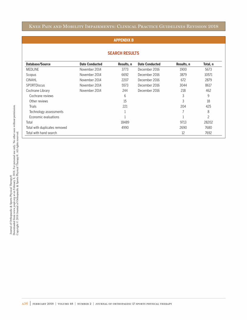
Knee Pain and Mobility Impairments: Clinical Practice Guidelines Revision 2018
a36 | february 2018 | volume 48 | number 2 | journal of orthopaedic & sports physical therapy
APPENDIX B
SEARCH RESULTS
Database/Source Date Conducted Results, n Date Conducted Results, n Total, nMEDLINE November 2014 3773 December 2016 1900 5673
Scopus November 2014 6692 December 2016 3879 10571
CINAHL November 2014 2207 December 2016 672 2879
SPORTDiscus November 2014 5573 December 2016 3044 8617
Cochrane Library November 2014 244 December 2016 218 462
Cochrane reviews 6 3 9
Other reviews 15 3 18
Trials 221 204 425
Technology assessments 1 7 8
Economic evaluations 1 1 2
Total 18489 9713 28202
Total with duplicates removed 4990 2690 7680
Total with hand search 12 7692
Jour
nal o
f Orth
opae
dic
& S
ports
Phy
sica
l The
rapy
®
Dow
nloa
ded
from
ww
w.jo
spt.o
rg a
t on
Febr
uary
8, 2
018.
For
per
sona
l use
onl
y. N
o ot
her u
ses w
ithou
t per
mis
sion
. C
opyr
ight
© 2
018
Jour
nal o
f Orth
opae
dic
& S
ports
Phy
sica
l The
rapy
®. A
ll rig
hts r
eser
ved.

Knee Pain and Mobility Impairments: Clinical Practice Guidelines Revision 2018
journal of orthopaedic & sports physical therapy | volume 48 | number 2 | february 2018 | a37
APPENDIX C
CRITERIA FOR INCLUSION AND EXCLUSION OF STUDIES FOR REVIEWArticles published in peer-reviewed journals that include studies of the following types: systematic reviews, meta-analyses, experimental and quasi-experimental, cohort, case series, and cross-sectional studies were included.
Exclusions: meeting abstracts, press releases, theses, nonsystematic review articles, case reports, and articles that cannot be retrieved in English.
Inclusion CriteriaArticles reporting on isolated and combined injuries for meniscus and articular cartilage injuries:• The functional anatomy of the menisci and articular cartilage of
the tibiofemoral jointOR• Tests and measures for diagnosis and/or differential diagnosis
of meniscal and chondral lesions within the scope of physical therapist practice, including but not limited to “specific tests and measures”
OR• Measurement properties of instruments and tests specific to mea-
suring meniscal and chondral lesion–related outcomes (including but not limited to symptoms, functions, activity, and participation)
OR• Measurement properties of instruments that are not specific to
meniscal and chondral lesions BUT are specific to lower extremity outcomes
OR• Measurement properties of instruments using data from a sample
of patients with meniscal and chondral lesions
OR• Primarily adolescents and adults (12 years old or older)
- Studies reporting on persons younger than 12 years old IF the proportion in the sample is small (less than 5%) OR with separate data available for adults
AND• Meniscal and chondral lesions, including the following topics:
- Risk of meniscal and chondral lesions- Diagnostic characteristics of meniscal and chondral lesions,
including but not limited to location, duration, and quality, and related impairments and functional limitations
- Interventions within the scope of practice of physical therapists for meniscal and chondral lesions
All outcome studies were included.
Exclusion CriteriaArticles reporting on:• Osteochondritis dissecans lesions• Primarily infants and children (younger than 12 years old)• Ligament-related injuries of the tibiofemoral joint• Patellofemoral pain, patellar tendinopathy/tendon pain, or
iliotibial band• Nonmusculoskeletal tibiofemoral pain
- Diabetes- Ulcers- Primary peripheral nerve entrapment
• Topics outside the scope of physical therapist practice- Decisions to order radiologic tests- Pharmacological interventions
• Biomechanical studies
Jour
nal o
f Orth
opae
dic
& S
ports
Phy
sica
l The
rapy
®
Dow
nloa
ded
from
ww
w.jo
spt.o
rg a
t on
Febr
uary
8, 2
018.
For
per
sona
l use
onl
y. N
o ot
her u
ses w
ithou
t per
mis
sion
. C
opyr
ight
© 2
018
Jour
nal o
f Orth
opae
dic
& S
ports
Phy
sica
l The
rapy
®. A
ll rig
hts r
eser
ved.
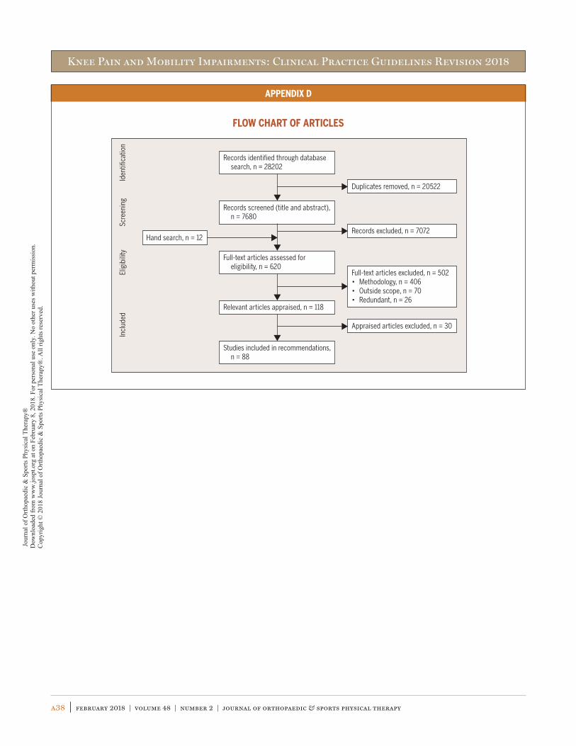
Knee Pain and Mobility Impairments: Clinical Practice Guidelines Revision 2018
a38 | february 2018 | volume 48 | number 2 | journal of orthopaedic & sports physical therapy
APPENDIX D
FLOW CHART OF ARTICLES
Records identified through database search, n = 28202
Records screened (title and abstract), n = 7680
Duplicates removed, n = 20522
Full-text articles assessed for eligibility, n = 620
Records excluded, n = 7072
Relevant articles appraised, n = 118
Full-text articles excluded, n = 502• Methodology, n = 406• Outside scope, n = 70• Redundant, n = 26
Appraised articles excluded, n = 30
Studies included in recommendations, n = 88
Iden
tifica
tion
Elig
ibili
tySc
reen
ing
Incl
uded
Hand search, n = 12
Jour
nal o
f Orth
opae
dic
& S
ports
Phy
sica
l The
rapy
®
Dow
nloa
ded
from
ww
w.jo
spt.o
rg a
t on
Febr
uary
8, 2
018.
For
per
sona
l use
onl
y. N
o ot
her u
ses w
ithou
t per
mis
sion
. C
opyr
ight
© 2
018
Jour
nal o
f Orth
opae
dic
& S
ports
Phy
sica
l The
rapy
®. A
ll rig
hts r
eser
ved.
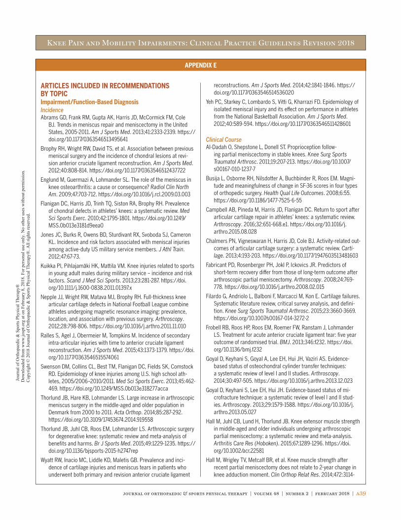
Knee Pain and Mobility Impairments: Clinical Practice Guidelines Revision 2018
journal of orthopaedic & sports physical therapy | volume 48 | number 2 | february 2018 | a39
APPENDIX E
ARTICLES INCLUDED IN RECOMMENDATIONS BY TOPICImpairment/Function-Based DiagnosisIncidenceAbrams GD, Frank RM, Gupta AK, Harris JD, McCormick FM, Cole
BJ. Trends in meniscus repair and meniscectomy in the United States, 2005-2011. Am J Sports Med. 2013;41:2333-2339. https://doi.org/10.1177/0363546513495641
Brophy RH, Wright RW, David TS, et al. Association between previous meniscal surgery and the incidence of chondral lesions at revi-sion anterior cruciate ligament reconstruction. Am J Sports Med. 2012;40:808-814. https://doi.org/10.1177/0363546512437722
Englund M, Guermazi A, Lohmander SL. The role of the meniscus in knee osteoarthritis: a cause or consequence? Radiol Clin North Am. 2009;47:703-712. https://doi.org/10.1016/j.rcl.2009.03.003
Flanigan DC, Harris JD, Trinh TQ, Siston RA, Brophy RH. Prevalence of chondral defects in athletes’ knees: a systematic review. Med Sci Sports Exerc. 2010;42:1795-1801. https://doi.org/10.1249/MSS.0b013e3181d9eea0
Jones JC, Burks R, Owens BD, Sturdivant RX, Svoboda SJ, Cameron KL. Incidence and risk factors associated with meniscal injuries among active-duty US military service members. J Athl Train. 2012;47:67-73.
Kuikka PI, Pihlajamäki HK, Mattila VM. Knee injuries related to sports in young adult males during military service – incidence and risk factors. Scand J Med Sci Sports. 2013;23:281-287. https://doi.org/10.1111/j.1600-0838.2011.01397.x
Nepple JJ, Wright RW, Matava MJ, Brophy RH. Full-thickness knee articular cartilage defects in National Football League combine athletes undergoing magnetic resonance imaging: prevalence, location, and association with previous surgery. Arthroscopy. 2012;28:798-806. https://doi.org/10.1016/j.arthro.2011.11.010
Ralles S, Agel J, Obermeier M, Tompkins M. Incidence of secondary intra-articular injuries with time to anterior cruciate ligament reconstruction. Am J Sports Med. 2015;43:1373-1379. https://doi.org/10.1177/0363546515574061
Swenson DM, Collins CL, Best TM, Flanigan DC, Fields SK, Comstock RD. Epidemiology of knee injuries among U.S. high school ath-letes, 2005/2006–2010/2011. Med Sci Sports Exerc. 2013;45:462-469. https://doi.org/10.1249/MSS.0b013e318277acca
Thorlund JB, Hare KB, Lohmander LS. Large increase in arthroscopic meniscus surgery in the middle-aged and older population in Denmark from 2000 to 2011. Acta Orthop. 2014;85:287-292. https://doi.org/10.3109/17453674.2014.919558
Thorlund JB, Juhl CB, Roos EM, Lohmander LS. Arthroscopic surgery for degenerative knee: systematic review and meta-analysis of benefits and harms. Br J Sports Med. 2015;49:1229-1235. https://doi.org/10.1136/bjsports-2015-h2747rep
Wyatt RW, Inacio MC, Liddle KD, Maletis GB. Prevalence and inci-dence of cartilage injuries and meniscus tears in patients who underwent both primary and revision anterior cruciate ligament
reconstructions. Am J Sports Med. 2014;42:1841-1846. https://doi.org/10.1177/0363546514536020
Yeh PC, Starkey C, Lombardo S, Vitti G, Kharrazi FD. Epidemiology of isolated meniscal injury and its effect on performance in athletes from the National Basketball Association. Am J Sports Med. 2012;40:589-594. https://doi.org/10.1177/0363546511428601
Clinical CourseAl-Dadah O, Shepstone L, Donell ST. Proprioception follow-
ing partial meniscectomy in stable knees. Knee Surg Sports Traumatol Arthrosc. 2011;19:207-213. https://doi.org/10.1007/s00167-010-1237-7
Busija L, Osborne RH, Nilsdotter A, Buchbinder R, Roos EM. Magni-tude and meaningfulness of change in SF-36 scores in four types of orthopedic surgery. Health Qual Life Outcomes. 2008;6:55. https://doi.org/10.1186/1477-7525-6-55
Campbell AB, Pineda M, Harris JD, Flanigan DC. Return to sport after articular cartilage repair in athletes’ knees: a systematic review. Arthroscopy. 2016;32:651-668.e1. https://doi.org/10.1016/j.arthro.2015.08.028
Chalmers PN, Vigneswaran H, Harris JD, Cole BJ. Activity-related out-comes of articular cartilage surgery: a systematic review. Carti-lage. 2013;4:193-203. https://doi.org/10.1177/1947603513481603
Fabricant PD, Rosenberger PH, Jokl P, Ickovics JR. Predictors of short-term recovery differ from those of long-term outcome after arthroscopic partial meniscectomy. Arthroscopy. 2008;24:769-778. https://doi.org/10.1016/j.arthro.2008.02.015
Filardo G, Andriolo L, Balboni F, Marcacci M, Kon E. Cartilage failures. Systematic literature review, critical survey analysis, and defini-tion. Knee Surg Sports Traumatol Arthrosc. 2015;23:3660-3669. https://doi.org/10.1007/s00167-014-3272-2
Frobell RB, Roos HP, Roos EM, Roemer FW, Ranstam J, Lohmander LS. Treatment for acute anterior cruciate ligament tear: five year outcome of randomised trial. BMJ. 2013;346:f232. https://doi.org/10.1136/bmj.f232
Goyal D, Keyhani S, Goyal A, Lee EH, Hui JH, Vaziri AS. Evidence-based status of osteochondral cylinder transfer techniques: a systematic review of level I and II studies. Arthroscopy. 2014;30:497-505. https://doi.org/10.1016/j.arthro.2013.12.023
Goyal D, Keyhani S, Lee EH, Hui JH. Evidence-based status of mi-crofracture technique: a systematic review of level I and II stud-ies. Arthroscopy. 2013;29:1579-1588. https://doi.org/10.1016/j.arthro.2013.05.027
Hall M, Juhl CB, Lund H, Thorlund JB. Knee extensor muscle strength in middle-aged and older individuals undergoing arthroscopic partial meniscectomy: a systematic review and meta-analysis. Arthritis Care Res (Hoboken). 2015;67:1289-1296. https://doi.org/10.1002/acr.22581
Hall M, Wrigley TV, Metcalf BR, et al. Knee muscle strength after recent partial meniscectomy does not relate to 2-year change in knee adduction moment. Clin Orthop Relat Res. 2014;472:3114-
Jour
nal o
f Orth
opae
dic
& S
ports
Phy
sica
l The
rapy
®
Dow
nloa
ded
from
ww
w.jo
spt.o
rg a
t on
Febr
uary
8, 2
018.
For
per
sona
l use
onl
y. N
o ot
her u
ses w
ithou
t per
mis
sion
. C
opyr
ight
© 2
018
Jour
nal o
f Orth
opae
dic
& S
ports
Phy
sica
l The
rapy
®. A
ll rig
hts r
eser
ved.
Knee Pain and Mobility Impairments: Clinical Practice Guidelines Revision 2018
a40 | february 2018 | volume 48 | number 2 | journal of orthopaedic & sports physical therapy
APPENDIX E
3120. https://doi.org/10.1007/s11999-014-3737-0
Hall M, Wrigley TV, Metcalf BR, et al. A longitudinal study of impact and early stance loads during gait following arthroscopic par-tial meniscectomy. J Biomech. 2014;47:2852-2857. https://doi.org/10.1016/j.jbiomech.2014.07.029
Hall M, Wrigley TV, Metcalf BR, et al. A longitudinal study of strength and gait after arthroscopic partial meniscectomy. Med Sci Sports Exerc. 2013;45:2036-2043. https://doi.org/10.1249/MSS.0b013e318299982a
Harris JD, Siston RA, Brophy RH, Lattermann C, Carey JL, Flanigan DC. Failures, re-operations, and complications after autologous chondrocyte implantation – a systematic review. Osteoar-thritis Cartilage. 2011;19:779-791. https://doi.org/10.1016/j.joca.2011.02.010
Howard JS, Mattacola CG, Mullineaux DR, English RA, Lattermann C. Patient-oriented and performance-based outcomes after knee autologous chondrocyte implantation: a timeline for the first year of recovery. J Sport Rehabil. 2014;23:223-234. https://doi.org/10.1123/jsr.2013-0094
Katz JN, Brophy RH, Chaisson CE, et al. Surgery versus physical therapy for a meniscal tear and osteoarthritis. N Engl J Med. 2013;368:1675-1684. https://doi.org/10.1056/NEJMoa1301408
Kijowski R, Woods MA, McGuine TA, Wilson JJ, Graf BK, De Smet AA. Arthroscopic partial meniscectomy: MR imaging for predic-tion of outcome in middle-aged and elderly patients. Radiology. 2011;259:203-212. https://doi.org/10.1148/radiol.11101392
Kim SG, Nagao M, Kamata K, Maeda K, Nozawa M. Return to sport af-ter arthroscopic meniscectomy on stable knees. BMC Sports Sci Med Rehabil. 2013;5:23. https://doi.org/10.1186/2052-1847-5-23
McLeod MM, Gribble P, Pfile KR, Pietrosimone BG. Effects of ar-throscopic partial meniscectomy on quadriceps strength: a systematic review. J Sport Rehabil. 2012;21:285-295. https://doi.org/10.1123/jsr.21.3.285
Mithoefer K, Hambly K, Della Villa S, Silvers H, Mandelbaum BR. Return to sports participation after articular cartilage repair in the knee: scientific evidence. Am J Sports Med. 2009;37 suppl 1:167S-176S. https://doi.org/10.1177/0363546509351650
Østerås H, Østerås B, Torstensen TA. Medical exercise therapy, and not arthroscopic surgery, resulted in decreased depres-sion and anxiety in patients with degenerative meniscus injury. J Bodyw Mov Ther. 2012;16:456-463. https://doi.org/10.1016/j.jbmt.2012.04.003
Scanzello CR, Albert AS, DiCarlo E, et al. The influence of synovial inflammation and hyperplasia on symptomatic outcomes up to 2 years post-operatively in patients undergoing partial meniscec-tomy. Osteoarthritis Cartilage. 2013;21:1392-1399. https://doi.org/10.1016/j.joca.2013.05.011
Stein T, Mehling AP, Welsch F, von Eisenhart-Rothe R, Jäger A. Long-term outcome after arthroscopic meniscal repair versus arthroscopic partial meniscectomy for traumatic menis-cal tears. Am J Sports Med. 2010;38:1542-1548. https://doi.
org/10.1177/0363546510364052
Thorlund JB, Aagaard P, Roos EM. Muscle strength and functional performance in patients at high risk of knee osteoarthritis: a follow-up study. Knee Surg Sports Traumatol Arthrosc. 2012;20:1110-1117. https://doi.org/10.1007/s00167-011-1719-2
Thorlund JB, Creaby MW, Wrigley TV, Metcalf BR, Bennell KL. Knee joint laxity and passive stiffness in meniscectomized patients compared with healthy controls. Knee. 2014;21:886-890. https://doi.org/10.1016/j.knee.2014.06.001
Xu C, Zhao J. A meta-analysis comparing meniscal repair with men-iscectomy in the treatment of meniscal tears: the more menis-cus, the better outcome? Knee Surg Sports Traumatol Arthrosc. 2015;23:164-170. https://doi.org/10.1007/s00167-013-2528-6
Zaffagnini S, Marcheggiani Muccioli GM, Lopomo N, et al. Prospec-tive long-term outcomes of the medial collagen meniscus im-plant versus partial medial meniscectomy: a minimum 10-year follow-up study. Am J Sports Med. 2011;39:977-985. https://doi.org/10.1177/0363546510391179
Risk FactorsBrambilla L, Pulici L, Carimati G, et al. Prevalence of associated
lesions in anterior cruciate ligament reconstruction: correla-tion with surgical timing and with patient age, sex, and body mass index. Am J Sports Med. 2015;43:2966-2973. https://doi.org/10.1177/0363546515608483
Chhadia AM, Inacio MC, Maletis GB, Csintalan RP, Davis BR, Funa-hashi TT. Are meniscus and cartilage injuries related to time to anterior cruciate ligament reconstruction? Am J Sports Med. 2011;39:1894-1899. https://doi.org/10.1177/0363546511410380
Crema MD, Guermazi A, Li L, et al. The association of prevalent medial meniscal pathology with cartilage loss in the medial tibio-femoral compartment over a 2-year period. Osteoarthritis Carti-lage. 2010;18:336-343. https://doi.org/10.1016/j.joca.2009.11.003
Crema MD, Roemer FW, Felson DT, et al. Factors associated with meniscal extrusion in knees with or at risk for osteoarthritis: the Multicenter Osteoarthritis study. Radiology. 2012;264:494-503. https://doi.org/10.1148/radiol.12110986
Ding J, Zhao J, He Y, Huangfu X, Zeng B. Risk factors for articular cartilage lesions in symptomatic discoid lateral meniscus. Arthroscopy. 2009;25:1423-1426. https://doi.org/10.1016/j.arthro.2009.06.024
Ebert JR, Smith A, Edwards PK, Hambly K, Wood DJ, Ackland TR. Factors predictive of outcome 5 years after matrix-induced autologous chondrocyte implantation in the tibiofemoral joint. Am J Sports Med. 2013;41:1245-1254. https://doi.org/10.1177/0363546513484696
Englund M, Guermazi A, Roemer FW, et al. Meniscal tear in knees without surgery and the development of radiographic osteoar-thritis among middle-aged and elderly persons: the Multicenter Osteoarthritis Study. Arthritis Rheum. 2009;60:831-839. https://doi.org/10.1002/art.24383
Jour
nal o
f Orth
opae
dic
& S
ports
Phy
sica
l The
rapy
®
Dow
nloa
ded
from
ww
w.jo
spt.o
rg a
t on
Febr
uary
8, 2
018.
For
per
sona
l use
onl
y. N
o ot
her u
ses w
ithou
t per
mis
sion
. C
opyr
ight
© 2
018
Jour
nal o
f Orth
opae
dic
& S
ports
Phy
sica
l The
rapy
®. A
ll rig
hts r
eser
ved.
Knee Pain and Mobility Impairments: Clinical Practice Guidelines Revision 2018
journal of orthopaedic & sports physical therapy | volume 48 | number 2 | february 2018 | a41
APPENDIX E
Henry S, Mascarenhas R, Kowalchuk D, Forsythe B, Irrgang JJ, Harner CD. Medial meniscus tear morphology and chondral degeneration of the knee: is there a relationship? Arthroscopy. 2012;28:1124-1134.e2. https://doi.org/10.1016/j.arthro.2011.12.020
Hwang BY, Kim SJ, Lee SW, et al. Risk factors for medial meniscus posterior root tear. Am J Sports Med. 2012;40:1606-1610. https://doi.org/10.1177/0363546512447792
Jacob KM, Oommen AT. A retrospective analysis of risk fac-tors for meniscal co-morbidities in anterior cruciate liga-ment injuries. Indian J Orthop. 2012;46:566-569. https://doi.org/10.4103/0019-5413.101038
Jaiswal PK, Bentley G, Carrington RW, Skinner JA, Briggs TW. The adverse effect of elevated body mass index on outcome after autologous chondrocyte implanta-tion. J Bone Joint Surg Br. 2012;94:1377-1381. https://doi.org/10.1302/0301-620X.94B10.29388
Jungmann PM, Salzmann GM, Schmal H, Pestka JM, Südkamp NP, Niemeyer P. Autologous chondrocyte implantation for treat-ment of cartilage defects of the knee: what predicts the need for reintervention? Am J Sports Med. 2012;40:58-67. https://doi.org/10.1177/0363546511423522
Kluczynski MA, Marzo JM, Bisson LJ. Factors associated with meniscal tears and chondral lesions in patients undergo-ing anterior cruciate ligament reconstruction: a prospective study. Am J Sports Med. 2013;41:2759-2765. https://doi.org/10.1177/0363546513503448
Lyman S, Hidaka C, Valdez AS, et al. Risk factors for meniscectomy after meniscal repair. Am J Sports Med. 2013;41:2772-2778. https://doi.org/10.1177/0363546513503444
Papalia R, Del Buono A, Osti L, Denaro V, Maffulli N. Meniscectomy as a risk factor for knee osteoarthritis: a systematic review. Br Med Bull. 2011;99:89-106. https://doi.org/10.1093/bmb/ldq043
Pestka JM, Bode G, Salzmann G, et al. Clinical outcomes after cell-seeded autologous chondrocyte implantation of the knee: when can success or failure be predicted? Am J Sports Med. 2014;42:208-215. https://doi.org/10.1177/0363546513507768
Petty CA, Lubowitz JH. Does arthroscopic partial meniscectomy result in knee osteoarthritis? A systematic review with a minimum of 8 years’ follow-up. Arthroscopy. 2011;27:419-424. https://doi.org/10.1016/j.arthro.2010.08.016
Rosenberger PH, Dhabhar FS, Epel E, Jokl P, Ickovics JR. Sex dif-ferences in factors influencing recovery from arthroscopic knee surgery. Clin Orthop Relat Res. 2010;468:3399-3405. https://doi.org/10.1007/s11999-010-1562-7
Salzmann GM, Sah B, Südkamp NP, Niemeyer P. Reoperative char-acteristics after microfracture of knee cartilage lesions in 454 patients. Knee Surg Sports Traumatol Arthrosc. 2013;21:365-371. https://doi.org/10.1007/s00167-012-1973-y
Snoeker BA, Bakker EW, Kegel CA, Lucas C. Risk factors for menis-cal tears: a systematic review including meta-analysis. J Orthop Sports Phys Ther. 2013;43:352-367. https://doi.org/10.2519/
jospt.2013.4295
Wu J, Huang JM, Zhao B, Cao JG, Chen X. Risk factors comparison for radial and horizontal tears. J Knee Surg. 2016;29:679-683. https://doi.org/10.1055/s-0036-1572415
DIAGNOSIS/CLASSIFICATION: DIFFERENTIAL DIAGNOSISExaminationOutcome MeasuresAlmangoush A, Herrington L, Attia I, et al. Cross-cultural adaptation,
reliability, internal consistency and validation of the Arabic ver-sion of the Knee injury and Osteoarthritis Outcome Score (KOOS) for Egyptian people with knee injuries. Osteoarthritis Cartilage. 2013;21:1855-1864. https://doi.org/10.1016/j.joca.2013.09.010
Balain B, Ennis O, Kanes G, et al. Response shift in self-reported functional scores after knee microfracture for full thickness carti-lage lesions. Osteoarthritis Cartilage. 2009;17:1009-1013. https://doi.org/10.1016/j.joca.2009.02.007
Celik D, Demirel M, Ku G, Erdil M, Özdinçler AR. Translation, cross-cultural adaptation, reliability and validity of the Turkish version of the Western Ontario Meniscal Evaluation Tool (WOMET). Knee Surg Sports Traumatol Arthrosc. 2015;23:816-825. https://doi.org/10.1007/s00167-013-2753-z
Engelhart L, Nelson L, Lewis S, et al. Validation of the Knee Injury and Osteoarthritis Outcome Score subscales for patients with articu-lar cartilage lesions of the knee. Am J Sports Med. 2012;40:2264-2272. https://doi.org/10.1177/0363546512457646
Garratt AM, Brealey S, Robling M, et al. Development of the Knee Quality of Life (KQoL-26) 26-item questionnaire: data quality, reli-ability, validity and responsiveness. Health Qual Life Outcomes. 2008;6:48. https://doi.org/10.1186/1477-7525-6-48
Goodwin PC, Ratcliffe J, Morris J, Morrissey MC. Using the knee-spe-cific Hughston Clinic Questionnaire, EQ-5D and SF-6D following arthroscopic partial meniscectomy surgery: a comparison of psy-chometric properties. Qual Life Res. 2011;20:1437-1446. https://doi.org/10.1007/s11136-011-9880-0
Salavati M, Mazaheri M, Negahban H, et al. Validation of a Persian-version of Knee injury and Osteoarthritis Outcome Score (KOOS) in Iranians with knee injuries. Osteoarthritis Cartilage. 2008;16:1178-1182. https://doi.org/10.1016/j.joca.2008.03.004
Smith HJ, Richardson JB, Tennant A. Modification and validation of the Lysholm Knee Scale to assess articular cartilage damage. Osteoarthritis Cartilage. 2009;17:53-58. https://doi.org/10.1016/j.joca.2008.05.002
van de Graaf VA, Wolterbeek N, Scholtes VA, Mutsaerts EL, Poolman RW. Reliability and validity of the IKDC, KOOS, and WOMAC for patients with meniscal injuries. Am J Sports Med. 2014;42:1408-1416. https://doi.org/10.1177/0363546514524698
Vaquero J, Longo UG, Forriol F, Martinelli N, Vethencourt R, Denaro V. Reliability, validity and responsiveness of the Spanish version of the Knee Injury and Osteoarthritis Outcome Score (KOOS) in patients with chondral lesion of the knee. Knee Surg Sports
Jour
nal o
f Orth
opae
dic
& S
ports
Phy
sica
l The
rapy
®
Dow
nloa
ded
from
ww
w.jo
spt.o
rg a
t on
Febr
uary
8, 2
018.
For
per
sona
l use
onl
y. N
o ot
her u
ses w
ithou
t per
mis
sion
. C
opyr
ight
© 2
018
Jour
nal o
f Orth
opae
dic
& S
ports
Phy
sica
l The
rapy
®. A
ll rig
hts r
eser
ved.
Knee Pain and Mobility Impairments: Clinical Practice Guidelines Revision 2018
a42 | february 2018 | volume 48 | number 2 | journal of orthopaedic & sports physical therapy
APPENDIX E
Traumatol Arthrosc. 2014;22:104-108. https://doi.org/10.1007/s00167-012-2290-1
Physical Impairment MeasuresBlyth M, Anthony I, Francq B, et al. Diagnostic accuracy of the
Thessaly test, standardised clinical history and other clinical examination tests (Apley’s, McMurray’s and joint line tenderness) for meniscal tears in comparison with magnetic resonance imag-ing diagnosis. Health Technol Assess. 2015;19:1-62. https://doi.org/10.3310/hta19620
Campbell J, Harte A, Kerr DP, Murray P. The location of knee pain and pathology in patients with a presumed meniscus tear: preopera-tive symptoms compared to arthroscopic findings. Ir J Med Sci. 2014;183:23-31. https://doi.org/10.1007/s11845-013-0965-3
Décary S, Ouellet P, Vendittoli PA, Desmeules F. Reliability of physical examination tests for the diagnosis of knee disorders: evidence from a systematic review. Man Ther. 2016;26:172-182. https://doi.org/10.1016/j.math.2016.09.007
Haviv B, Bronak S, Kosashvili Y, Thein R. Gender differences in the accuracy of joint line tenderness for arthroscopically confirmed meniscal tears. Arch Orthop Trauma Surg. 2015;135:1567-1570. https://doi.org/10.1007/s00402-015-2305-8
Snoeker BA, Lindeboom R, Zwinderman AH, Vincken PW, Jansen JA, Lucas C. Detecting meniscal tears in primary care: reproducibility and accuracy of 2 weight-bearing tests and 1 non–weight-bearing test. J Orthop Sports Phys Ther. 2015;45:693-702. https://doi.org/10.2519/jospt.2015.5712
InterventionProgressive Knee MotionFazalare JA, Griesser MJ, Siston RA, Flanigan DC. The use of con-
tinuous passive motion following knee cartilage defect surgery: a systematic review. Orthopedics. 2010;33:878. https://doi.org/10.3928/01477447-20101021-16
Kelln BM, Ingersoll CD, Saliba S, Miller MD, Hertel J. Effect of early active range of motion rehabilitation on outcome measures after partial meniscectomy. Knee Surg Sports Traumatol Arthrosc. 2009;17:607-616. https://doi.org/10.1007/s00167-009-0723-2
Progressive Weight BearingEbert JR, Robertson WB, Lloyd DG, Zheng MH, Wood DJ, Ackland
T. Traditional vs accelerated approaches to post-operative re-habilitation following matrix-induced autologous chondrocyte implantation (MACI): comparison of clinical, biomechanical and radiographic outcomes. Osteoarthritis Cartilage. 2008;16:1131-1140. https://doi.org/10.1016/j.joca.2008.03.010
Edwards PK, Ackland TR, Ebert JR. Accelerated weightbearing rehabilitation after matrix-induced autologous chondrocyte im-plantation in the tibiofemoral joint: early clinical and radiological outcomes. Am J Sports Med. 2013;41:2314-2324. https://doi.org/10.1177/0363546513495637
Elbaz A, Beer Y, Rath E, et al. A unique foot-worn device for pa-
tients with degenerative meniscal tear. Knee Surg Sports Traumatol Arthrosc. 2013;21:380-387. https://doi.org/10.1007/s00167-012-2026-2
Wondrasch B, Zak L, Welsch GH, Marlovits S. Effect of accelerated weightbearing after matrix-associated autologous chondrocyte implantation on the femoral condyle on radiographic and clinical outcome after 2 years: a prospective, randomized controlled pilot study. Am J Sports Med. 2009;37 suppl 1:88S-96S. https://doi.org/10.1177/0363546509351272
Progressive Return to ActivityDias JM, Mazuquin BF, Mostagi FQ, et al. The effectiveness of
postoperative physical therapy treatment in patients who have undergone arthroscopic partial meniscectomy: systematic review with meta-analysis. J Orthop Sports Phys Ther. 2013;43:560-576. https://doi.org/10.2519/jospt.2013.4255
Reid D, Rydwanski J, Hing W, White S. The effectiveness of post-op-erative rehabilitation following partial meniscectomy of the knee. Phys Ther Rev. 2012;17:45-54. https://doi.org/10.1179/1743288X11Y.0000000046
Supervised RehabilitationCoppola SM, Collins SM. Is physical therapy more beneficial than
unsupervised home exercise in treatment of post surgical knee disorders? A systematic review. Knee. 2009;16:171-175. https://doi.org/10.1016/j.knee.2008.09.001
Papalia R, Vasta S, Tecame A, D’Adamio S, Maffulli N, Denaro V. Home-based vs supervised rehabilitation programs following knee surgery: a systematic review. Br Med Bull. 2013;108:55-72. https://doi.org/10.1093/bmb/ldt014
Therapeutic ExercisesAssche DV, Caspel DV, Staes F, et al. Implementing one standardized
rehabilitation protocol following autologous chondrocyte implan-tation or microfracture in the knee results in comparable physical therapy management. Physiother Theory Pract. 2011;27:125-136. https://doi.org/10.3109/09593981003681046
Della Villa S, Kon E, Filardo G, et al. Does intensive rehabilitation permit early return to sport without compromising the clinical outcome after arthroscopic autologous chondrocyte implantation in highly competitive athletes? Am J Sports Med. 2010;38:68-77. https://doi.org/10.1177/0363546509348490
Hall M, Hinman RS, Wrigley TV, et al. Neuromuscular exercise post partial medial meniscectomy: randomized controlled trial. Med Sci Sports Exerc. 2015;47:1557-1566. https://doi.org/10.1249/MSS.0000000000000596
Kise NJ, Risberg MA, Stensrud S, Ranstam J, Engebretsen L, Roos EM. Exercise therapy versus arthroscopic partial meniscec-tomy for degenerative meniscal tear in middle aged patients: randomised controlled trial with two year follow-up. BMJ. 2016;354:i3740. https://doi.org/10.1136/bmj.i3740
Koutras G, Letsi M, Papadopoulos P, Gigis I, Pappas E. A randomized
Jour
nal o
f Orth
opae
dic
& S
ports
Phy
sica
l The
rapy
®
Dow
nloa
ded
from
ww
w.jo
spt.o
rg a
t on
Febr
uary
8, 2
018.
For
per
sona
l use
onl
y. N
o ot
her u
ses w
ithou
t per
mis
sion
. C
opyr
ight
© 2
018
Jour
nal o
f Orth
opae
dic
& S
ports
Phy
sica
l The
rapy
®. A
ll rig
hts r
eser
ved.
Knee Pain and Mobility Impairments: Clinical Practice Guidelines Revision 2018
journal of orthopaedic & sports physical therapy | volume 48 | number 2 | february 2018 | a43
APPENDIX E
trial of isokinetic versus isotonic rehabilitation program after ar-throscopic meniscectomy. Int J Sports Phys Ther. 2012;7:31-38.
Lim HC, Bae JH, Wang JH, Seok CW, Kim MK. Non-operative treat-ment of degenerative posterior root tear of the medial meniscus. Knee Surg Sports Traumatol Arthrosc. 2010;18:535-539. https://doi.org/10.1007/s00167-009-0891-0
Lind M, Nielsen T, Faunø P, Lund B, Christiansen SE. Free reha-bilitation is safe after isolated meniscus repair: a prospective randomized trial comparing free with restricted rehabilitation regimens. Am J Sports Med. 2013;41:2753-2758. https://doi.org/10.1177/0363546513505079
Neogi DS, Kumar A, Rijal L, Yadav CS, Jaiman A, Nag HL. Role of non-operative treatment in managing degenerative tears of the medial meniscus posterior root. J Orthop Traumatol. 2013;14:193-199. https://doi.org/10.1007/s10195-013-0234-2
Østerås H. A 12-week medical exercise therapy program leads to significant improvement in knee function after degenerative men-iscectomy: a randomized controlled trial with one year follow-up. J Bodyw Mov Ther. 2014;18:374-382. https://doi.org/10.1016/j.jbmt.2013.11.015
Østerås H, Østerås B, Torstensen TA. Is postoperative exercise thera-py necessary in patients with degenerative meniscus? A random-ized controlled trial with one year follow-up. Knee Surg Sports Traumatol Arthrosc. 2014;22:200-206. https://doi.org/10.1007/s00167-012-2354-2
Wondrasch B, Årøen A, Røtterud JH, Høysveen T, Bølstad K, Risberg MA. The feasibility of a 3-month active rehabilitation program for patients with knee full-thickness articular cartilage lesions: the Oslo Cartilage Active Rehabilitation and Education Study. J Or-thop Sports Phys Ther. 2013;43:310-324. https://doi.org/10.2519/jospt.2013.4354
Neuromuscular Electrical StimulationAkkaya N, Ardic F, Ozgen M, Akkaya S, Sahin F, Kilic A. Efficacy
of electromyographic biofeedback and electrical stimulation following arthroscopic partial meniscectomy: a randomized controlled trial. Clin Rehabil. 2012;26:224-236. https://doi.org/10.1177/0269215511419382
Oravitan M, Avram C. The effectiveness of electromyographic biofeed-back as part of a meniscal repair rehabilitation programme. J Sports Sci Med. 2013;12:526-532.
Jour
nal o
f Orth
opae
dic
& S
ports
Phy
sica
l The
rapy
®
Dow
nloa
ded
from
ww
w.jo
spt.o
rg a
t on
Febr
uary
8, 2
018.
For
per
sona
l use
onl
y. N
o ot
her u
ses w
ithou
t per
mis
sion
. C
opyr
ight
© 2
018
Jour
nal o
f Orth
opae
dic
& S
ports
Phy
sica
l The
rapy
®. A
ll rig
hts r
eser
ved.
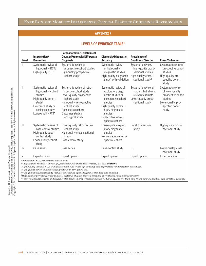
Knee Pain and Mobility Impairments: Clinical Practice Guidelines Revision 2018
a44 | february 2018 | volume 48 | number 2 | journal of orthopaedic & sports physical therapy
APPENDIX F
LEVELS OF EVIDENCE TABLE*
LevelIntervention/Prevention
Pathoanatomic/Risk/Clinical Course/Prognosis/Differential Diagnosis
Diagnosis/Diagnostic Accuracy
Prevalence of Condition/Disorder Exam/Outcomes
I Systematic review of high-quality RCTs
High-quality RCT†
Systematic review of prospective cohort studies
High-quality prospective cohort study‡
Systematic review of high-quality diagnostic studies
High-quality diagnostic study§ with validation
Systematic review, high-quality cross-sectional studies
High-quality cross-sectional study
Systematic review of prospective cohort studies
High-quality pro-spective cohort study
II Systematic review of high-quality cohort studies
High-quality cohort study‡
Outcomes study or ecological study
Lower-quality RCT¶
Systematic review of retro-spective cohort study
Lower-quality prospective cohort study
High-quality retrospective cohort study
Consecutive cohortOutcomes study or
ecological study
Systematic review of exploratory diag-nostic studies or consecutive cohort studies
High-quality explor-atory diagnostic studies
Consecutive retro-spective cohort
Systematic review of studies that allows relevant estimate
Lower-quality cross-sectional study
Systematic review of lower-quality prospective cohort studies
Lower-quality pro-spective cohort study
III Systematic reviews of case-control studies
High-quality case-control study
Lower-quality cohort study
Lower-quality retrospective cohort study
High-quality cross-sectional study
Case-control study
Lower-quality explor-atory diagnostic studies
Nonconsecutive retro-spective cohort
Local nonrandom study
High-quality cross-sectional study
IV Case series Case series Case-control study … Lower-quality cross-sectional study
V Expert opinion Expert opinion Expert opinion Expert opinion Expert opinionAbbreviation: RCT, randomized clinical trial.*Adapted from Phillips et al114 (http://www.cebm.net/index.aspx?o=1025). See also APPENDIX G.†High quality includes RCTs with greater than 80% follow-up, blinding, and appropriate randomization procedures.‡High-quality cohort study includes greater than 80% follow-up.§High-quality diagnostic study includes consistently applied reference standard and blinding.High-quality prevalence study is a cross-sectional study that uses a local and current random sample or censuses.
¶Weaker diagnostic criteria and reference standards, improper randomization, no blinding, and less than 80% follow-up may add bias and threats to validity.
Jour
nal o
f Orth
opae
dic
& S
ports
Phy
sica
l The
rapy
®
Dow
nloa
ded
from
ww
w.jo
spt.o
rg a
t on
Febr
uary
8, 2
018.
For
per
sona
l use
onl
y. N
o ot
her u
ses w
ithou
t per
mis
sion
. C
opyr
ight
© 2
018
Jour
nal o
f Orth
opae
dic
& S
ports
Phy
sica
l The
rapy
®. A
ll rig
hts r
eser
ved.
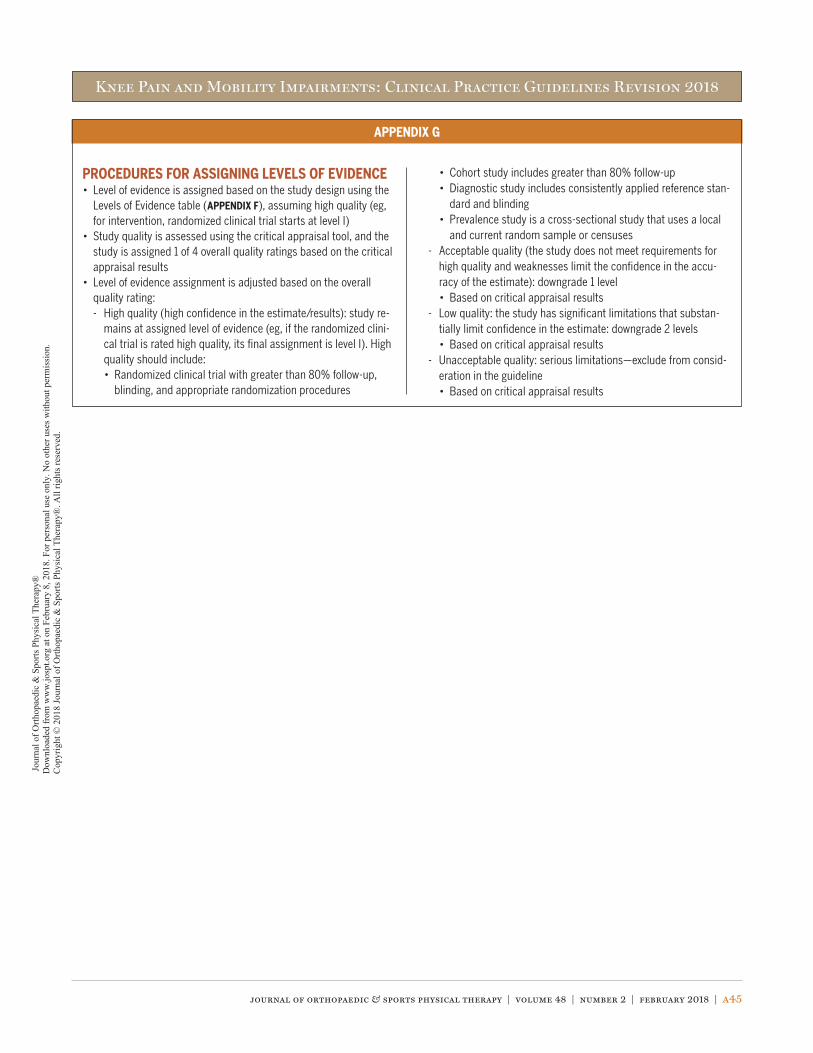
Knee Pain and Mobility Impairments: Clinical Practice Guidelines Revision 2018
journal of orthopaedic & sports physical therapy | volume 48 | number 2 | february 2018 | a45
APPENDIX G
PROCEDURES FOR ASSIGNING LEVELS OF EVIDENCE• Level of evidence is assigned based on the study design using the
Levels of Evidence table (APPENDIX F), assuming high quality (eg, for intervention, randomized clinical trial starts at level I)
• Study quality is assessed using the critical appraisal tool, and the study is assigned 1 of 4 overall quality ratings based on the critical appraisal results
• Level of evidence assignment is adjusted based on the overall quality rating:- High quality (high confidence in the estimate/results): study re-
mains at assigned level of evidence (eg, if the randomized clini-cal trial is rated high quality, its final assignment is level I). High quality should include:• Randomized clinical trial with greater than 80% follow-up,
blinding, and appropriate randomization procedures
• Cohort study includes greater than 80% follow-up• Diagnostic study includes consistently applied reference stan-
dard and blinding• Prevalence study is a cross-sectional study that uses a local
and current random sample or censuses- Acceptable quality (the study does not meet requirements for
high quality and weaknesses limit the confidence in the accu-racy of the estimate): downgrade 1 level• Based on critical appraisal results
- Low quality: the study has significant limitations that substan-tially limit confidence in the estimate: downgrade 2 levels• Based on critical appraisal results
- Unacceptable quality: serious limitations—exclude from consid-eration in the guideline• Based on critical appraisal results
Jour
nal o
f Orth
opae
dic
& S
ports
Phy
sica
l The
rapy
®
Dow
nloa
ded
from
ww
w.jo
spt.o
rg a
t on
Febr
uary
8, 2
018.
For
per
sona
l use
onl
y. N
o ot
her u
ses w
ithou
t per
mis
sion
. C
opyr
ight
© 2
018
Jour
nal o
f Orth
opae
dic
& S
ports
Phy
sica
l The
rapy
®. A
ll rig
hts r
eser
ved.
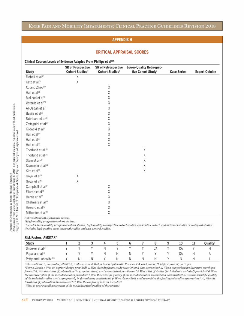
Knee Pain and Mobility Impairments: Clinical Practice Guidelines Revision 2018
a46 | february 2018 | volume 48 | number 2 | journal of orthopaedic & sports physical therapy
APPENDIX H
CRITICAL APPRAISAL SCORES
Clinical Course: Levels of Evidence Adapted From Phillips et al114
StudySR of Prospective Cohort Studies*
SR of Retrospective Cohort Studies†
Lower-Quality Retrospec-tive Cohort Study‡ Case Series Expert Opinion
Frobell et al52 X
Katz et al78 X
Xu and Zhao145 X
Hall et al61 X
McLeod et al97 X
Østerås et al109 X
Al-Dadah et al3 X
Busija et al26 X
Fabricant et al48 X
Zaffagnini et al147 X
Kijowski et al81 X
Hall et al64 X
Hall et al63 X
Hall et al62 X
Thorlund et al133 X
Thorlund et al132 X
Stein et al126 X
Scanzello et al122 X
Kim et al82 X
Goyal et al58 X
Goyal et al57 X
Campbell et al27 X
Filardo et al50 X
Harris et al65 X
Chalmers et al30 X
Howard et al70 X
Mithoefer et al99 XAbbreviation: SR, systematic review.*High-quality prospective cohort studies.†Includes lower-quality prospective cohort studies, high-quality retrospective cohort studies, consecutive cohort, and outcomes studies or ecological studies.‡Includes high-quality cross-sectional studies and case-control studies.
Risk Factors: AMSTAR*
Study 1 2 3 4 5 6 7 8 9 10 11 Quality†
Snoeker et al124 Y Y Y N Y Y Y CA Y CA Y H
Papalia et al110 Y Y Y N N N Y Y Y CA N A
Petty and Lubowitz 113 Y N N Y N N N N Y N N LAbbreviations: A, acceptable; AMSTAR, A Measurement Tool to Assess Systematic Reviews; CA, can’t access; H, high; L, low; N, no; Y, yes.*Yes/no. Items: 1, Was an a priori design provided? 2, Was there duplicate study selection and data extraction? 3, Was a comprehensive literature search per-formed? 4, Was the status of publication (ie, gray literature) used as an inclusion criterion? 5, Was a list of studies (included and excluded) provided? 6, Were the characteristics of the included studies provided? 7, Was the scientific quality of the included studies assessed and documented? 8, Was the scientific quality of the included studies used appropriately in formulating conclusions? 9, Were the methods used to combine the findings of studies appropriate? 10, Was the likelihood of publication bias assessed? 11, Was the conflict of interest included?†What is your overall assessment of the methodological quality of this review?
Jour
nal o
f Orth
opae
dic
& S
ports
Phy
sica
l The
rapy
®
Dow
nloa
ded
from
ww
w.jo
spt.o
rg a
t on
Febr
uary
8, 2
018.
For
per
sona
l use
onl
y. N
o ot
her u
ses w
ithou
t per
mis
sion
. C
opyr
ight
© 2
018
Jour
nal o
f Orth
opae
dic
& S
ports
Phy
sica
l The
rapy
®. A
ll rig
hts r
eser
ved.
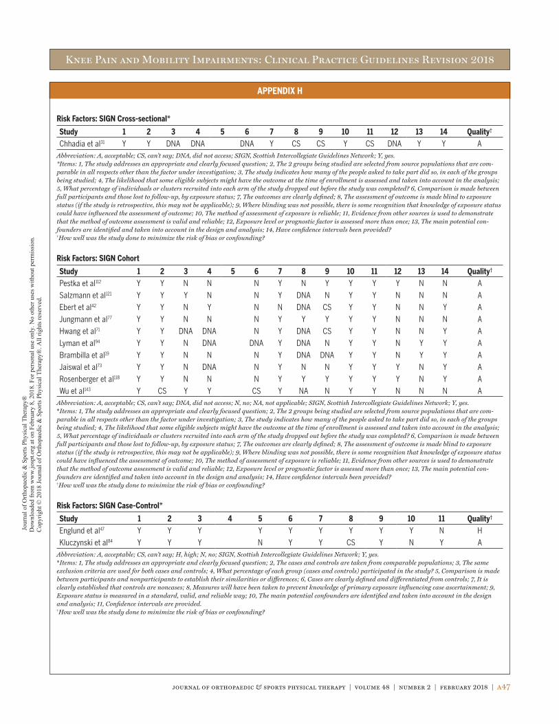
Knee Pain and Mobility Impairments: Clinical Practice Guidelines Revision 2018
journal of orthopaedic & sports physical therapy | volume 48 | number 2 | february 2018 | a47
APPENDIX H
Risk Factors: SIGN Cross-sectional*
Study 1 2 3 4 5 6 7 8 9 10 11 12 13 14 Quality†
Chhadia et al31 Y Y DNA DNA DNA Y CS CS Y CS DNA Y Y AAbbreviation: A, acceptable; CS, can’t say; DNA, did not access; SIGN, Scottish Intercollegiate Guidelines Network; Y, yes.*Items: 1, The study addresses an appropriate and clearly focused question; 2, The 2 groups being studied are selected from source populations that are com-parable in all respects other than the factor under investigation; 3, The study indicates how many of the people asked to take part did so, in each of the groups being studied; 4, The likelihood that some eligible subjects might have the outcome at the time of enrollment is assessed and taken into account in the analysis; 5, What percentage of individuals or clusters recruited into each arm of the study dropped out before the study was completed? 6, Comparison is made between full participants and those lost to follow-up, by exposure status; 7, The outcomes are clearly defined; 8, The assessment of outcome is made blind to exposure status (if the study is retrospective, this may not be applicable); 9, Where blinding was not possible, there is some recognition that knowledge of exposure status could have influenced the assessment of outcome; 10, The method of assessment of exposure is reliable; 11, Evidence from other sources is used to demonstrate that the method of outcome assessment is valid and reliable; 12, Exposure level or prognostic factor is assessed more than once; 13, The main potential con-founders are identified and taken into account in the design and analysis; 14, Have confidence intervals been provided?†How well was the study done to minimize the risk of bias or confounding?
Risk Factors: SIGN Cohort
Study 1 2 3 4 5 6 7 8 9 10 11 12 13 14 Quality†
Pestka et al112 Y Y N N N Y N Y Y Y Y N N A
Salzmann et al121 Y Y Y N N Y DNA N Y Y N N N A
Ebert et al42 Y Y N Y N N DNA CS Y Y N N Y A
Jungmann et al77 Y Y N N N Y Y Y Y Y N N N A
Hwang et al71 Y Y DNA DNA N Y DNA CS Y Y N N Y A
Lyman et al94 Y Y N DNA DNA Y DNA N Y Y N Y Y A
Brambilla et al19 Y Y N N N Y DNA DNA Y Y N Y Y A
Jaiswal et al73 Y Y N DNA N Y N N Y Y Y N Y A
Rosenberger et al118 Y Y N N N Y Y Y Y Y Y N Y A
Wu et al143 Y CS Y Y CS Y NA N Y Y N N N AAbbreviation: A, acceptable; CS, can’t say; DNA, did not access; N, no; NA, not applicable; SIGN, Scottish Intercollegiate Guidelines Network; Y, yes.*Items: 1, The study addresses an appropriate and clearly focused question; 2, The 2 groups being studied are selected from source populations that are com-parable in all respects other than the factor under investigation; 3, The study indicates how many of the people asked to take part did so, in each of the groups being studied; 4, The likelihood that some eligible subjects might have the outcome at the time of enrollment is assessed and taken into account in the analysis; 5, What percentage of individuals or clusters recruited into each arm of the study dropped out before the study was completed? 6, Comparison is made between full participants and those lost to follow-up, by exposure status; 7, The outcomes are clearly defined; 8, The assessment of outcome is made blind to exposure status (if the study is retrospective, this may not be applicable); 9, Where blinding was not possible, there is some recognition that knowledge of exposure status could have influenced the assessment of outcome; 10, The method of assessment of exposure is reliable; 11, Evidence from other sources is used to demonstrate that the method of outcome assessment is valid and reliable; 12, Exposure level or prognostic factor is assessed more than once; 13, The main potential con-founders are identified and taken into account in the design and analysis; 14, Have confidence intervals been provided?†How well was the study done to minimize the risk of bias or confounding?
Risk Factors: SIGN Case-Control*
Study 1 2 3 4 5 6 7 8 9 10 11 Quality†
Englund et al47 Y Y Y Y Y Y Y Y Y N H
Kluczynski et al84 Y Y Y N Y Y CS Y N Y AAbbreviation: A, acceptable; CS, can’t say; H, high; N, no; SIGN, Scottish Intercollegiate Guidelines Network; Y, yes.*Items: 1, The study addresses an appropriate and clearly focused question; 2, The cases and controls are taken from comparable populations; 3, The same exclusion criteria are used for both cases and controls; 4, What percentage of each group (cases and controls) participated in the study? 5, Comparison is made between participants and nonparticipants to establish their similarities or differences; 6, Cases are clearly defined and differentiated from controls; 7, It is clearly established that controls are noncases; 8, Measures will have been taken to prevent knowledge of primary exposure influencing case ascertainment; 9, Exposure status is measured in a standard, valid, and reliable way; 10, The main potential confounders are identified and taken into account in the design and analysis; 11, Confidence intervals are provided.†How well was the study done to minimize the risk of bias or confounding?
Jour
nal o
f Orth
opae
dic
& S
ports
Phy
sica
l The
rapy
®
Dow
nloa
ded
from
ww
w.jo
spt.o
rg a
t on
Febr
uary
8, 2
018.
For
per
sona
l use
onl
y. N
o ot
her u
ses w
ithou
t per
mis
sion
. C
opyr
ight
© 2
018
Jour
nal o
f Orth
opae
dic
& S
ports
Phy
sica
l The
rapy
®. A
ll rig
hts r
eser
ved.
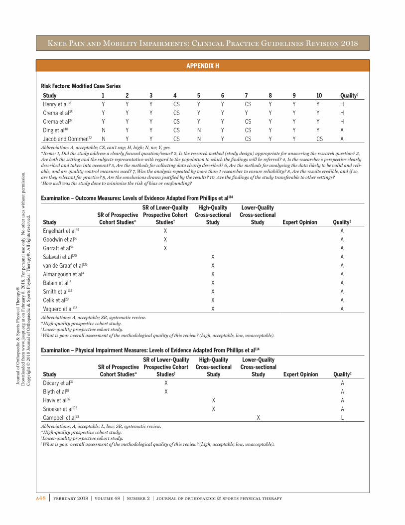
Knee Pain and Mobility Impairments: Clinical Practice Guidelines Revision 2018
a48 | february 2018 | volume 48 | number 2 | journal of orthopaedic & sports physical therapy
APPENDIX H
Risk Factors: Modified Case Series
Study 1 2 3 4 5 6 7 8 9 10 Quality†
Henry et al68 Y Y Y CS Y Y CS Y Y Y H
Crema et al35 Y Y Y CS Y Y Y Y Y Y H
Crema et al34 Y Y Y CS Y Y CS Y Y Y H
Ding et al40 N Y Y CS N Y CS Y Y Y A
Jacob and Oommen72 N Y Y CS N Y CS Y Y CS AAbbreviation: A, acceptable; CS, can’t say; H, high; N, no; Y, yes.*Items: 1, Did the study address a clearly focused question/issue? 2, Is the research method (study design) appropriate for answering the research question? 3, Are both the setting and the subjects representative with regard to the population to which the findings will be referred? 4, Is the researcher’s perspective clearly described and taken into account? 5, Are the methods for collecting data clearly described? 6, Are the methods for analyzing the data likely to be valid and reli-able, and are quality control measures used? 7, Was the analysis repeated by more than 1 researcher to ensure reliability? 8, Are the results credible, and if so, are they relevant for practice? 9, Are the conclusions drawn justified by the results? 10, Are the findings of the study transferable to other settings?†How well was the study done to minimize the risk of bias or confounding?
Examination – Outcome Measures: Levels of Evidence Adapted From Phillips et al114
StudySR of Prospective Cohort Studies*
SR of Lower-Quality Prospective Cohort
Studies†
High-Quality Cross-sectional
Study
Lower-Quality Cross-sectional
Study Expert Opinion Quality‡
Engelhart et al45 X A
Goodwin et al56 X A
Garratt et al54 X A
Salavati et al120 X A
van de Graaf et al136 X A
Almangoush et al4 X A
Balain et al13 X A
Smith et al123 X A
Celik et al29 X A
Vaquero et al137 X AAbbreviations: A, acceptable; SR, systematic review.*High-quality prospective cohort study.†Lower-quality prospective cohort study.‡What is your overall assessment of the methodological quality of this review? (high, acceptable, low, unacceptable).
Examination – Physical Impairment Measures: Levels of Evidence Adapted From Phillips et al114
StudySR of Prospective Cohort Studies*
SR of Lower-Quality Prospective Cohort
Studies†
High-Quality Cross-sectional
Study
Lower-Quality Cross-sectional
Study Expert Opinion Quality‡
Décary et al37 X A
Blyth et al18 X A
Haviv et al66 X A
Snoeker et al125 X A
Campbell et al28 X LAbbreviations: A, acceptable; L, low; SR, systematic review.*High-quality prospective cohort study.†Lower-quality prospective cohort study.‡What is your overall assessment of the methodological quality of this review? (high, acceptable, low, unacceptable).
Jour
nal o
f Orth
opae
dic
& S
ports
Phy
sica
l The
rapy
®
Dow
nloa
ded
from
ww
w.jo
spt.o
rg a
t on
Febr
uary
8, 2
018.
For
per
sona
l use
onl
y. N
o ot
her u
ses w
ithou
t per
mis
sion
. C
opyr
ight
© 2
018
Jour
nal o
f Orth
opae
dic
& S
ports
Phy
sica
l The
rapy
®. A
ll rig
hts r
eser
ved.
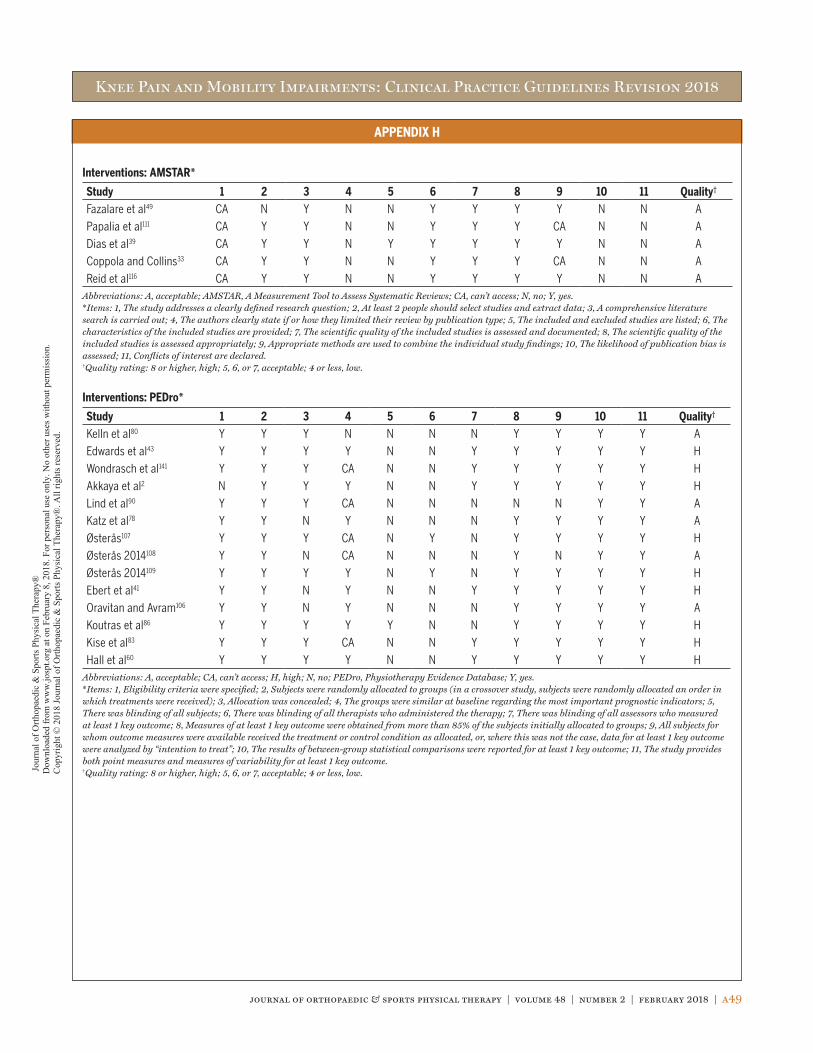
Knee Pain and Mobility Impairments: Clinical Practice Guidelines Revision 2018
journal of orthopaedic & sports physical therapy | volume 48 | number 2 | february 2018 | a49
APPENDIX H
Interventions: AMSTAR*
Study 1 2 3 4 5 6 7 8 9 10 11 Quality†
Fazalare et al49 CA N Y N N Y Y Y Y N N A
Papalia et al111 CA Y Y N N Y Y Y CA N N A
Dias et al39 CA Y Y N Y Y Y Y Y N N A
Coppola and Collins33 CA Y Y N N Y Y Y CA N N A
Reid et al116 CA Y Y N N Y Y Y Y N N AAbbreviations: A, acceptable; AMSTAR, A Measurement Tool to Assess Systematic Reviews; CA, can’t access; N, no; Y, yes.*Items: 1, The study addresses a clearly defined research question; 2, At least 2 people should select studies and extract data; 3, A comprehensive literature search is carried out; 4, The authors clearly state if or how they limited their review by publication type; 5, The included and excluded studies are listed; 6, The characteristics of the included studies are provided; 7, The scientific quality of the included studies is assessed and documented; 8, The scientific quality of the included studies is assessed appropriately; 9, Appropriate methods are used to combine the individual study findings; 10, The likelihood of publication bias is assessed; 11, Conflicts of interest are declared.†Quality rating: 8 or higher, high; 5, 6, or 7, acceptable; 4 or less, low.
Interventions: PEDro*
Study 1 2 3 4 5 6 7 8 9 10 11 Quality†
Kelln et al80 Y Y Y N N N N Y Y Y Y A
Edwards et al43 Y Y Y Y N N Y Y Y Y Y H
Wondrasch et al141 Y Y Y CA N N Y Y Y Y Y H
Akkaya et al2 N Y Y Y N N Y Y Y Y Y H
Lind et al90 Y Y Y CA N N N N N Y Y A
Katz et al78 Y Y N Y N N N Y Y Y Y A
Østerås107 Y Y Y CA N Y N Y Y Y Y H
Østerås 2014108 Y Y N CA N N N Y N Y Y A
Østerås 2014109 Y Y Y Y N Y N Y Y Y Y H
Ebert et al41 Y Y N Y N N Y Y Y Y Y H
Oravitan and Avram106 Y Y N Y N N N Y Y Y Y A
Koutras et al86 Y Y Y Y Y N N Y Y Y Y H
Kise et al83 Y Y Y CA N N Y Y Y Y Y H
Hall et al60 Y Y Y Y N N Y Y Y Y Y HAbbreviations: A, acceptable; CA, can’t access; H, high; N, no; PEDro, Physiotherapy Evidence Database; Y, yes.*Items: 1, Eligibility criteria were specified; 2, Subjects were randomly allocated to groups (in a crossover study, subjects were randomly allocated an order in which treatments were received); 3, Allocation was concealed; 4, The groups were similar at baseline regarding the most important prognostic indicators; 5, There was blinding of all subjects; 6, There was blinding of all therapists who administered the therapy; 7, There was blinding of all assessors who measured at least 1 key outcome; 8, Measures of at least 1 key outcome were obtained from more than 85% of the subjects initially allocated to groups; 9, All subjects for whom outcome measures were available received the treatment or control condition as allocated, or, where this was not the case, data for at least 1 key outcome were analyzed by “intention to treat”; 10, The results of between-group statistical comparisons were reported for at least 1 key outcome; 11, The study provides both point measures and measures of variability for at least 1 key outcome.†Quality rating: 8 or higher, high; 5, 6, or 7, acceptable; 4 or less, low. Jo
urna
l of O
rthop
aedi
c &
Spo
rts P
hysi
cal T
hera
py®
D
ownl
oade
d fr
om w
ww
.josp
t.org
at o
n Fe
brua
ry 8
, 201
8. F
or p
erso
nal u
se o
nly.
No
othe
r use
s with
out p
erm
issi
on.
Cop
yrig
ht ©
201
8 Jo
urna
l of O
rthop
aedi
c &
Spo
rts P
hysi
cal T
hera
py®
. All
right
s res
erve
d.
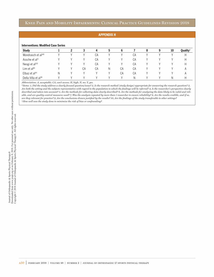
Knee Pain and Mobility Impairments: Clinical Practice Guidelines Revision 2018
a50 | february 2018 | volume 48 | number 2 | journal of orthopaedic & sports physical therapy
APPENDIX H
Interventions: Modified Case Series
Study 1 2 3 4 5 6 7 8 9 10 Quality†
Wondrasch et al140 Y Y Y CA Y Y CA Y Y Y H
Assche et al11 Y Y Y CA Y Y CA Y Y Y H
Neogi et al102 Y Y Y CA Y Y CA Y Y Y H
Lim et al89 Y Y CA CA N CA CA Y Y Y A
Elbaz et al44 N Y Y Y Y CA CA Y Y Y A
Della Villa et al38 Y Y Y Y Y Y N Y Y N HAbbreviation: A, acceptable; CA, can’t access; H, high; N, no; Y, yes.*Items: 1, Did the study address a clearly focused question/issue? 2, Is the research method (study design) appropriate for answering the research question? 3, Are both the setting and the subjects representative with regard to the population to which the findings will be referred? 4, Is the researcher’s perspective clearly described and taken into account? 5, Are the methods for collecting data clearly described? 6, Are the methods for analyzing the data likely to be valid and reli-able, and are quality control measures used? 7, Was the analysis repeated by more than 1 researcher to ensure reliability? 8, Are the results credible, and if so, are they relevant for practice? 9, Are the conclusions drawn justified by the results? 10, Are the findings of the study transferable to other settings?†How well was the study done to minimize the risk of bias or confounding?
Jour
nal o
f Orth
opae
dic
& S
ports
Phy
sica
l The
rapy
®
Dow
nloa
ded
from
ww
w.jo
spt.o
rg a
t on
Febr
uary
8, 2
018.
For
per
sona
l use
onl
y. N
o ot
her u
ses w
ithou
t per
mis
sion
. C
opyr
ight
© 2
018
Jour
nal o
f Orth
opae
dic
& S
ports
Phy
sica
l The
rapy
®. A
ll rig
hts r
eser
ved.