Embed Size (px)
Citation preview
Evaluation of Knee Problems
OBJECTIVES• Review knee anatomy• Explain tests to look for pathology• Briefly introduce knee problems
Only by a thorough knowledge of anatomy and functional testing can one make an
accurate diagnosis and direct effective care to an injured
knee.
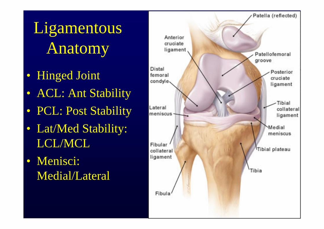
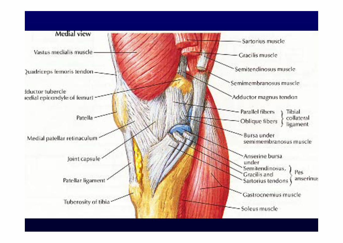
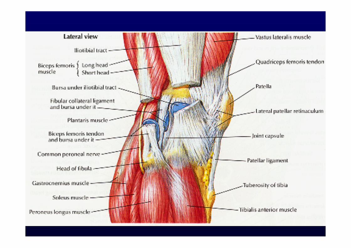
Ligamentous Anatomy
• Hinged Joint• ACL: Ant Stability• PCL: Post Stability• Lat/Med Stability:
LCL/MCL• Menisci:
Medial/Lateral
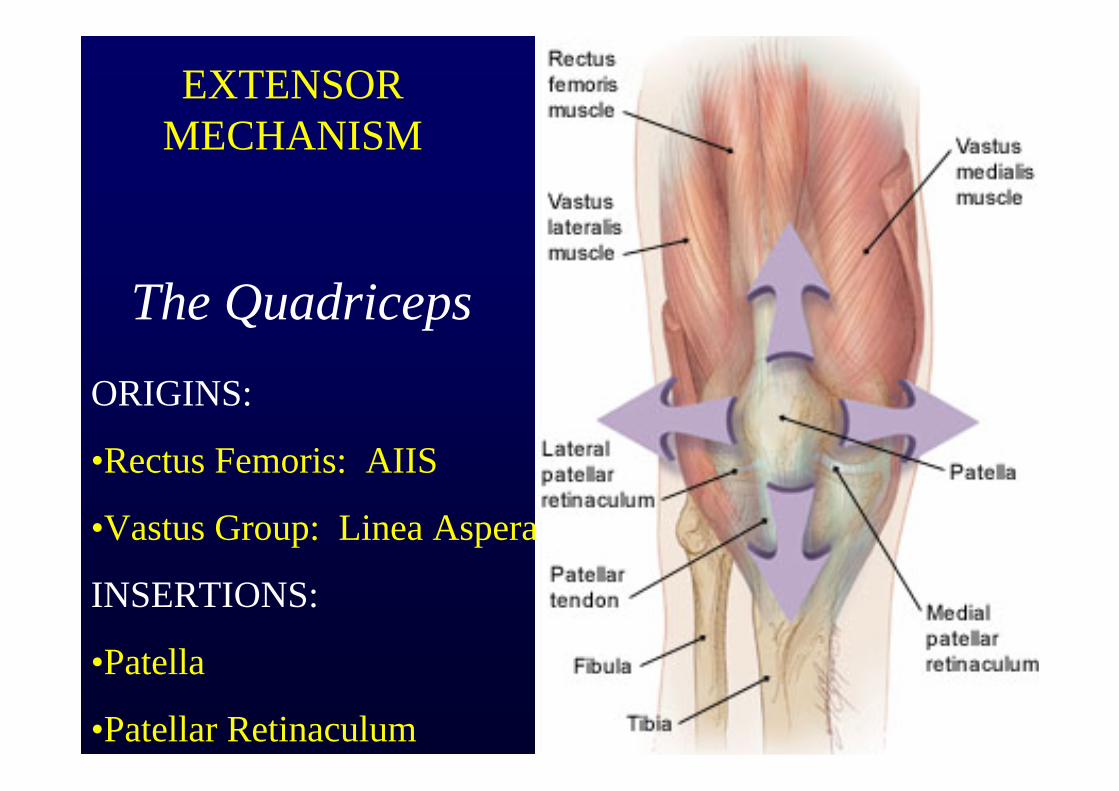
EXTENSOR MECHANISM
The QuadricepsORIGINS:
•Rectus Femoris: AIIS
•Vastus Group: Linea Aspera
INSERTIONS:
•Patella
•Patellar Retinaculum
Always have the patient perform a straight leg raise to rule out an extensor mechanism rupture after
acute trauma
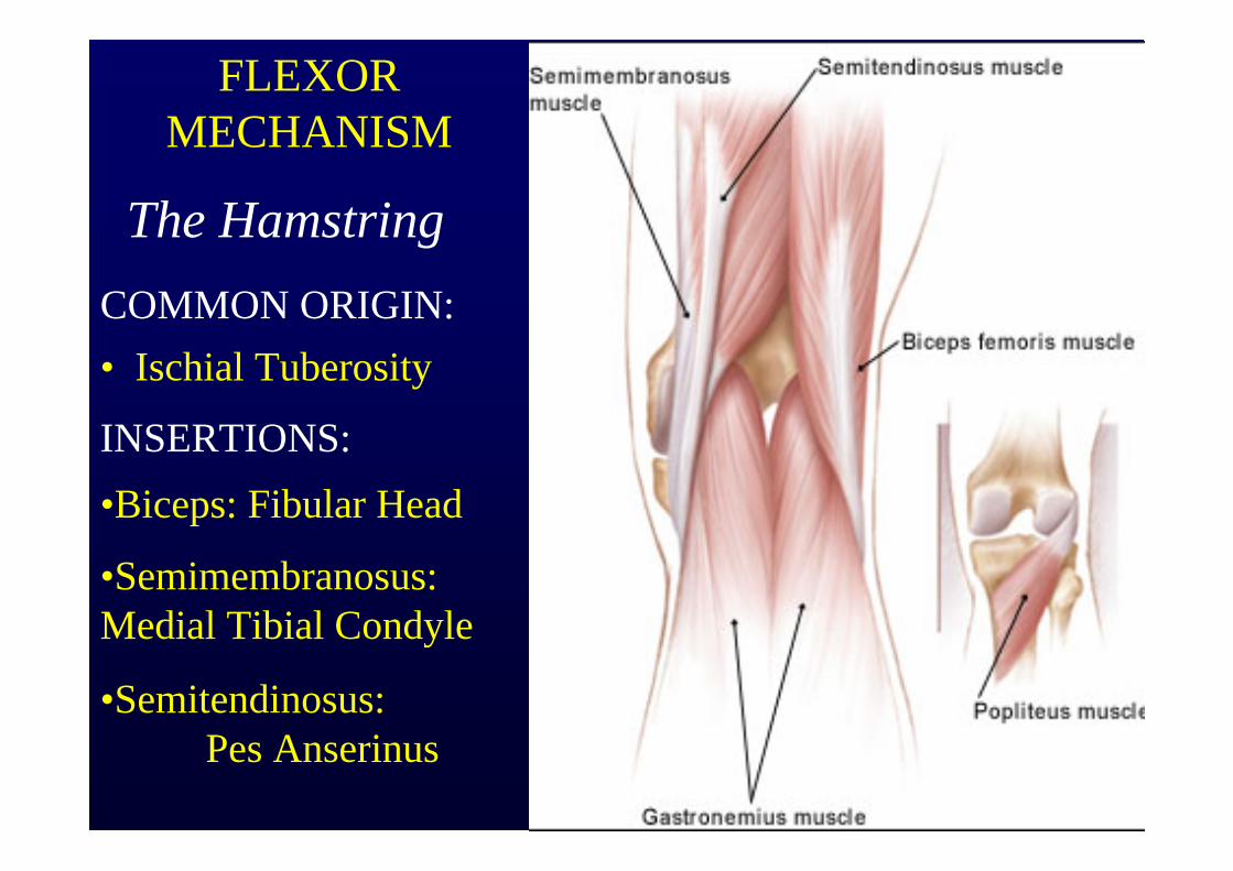
FLEXOR MECHANISM
The HamstringCOMMON ORIGIN:• Ischial Tuberosity
INSERTIONS:•Biceps: Fibular Head
•Semimembranosus: Medial Tibial Condyle
•Semitendinosus: Pes Anserinus
History• Chief Complaint• Antecedent event/Repetitive activity• Previous injuries to affected area• Attempted therapies• Review of symptoms/Past medical
history• Occupation/Treatment Goals
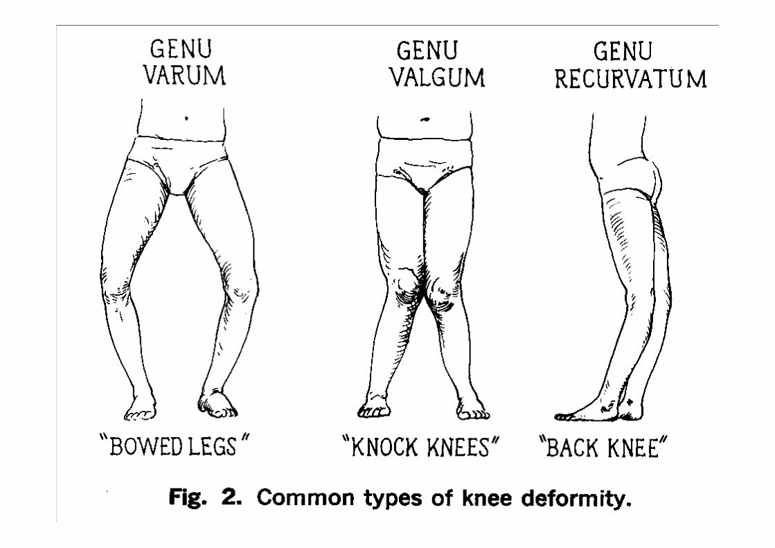
Inspection• Lower extremity alignment• Foot structure• Effusion/Erythmea• Q Angle• Thigh atrophy
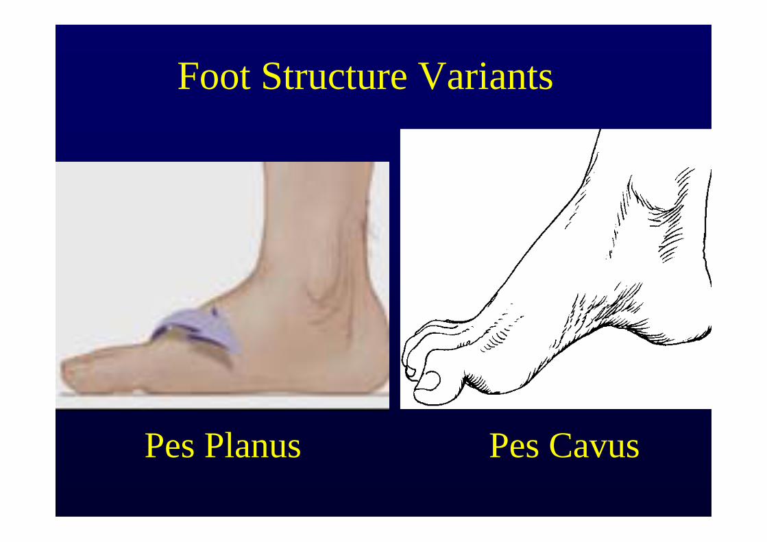
Foot Structure Variants
Pes Planus Pes Cavus
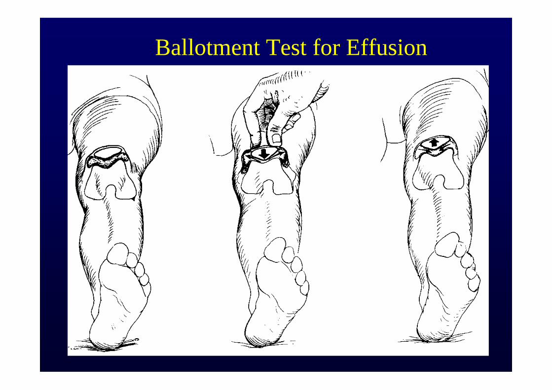
Ballotment Test for Effusion
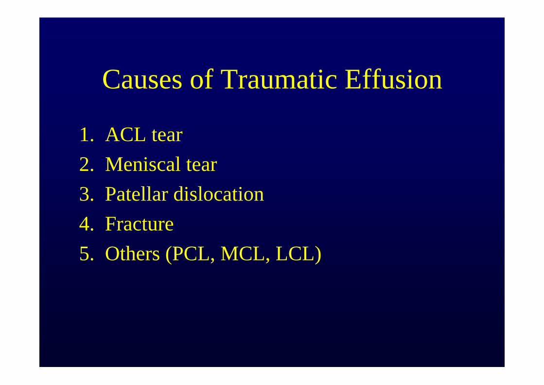
Causes of Traumatic Effusion
1. ACL tear2. Meniscal tear3. Patellar dislocation4. Fracture5. Others (PCL, MCL, LCL)
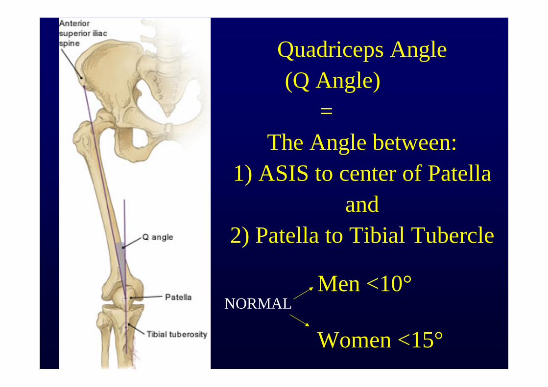
Quadriceps Angle(Q Angle)
=The Angle between:
1) ASIS to center of Patella and
2) Patella to Tibial Tubercle
NORMALMen <10°
Women <15°
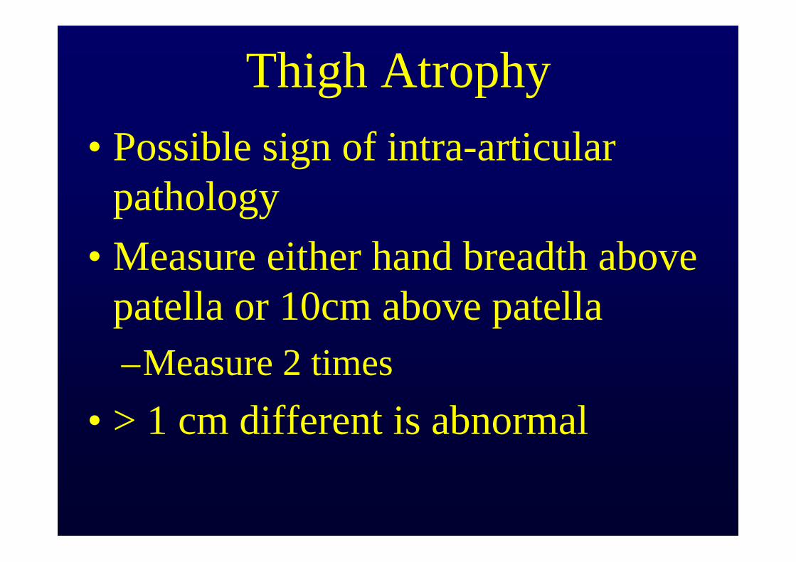
Thigh Atrophy• Possible sign of intra-articular
pathology• Measure either hand breadth above
patella or 10cm above patella –Measure 2 times
• > 1 cm different is abnormal
Leg Length
• FUNCTIONAL METHOD: Compare heights of ASIS & PSIS– Add foot shims in small adjustments until level
• ANATOMICAL METHOD: Measure from ASIS to Medial Malleous– > 1 cm difference is significant
• Pelvic Obliquity will confuse issue
• RADIOLOGIC METHOD: Scanogram (X-ray)– most definitive but usually not needed
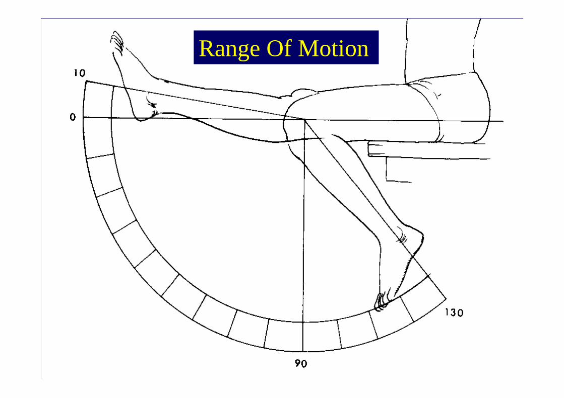
Range Of Motion
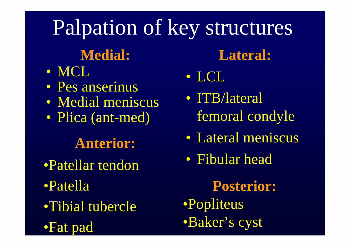
Palpation of key structuresMedial:
• MCL• Pes anserinus• Medial meniscus• Plica (ant-med)
Lateral:• LCL• ITB/lateral
femoral condyle• Lateral meniscus• Fibular head
Anterior:•Patellar tendon•Patella•Tibial tubercle•Fat pad
Posterior:•Popliteus•Baker’s cyst
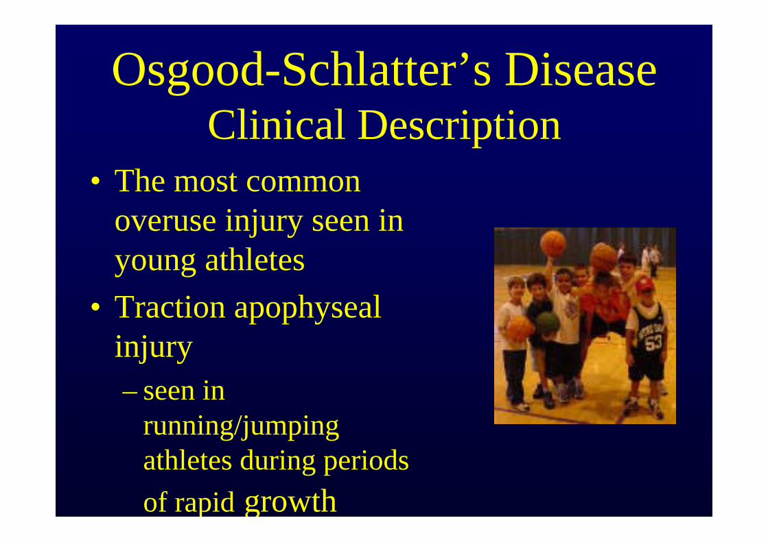
Osgood-Schlatter’s DiseaseClinical Description
• The most common overuse injury seen in young athletes
• Traction apophysealinjury– seen in
running/jumping athletes during periods of rapid growth
Osgood-Schlatter’s DiseaseClinical Features
• History – young athlete complains of painful
enlargement of the tibial tuberosity– pain worse with activity, esp. run/jump
• Exam– tender tibial tuberosity– tight quads +/- hamstrings
• Imaging: usually not necessary
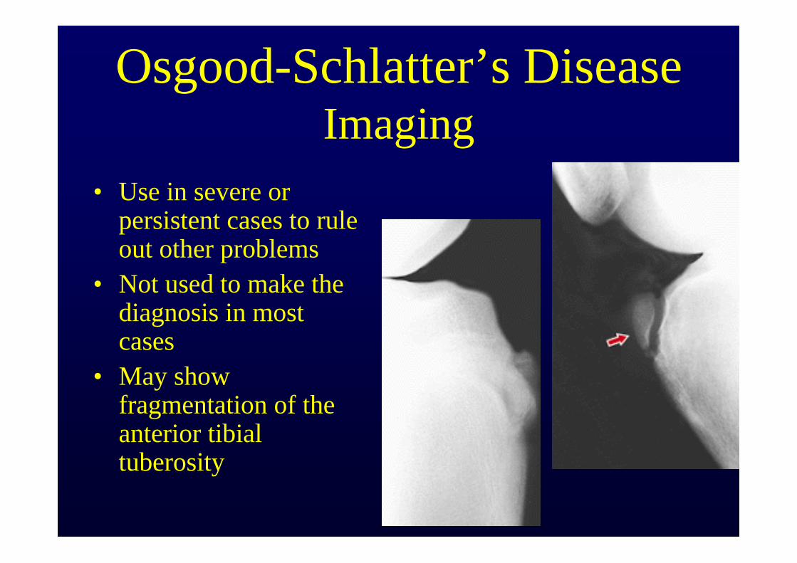
Osgood-Schlatter’s DiseaseImaging
• Use in severe or persistent cases to rule out other problems
• Not used to make the diagnosis in most cases
• May show fragmentation of the anterior tibial tuberosity
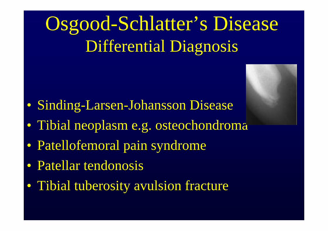
Osgood-Schlatter’s DiseaseDifferential Diagnosis
• Sinding-Larsen-Johansson Disease• Tibial neoplasm e.g. osteochondroma• Patellofemoral pain syndrome• Patellar tendonosis• Tibial tuberosity avulsion fracture
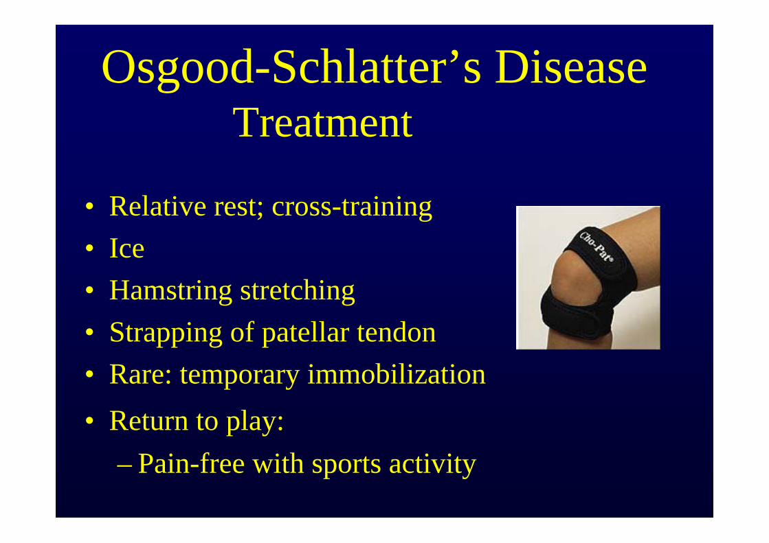
Osgood-Schlatter’s DiseaseTreatment
• Relative rest; cross-training • Ice • Hamstring stretching• Strapping of patellar tendon• Rare: temporary immobilization• Return to play:
– Pain-free with sports activity
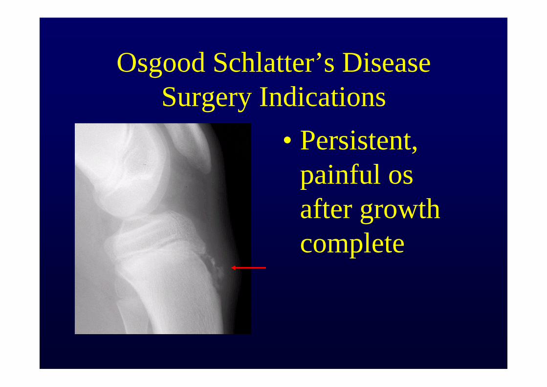
Osgood Schlatter’s Disease Surgery Indications
• Persistent, painful osafter growth complete
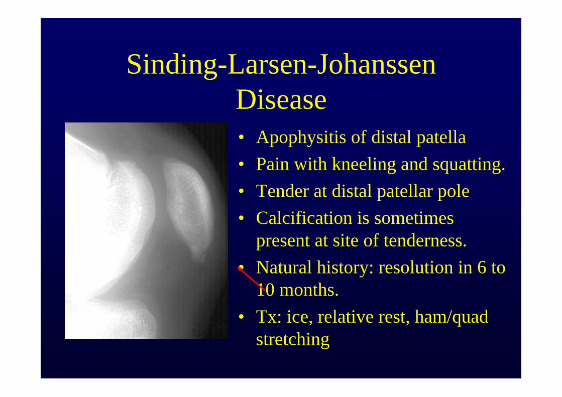
Sinding-Larsen-JohanssenDisease• Apophysitis of distal patella• Pain with kneeling and squatting.• Tender at distal patellar pole• Calcification is sometimes
present at site of tenderness.• Natural history: resolution in 6 to
10 months.• Tx: ice, relative rest, ham/quad
stretching
Patellar Grind Test
• Detects pain from patellar pressure against femur
• Compress patella against femoral groove– Gentle way: pressure with fingers– Most sensitive way: press down above patella;
have patient contract quads• POSITIVE:
– Pain – Crepitus
Management of Patello-Femoral Syndrome
• Cross-training; avoid painful activity• VMO strength ex’s• Flexibility ex’s (quad, hams, ITB, Achilles)• Retinaculum stretching • Patellar sleeve w/ cutout• Correct hyper-pronation• Referral:
– refractory cases w/ high Q angle, tight retinaculum, severe crepitus
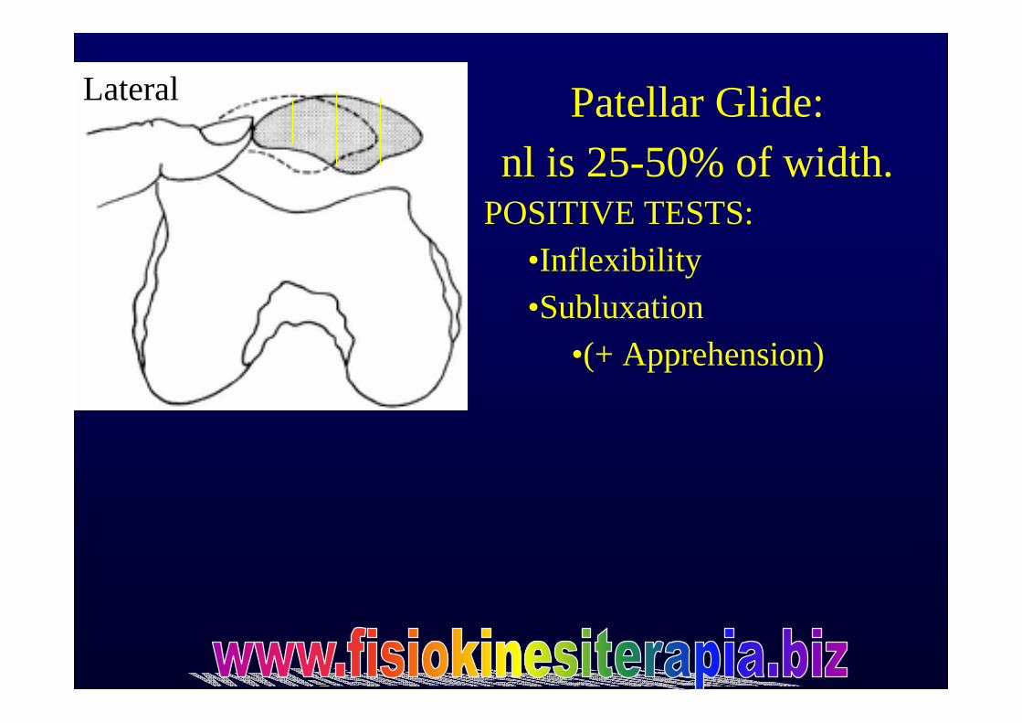
Patellar Glide: nl is 25-50% of width.
POSITIVE TESTS:•Inflexibility•Subluxation
•(+ Apprehension)
Lateral
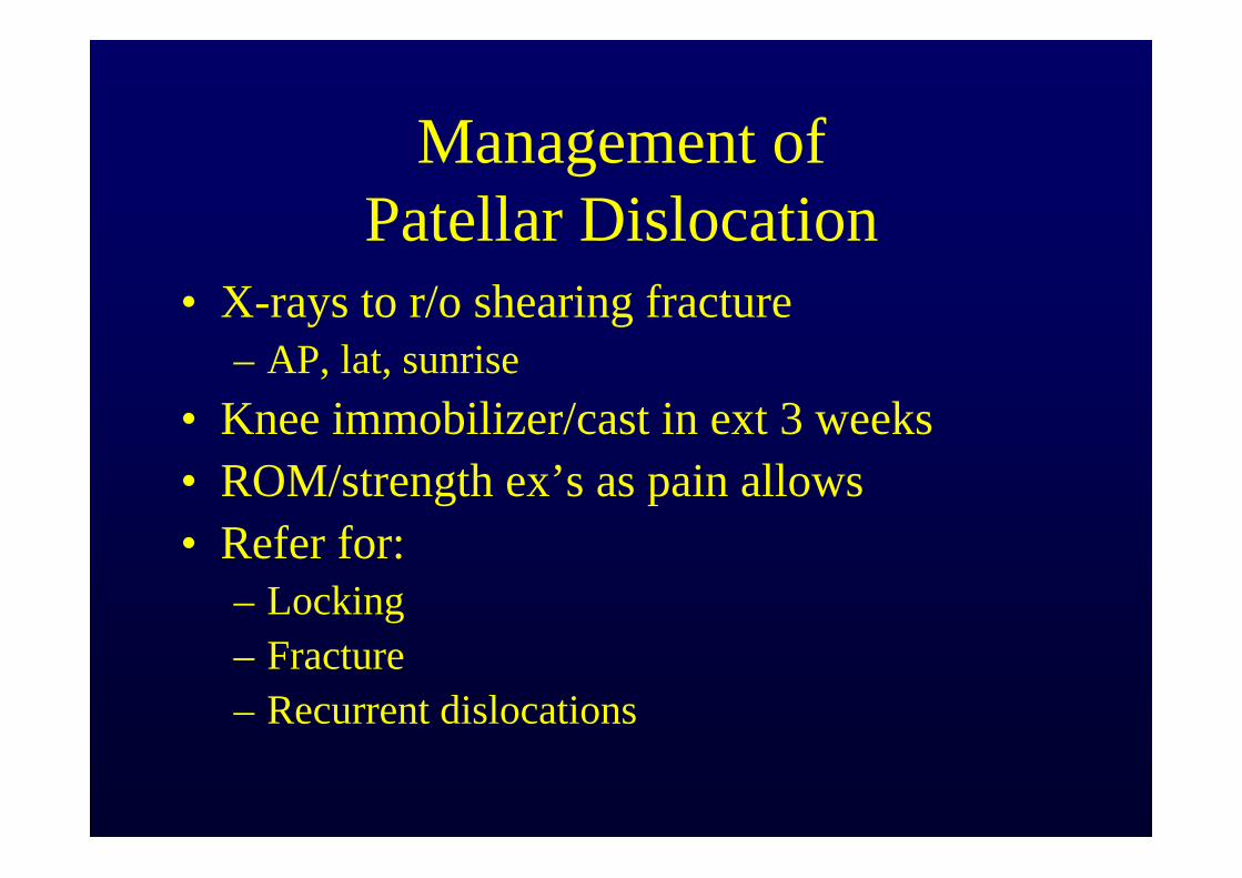
Management of Patellar Dislocation
• X-rays to r/o shearing fracture– AP, lat, sunrise
• Knee immobilizer/cast in ext 3 weeks• ROM/strength ex’s as pain allows• Refer for:
– Locking– Fracture– Recurrent dislocations

Medio-Patellar Plica
Management of Medio-Patellar Plica Syndrome
• Cross-training/relative rest• NSAID 1-2 weeks• Phonopheresis• Injection w/ anesthetic/steroid• Referral: failed 6 months tx
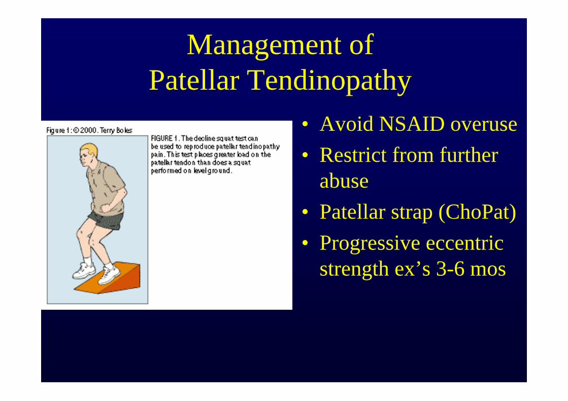
Management ofPatellar Tendinopathy
• Avoid NSAID overuse• Restrict from further
abuse• Patellar strap (ChoPat)• Progressive eccentric
strength ex’s 3-6 mos
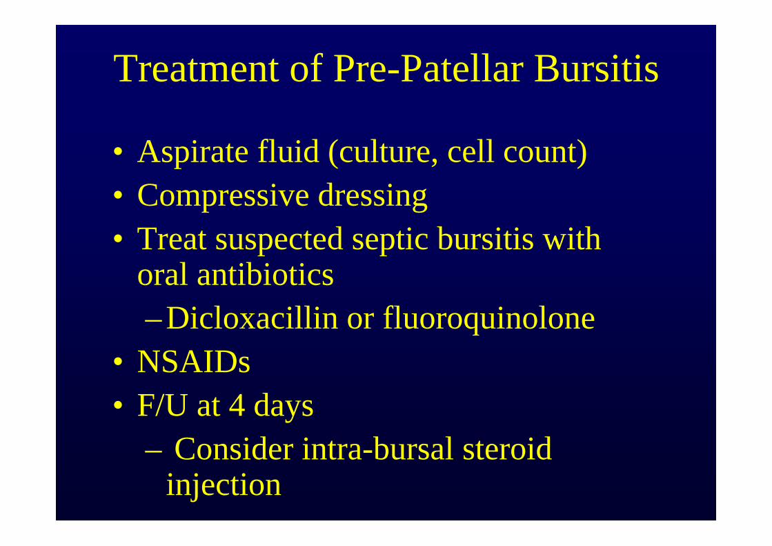
Treatment of Pre-Patellar Bursitis
• Aspirate fluid (culture, cell count)• Compressive dressing• Treat suspected septic bursitis with
oral antibiotics–Dicloxacillin or fluoroquinolone
• NSAIDs• F/U at 4 days
– Consider intra-bursal steroid injection
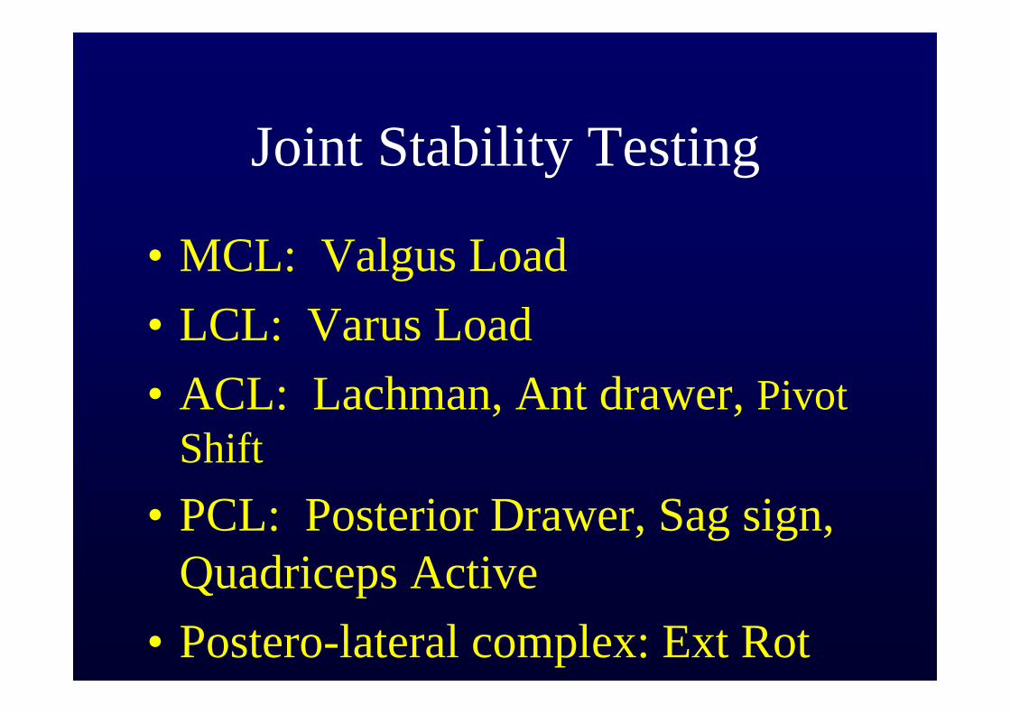
Joint Stability Testing
• MCL: Valgus Load• LCL: Varus Load• ACL: Lachman, Ant drawer, Pivot
Shift• PCL: Posterior Drawer, Sag sign,
Quadriceps Active• Postero-lateral complex: Ext Rot
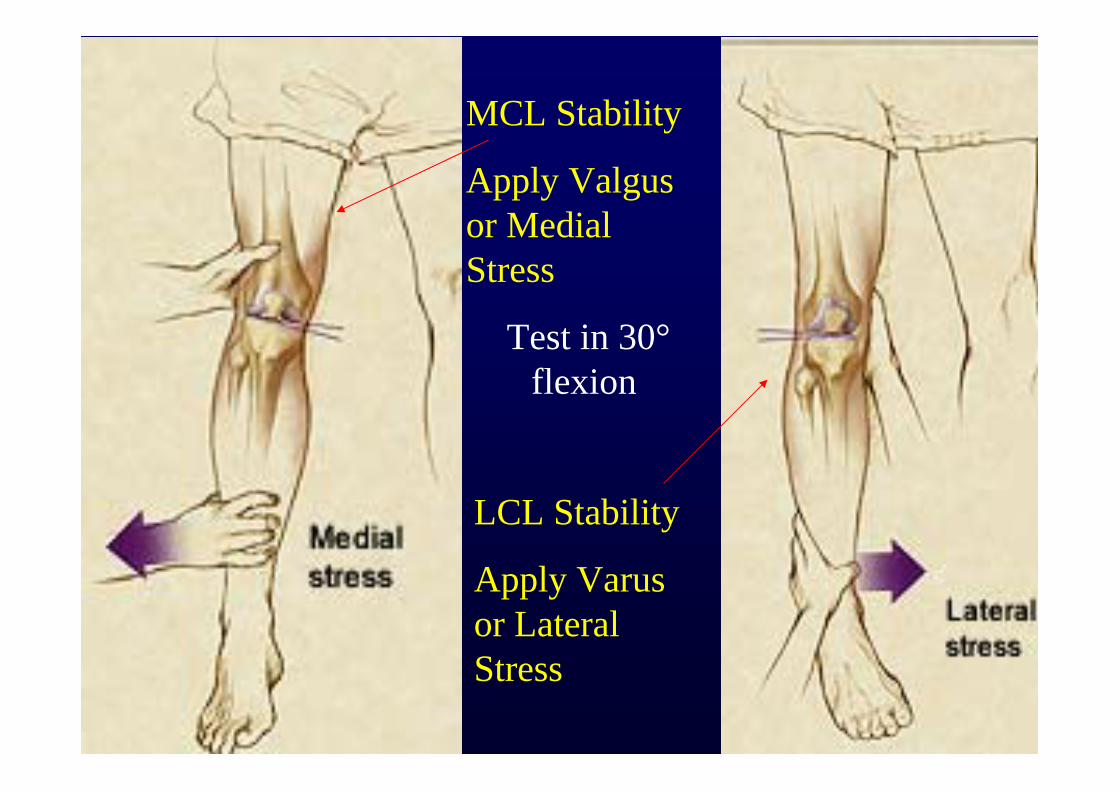
MCL Stability
Apply Valgus or Medial Stress
Test in 30°flexion
LCL Stability
Apply Varus or Lateral Stress
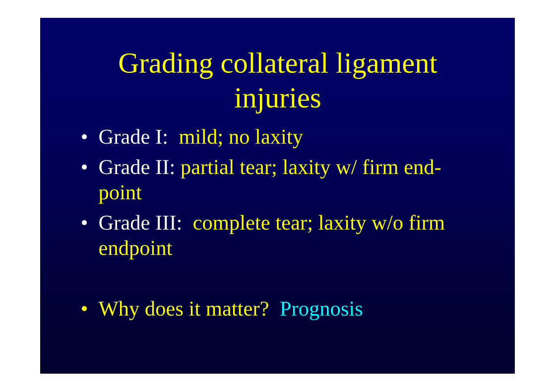
Grading collateral ligament injuries
• Grade I: mild; no laxity• Grade II: partial tear; laxity w/ firm end-
point• Grade III: complete tear; laxity w/o firm
endpoint
• Why does it matter? Prognosis
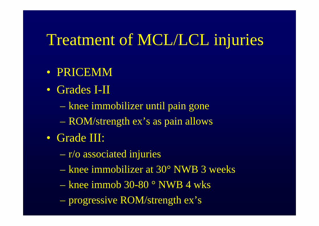
Treatment of MCL/LCL injuries
• PRICEMM• Grades I-II
– knee immobilizer until pain gone– ROM/strength ex’s as pain allows
• Grade III: – r/o associated injuries– knee immobilizer at 30° NWB 3 weeks– knee immob 30-80 ° NWB 4 wks– progressive ROM/strength ex’s
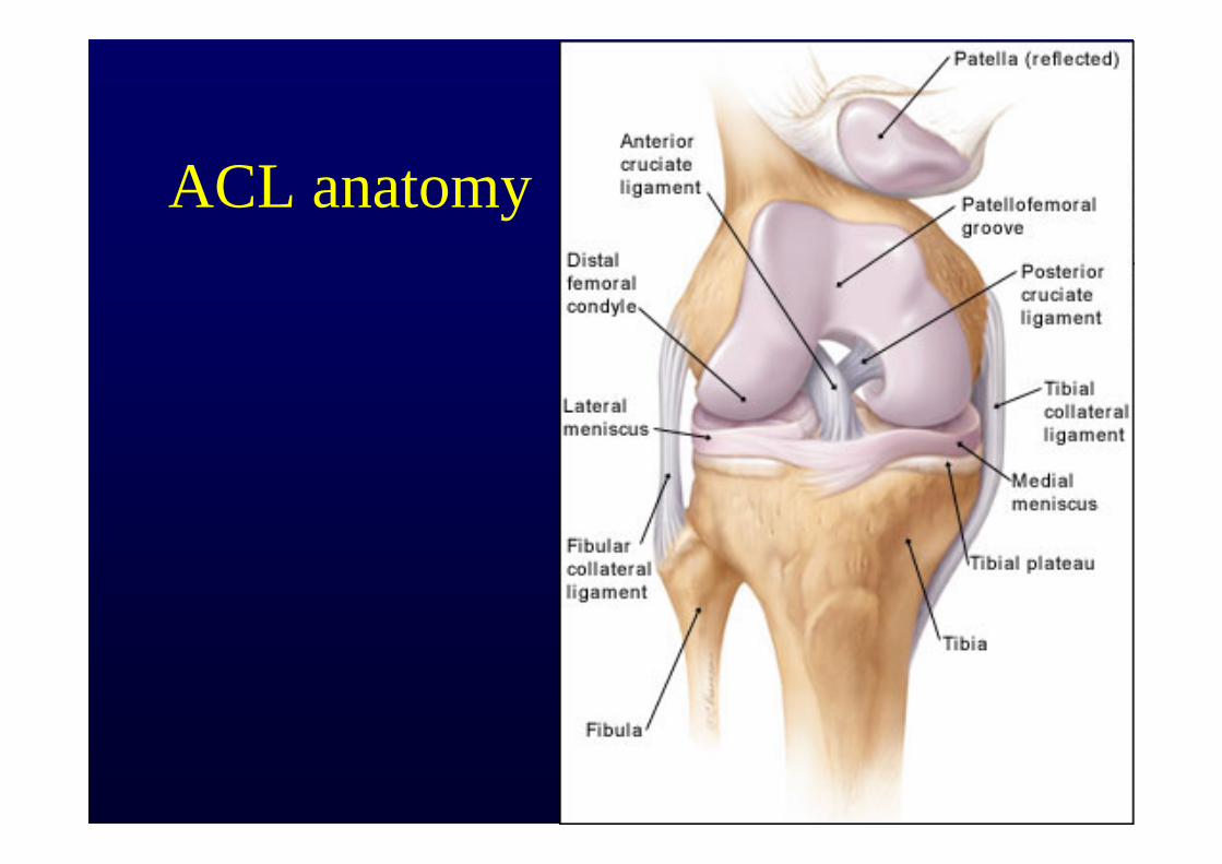
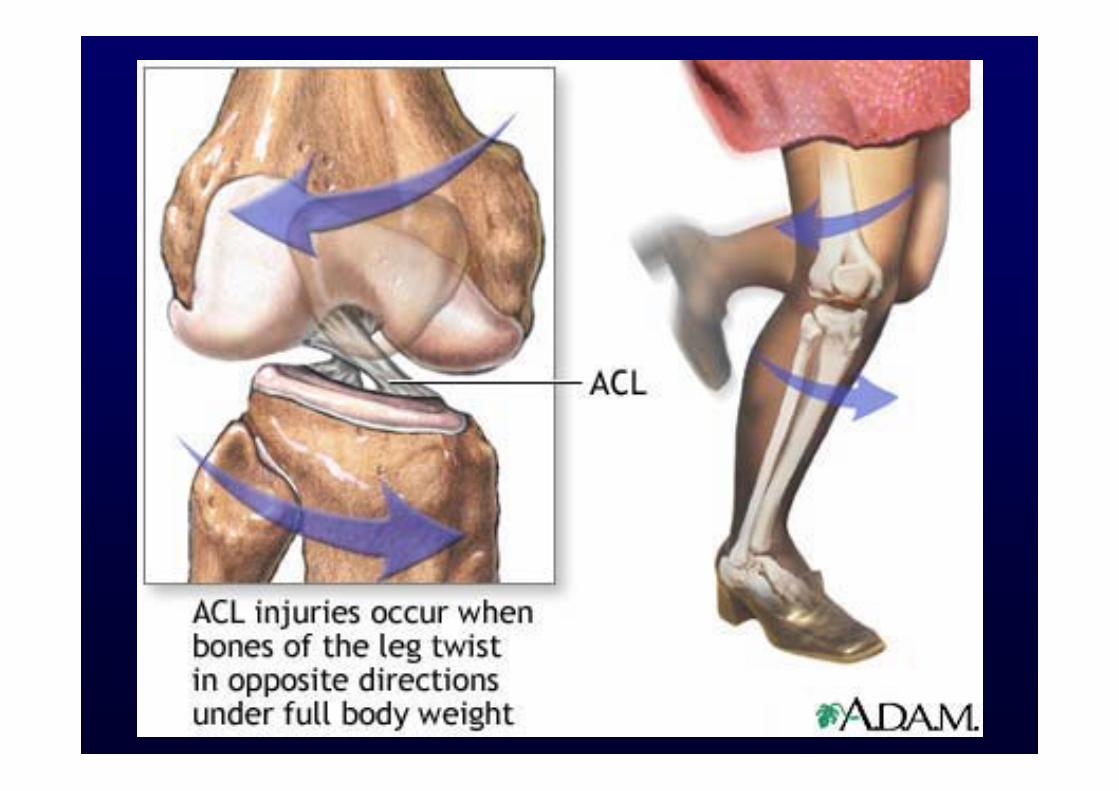
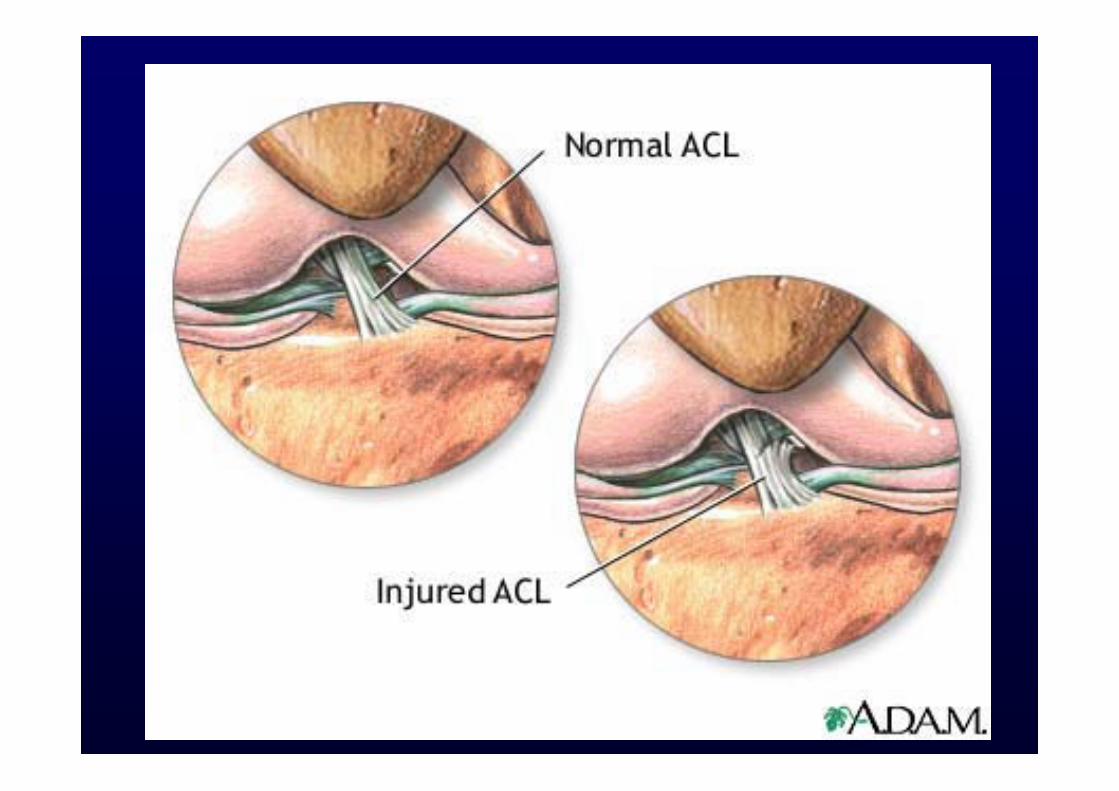
ACL anatomy
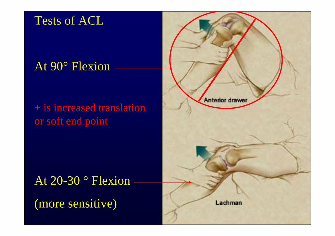
Tests of ACL
At 90° Flexion
At 20-30 ° Flexion
(more sensitive)
+ is increased translation or soft end point
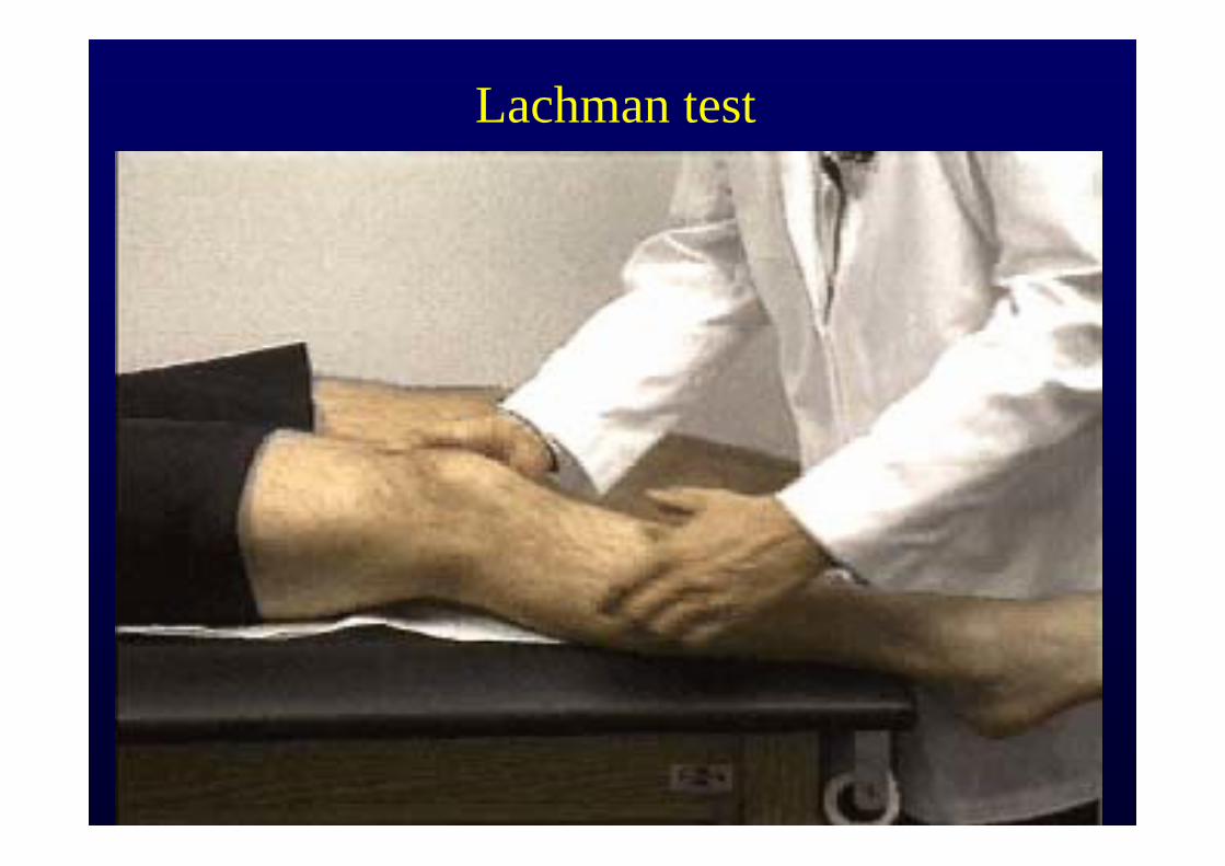
Lachman test

Pivot Shift: ACL Injury
1. Knee extended
2. Internally rotate tibia
3. Apply valgus load
4. Flex Knee
5. At 20-30°, if you feel a jerk at Ant/Lat proximal tibia, test +
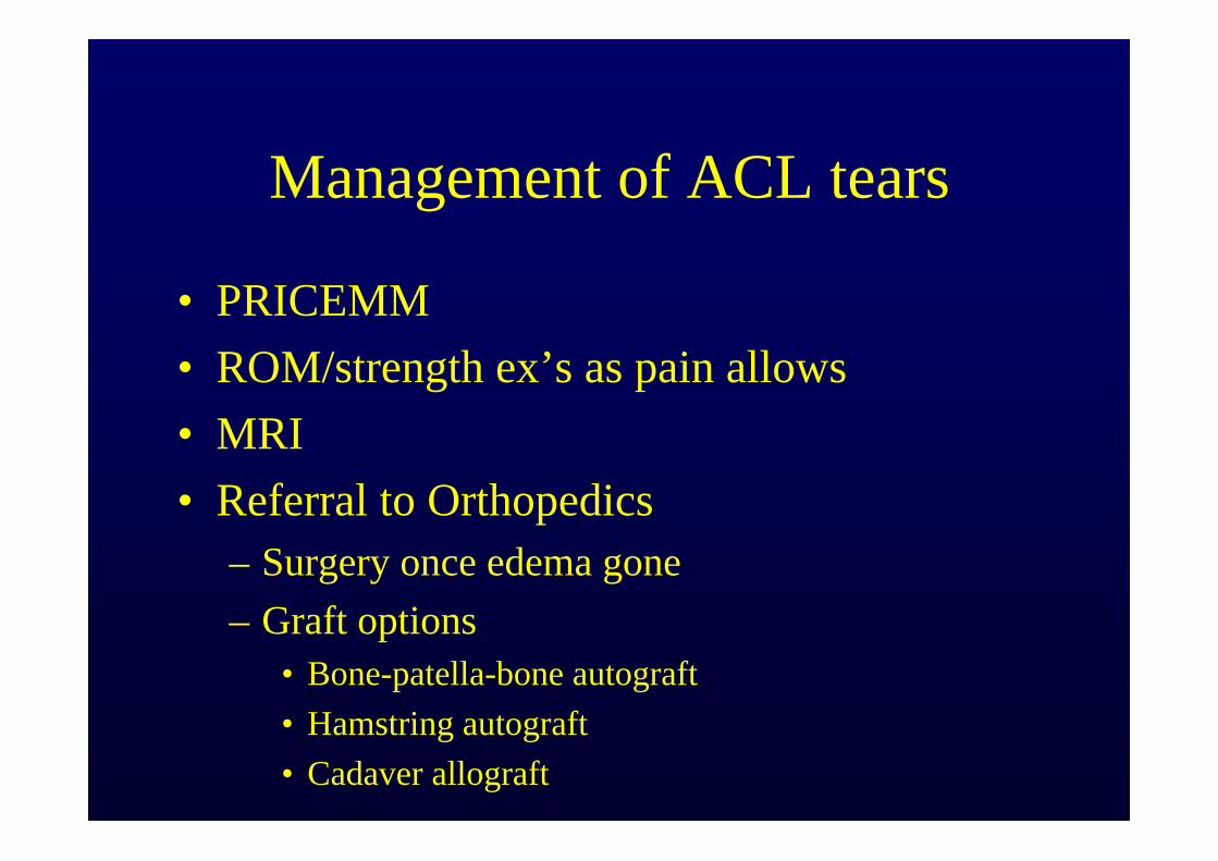
Management of ACL tears
• PRICEMM• ROM/strength ex’s as pain allows• MRI• Referral to Orthopedics
– Surgery once edema gone– Graft options
• Bone-patella-bone autograft• Hamstring autograft• Cadaver allograft
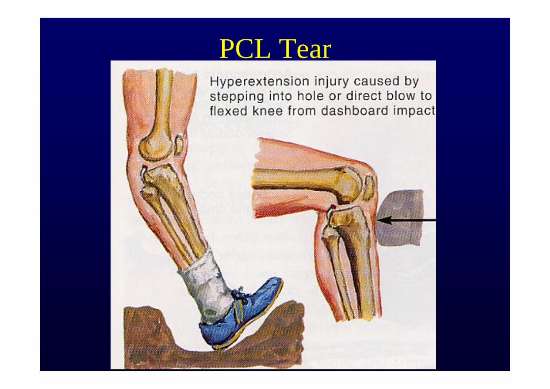
PCL Tear
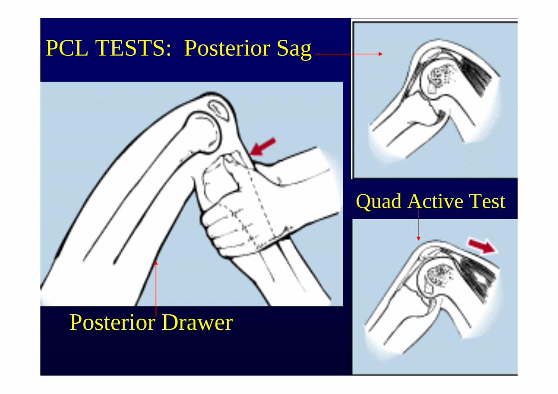
PCL TESTS: Posterior Sag
Quad Active Test
Posterior Drawer
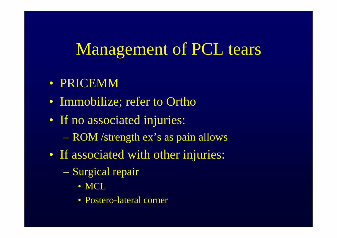
Management of PCL tears
• PRICEMM• Immobilize; refer to Ortho• If no associated injuries:
– ROM /strength ex’s as pain allows• If associated with other injuries:
– Surgical repair• MCL• Postero-lateral corner
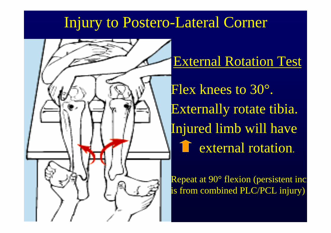
Flex knees to 30°.Externally rotate tibia.Injured limb will have
external rotation.
Repeat at 90° flexion (persistent incris from combined PLC/PCL injury)
Injury to Postero-Lateral Corner
External Rotation Test
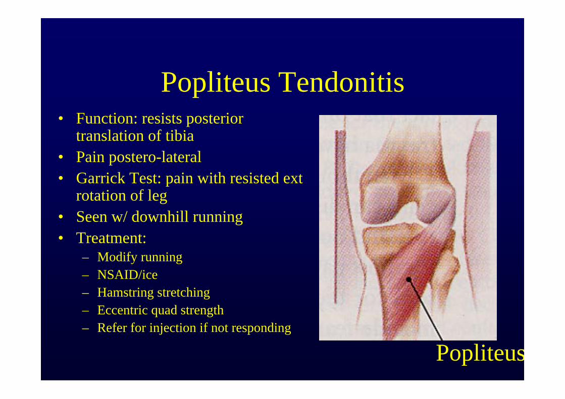
Popliteus Tendonitis• Function: resists posterior
translation of tibia• Pain postero-lateral• Garrick Test: pain with resisted ext
rotation of leg• Seen w/ downhill running• Treatment:
– Modify running– NSAID/ice– Hamstring stretching– Eccentric quad strength– Refer for injection if not responding
Popliteus
Flexibility testing
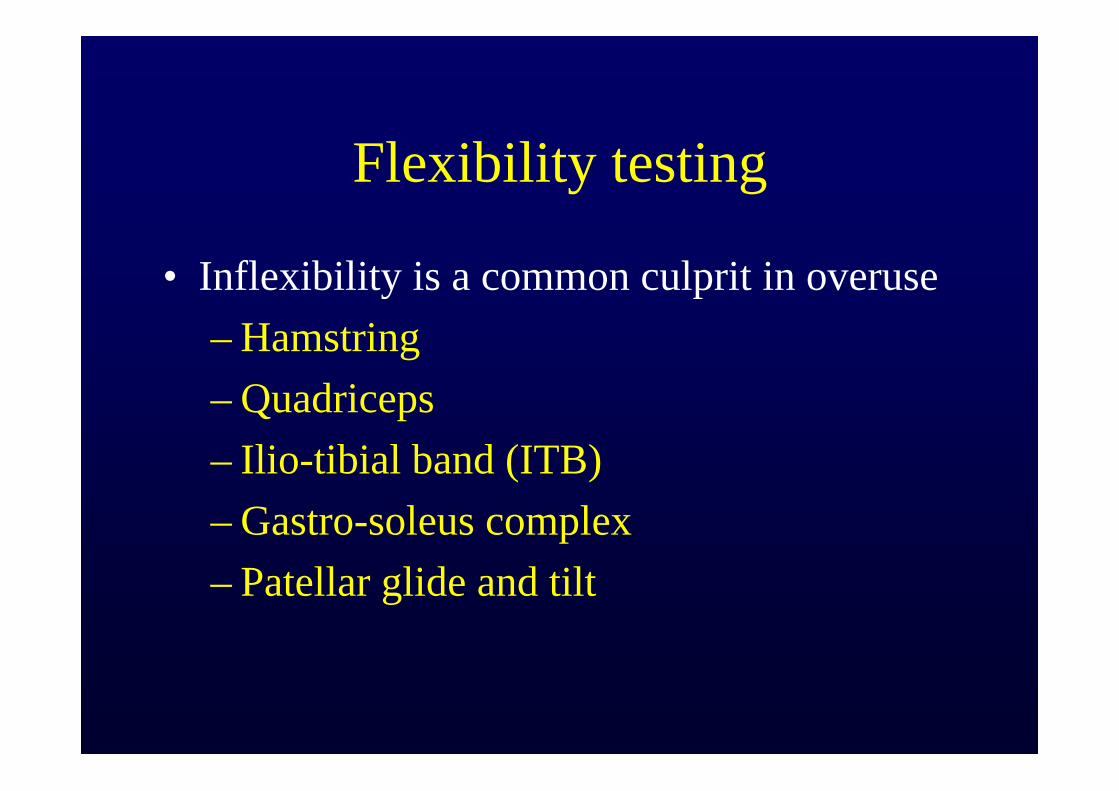
• Inflexibility is a common culprit in overuse– Hamstring– Quadriceps– Ilio-tibial band (ITB)– Gastro-soleus complex– Patellar glide and tilt

Quadriceps flexibility
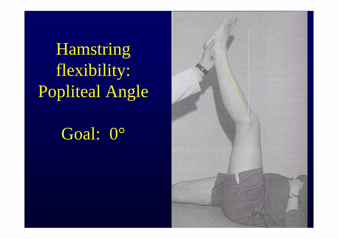
Hamstring flexibility:
Popliteal Angle
Goal: 0°
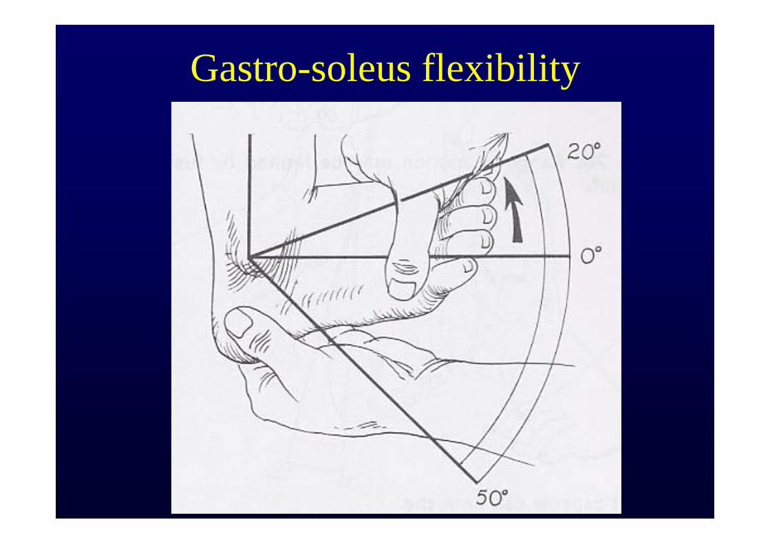
Gastro-soleus flexibility
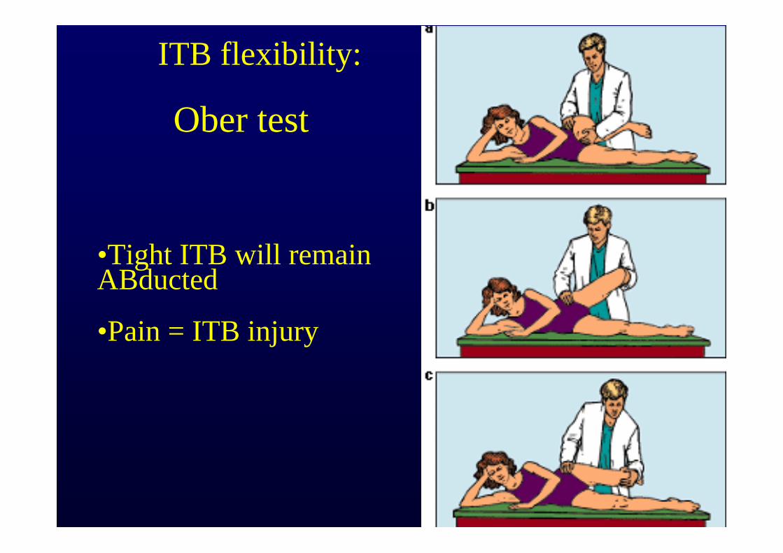
ITB flexibility:
Ober test
•Tight ITB will remain ABducted
•Pain = ITB injury
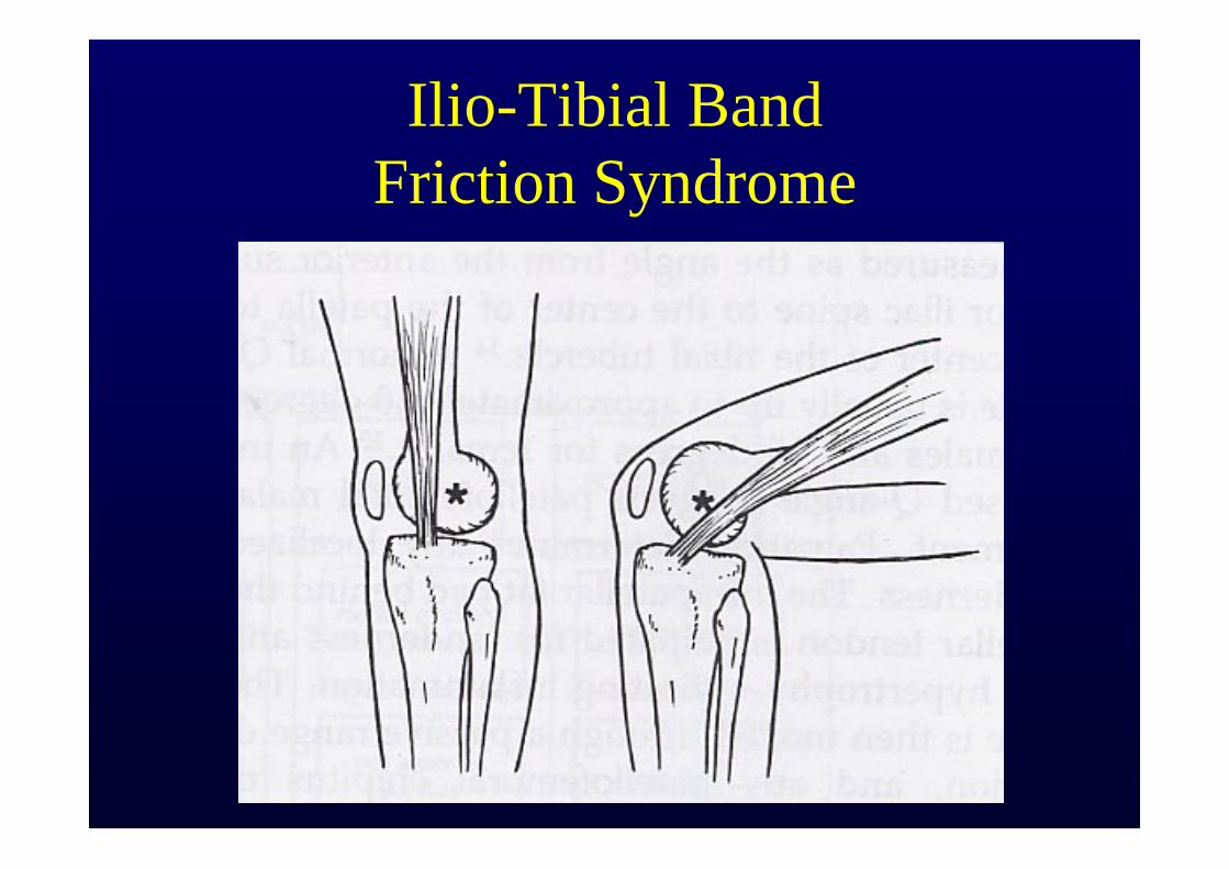
Ilio-Tibial Band Friction Syndrome
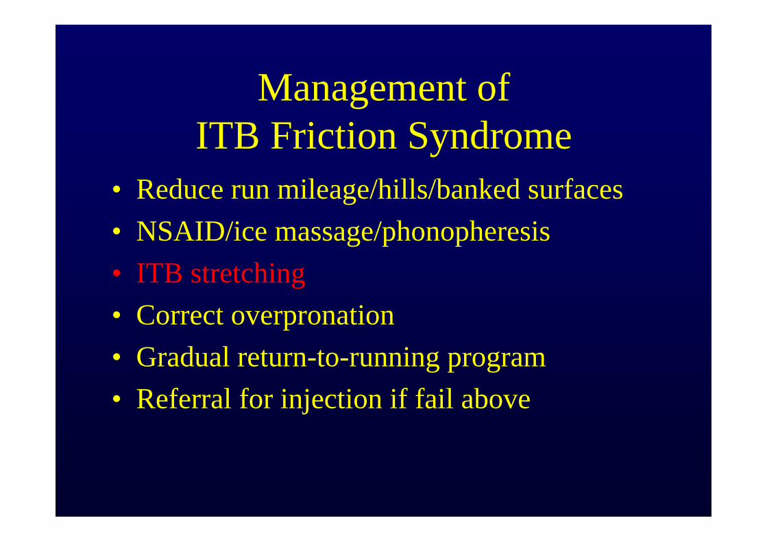
Management of ITB Friction Syndrome
• Reduce run mileage/hills/banked surfaces• NSAID/ice massage/phonopheresis• ITB stretching• Correct overpronation• Gradual return-to-running program• Referral for injection if fail above
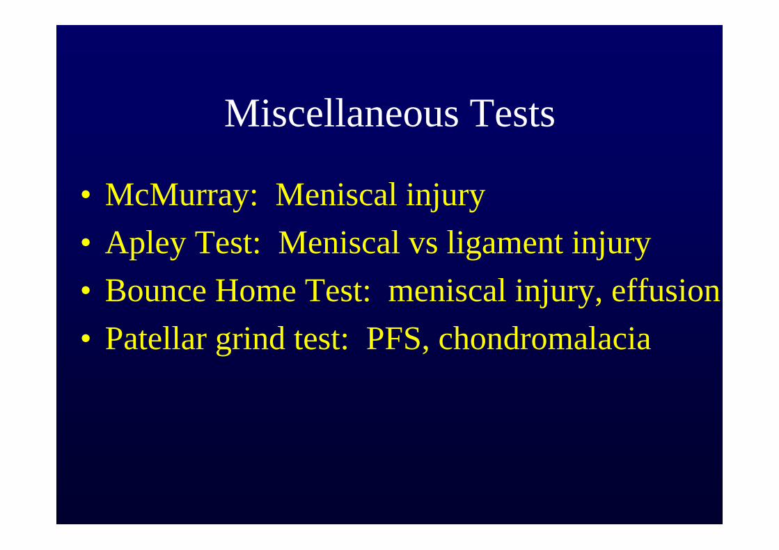
Miscellaneous Tests
• McMurray: Meniscal injury• Apley Test: Meniscal vs ligament injury • Bounce Home Test: meniscal injury, effusion• Patellar grind test: PFS, chondromalacia
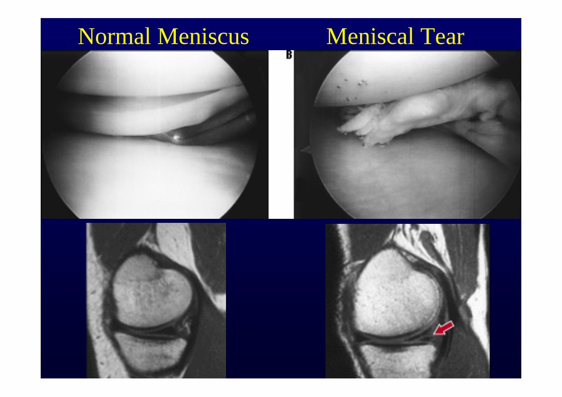
Normal Meniscus Meniscal Tear
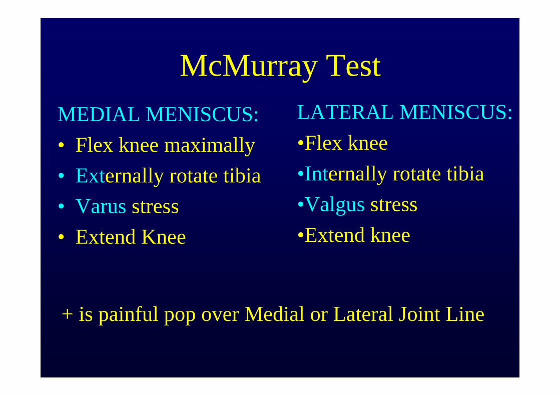
McMurray TestMEDIAL MENISCUS:• Flex knee maximally• Externally rotate tibia• Varus stress• Extend Knee
+ is painful pop over Medial or Lateral Joint Line
LATERAL MENISCUS:•Flex knee•Internally rotate tibia•Valgus stress•Extend knee
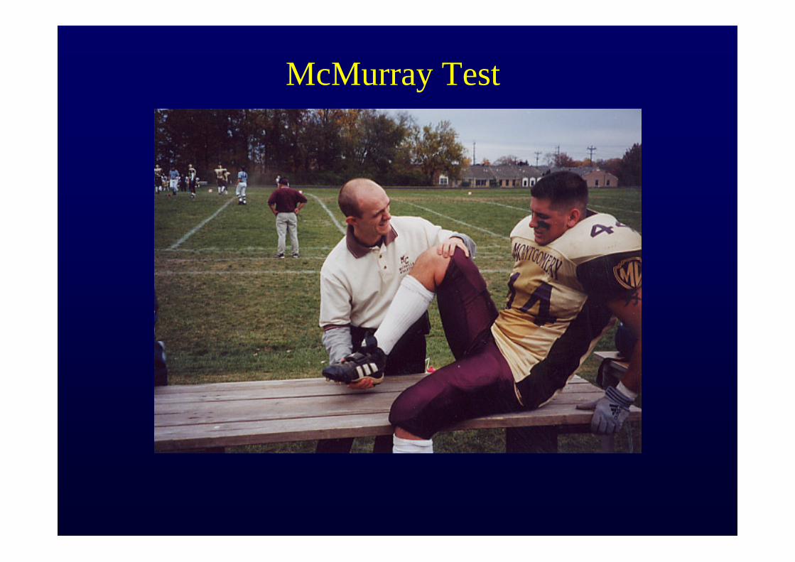
McMurray Test
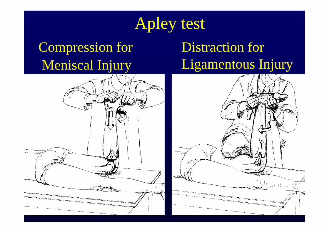
Apley testCompression forMeniscal Injury
Distraction for Ligamentous Injury
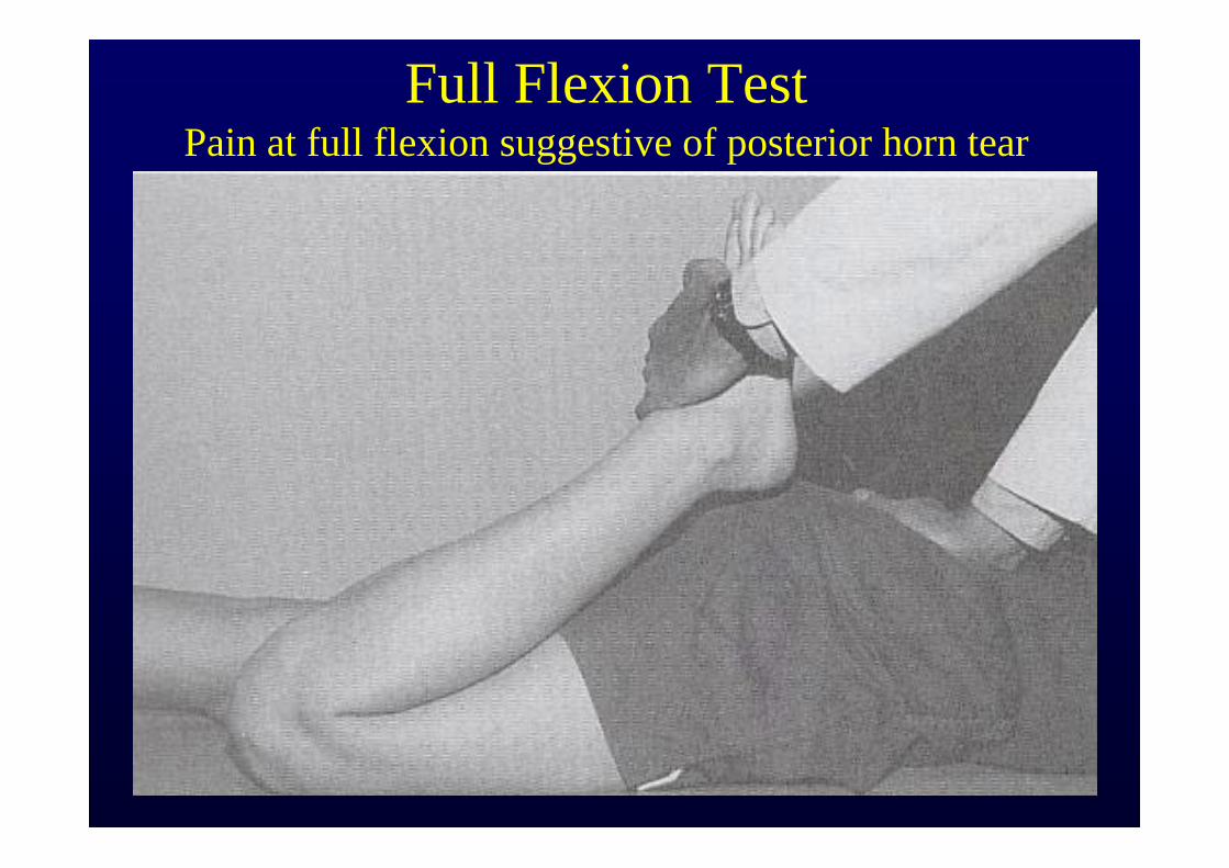
Full Flexion TestPain at full flexion suggestive of posterior horn tear
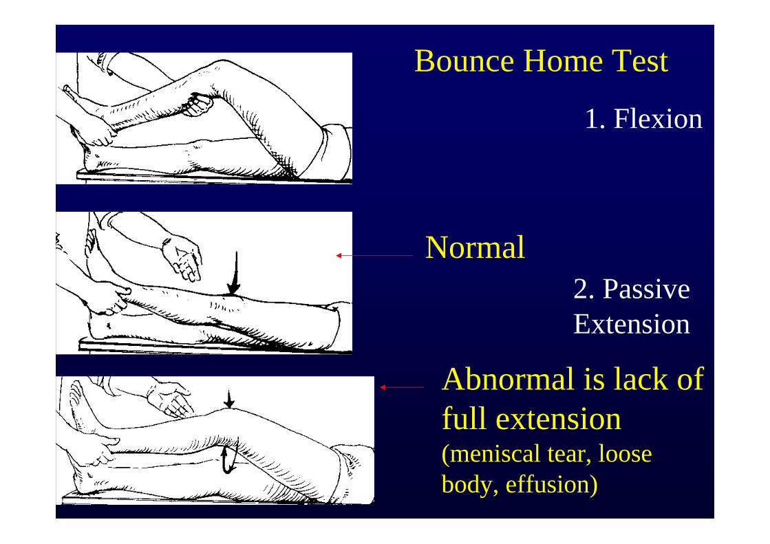
Bounce Home Test
Abnormal is lack of full extension (meniscal tear, loose body, effusion)
1. Flexion
2. Passive Extension
Normal
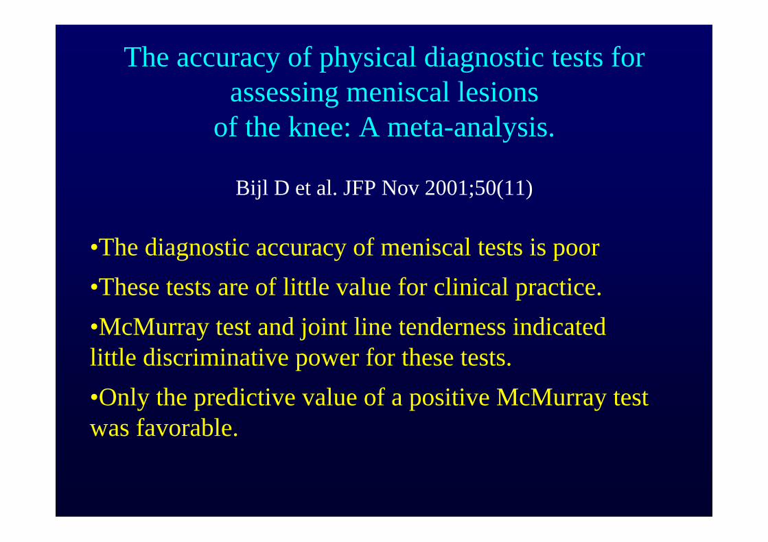
The accuracy of physical diagnostic tests for assessing meniscal lesions
of the knee: A meta-analysis.
Bijl D et al. JFP Nov 2001;50(11)
•The diagnostic accuracy of meniscal tests is poor•These tests are of little value for clinical practice.•McMurray test and joint line tenderness indicatedlittle discriminative power for these tests. •Only the predictive value of a positive McMurray test was favorable.
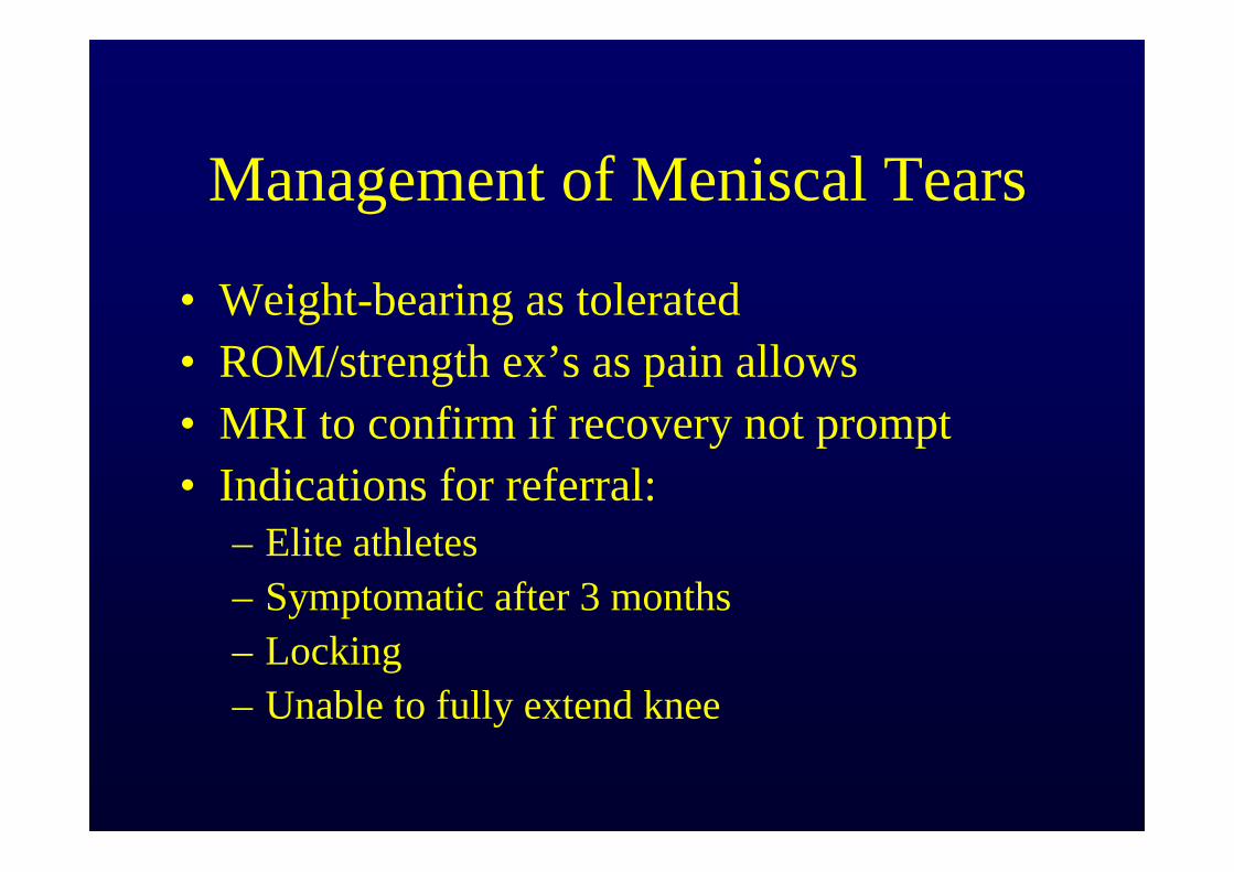
Management of Meniscal Tears
• Weight-bearing as tolerated• ROM/strength ex’s as pain allows• MRI to confirm if recovery not prompt• Indications for referral:
– Elite athletes– Symptomatic after 3 months– Locking– Unable to fully extend knee
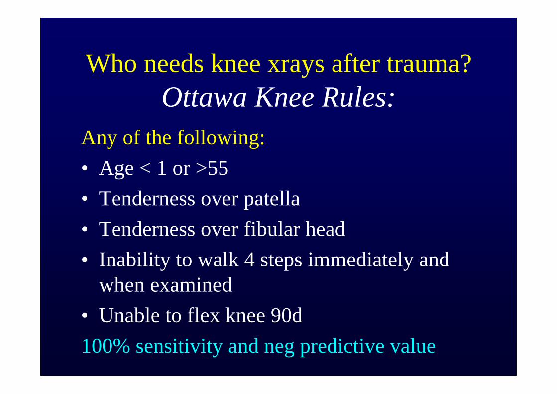
Who needs knee xrays after trauma?Ottawa Knee Rules:
Any of the following:• Age < 1 or >55• Tenderness over patella• Tenderness over fibular head • Inability to walk 4 steps immediately and
when examined• Unable to flex knee 90d100% sensitivity and neg predictive value
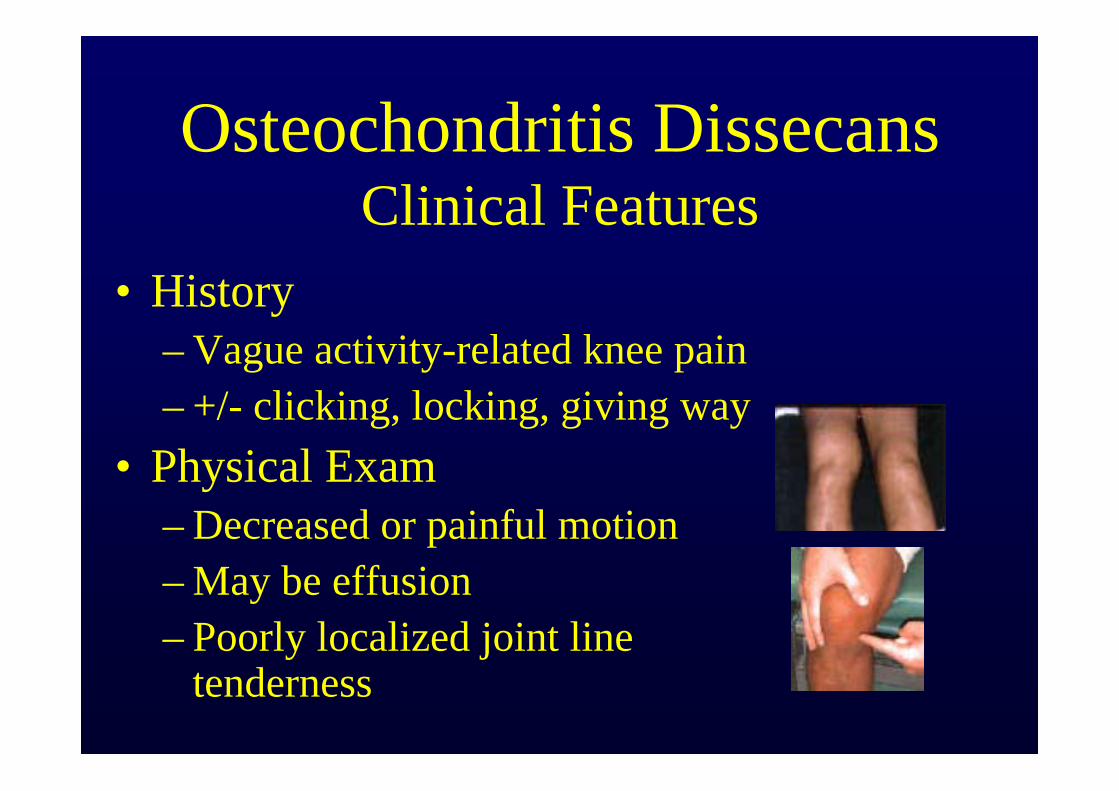
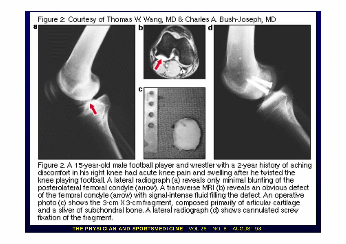
Osteochondritis DissecansClinical Features
• History– Vague activity-related knee pain– +/- clicking, locking, giving way
• Physical Exam– Decreased or painful motion– May be effusion– Poorly localized joint line
tenderness
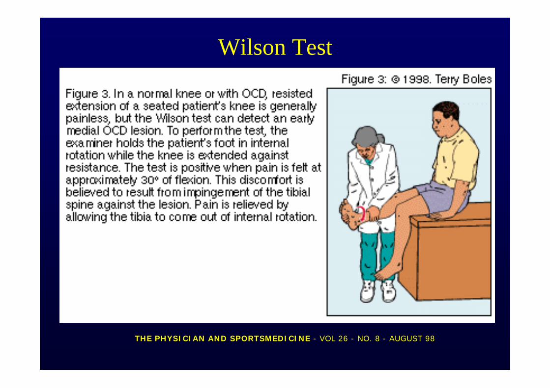
Wilson Test
THE PHYSICIAN AND SPORTSMEDICINE - VOL 26 - NO. 8 - AUGUST 98
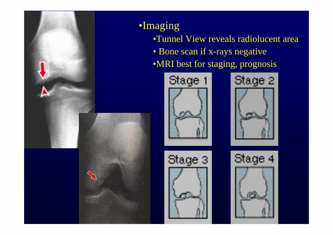
•Imaging•Tunnel View reveals radiolucent area• Bone scan if x-rays negative•MRI best for staging, prognosis
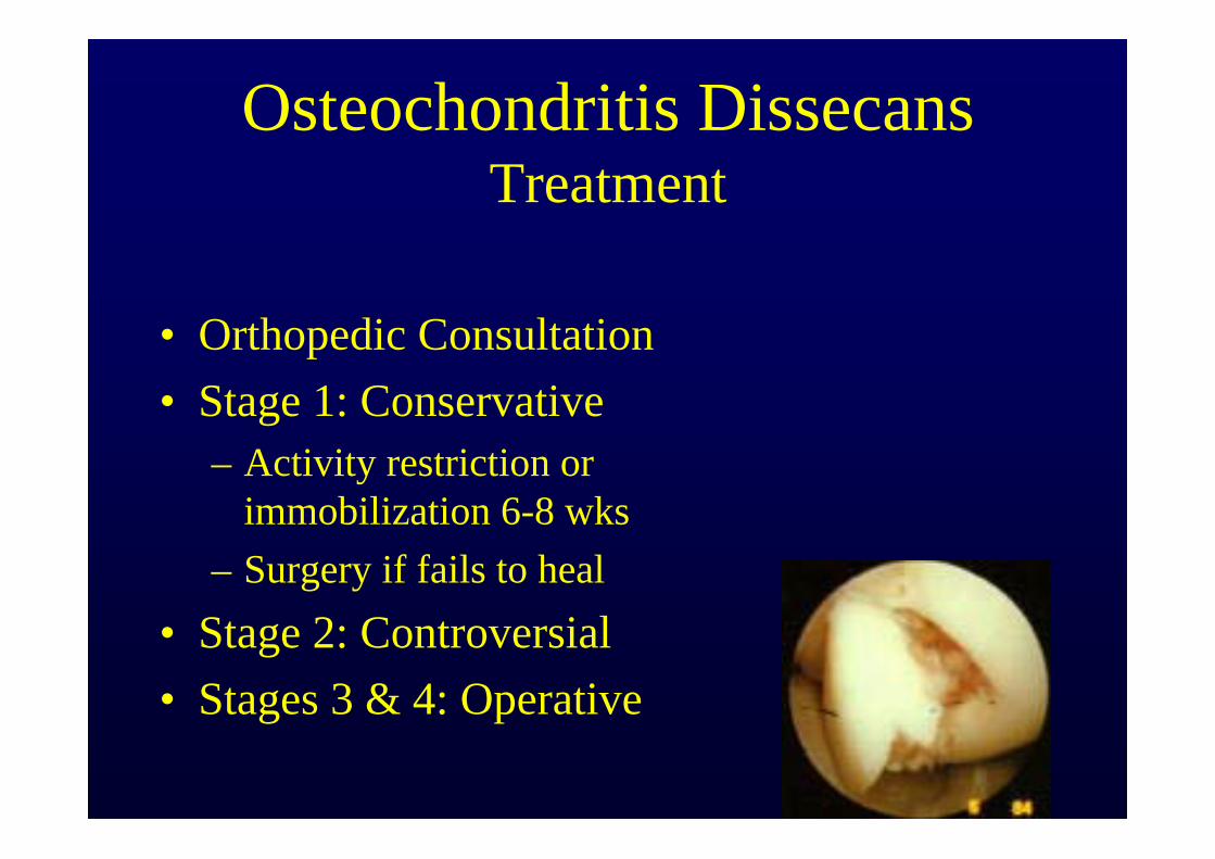
Osteochondritis DissecansTreatment
• Orthopedic Consultation• Stage 1: Conservative
– Activity restriction or immobilization 6-8 wks
– Surgery if fails to heal• Stage 2: Controversial• Stages 3 & 4: Operative
THE PHYSICIAN AND SPORTSMEDICINE - VOL 26 - NO. 8 - AUGUST 98
Review
Only by a thorough knowledge of anatomy and functional testing can one make an
accurate diagnosis and direct effective care to an injured
knee.