Embed Size (px)
Citation preview
Cartilage Intermediate Layer Protein 2 (CILP-2) Is Expressedin Articular and Meniscal Cartilage and Down-regulated inExperimental Osteoarthritis*□S
Received for publication, April 5, 2011, and in revised form, August 12, 2011 Published, JBC Papers in Press, August 31, 2011, DOI 10.1074/jbc.M111.248039
Bianca C. Bernardo‡§1, Daniele Belluoccio‡, Lynn Rowley‡, Christopher B. Little¶, Uwe Hansen�,and John F. Bateman‡**2
From the ‡Murdoch Childrens Research Institute and Departments of §Paediatrics and **Biochemistry and Molecular Biology,University of Melbourne, Parkville VIC 3052, Australia, the ¶Raymond Purves Bone and Joint Research Laboratories, Kolling Instituteof Medical Research, University of Sydney at Royal North Shore Hospital, St. Leonards, NSW 2065, Australia, and the �Institute forPhysiological Chemistry and Pathobiochemistry, University Hospital of Muenster, Muenster D-48149, Germany
Using transcriptome profiling to determine differential geneexpression between the permanent mouse articular cartilageand the transient growth plate cartilage, we identified a highlyexpressed gene,Cilp2,which is expressed differentially by artic-ular chondrocytes. CILP-2 is highly homologous toCILP-1 (car-tilage intermediate layer protein 1), which is expressed in theintermediate zone of articular cartilage and has been linked tocartilage degenerative diseases. We demonstrated that Cilp2has a restricted mRNA distribution at the surface of the mousearticular cartilage during development, becoming localized tothe intermediate zone of articular cartilage and meniscal carti-lage with maturity. Although the extracellular CILP-2 proteinlocalization is broadly similar to CILP-1, CILP-2 appears to bemore localized in the deeper intermediate zone of the articularcartilage extracellular matrix at maturity. CILP-2 was shown tobe proteolytically processed, N-glycosylated, and present inhumanarticular cartilage. In surgically inducedosteoarthritis inmice, Cilp1 and Cilp2 gene expression was dysregulated. How-ever, whereas Cilp1 expression was increased, Cilp2 geneexpression was down-regulated demonstrating a differentialresponse to mechanically induced joint destabilization. CILP-2proteinwas reduced in themouse osteoarthritic cartilage.Ultra-structural analysis also suggested that CILP-2may be associatedwith collagenVImicrofibrils and thusmaymediate interactionsbetween matrix components in the territorial and inter-territo-rial articular cartilage matrix. mRNA expression analysis indi-cated that whereas Cilp1 and Cilp2 are expressed most abun-dantly in cartilaginous tissues, expression can be detected inmuscle and heart.
During limb development, chondrocytes follow alternativedevelopmental pathways. Chondrocytes that form at the epiph-
yseal surface of long bones develop into permanent articularchondrocytes and form the smooth articular cartilage neces-sary for effective weight-bearing and joint movement. Theother group of chondrocytes, the transient epiphyseal chondro-cytes, are organized into growth plates, and undergo a sequen-tial maturation program from resting cells to proliferative,prehypertrophic, and ultimately hypertrophic end-stage chon-drocytes that are replacedwith bone during endochondral ossi-fication. Both populations of chondrocytes are critical for theformation of the skeleton and disturbances to normal chondro-cyte development and function results in chondrodysplasias(reviewed by Refs. 1–3).One of the important determinants of cartilage function is
extracellular matrix (ECM)3 structure and composition. Toidentify new ECM determinants of the articular cartilage, wecompared the gene expression profile of mouse articular carti-lage with growth plate cartilage. These studies revealed that themost highly differentially expressed cDNA on the array was aRiken clone that corresponded toCilp2 (cartilage intermediate-layer protein, isoform 2), recently reported as a product of car-tilage cells (4, 5). CILP-2 is highly homologous to a known car-tilage protein, CILP-1 (5). CILP-1 is a large secretedglycoprotein that is thought to play a role in cartilage scaffold-ing (6). It was isolated from human articular cartilage and itsexpression has been reported to be localized to the intermedi-ate zone of articular cartilage in the territorial matrix (6).CILP-1 is a pro-form of two polypeptides, and is cleaved intodistinct N- and C-terminal fragments at a furin endoproteaseconsensus site (7). TheN-terminal of CILP-1 has been shown tobind to and inhibit TGF�1 in vitro (8), and CILP1 mRNA isinduced by TGF�1 (9). CILP-1 levels have been shown toincrease with age (6, 10) and in patients with early stage osteo-arthritis (11). The association of single nucleotide polymor-phisms in the CILP1 gene with musculoskeletal disordersincluding osteoarthritis (12–14), and lumbar disc disease in aJapanese population (8) implies that CILP proteins may beimportant in cartilage structure and disease. In contrast toCILP-1, little is known about themRNAand protein expression
* This work was supported in part by National Health and Medical ResearchCouncil of Australia Grant 607399), the Murdoch Childrens Research Insti-tute, the Victorian Government’s Operational Infrastructure Support Pro-gram, and Sonderforschungsbereich Grant SFB 492, TP A2.
□S The on-line version of this article (available at http://www.jbc.org) containsTables S1 and S2 and Figs. S1–S4.
1 Recipient of an Australian Postgraduate Award.2 To whom correspondence should be addressed: Murdoch Childrens
Research Institute, Royal Children’s Hospital, Parkville, Victoria 3052, Aus-tralia. Fax: 61-3-8341-6429; E-mail: [email protected].
3 The abbreviations used are: ECM, extracellular matrix; CILP-2, cartilage inter-mediate layer protein 2; qPCR, quantitative PCR; TSP-1, thrombospondintype 1; DMM, medial meniscal destabilization; OA, osteoarthritis; P14, post-natal 14.
THE JOURNAL OF BIOLOGICAL CHEMISTRY VOL. 286, NO. 43, pp. 37758 –37767, October 28, 2011© 2011 by The American Society for Biochemistry and Molecular Biology, Inc. Printed in the U.S.A.
37758 JOURNAL OF BIOLOGICAL CHEMISTRY VOLUME 286 • NUMBER 43 • OCTOBER 28, 2011
by guest on February 21, 2020http://w
ww
.jbc.org/D
ownloaded from

of CILP-2 in cartilage and noncartilaginous tissues. Our studiesshow that Cilp2 has a restricted mRNA distribution at the sur-face of the mouse articular cartilage during development,becoming localized to the intermediate zone of articular carti-lage andmeniscal cartilagewithmaturity.We show thatCILP-2is a glycoprotein and is proteolytically processed, similar toCILP-1. CILP-2 protein is present in human articular cartilageand ultrastructural studies demonstrated that CILP-2 may beassociated with collagen VI containing suprastructures. Impor-tantly, our studies show that in a mouse experimental model ofmechanically induced osteoarthritis Cilp2 gene expression isdown-regulated suggesting a role for loss of CILP-2 in thepathophysiology of arthritis. In addition, our studies show forthe first time the expression ofCilp1 andCilp2 in skeletal mus-cle, suggesting that the CILPs can have additional roles in non-cartilaginous tissue ECM structure and function.
EXPERIMENTAL PROCEDURES
Dissection and RNA Preparation—Dissection and RNAextraction from mouse articular cartilage was performed aspreviously described for growth plate cartilage (15). Briefly,14-day-old (P14) Swiss white mice were sacrificed in accord-ance with Institutional Animal Ethics guidelines and femurswere dissected. The tissue was immersed in Tissue-Tek OCTembedding compound (Sakura Finetechnical), sectioned (5�m) on a cryostat (Reichert-Jung), dehydrated in graded etha-nol series, and air-dried. Slides were then immobilized on aninverted microscope (Leica) and the articular cartilage, andproliferative, prehypertrophic, and hypertrophic growth platecartilage was dissected using an ophthalmic scalpel (Feather)(supplemental Fig. S1). Total RNA was extracted using thePicoPure RNA isolation kit (Arcturus Bioscience) and linearlyamplified in two rounds using the MessageAmp aRNA kit(Ambion) following the manufacturer’s instructions.Mouse Osteoarthritis Model—Animal experimentation was
approved by the Institutional Animal Ethics Committee.Osteoarthritis (OA) was induced in 10-week-old male C57BL6mice by medial meniscal destabilization (DMM) of the rightknee (16). Joints subjected to sham-operation (exposure of themedial menisco-tibial ligament but no transection) were usedas controls. Animals were sacrificed at 2 and 6 weeks after sur-gery (n� 4 per time point). The joints were dissected to exposethe articular cartilage, tibial epiphyses were isolated and placedin RNALater (Ambion) containing 20% EDTA, decalcified at4 °C for 72 h, and then embedded inOCT and stored at�80 °C.Serial 7-�m coronal cryosections were fixed in ethanol, air-dried, and noncalcified medial tibial plateau articular cartilagefrom previously assigned areas of cartilage fibrillation and lossof toluidine blue stainingwere laser-microdissected (Arcturus).Total RNA was extracted and amplified as described above.RT PCR Analysis—5 ng of aRNA from articular cartilage and
growth plate cartilage was reverse transcribed (RT) in a reac-tion volume of 20 �l using SuperScript III reverse transcriptase(Invitrogen) at 50 °C for 1 h. PCR was performed as previouslydescribed (15) except that amplification consisted of 35 cyclesof denaturation for 30 s at 94 °C, annealing for 30 s at 58 °C, andextension for 30 s at 72 °C. Primer sequences for Cilp1 andCilp2 are listed under supplemental Table S1. For multitissue
RT-PCR to compare Cilp1 and Cilp2 expression, tissues weredissected from outbred Swiss white mice and total was RNAisolated using the RNeasy Mini Kit (Qiagen) according to themanufacturer’s directions. RNA quality was assessed on anRNA pico/nano labchip (Agilent Technology Bioanalyzer).Microarray Expression Profiling—cDNAmicroarray analysis
of the individually microdissected proliferative, prehypertro-phic, and hypertophic zones of the growth plate cartilage hasbeen previously described (15). Microdissected articular carti-lage analysis was performed similarly using microarray slidesprinted with the 15k NIA cDNA mouse clone set (AustralianGenome Research Facility). Microarray expression profilingwas performed on amplified RNA from the DMM-operatedand Sham-operated mouse cartilage using duplicate microar-rays (Cy3/Cy5 dye-swap with replicate RNA samples). LabeledRNA was hybridized to 44k whole genome oligo microarrays(G4122A, Agilent Technologies). All arrays were scanned on anAxon 4000B scanner and features were extracted with GenePixPro 4.1 software (Axon Instruments). Raw data were processedusing a print-tip Loess normalization (17) using limmaGUIsoftware (18, 19).Mean log2-transformed expression ratios andB-statistic values (log posterior odds ratio (17, 20) were calcu-lated for all direct comparisons.Quantitative Real Time PCR—Quantitative real time PCR
(qPCR) was used for validation of differential expression ofCilp2 in articular and on the pooled amplified aRNA of thedifferent zones of the growth plate cartilage. Gene-specificprimers and prevalidated probes were designed using onlinesoftware (from Roche Applied Science) and are listed undersupplemental Table S1. qPCRwas performed using the Univer-sal ProbeLibrary System (Roche Applied Science) as previouslydescribed (22). mRNA was quantitated using the comparativeCTmethod (23), usingMapk1 as the internal control because itwas not differentially expressed between the growth plate zonesand articular cartilage. In expression studies on DMM mousecartilage, gene expression values were normalized using thegeometric mean expression of two housekeeping genes, Atp5bandRpl10.These housekeeper geneswere shownbymicroarrayexpression profiling to be unchanged during the onset and pro-gression of OA in the DMMmouse model (data not shown).In SituHybridization—Knee joints of 14-day-old Swiss white
mice were embedded and 8-�m cryosections were mountedonto SuperFrost Plus slides and in situ hybridization was per-formed using antisense [35S]CTP-RNA probes as describedpreviously (15). To prepare the probes PCR amplification wasperformed and consisted of 35 cycles of denaturation for 1 minat 94 °C, annealing for 1min at 58 °C, and extension for 1min at72 °C using specific primer sets for Cilp1 and Cilp2 (supple-mental Table S1). Hybridization and autoradiography was per-formed as previously described (15).Antibodies—Polyclonal antisera against the CILP-2 mouse
sequenceTLLDRRQQGSPHELEwas produced in rabbits (Mil-lipore). The CILP-2 antisera bound to the peptide antigen in adose-dependent manner by standard enzyme-linked immu-nosorbent assay and immunodot blots (data not shown). TheCILP-2 polyclonal antibodywas further affinity purified againstthe antigen using the 1-ml HiTrapTM NHS-activated pre-packed column (GE Healthcare) according to the manufactur-
CILP-2 Is a Component of the Cartilage ECM
OCTOBER 28, 2011 • VOLUME 286 • NUMBER 43 JOURNAL OF BIOLOGICAL CHEMISTRY 37759
by guest on February 21, 2020http://w
ww
.jbc.org/D
ownloaded from
er’s instructions. The purified CILP-2 polyclonal antibody wastested by standard enzyme-linked immunosorbent assay andimmunoblotting and was found to bind to antigen and CILP-2protein in cartilage extracts (data not shown). Serum from thesame rabbit, taken prior to immunization with the CILP-2 pep-tide antigen, was used as a negative control. The rabbit anti-humanCILP-1 antibody raised against peptide sequences in theN-terminal region of CILP-1 was generously provided by Dr.Pilar Lorenzo, Lund University, Sweden.SDS-PAGE and Immunoblotting—14-day-old Swiss white
mice were sacrificed and the femoral heads were harvestedfrom the hip joints. Protein extraction from cartilage was per-formed as previously described (24). Briefly, 10 femoral headswere solubilized in 1 ml of 4 M guanidine HCl, 50 mM sodiumacetate, pH 5.8, containing 10 mM EDTA, and mini-proteaseinhibitor mixture (Roche Applied Science) at 4 °C for 24 h.Insoluble material was removed by centrifugation at 13,0006 �g for 20min at 4 °C. The supernatantwas precipitated overnightat �20 °C using 9 volumes of ice-cold ethanol, followed by onewash in 70% ethanol to remove residual guanidine HCl andother salts. The precipitate was resuspended and denatured inreducing Laemmli sample buffer (Bio-Rad), separated on a 10%polyacrylamide gel, and transferred to ImmobilonTM-P polyvi-nylidene difluoride membrane (Millipore). Immunoblottingwas performed as previously described (25) with some modifi-cations. Themembranewas blocked in 5% skimmilk powder inPBS for 1 h and then incubated in antibody buffer (0.5% skim-milk powder in PBS with 0.1% Tween 20) containing rabbitanti-mouse CILP-2 antibody (10 �g/ml) for 1 h. Following sixwashes in PBSwith 0.1%Tween 20, anti-rabbit IgG horseradishperoxidase secondary antibody (Dako) was added at a dilutionof 1:10,000 in antibody buffer and incubated for 1 h. Followingwashing, the signal was developed with the ECL Plus Westernblotting detection system (GE Healthcare) and autoradiogra-phy using X-Omat film.N-Glycosidase F Treatment—For N-glycosidase F digestion,
40 �g of protein extracted from cartilage (described above) wasresuspended in 25 �l of phosphate buffer (0.02 M sodium phos-phate, pH 7.0, 0.1% SDS, 0.05 M EDTA, and 0.5%Nonidet P-40)and boiled for 2 min. Then 5 units of N-glycosidase F (RocheApplied Science) enzyme was added to the sample. Deglycosy-lation was performed in a heating block at 37 °C for 8 h. Follow-ing deglycosylation, samples were denatured in reducingLaemmli sample buffer (Bio-Rad), separated on a 7.5% poly-acrylamide gel, and transferred to Immobilon-P polyvinylidenedifluoride membrane. Immunoblotting was performed asdescribed above.Immunohistochemistry—Human articular cartilage from
cadaveric donors aged 60 to 75 years were obtained from TheInternational Institute of Advancement in Medicine (Jessup,PA; a division of the Musculoskeletal Foundation). Formalin-fixed paraffin-embedded tissue sectionswere de-paraffinized inxylene and rehydrated. To facilitate antibody penetration sec-tions were treated with 30�g/ml of Proteinase K in 50mMTris,pH 6.0, 5mMCaCl2 at 37 °C for 30min, and 0.2% hyaluronidase(bovine, type IV; Sigma) for 1 h at 37 °C. Sections were thentreated with 0.3% H2O2 in PBS to inactivate endogenous per-oxidases. After blocking with 1% goat serum in 1% BSA in PBS
overnight at 4 °C, the sections were immunolabeled with rabbitanti-human CILP-2 antisera or nonimmune serum (dilution1:200 in 1%BSA inPBS; antibodies a gift fromDr. Pilar Lorenzo,University of Lund, Sweden) and detected using the VectastainElite ABC kit (Vector Laboratories). Bound antibodies werevisualized using Sigma FASTTM DAB tablets (Sigma), and sec-tions were counterstained with hematoxylin. For immunohis-tochemistry on mouse tissue, serial sections of frozen, unfixedtissue were cut at 8-�m thickness on a Leica CM1580 cryostatat �20 °C, and thaw-mounted onto superfrost plus glass slides.The sections were fixed in HistoChoice� Tissue Fixative(Amersco) for 15 min at 4 °C then washed in 3 changes of PBS.Immunohistochemistry was performed as described for paraf-fin sections, except the sections were immunolabeled with rab-bit anti-mouse CILP-2 antibody or preimmune serum diluted1:500 in 1% BSA (w/v) in PBS.Immunoelectron Microscopy—Fragments of suprastructural
aggregates were obtained from human articular cartilage aspreviously described (26). In brief, slices of cartilage were twicehomogenized in 15 volumes of 150 mM NaCl, 2 mM sodiumphosphate buffer, pH 7.4 (PBS), containing a mixture of prote-ase inhibitors, and centrifuged at low speed for clarification.This procedure was repeated twice. Aliquots of supramolecularfragments were spotted onto sheets of parafilm. Nickel gridscovered with formvar and coated with carbon were floated onthe drops for 10 min to allow adsorption of material, subse-quently washed with PBS, and treated for 30 min with 2% (w/v)dried skim milk in PBS. Next, the adsorbed material wasallowed to react for 2 h with polyclonal antibodies againstCILP-2 and monoclonal antibodies against collagen VI (AF6210,Medicorp) diluted 1:100 in PBS containing 0.2% drymilk.After washing five times with PBS, the grids were put on dropsof 0.2% (w/v) milk solution containing colloidal gold particlescoated with antibodies to rabbit (18-nm gold particles, JacksonImmunoResearch) and mouse immunoglobulins (12-nm goldparticles, Jackson ImmunoResearch). Finally, the grids werewashed with distilled water and negatively stained with 2% ura-nyl acetate for 7 min. Control experiments were undertakenwith the first antibody omitted. Electron micrographs weretaken at 80 kV with a Philips EM 410 electron microscope.
RESULTS
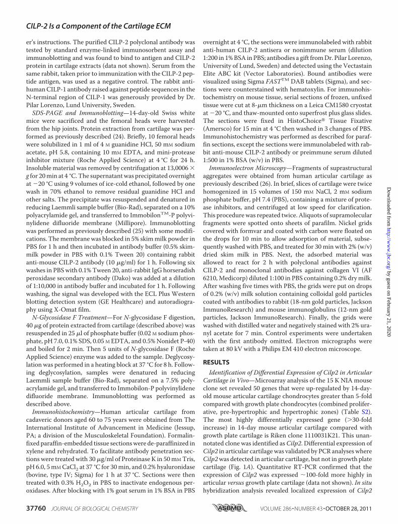
Identification of Differential Expression of Cilp2 in ArticularCartilage in Vivo—Microarray analysis of the 15 K NIA mouseclone set revealed 50 genes that were up-regulated by 14-day-old mouse articular cartilage chondrocytes greater than 5-foldcompared with growth plate chondrocytes (combined prolifer-ative, pre-hypertrophic and hypertrophic zones) (Table S2).The most highly differentially expressed gene (�30-foldincrease) in 14-day mouse articular cartilage compared withgrowth plate cartilage is Riken clone 1110031K21. This unan-notated clone was identified asCilp2. Differential expression ofCilp2 in articular cartilagewas validated by PCR analyses whereCilp2was detected in articular cartilage, but not in growth platecartilage (Fig. 1A). Quantitative RT-PCR confirmed that theexpression of Cilp2 was expressed �100-fold more highly inarticular versus growth plate cartilage (data not shown). In situhybridization analysis revealed localized expression of Cilp2
CILP-2 Is a Component of the Cartilage ECM
37760 JOURNAL OF BIOLOGICAL CHEMISTRY VOLUME 286 • NUMBER 43 • OCTOBER 28, 2011
by guest on February 21, 2020http://w
ww
.jbc.org/D
ownloaded from
mRNA to the superficial and intermediate zones of 14-day-oldmouse articular cartilage, as well as throughout the meniscusbut was not detected in growth plate cartilage (Fig. 1B).Sequence analysis demonstrated that CILP-2 is highly homol-ogous to a cartilage matrix molecule called CILP. CILP wasoriginally isolated from human articular cartilage, is a largesecreted glycoprotein, and is thought to play a role in cartilagescaffolding (6). CILP has been associated with osteoarthritis(12–14), and more recently with lumbar disc disease in a Japa-nese population (8), and will be referred to hereafter as CILP1(5). We and others have shown CILP-1 and CILP-2 to be struc-turally similar (5) (supplemental Fig. S2), with both polypep-tides containing a thrombospondin type 1 (TSP-1) repeatdomain, and an immunoglobulin-like domain. Both proteinscontain a signal peptide, putativeN-glycosylation sites, cysteine
residues forming disulfide bonds, and a furin endoproteaseconsensus site, but only CILP-1 has a putative phosphate-bind-ing loop (P-loop) motif.Cilp1 and Cilp2 Have Distinct mRNA and Protein Expression
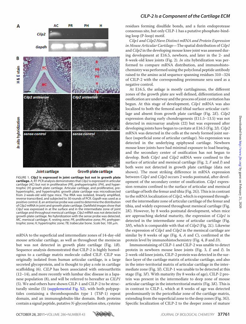
inMouse Articular Cartilage—The spatial distribution ofCilp1andCilp2 in the developingmouse knee joint was assessed dur-ing development at E16.5, newborn, and later in the 2- and8-week-old knee joints (Fig. 2). In situ hybridization was per-formed to compare mRNA distribution, and immunohisto-chemistrywas performedusing the polyclonal peptide antibodyraised to the amino acid sequence spanning residues 310–324of CILP-2 with the corresponding preimmune sera used as anegative control.At E16.5, the anlage is mostly cartilaginous, the different
zones of the growth plate are well defined, differentiation andossification are underway and the process of joint cavitation hasbegun. At this stage of development, Cilp1 mRNA was alsolocalized to both the femoral and tibial surface articular carti-lage and absent from growth plate cartilage (Fig. 2E). Cilp2expression during early chondrogenesis (E11.5–13.5) was notdetected in microarray analysis (22) but was expressed afterdeveloping joints have begun to cavitate at E16.5 (Fig. 2I).Cilp2mRNA was detected in the cells at the newly formed joint sur-face (superficial zone of articular cartilage). No expression wasdetected in the underlying epiphyseal cartilage. Newbornmouse knee joints have had minimal exposure to load bearing,and the secondary center of ossification has not begun todevelop. Both Cilp1 and Cilp2 mRNA were confined to thesurface of articular and meniscal cartilage (Fig. 2, F and J) andboth were not detected in growth plate cartilage (data notshown). The most striking difference in mRNA expressionbetween Cilp1 and Cilp2 occurs 2 weeks postnatal, after devel-opment of the secondary center of ossification. Cilp1 expres-sion remains confined to the surface of articular and meniscalcartilage of both the femur and tibia (Fig. 2G). This is in contrastto themRNA localization ofCilp2,which is expressed through-out the intermediate zone of articular cartilage of the femur andtibia, and widely expressed throughout meniscal cartilage (Fig.2K). However, by 8 weeks postnatal development, when miceare approaching skeletal maturity, the expression of Cilp1 isdetected in the intermediate zone of articular cartilage (Fig.3H), which is comparable with that of Cilp2 (Fig. 2L). Likewisethe expression of Cilp1 and Cilp2 in the meniscal cartilage aresimilar by 8 weeks of age (Fig. 4, A and C), confirmed at theprotein level by immunohistochemistry (Fig. 4, B and D).Immunostaining of CILP-1 and CILP-2 was unable to detect
protein in newborn mouse knee joints (Fig. 3, E and I). In2-week-old knee joints, CILP-2 protein was detected in the sur-face layer of the cartilage matrix of articular cartilage, and alsoin the inter-territorial matrix of articular cartilage in the inter-mediate zone (Fig. 3J). CILP-1 was unable to be detected at thisstage (Fig. 3F). With maturity (by 8 weeks of age), CILP-2 pro-tein was present in the intermediate to deep zone of mousearticular cartilage in the interterritorial matrix (Fig. 3K). This isin contrast to CILP-1, which at 8 weeks of age was detectedpredominantly in the pericellular zone of the cartilage matrix,extending from the superficial zone to the deep zones (Fig. 3G).Specific localization of CILP-2 to the deeper zones of mature
FIGURE 1. Cilp2 is expressed in joint cartilage but not in growth platecartilage. A, RT-PCR analysis demonstrates that Cilp2 is expressed in articularcartilage (AC) but not in proliferative (PR), prehypertrophic (PH), and hyper-trophic (H) growth plate cartilage. Articular cartilage, and proliferative, pre-hypertrophic, and hypertrophic growth plate cartilage was microdissectedfrom 2-week-old wild type mice. The RNA was isolated, linearly amplified,reverse transcribed, and subjected to 30 rounds of PCR. Gapdh was used as apositive control. B, an antisense probe was used to determine the distributionof Cilp2 mRNA in joint and growth plate cartilage. Darkfield images show thatCilp2 was expressed in at the surface and in the intermediate zone of jointcartilage and throughout meniscal cartilage. Cilp2 mRNA was not detected ingrowth plate cartilage. No hybridization with the sense probe was detected.MC, meniscal cartilage; R, resting zone; PR, proliferative zone; PH, prehyper-trophic zone; H, hypertrophic zone; TB, trabecular bone. Scale bar, 100 �m.
CILP-2 Is a Component of the Cartilage ECM
OCTOBER 28, 2011 • VOLUME 286 • NUMBER 43 JOURNAL OF BIOLOGICAL CHEMISTRY 37761
by guest on February 21, 2020http://w
ww
.jbc.org/D
ownloaded from
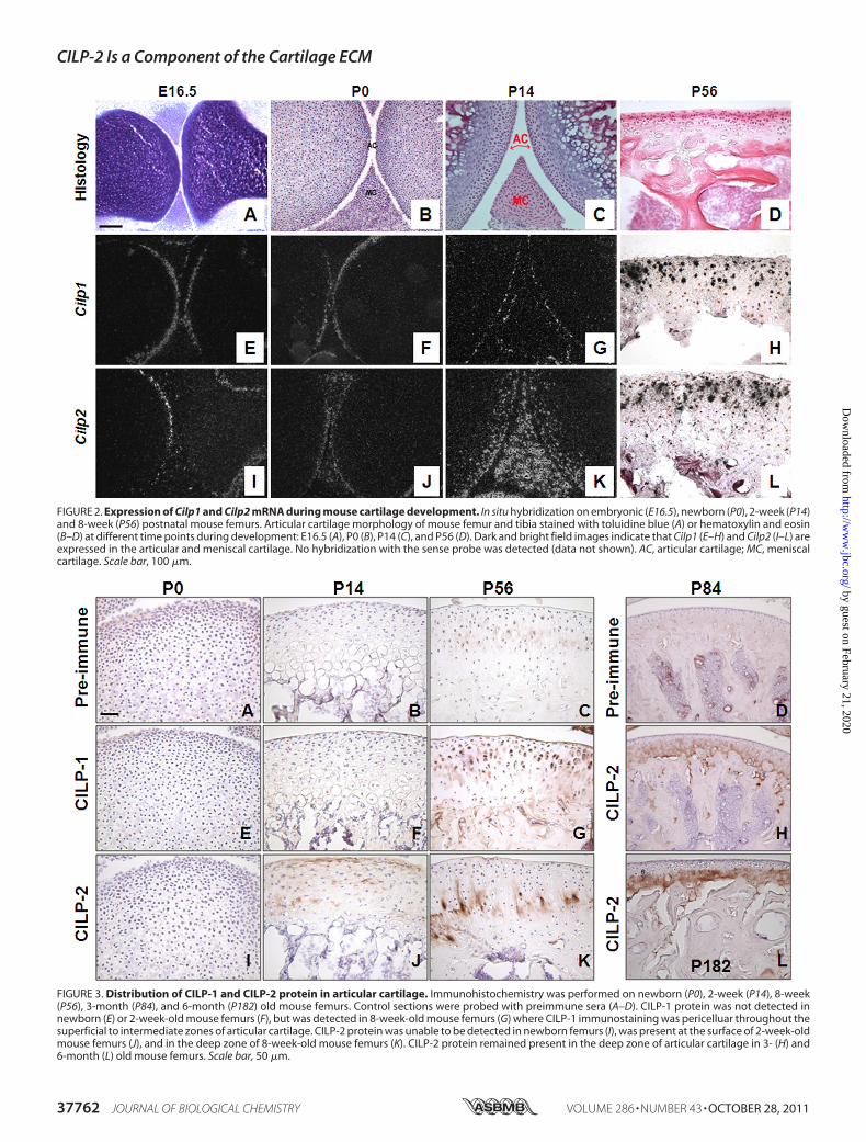
FIGURE 2. Expression of Cilp1 and Cilp2 mRNA during mouse cartilage development. In situ hybridization on embryonic (E16.5), newborn (P0), 2-week (P14)and 8-week (P56) postnatal mouse femurs. Articular cartilage morphology of mouse femur and tibia stained with toluidine blue (A) or hematoxylin and eosin(B–D) at different time points during development: E16.5 (A), P0 (B), P14 (C), and P56 (D). Dark and bright field images indicate that Cilp1 (E–H) and Cilp2 (I–L) areexpressed in the articular and meniscal cartilage. No hybridization with the sense probe was detected (data not shown). AC, articular cartilage; MC, meniscalcartilage. Scale bar, 100 �m.
FIGURE 3. Distribution of CILP-1 and CILP-2 protein in articular cartilage. Immunohistochemistry was performed on newborn (P0), 2-week (P14), 8-week(P56), 3-month (P84), and 6-month (P182) old mouse femurs. Control sections were probed with preimmune sera (A–D). CILP-1 protein was not detected innewborn (E) or 2-week-old mouse femurs (F), but was detected in 8-week-old mouse femurs (G) where CILP-1 immunostaining was pericelluar throughout thesuperficial to intermediate zones of articular cartilage. CILP-2 protein was unable to be detected in newborn femurs (I), was present at the surface of 2-week-oldmouse femurs (J), and in the deep zone of 8-week-old mouse femurs (K). CILP-2 protein remained present in the deep zone of articular cartilage in 3- (H) and6-month (L) old mouse femurs. Scale bar, 50 �m.
CILP-2 Is a Component of the Cartilage ECM
37762 JOURNAL OF BIOLOGICAL CHEMISTRY VOLUME 286 • NUMBER 43 • OCTOBER 28, 2011
by guest on February 21, 2020http://w
ww
.jbc.org/D
ownloaded from
cartilage was further confirmed in 3- and 6-month-old mice(Fig. 3, H and L). The localization of CILP-1 and CILP-2 toarticular and meniscal cartilage suggests that these ECM pro-teins may be components of such permanent cartilages, ratherthan the transient growth plate cartilage.CILP-2 Is Proteolytically Processed, Glycosylated, and Inte-
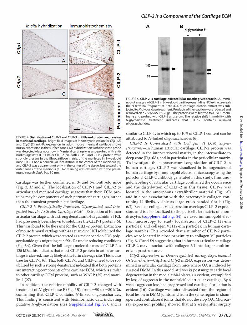
grated into the Articular Cartilage ECM—Extraction of humanarticular cartilage with a strong denaturant, 4 M guanidine HCl,had previously been shown to solubilize the CILP-1 protein (6).This was found to be the same for the CILP-2 protein. Extractionofmouse femoral cartilagewith 4M guanidineHCl solubilized theCILP-2 protein,whichwas detected as amajor bandonSDS-poly-acrylamide gels migrating at �90 kDa under reducing conditions(Fig. 5A). Given that the full-length molecular mass of CILP-2 is125 kDa, this indicates that most CILP-2 protein in articular car-tilage is cleaved,mostly likely at the furin cleavage site. This is alsotrue for CILP-1 (6). That both CILP-1 and CILP-2 need to be sol-ubilized by such a strong denaturant indicated that both proteinsare interacting components of the cartilage ECM,which is similarto other cartilage ECM proteins, such as WARP (25) and matri-lin-1 (27).In addition, the relative mobility of CILP-2 changed with
treatment of N-glycosidase F (Fig. 5B), from �90 to �80 kDa,confirming that CILP-2 contains N-linked oligosaccharides.This finding is consistent with bioinformatic data indicatingputative N-glycosylation sites (supplemental Fig. S3), and is
similar to CILP-1, in which up to 10% of CILP-1 content can beattributed to N-linked oligosaccharides (6).CILP-2 Is Co-localized with Collagen VI ECM Supra-
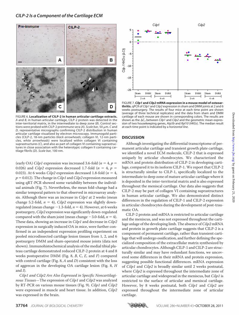
structures—In human articular cartilage, CILP-2 protein wasdetected in the inter-territorial matrix, in the intermediate todeep zone (Fig. 6B), and in particular in the pericellular matrix.To investigate the suprastructural organization of CILP-2 inhuman cartilage, CILP-2 was visualized in homogenates ofhuman cartilage by immunogold electronmicroscopy using thepolyclonal CILP-2 antibody generated in this study. Immuno-gold labeling of articular cartilage confirmed the occurrenceand the distribution of CILP-2 in this tissue. CILP-2 waslocated in the amorphous extrafibriller material (Fig. 6C)and in close association with the heterotypic collagen con-taining II fibrils, visible as large cross-banded fibrils (Fig.6D). Because collagen VI expression overlaps CILP-2 expres-sion, and is also localized to the pericellular matrix of chon-drocytes (supplemental Fig. S4), we used immunogold elec-tron microscopy to study localization of CILP-2 (18-nmparticles) and collagen VI (12-nm particles) in human carti-lage samples. This revealed that a number of CILP-2 parti-cles were located in close proximity to collagen VI particles(Fig. 6, C and D) suggesting that in human articular cartilageCILP-2 may associate with collagen VI into larger multim-eric suprastructures.Cilp2 Expression Is Down-regulated during Experimental
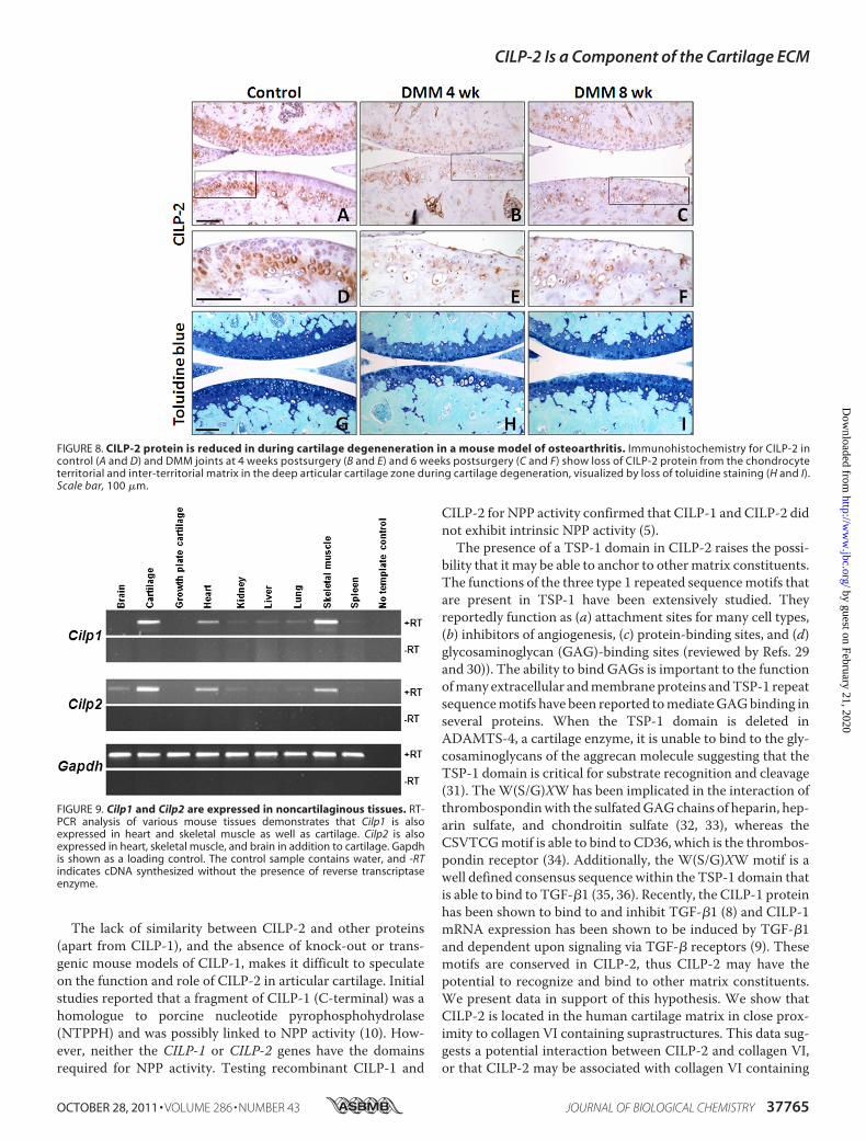
Osteoarthritis—Cilp1 and Cilp2 mRNA expression was deter-mined in articular cartilage frommice where OA is induced bysurgical DMM. In this model at 2 weeks postsurgery early focaldegeneration in themedial tibial plateau is evident, exemplifiedby loss of aggrecan in the noncalcified articular cartilage. By 6weeks aggrecan loss had progressed and cartilage fibrillation isevident (16). Cartilage was microdissected from the region offocal lesion in DMM joints, and from the same region in sham-operated contralateral joints that do not develop OA. Microar-ray expression profiling showed that at 2 weeks after surgery
FIGURE 4. Distribution of CILP-1 and CILP-2 mRNA and protein expressionin meniscal cartilage. Bright field images of in situ hybridization for Cilp1 (A)and Cilp2 (C) mRNA expression in adult mouse meniscal cartilage showsmRNA expression in the surface zones. No hybridization with the sense probewas detected (data not shown). Meniscal cartilage was also probed with anti-bodies against CILP-1 (B) or CILP-2 (D). Both CILP-1 and CILP-2 protein werestrongly present in the fibrocartilage matrix of the meniscus in 8-week-oldmice. CILP-1 had a pericellular localization in the center of the meniscus (B),and CILP-2 was apparent not only in the center of the tissue, but toward theouter zones of the meniscus (C). No staining was observed with the preim-mune sera (E). Scale bar, 50 �m.
FIGURE 5. CILP-2 is cartilage extracellular matrix glycoprotein. A, immu-noblot analysis of CILP-2 in 2-week-old cartilage guanidine HCl extract revealsthe N-terminal fragment at �90 kDa. B, cartilage protein extract was sub-jected to N-glycosidase treatment. Products of the reaction were reduced andresolved on a 7.5% SDS-PAGE gel. The proteins were blotted to a PVDF mem-brane and probed with CILP-2 antiserum. The relative shift in mobility withN-glycosidase treatment indicates that CILP-2 contains N-linkedoligosaccharides.
CILP-2 Is a Component of the Cartilage ECM
OCTOBER 28, 2011 • VOLUME 286 • NUMBER 43 JOURNAL OF BIOLOGICAL CHEMISTRY 37763
by guest on February 21, 2020http://w
ww
.jbc.org/D
ownloaded from
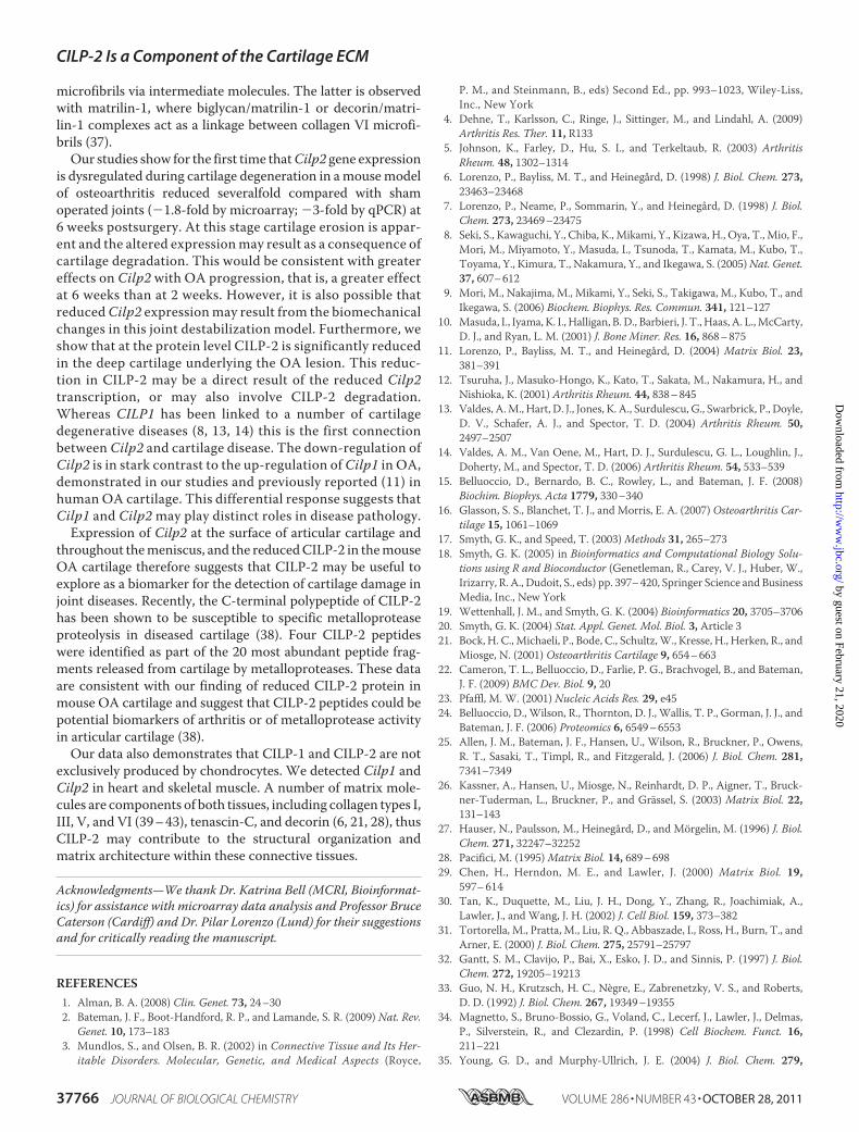
(early OA) Cilp1 expression was increased 3.6-fold (n � 4, p �0.026) and Cilp2 expression decreased 1.7-fold (n � 4, p �0.023). At 6 weeks Cilp2 expression decreased 1.8-fold (n � 4,p� 0.012). The change inCilp1 andCilp2 expressionmeasuredusing qRT-PCR showed some variability between the individ-ual animals (Fig. 7). Nevertheless, the mean fold-change had asimilar temporal pattern to that observed in microarray analy-sis. Although there was an increase in Cilp1 at 2 weeks (meanchange 5.2-fold, n � 4), Cilp2 expression was slightly down-regulated (mean change�1.3-fold, n� 4). However, at 6 weekspostsurgery,Cilp2 expressionwas significantly down-regulatedcompared with the sham joint (mean change �3.0-fold, n � 4).These data, showing an increase in Cilp1 and decrease in Cilp2expression in surgically induced OA in mice, were further con-firmed in an independent expression profiling experiment onpooled microdissected cartilage lesion tissues from 1, 2, and 6postsurgery DMM and sham-operated mouse joints (data notshown). Immunohistochemical analysis of themedial tibial pla-teau cartilage demonstrated reduced CILP-2 protein at 4 and 8weeks postoperative DMM (Fig. 8, B, C, E, and F) comparedwith control cartilage (Fig. 8, A and D) consistent with the lossof aggrecan in the developing OA cartilage lesion (Fig. 8, Hand I).Cilp1 and Cilp2 Are Also Expressed in Specific Noncartilagi-
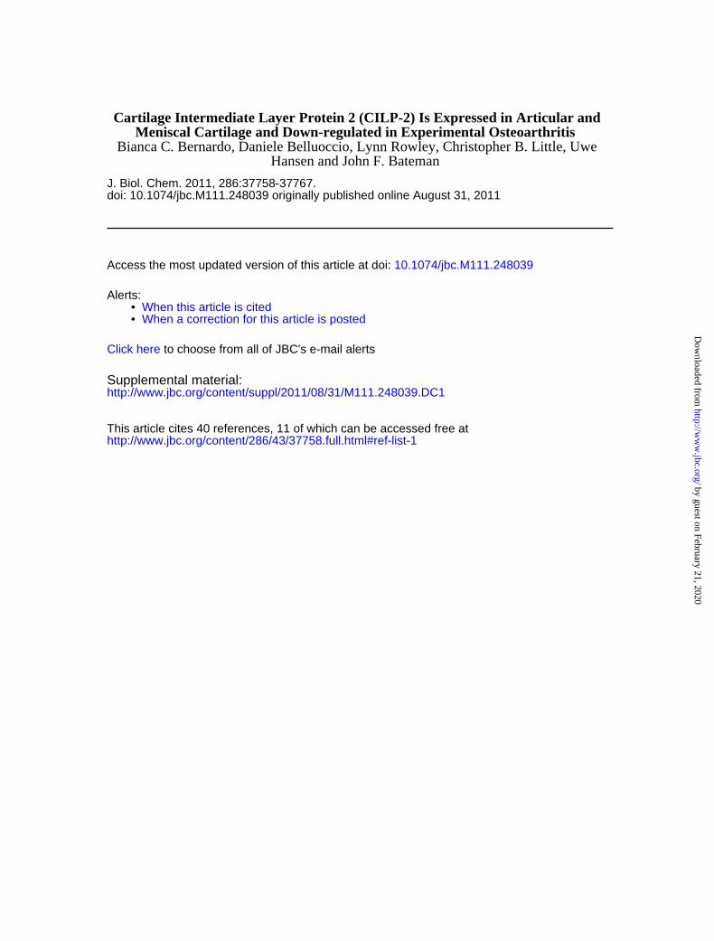
nous Tissues—The expression of Cilp1 and Cilp2 was analyzedby RT-PCR on various mouse tissues (Fig. 9). Cilp1 and Cilp2were expressed in muscle and heart tissue. In addition, Cilp2was expressed in the brain.
DISCUSSION
Although investigating the differential transcriptome of per-manent articular cartilage and transient growth plate cartilage,we identified a novel ECM molecule, CILP-2 that is expresseduniquely by articular chondrocytes. We characterized themRNA and protein distribution of CILP-2 in developing carti-lage, compared it to its isoform CILP-1. We report that CILP-2is structurally similar to CILP-1, specifically localized to theintermediate to deep zone ofmature articular cartilage where itis deposited in the inter-territorial matrix and is also expressedthroughout the meniscal cartilage. Our data also suggests thatCILP-2 may be part of collagen VI containing suprastructuresin human articular cartilage. We also demonstrated distinctdifferences in the regulation of CILP-1 and CILP-2 expressionin articular chondrocytes during the development of post-trau-matic OA.CILP-2 protein and mRNA is restricted to articular cartilage
and the meniscus, and was not expressed throughout the carti-lage anlage of the developing limb. The absence ofCilp2mRNAand protein in growth plate cartilage suggests that CILP-2 is acomponent of permanent cartilage, rather than transient carti-lage that will undergo ossification, and further defining the spe-cialized composition of the extracellular matrix synthesized byarticular chondrocytes. AlthoughCILP-1 andCILP-2 are struc-turally similar and may have redundant functions, we uncov-ered some differences in their mRNA and protein expression,suggesting possible functional differences. mRNA expressionof Cilp1 and Cilp2 is broadly similar until 2 weeks postnatal,where Cilp2 is expressed throughout the intermediate zone ofarticular cartilage and widespread in the meniscus, but Cilp1 isrestricted to the surface of articular and meniscal cartilage.However, by 8 weeks postnatal, both Cilp1 and Cilp2 areexpressed throughout the intermediate zone of articularcartilage.
FIGURE 6. Localization of CILP-2 in human articular cartilage extracts.A and B, in human articular cartilage, CILP-2 protein was detected in theinter-territorial matrix, in the intermediate to deep zone (B). Control sec-tions were probed with CILP-2 preimmune sera (A). Scale bar, 50 �m. C andD, representative micrographs confirming CILP-2 distribution in humanarticular cartilage visualized by electron microscopy. Immunogold parti-cles (CILP-2, 18-nm particles black arrowheads; collagen VI, 12-nm parti-cles, white arrowheads) were localized within collagen VI containingsuprastructures (C), and also as part of collagen VI containing suprastruc-tures in close association with the heterotypic collagen II containing car-tilage fibrils (D). Scale bar, 100 nm.
FIGURE 7. Cilp1 and Cilp2 mRNA expression in a mouse model of osteoar-thritis. qPCR of Cilp1 and Cilp2 expression in sham and DMM joints at 2 and 6weeks postsurgery. The results of four mice at each time point are shown(average of three technical replicates) and the data from sham and DMMcartilage of each mouse are shown in corresponding colors. The results areshown as the �CT between Cilp1 and Cilp2 and the geometric mean expres-sion of two housekeeping genes, Atp5b and Rpl10 (HKGs). The median resultat each time point is indicated by a horizontal line.
CILP-2 Is a Component of the Cartilage ECM
37764 JOURNAL OF BIOLOGICAL CHEMISTRY VOLUME 286 • NUMBER 43 • OCTOBER 28, 2011
by guest on February 21, 2020http://w
ww
.jbc.org/D
ownloaded from
The lack of similarity between CILP-2 and other proteins(apart from CILP-1), and the absence of knock-out or trans-genic mouse models of CILP-1, makes it difficult to speculateon the function and role of CILP-2 in articular cartilage. Initialstudies reported that a fragment of CILP-1 (C-terminal) was ahomologue to porcine nucleotide pyrophosphohydrolase(NTPPH) and was possibly linked to NPP activity (10). How-ever, neither the CILP-1 or CILP-2 genes have the domainsrequired for NPP activity. Testing recombinant CILP-1 and
CILP-2 for NPP activity confirmed that CILP-1 and CILP-2 didnot exhibit intrinsic NPP activity (5).The presence of a TSP-1 domain in CILP-2 raises the possi-
bility that it may be able to anchor to othermatrix constituents.The functions of the three type 1 repeated sequencemotifs thatare present in TSP-1 have been extensively studied. Theyreportedly function as (a) attachment sites for many cell types,(b) inhibitors of angiogenesis, (c) protein-binding sites, and (d)glycosaminoglycan (GAG)-binding sites (reviewed by Refs. 29and 30)). The ability to bind GAGs is important to the functionofmany extracellular andmembrane proteins andTSP-1 repeatsequencemotifs have been reported tomediateGAGbinding inseveral proteins. When the TSP-1 domain is deleted inADAMTS-4, a cartilage enzyme, it is unable to bind to the gly-cosaminoglycans of the aggrecan molecule suggesting that theTSP-1 domain is critical for substrate recognition and cleavage(31). TheW(S/G)XWhas been implicated in the interaction ofthrombospondinwith the sulfatedGAGchains of heparin, hep-arin sulfate, and chondroitin sulfate (32, 33), whereas theCSVTCGmotif is able to bind to CD36, which is the thrombos-pondin receptor (34). Additionally, the W(S/G)XW motif is awell defined consensus sequence within the TSP-1 domain thatis able to bind to TGF-�1 (35, 36). Recently, the CILP-1 proteinhas been shown to bind to and inhibit TGF-�1 (8) and CILP-1mRNA expression has been shown to be induced by TGF-�1and dependent upon signaling via TGF-� receptors (9). Thesemotifs are conserved in CILP-2, thus CILP-2 may have thepotential to recognize and bind to other matrix constituents.We present data in support of this hypothesis. We show thatCILP-2 is located in the human cartilage matrix in close prox-imity to collagen VI containing suprastructures. This data sug-gests a potential interaction between CILP-2 and collagen VI,or that CILP-2 may be associated with collagen VI containing
FIGURE 8. CILP-2 protein is reduced in during cartilage degeneneration in a mouse model of osteoarthritis. Immunohistochemistry for CILP-2 incontrol (A and D) and DMM joints at 4 weeks postsurgery (B and E) and 6 weeks postsurgery (C and F) show loss of CILP-2 protein from the chondrocyteterritorial and inter-territorial matrix in the deep articular cartilage zone during cartilage degeneration, visualized by loss of toluidine staining (H and I).Scale bar, 100 �m.
FIGURE 9. Cilp1 and Cilp2 are expressed in noncartilaginous tissues. RT-PCR analysis of various mouse tissues demonstrates that Cilp1 is alsoexpressed in heart and skeletal muscle as well as cartilage. Cilp2 is alsoexpressed in heart, skeletal muscle, and brain in addition to cartilage. Gapdhis shown as a loading control. The control sample contains water, and -RTindicates cDNA synthesized without the presence of reverse transcriptaseenzyme.
CILP-2 Is a Component of the Cartilage ECM
OCTOBER 28, 2011 • VOLUME 286 • NUMBER 43 JOURNAL OF BIOLOGICAL CHEMISTRY 37765
by guest on February 21, 2020http://w
ww
.jbc.org/D
ownloaded from
microfibrils via intermediate molecules. The latter is observedwith matrilin-1, where biglycan/matrilin-1 or decorin/matri-lin-1 complexes act as a linkage between collagen VI microfi-brils (37).Our studies show for the first time thatCilp2 gene expression
is dysregulated during cartilage degeneration in amousemodelof osteoarthritis reduced severalfold compared with shamoperated joints (�1.8-fold by microarray; �3-fold by qPCR) at6 weeks postsurgery. At this stage cartilage erosion is appar-ent and the altered expressionmay result as a consequence ofcartilage degradation. This would be consistent with greatereffects on Cilp2 with OA progression, that is, a greater effectat 6 weeks than at 2 weeks. However, it is also possible thatreducedCilp2 expressionmay result from the biomechanicalchanges in this joint destabilization model. Furthermore, weshow that at the protein level CILP-2 is significantly reducedin the deep cartilage underlying the OA lesion. This reduc-tion in CILP-2 may be a direct result of the reduced Cilp2transcription, or may also involve CILP-2 degradation.Whereas CILP1 has been linked to a number of cartilagedegenerative diseases (8, 13, 14) this is the first connectionbetween Cilp2 and cartilage disease. The down-regulation ofCilp2 is in stark contrast to the up-regulation of Cilp1 in OA,demonstrated in our studies and previously reported (11) inhuman OA cartilage. This differential response suggests thatCilp1 and Cilp2may play distinct roles in disease pathology.Expression of Cilp2 at the surface of articular cartilage and
throughout themeniscus, and the reducedCILP-2 in themouseOA cartilage therefore suggests that CILP-2 may be useful toexplore as a biomarker for the detection of cartilage damage injoint diseases. Recently, the C-terminal polypeptide of CILP-2has been shown to be susceptible to specific metalloproteaseproteolysis in diseased cartilage (38). Four CILP-2 peptideswere identified as part of the 20 most abundant peptide frag-ments released from cartilage by metalloproteases. These dataare consistent with our finding of reduced CILP-2 protein inmouse OA cartilage and suggest that CILP-2 peptides could bepotential biomarkers of arthritis or of metalloprotease activityin articular cartilage (38).Our data also demonstrates that CILP-1 and CILP-2 are not
exclusively produced by chondrocytes. We detected Cilp1 andCilp2 in heart and skeletal muscle. A number of matrix mole-cules are components of both tissues, including collagen types I,III, V, and VI (39–43), tenascin-C, and decorin (6, 21, 28), thusCILP-2 may contribute to the structural organization andmatrix architecture within these connective tissues.
Acknowledgments—We thank Dr. Katrina Bell (MCRI, Bioinformat-ics) for assistance with microarray data analysis and Professor BruceCaterson (Cardiff) and Dr. Pilar Lorenzo (Lund) for their suggestionsand for critically reading the manuscript.
REFERENCES1. Alman, B. A. (2008) Clin. Genet. 73, 24–302. Bateman, J. F., Boot-Handford, R. P., and Lamande, S. R. (2009) Nat. Rev.
Genet. 10, 173–1833. Mundlos, S., and Olsen, B. R. (2002) in Connective Tissue and Its Her-
itable Disorders. Molecular, Genetic, and Medical Aspects (Royce,
P. M., and Steinmann, B., eds) Second Ed., pp. 993–1023, Wiley-Liss,Inc., New York
4. Dehne, T., Karlsson, C., Ringe, J., Sittinger, M., and Lindahl, A. (2009)Arthritis Res. Ther. 11, R133
5. Johnson, K., Farley, D., Hu, S. I., and Terkeltaub, R. (2003) ArthritisRheum. 48, 1302–1314
6. Lorenzo, P., Bayliss, M. T., and Heinegård, D. (1998) J. Biol. Chem. 273,23463–23468
7. Lorenzo, P., Neame, P., Sommarin, Y., and Heinegård, D. (1998) J. Biol.Chem. 273, 23469–23475
8. Seki, S., Kawaguchi, Y., Chiba, K., Mikami, Y., Kizawa, H., Oya, T., Mio, F.,Mori, M., Miyamoto, Y., Masuda, I., Tsunoda, T., Kamata, M., Kubo, T.,Toyama, Y., Kimura, T., Nakamura, Y., and Ikegawa, S. (2005)Nat. Genet.37, 607–612
9. Mori, M., Nakajima, M., Mikami, Y., Seki, S., Takigawa, M., Kubo, T., andIkegawa, S. (2006) Biochem. Biophys. Res. Commun. 341, 121–127
10. Masuda, I., Iyama, K. I., Halligan, B. D., Barbieri, J. T., Haas, A. L.,McCarty,D. J., and Ryan, L. M. (2001) J. Bone Miner. Res. 16, 868–875
11. Lorenzo, P., Bayliss, M. T., and Heinegård, D. (2004) Matrix Biol. 23,381–391
12. Tsuruha, J., Masuko-Hongo, K., Kato, T., Sakata, M., Nakamura, H., andNishioka, K. (2001) Arthritis Rheum. 44, 838–845
13. Valdes, A.M., Hart, D. J., Jones, K. A., Surdulescu, G., Swarbrick, P., Doyle,D. V., Schafer, A. J., and Spector, T. D. (2004) Arthritis Rheum. 50,2497–2507
14. Valdes, A. M., Van Oene, M., Hart, D. J., Surdulescu, G. L., Loughlin, J.,Doherty, M., and Spector, T. D. (2006) Arthritis Rheum. 54, 533–539
15. Belluoccio, D., Bernardo, B. C., Rowley, L., and Bateman, J. F. (2008)Biochim. Biophys. Acta 1779, 330–340
16. Glasson, S. S., Blanchet, T. J., and Morris, E. A. (2007) Osteoarthritis Car-tilage 15, 1061–1069
17. Smyth, G. K., and Speed, T. (2003)Methods 31, 265–27318. Smyth, G. K. (2005) in Bioinformatics and Computational Biology Solu-
tions using R and Bioconductor (Genetleman, R., Carey, V. J., Huber, W.,Irizarry, R. A., Dudoit, S., eds) pp. 397–420, Springer Science and BusinessMedia, Inc., New York
19. Wettenhall, J. M., and Smyth, G. K. (2004) Bioinformatics 20, 3705–370620. Smyth, G. K. (2004) Stat. Appl. Genet. Mol. Biol. 3, Article 321. Bock, H. C., Michaeli, P., Bode, C., Schultz,W., Kresse, H., Herken, R., and
Miosge, N. (2001) Osteoarthritis Cartilage 9, 654–66322. Cameron, T. L., Belluoccio, D., Farlie, P. G., Brachvogel, B., and Bateman,
J. F. (2009) BMC Dev. Biol. 9, 2023. Pfaffl, M. W. (2001) Nucleic Acids Res. 29, e4524. Belluoccio, D., Wilson, R., Thornton, D. J., Wallis, T. P., Gorman, J. J., and
Bateman, J. F. (2006) Proteomics 6, 6549–655325. Allen, J. M., Bateman, J. F., Hansen, U., Wilson, R., Bruckner, P., Owens,
R. T., Sasaki, T., Timpl, R., and Fitzgerald, J. (2006) J. Biol. Chem. 281,7341–7349
26. Kassner, A., Hansen, U., Miosge, N., Reinhardt, D. P., Aigner, T., Bruck-ner-Tuderman, L., Bruckner, P., and Grässel, S. (2003) Matrix Biol. 22,131–143
27. Hauser, N., Paulsson, M., Heinegârd, D., and Mörgelin, M. (1996) J. Biol.Chem. 271, 32247–32252
28. Pacifici, M. (1995)Matrix Biol. 14, 689–69829. Chen, H., Herndon, M. E., and Lawler, J. (2000) Matrix Biol. 19,
597–61430. Tan, K., Duquette, M., Liu, J. H., Dong, Y., Zhang, R., Joachimiak, A.,
Lawler, J., and Wang, J. H. (2002) J. Cell Biol. 159, 373–38231. Tortorella, M., Pratta, M., Liu, R. Q., Abbaszade, I., Ross, H., Burn, T., and
Arner, E. (2000) J. Biol. Chem. 275, 25791–2579732. Gantt, S. M., Clavijo, P., Bai, X., Esko, J. D., and Sinnis, P. (1997) J. Biol.
Chem. 272, 19205–1921333. Guo, N. H., Krutzsch, H. C., Nègre, E., Zabrenetzky, V. S., and Roberts,
D. D. (1992) J. Biol. Chem. 267, 19349–1935534. Magnetto, S., Bruno-Bossio, G., Voland, C., Lecerf, J., Lawler, J., Delmas,
P., Silverstein, R., and Clezardin, P. (1998) Cell Biochem. Funct. 16,211–221
35. Young, G. D., and Murphy-Ullrich, J. E. (2004) J. Biol. Chem. 279,
CILP-2 Is a Component of the Cartilage ECM
37766 JOURNAL OF BIOLOGICAL CHEMISTRY VOLUME 286 • NUMBER 43 • OCTOBER 28, 2011
by guest on February 21, 2020http://w
ww
.jbc.org/D
ownloaded from

47633–4764236. Murphy-Ullrich, J. E., and Poczatek, M. (2000) Cytokine Growth Factor
Rev. 11, 59–6937. Wiberg, C., Klatt, A. R., Wagener, R., Paulsson, M., Bateman, J. F., Hei-
negård, D., and Mörgelin, M. (2003) J. Biol. Chem. 278, 37698–3770438. Zhen, E. Y., Brittain, I. J., Laska, D.A.,Mitchell, P. G., Sumer, E. U., Karsdal,
M. A., and Duffin, K. L. (2008) Arthritis Rheum. 58, 2420–2431
39. Eyre, D. (2002) Arthritis Res. 4, 30–3540. Light, N., and Champion, A. E. (1984) Biochem. J. 219, 1017–102641. Listrat, A., Lethias, C., Hocquette, J. F., Renand, G., Ménissier, F., Geay, Y.,
and Picard, B. (2000) Histochem. J. 32, 349–35642. Listrat, A., Picard, B., and Geay, Y. (1999) Tissue Cell 31, 17–2743. Passerieux, E., Rossignol, R., Chopard, A., Carnino, A., Marini, J. F., Le-
tellier, T., and Delage, J. P. (2006) J. Struct. Biol. 154, 206–216
CILP-2 Is a Component of the Cartilage ECM
OCTOBER 28, 2011 • VOLUME 286 • NUMBER 43 JOURNAL OF BIOLOGICAL CHEMISTRY 37767
by guest on February 21, 2020http://w
ww
.jbc.org/D
ownloaded from
Hansen and John F. BatemanBianca C. Bernardo, Daniele Belluoccio, Lynn Rowley, Christopher B. Little, Uwe
Meniscal Cartilage and Down-regulated in Experimental OsteoarthritisCartilage Intermediate Layer Protein 2 (CILP-2) Is Expressed in Articular and
doi: 10.1074/jbc.M111.248039 originally published online August 31, 20112011, 286:37758-37767.J. Biol. Chem.
10.1074/jbc.M111.248039Access the most updated version of this article at doi:
Alerts:
When a correction for this article is posted•
When this article is cited•
to choose from all of JBC's e-mail alertsClick here
Supplemental material:
http://www.jbc.org/content/suppl/2011/08/31/M111.248039.DC1
http://www.jbc.org/content/286/43/37758.full.html#ref-list-1
This article cites 40 references, 11 of which can be accessed free at
by guest on February 21, 2020http://w
ww
.jbc.org/D
ownloaded from