Embed Size (px)
Citation preview
King’s Research Portal
DOI:10.1016/j.cortex.2016.02.005
Document VersionPeer reviewed version
Link to publication record in King's Research Portal
Citation for published version (APA):Cassel, A., Morris, R., Koutroumanidis, M., & Kopelman, M. D. (2016). Forgetting in temporal lobe epilepsy:when does it become accelerated? Cortex, 78, 70–84. DOI: 10.1016/j.cortex.2016.02.005
Citing this paperPlease note that where the full-text provided on King's Research Portal is the Author Accepted Manuscript or Post-Print version this maydiffer from the final Published version. If citing, it is advised that you check and use the publisher's definitive version for pagination,volume/issue, and date of publication details. And where the final published version is provided on the Research Portal, if citing you areagain advised to check the publisher's website for any subsequent corrections.
General rightsCopyright and moral rights for the publications made accessible in the Research Portal are retained by the authors and/or other copyrightowners and it is a condition of accessing publications that users recognize and abide by the legal requirements associated with these rights.
•Users may download and print one copy of any publication from the Research Portal for the purpose of private study or research.•You may not further distribute the material or use it for any profit-making activity or commercial gain•You may freely distribute the URL identifying the publication in the Research Portal
Take down policyIf you believe that this document breaches copyright please contact [email protected] providing details, and we will remove access tothe work immediately and investigate your claim.
Download date: 20. May. 2018
Accepted Manuscript
Forgetting in temporal lobe epilepsy: when does it become accelerated?
Anneli Cassel, Dr, Robin Morris, Michael Koutroumanidis, Michael Kopelman
PII: S0010-9452(16)30007-7
DOI: 10.1016/j.cortex.2016.02.005
Reference: CORTEX 1683
To appear in: Cortex
Received Date: 31 August 2015
Revised Date: 5 January 2016
Accepted Date: 8 February 2016
Please cite this article as: Cassel A, Morris R, Koutroumanidis M, Kopelman M, Forgetting in temporallobe epilepsy: when does it become accelerated?, CORTEX (2016), doi: 10.1016/j.cortex.2016.02.005.
This is a PDF file of an unedited manuscript that has been accepted for publication. As a service toour customers we are providing this early version of the manuscript. The manuscript will undergocopyediting, typesetting, and review of the resulting proof before it is published in its final form. Pleasenote that during the production process errors may be discovered which could affect the content, and alllegal disclaimers that apply to the journal pertain.
MANUSCRIP
T
ACCEPTED
ACCEPTED MANUSCRIPT
Forgetting in temporal lobe epilepsy: when does it become
accelerated?
Anneli Cassela, Robin Morrisa,b, Michael Koutroumanidisc and Michael
Kopelmana,d
aKing’s College London, Institute of Psychiatry, Psychology and Neuroscience,
London, SE5 8AF, UK
bDepartment of Clinical Neuropsychology, King’s College Hospital, London, UK
cDepartment of Clinical Neurophysiology and Epilepsies, St Thomas’ Hospital,
London, SE1 7AH, UK
dNeuropsychiatry and Memory Disorders Clinic, St Thomas’ Hospital, London, SE1
7AH, UK
Correspondence Details:
Dr Anneli Cassel
Present Address: Moving Ahead Centre for Research Excellence in Brain Recovery,
School of Psychology, University of New South Wales, Randwick NSW 2052,
Australia
MANUSCRIP
T
ACCEPTED
ACCEPTED MANUSCRIPT
1
Abstract
The notion of ‘accelerated long-term forgetting’ has often been attributed to disrupted
‘late’ memory consolidation. Nevertheless, methodological issues in the literature have
left this theory unproven, leading some to suggest such findings may be reflective of
subtle acquisition or early retention deficits. This study attempts to address such issues,
and also to explore which pathophysiological variables are associated with forgetting
rates. Eighteen participants with temporal lobe epilepsy (TLE) and eighteen matched
controls completed background neuropsychological measurement of immediate and
short-delay memory that showed comparable performance, both on verbal and visual
tests. Using two novel experimental tasks to measure long-term forgetting, cued recall
of verbal and visuospatial material was tested 30 seconds, 10 minutes, one day, and one
week after learning. Forgetting of verbal material was found to be progressively faster
during the course of a week in the TLE group. For visuospatial memory, participants in
the TLE group exhibited faster early forgetting in the first 10 minutes after learning, as
indicated by planned comparisons, with comparable forgetting rates thereafter. Our
findings provide evidence for two patterns of disruption to ‘early’ memory
consolidation in this population, occurring either at the initial delay only or continuing
progressively through time. Differences in how soon after learning accelerated
forgetting was detectable were related to factors associated with greater severity of
epilepsy, such as presence of medial temporal lobe sclerosis on MRI and use of
multiple anti-epileptic agents.
Keywords: temporal lobe epilepsy; forgetting; accelerated long-term forgetting; memory
consolidation; anterograde memory measures
MANUSCRIP
T
ACCEPTED
ACCEPTED MANUSCRIPT
2
1. Introduction
Interest in forgetting rates in temporal lobe epilepsy (TLE) has helped inform our knowledge
of memory consolidation processes. Such patients are often studied in this regard as medial
temporal lobe disruption and associated damage provides a useful paradigm for investigating
the mnemonic function of this brain region. Consolidation can be defined as the stabilisation
of long-term declarative memories post-acquisition, thought to occur as a dual process,
involving synaptic (‘early’) and systems (‘late’) consolidation (Dudai, 2004). Within this
theoretical framework, synaptic modification of memory neural networks occurs in the first
minutes to hours after learning within the hippocampal network, whilst systems consolidation
involves the reorganisation of medial temporal and neocortical structures over much longer
timescales (Dudai, 2004). The extent to which declarative memory engrams eventually
become hippocampal-independent, or continue to rely on this region each time traces are
activated, is a controversial issue, with a number of competing theories in existence (Alvarez
& Squire, 1994; Nadal & Moscovitch, 1997; Winocur & Moscovitch, 2011).
Some have described a pattern of memory decay known as ‘accelerated long-term
forgetting’, thought to be related to deficits in memory consolidation (Butler, Mulhert, &
Zeman, 2010; Butler & Zeman, 2008b; Fitzgerald, Mohamed, Ricci, Thayer, & Miller, 2013;
Hoefeijzers, Dewar, Della Sala, Zeman, & Butler, 2013). This notion refers to findings that
people with TLE can appear to perform ‘normally’ on standard neuropsychological
anterograde memory tests (where recall is typically assessed within 30 to 45-minutes
following new learning) yet show evidence of faster forgetting at later, ‘long-term’, delay
intervals. It has been argued that this phenomenon is indicative of disrupted ‘late’ memory
consolidation but, at present, this theory remains unproven (Hoefeijzers et al., 2013).
Exploring what pathophysiological variables are implicated in this type of forgetting could
further provide insight into this phenomenon: clinical and subclinical seizure activity (Jokeit,
MANUSCRIP
T
ACCEPTED
ACCEPTED MANUSCRIPT
3
Daamen, Zang, Janszky, & Ebner, 2001; Mameniskiene, Jatuzis, Kaubrys, & Budrys, 2006;
Wilkinson et al., 2012); sclerosis in the medial temporal lobe (Mulhert et al., 2011;
Wilkinson et al., 2012); and use of anti-epileptic medication (Jokeit, Krämer, & Ebner, 2005)
have all been shown to be associated with accelerated forgetting rates in epilepsy patients.
However, the extent to which these variables contribute to forgetting is not clear; findings are
heterogeneous because of the wide variability of clinical features and cognitive profiles in
this population (Butler et al., 2010; Fitzgerald, Mohamed, et al., 2013; Kwan & Brodie,
2001).
Despite this growing literature base, research on forgetting in healthy participants and
non-epilepsy patient groups has long highlighted important aspects of method or technique,
which need to be addressed before inferences can be drawn about forgetting rates. These
include: (1) the need to ‘match’ the starting point from which forgetting is measured; (2) the
advantages/disadvantages of different techniques for this matching; (3) avoiding ceiling and
floor effects; (4) consideration about whether forgetting should start being measured during
or immediately following stimulus presentation; (5) the nature of the distraction activity
between test intervals; and (6) whether repeated or equivalent material should be tested at
different delay intervals (Brooks & Baddeley, 1976; Green & Kopelman, 2002; Huppert &
Piercy, 1977, 1978; Isaac & Mayes, 1999a, 1999b; Kopelman, 1985, 1997, 2000b; Kopelman
& Stanhope, 1997; Mayes, 1988; Mayes & Downes, 1997; McKee & Squire, 1992; Slamecka
& McElree, 1983). Reviewing the epilepsy literature, Elliott, Isaac, and Mulhert (2014)
published a methodological critique of forgetting studies, which additionally included
comments on the need to use both verbal and visual forgetting measures, and the importance
of appropriate matching of groups on demographic and cognitive variables. Elliott et al.
(2014) noted that very few of these epilepsy studies have been methodologically robust. This
MANUSCRIP
T
ACCEPTED
ACCEPTED MANUSCRIPT
4
seriously limits the validity of the epilepsy findings, and conclusions made within the current
literature with regards to when, during stabilisation, declarative memory traces are disrupted.
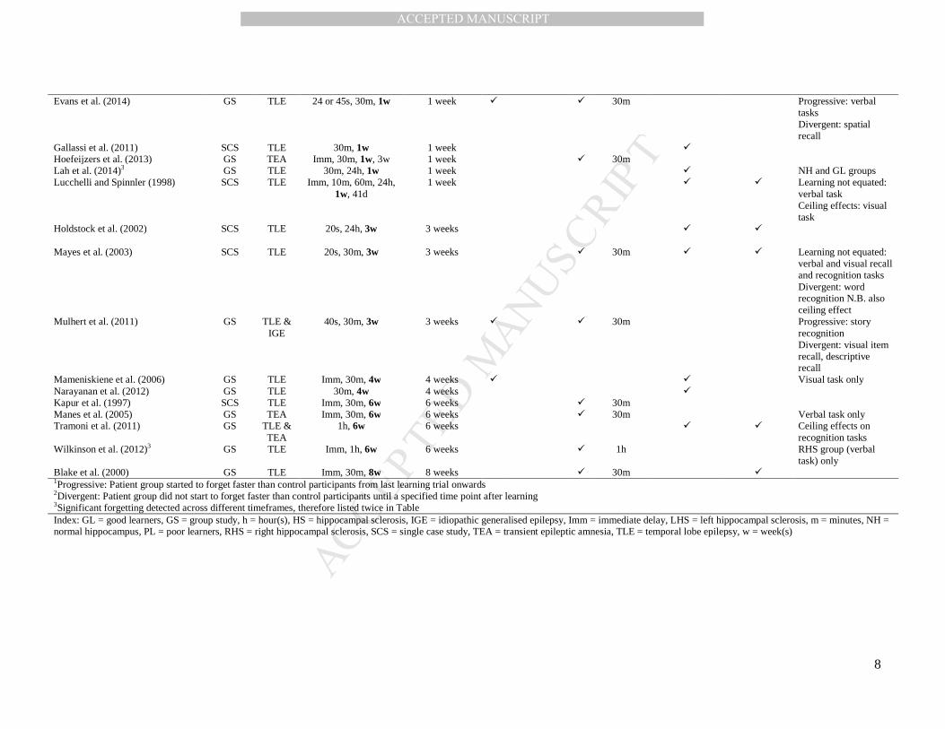
Another factor important in the design of forgetting studies, and subsequent
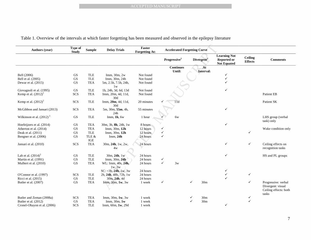
conclusions made, concerns the delays over which long-term memory is assessed. Table 1
summarises studies in TLE (including participants with transient epileptic amnesia). It
includes information regarding the delay periods measured, whether significant accelerated
forgetting was observed, and observations on the forgetting rate curves obtained in these
studies. This Table indicates that there is great variability in the literature regarding when
memory is assessed and the number of delay intervals used. Further, it is evident in the
majority of studies that the precise period over which accelerated forgetting manifested was
often reflective of the time points measured: most found faster forgetting by the first or
second delay interval measured after learning of new material (see Table 1, Delay Trials
column). Moreover, some studies did not report learning performance (Dewar, Hoefeijzers,
Zeman, Butler, & Della Sala, 2015; Gallassi et al., 2011; Hoefeijzers, Dewar, Dalla Sala,
Butler, & Zeman, 2014; Jansari, Davis, McGibbon, Firminger, & Kapur, 2010; Lah,
Mohamed, Thayer, Miller, & Diamond, 2014; McGibbon & Jansari, 2013; Narayanan et al.,
2012; O'Connor, Sieggreen, Ahern, Schomer, & Mesulam, 1997; Ricci, Mohamed, Savage,
Boserio, & Miller, 2015; Tramoni et al., 2011). In others, learning performance was not
equated (Bell, 2006; Bell, Fine, Dow, Seidenberg, & Hermann, 2005; Cronel-Ohayon et al.,
2006; Giovagnoli, Casazza, & Avanzini, 1995; Holdstock, Mayes, Isaac, Gong, & Roberts,
2002; Lucchelli & Spinnler, 1998; Mameniskiene et al., 2006; Mayes et al., 2003). These
omissions or oversights limit the implications of these studies, as the role of subtle
acquisition deficits cannot be excluded.
Of the studies in Table 1 that measured recall at multiple delay intervals, visual
inspection of forgetting curves can provide some insight into the point at which memory
MANUSCRIP
T
ACCEPTED
ACCEPTED MANUSCRIPT
5
consolidation is disrupted. For instance, a progressive pattern of forgetting, in which patients
start forgetting faster than controls immediately after learning, which becomes more
pronounced with time, would suggest an impairment in consolidation from the ‘early’ stages
onwards (even if between-group interactions do not become significant until later time-
points). On the other hand, forgetting curves that are parallel (or identical) for a period of
time, but then diverge would be indicative of a disruption to ‘late’ memory consolidation.
Reviewing the studies listed in Table 1, approximately half exhibited progressive forgetting
soon after learning that eventually became statistically significant at longer delays (Atherton,
Nobre, Zeman, & Butler, 2014; Bengner et al., 2006; Butler et al., 2007; Deak, Stickgold,
Pietras, Nelson, & Bubrick, 2011; Evans, Elliott, Reynders, & Isaac, 2014; Kemp, Illman,
Moulin, & Baddeley, 2012; Mameniskiene et al., 2006; Martin et al., 1991; Mulhert et al.,
2011; Mulhert, Milton, Butler, Kapur, & Zeman, 2010; Wilkinson et al., 2012). Other studies
showed a divergent pattern of forgetting, although some of these also exhibited ceiling effects
(Blake, Wroe, Breen, & McCarthy, 2000; Butler et al., 2007; Butler, Kapur, Zeman, Weller,
& Connelly, 2012; Butler & Zeman, 2008a; Evans et al., 2014; Hoefeijzers et al., 2013;
Kapur et al., 1997; Manes, Graham, Zeman, de Luján Calcagno, & Hodges, 2005; Mayes et
al., 2003; Mulhert et al., 2011; Wilkinson et al., 2012). The influence of ceiling effects is
particularly important in these cases because of the potential for overlearning, which may
mask any (early) differential forgetting effects between groups.
In light of such findings, some have argued that accelerated long-term forgetting may
reflect a subtle acquisition deficit, or an early consolidation deficit, which subsequently
affects long-term memory retention (Bell et al., 2005; Kopelman, 2000a, 2002). In this study,
therefore, we aimed to investigate (after appropriate matching of initial learning) whether and
when faster forgetting would be observed in a sample of TLE patients, compared with
healthy controls. We hypothesised that:
MANUSCRIP
T
ACCEPTED
ACCEPTED MANUSCRIPT
6
(1) TLE participants would forget newly learned (verbal and visual) material faster
than control participants;
(2) on examining epilepsy-related variables, more severe TLE cases would show
faster forgetting than milder TLE cases (as indicated by such factors as experience of
manifest seizures, polypharmacy, and medial temporal sclerosis on MRI); and
(3) any differences in forgetting rate would arise soon after learning, reflecting a
deficit in ‘early’ consolidation in TLE patients, rather than arising de novo after a period of
‘normal’ forgetting (which would reflect a deficit in ‘late’ consolidation).
MANUSCRIP
T
ACCEPTED
ACCEPTED MANUSCRIPT
7
Table 1. Overview of the intervals at which faster forgetting has been measured and observed in the epilepsy literature
Authors (year) Type of Study Sample Delay Trials
Faster Forgetting At: Accelerated Forgetting Curve
Progressive1 Divergent2 Learning Not Reported or Not Equated
Ceiling Effects Comments
Continues
Until: At
Interval:
Bell (2006) GS TLE Imm, 30m, 2w Not found � Bell et al. (2005) GS TLE Imm, 30m, 24h Not found � Dewar et al. (2015) GS TEA 5m, 2.5h, 7.5h, 24h,
1w Not found �
Giovagnoli et al. (1995) GS TLE 1h, 24h, 3d, 6d, 13d Not found � Kemp et al. (2012)3 SCS TEA Imm, 20m, 4d, 11d,
30d Not found Patient EB
Kemp et al. (2012)3 SCS TLE Imm, 20m, 4d, 11d, 30d
20 minutes � 11d Patient SK
McGibbon and Jansari (2013) SCS TEA 5m, 30m, 55m, 4h, 24h
55 minutes �
Wilkinson et al. (2012) 3 GS TLE Imm, 1h, 6w 1 hour � 6w LHS group (verbal task) only
Hoefeijzers et al. (2014) GS TEA 30m, 3h, 8h, 24h, 1w 8 hours � Atherton et al. (2014) GS TEA Imm, 30m, 12h 12 hours � Wake condition only Deak et al. (2011) GS TLE Imm, 30m, 12h 12 hours � � Bengner et al. (2006) GS TLE &
IGE Imm, 24h 24 hours �
Jansari et al. (2010) SCS TEA 30m, 24h, 1w, 2w, 4w
24 hours
� � Ceiling effects on recognition tasks
Lah et al. (2014)3 GS TLE 30m, 24h, 1w 24 hours � HS and PL groups Martin et al. (1991) GS TLE Imm, 30m, 24h 24 hours � Mulhert et al. (2010) GS TEA WL: Imm, 40s, 24h,
1w, 3w SC: ≈3h, 24h, 1w, 3w
24 hours
24 hours
�
3w �
O'Connor et al. (1997) SCS TLE 2h, 24h, 48h, 72h, 1w 24 hours � � Ricci et al. (2015) GS TLE 30m, 24h, 4d 24 hours � Butler et al. (2007) GS TEA Imm, 30m, 1w, 3w 1 week �
� 30m � Progressive: verbal
Divergent: visual Ceiling effects: both tasks
Butler and Zeman (2008a) SCS TEA Imm, 30m, 1w, 3w 1 week � 30m � Butler et al. (2012) GS TEA Imm, 30m, 1w 1 week � 30m � Cronel-Ohayon et al. (2006) SCS TLE Imm, 60m, 1w, 29d 1 week �
MANUSCRIP
T
ACCEPTED
ACCEPTED MANUSCRIPT
8
Evans et al. (2014) GS TLE 24 or 45s, 30m, 1w 1 week � � 30m Progressive: verbal tasks Divergent: spatial recall
Gallassi et al. (2011) SCS TLE 30m, 1w 1 week � Hoefeijzers et al. (2013) GS TEA Imm, 30m, 1w, 3w 1 week � 30m Lah et al. (2014)3 GS TLE 30m, 24h, 1w 1 week � NH and GL groups Lucchelli and Spinnler (1998) SCS TLE Imm, 10m, 60m, 24h,
1w, 41d 1 week � � Learning not equated:
verbal task Ceiling effects: visual task
Holdstock et al. (2002) SCS TLE 20s, 24h, 3w
3 weeks � �
Mayes et al. (2003) SCS TLE 20s, 30m, 3w
3 weeks � 30m � � Learning not equated: verbal and visual recall and recognition tasks Divergent: word recognition N.B. also ceiling effect
Mulhert et al. (2011) GS TLE & IGE
40s, 30m, 3w 3 weeks � � 30m Progressive: story recognition Divergent: visual item recall, descriptive recall
Mameniskiene et al. (2006) GS TLE Imm, 30m, 4w 4 weeks � � Visual task only Narayanan et al. (2012) GS TLE 30m, 4w 4 weeks � Kapur et al. (1997) SCS TLE Imm, 30m, 6w 6 weeks � 30m Manes et al. (2005) GS TEA Imm, 30m, 6w 6 weeks � 30m Verbal task only Tramoni et al. (2011) GS TLE &
TEA 1h, 6w 6 weeks � � Ceiling effects on
recognition tasks Wilkinson et al. (2012)3 GS TLE Imm, 1h, 6w 6 weeks � 1h RHS group (verbal
task) only Blake et al. (2000) GS TLE Imm, 30m, 8w 8 weeks � 30m � 1Progressive: Patient group started to forget faster than control participants from last learning trial onwards 2Divergent: Patient group did not start to forget faster than control participants until a specified time point after learning 3Significant forgetting detected across different timeframes, therefore listed twice in Table Index: GL = good learners, GS = group study, h = hour(s), HS = hippocampal sclerosis, IGE = idiopathic generalised epilepsy, Imm = immediate delay, LHS = left hippocampal sclerosis, m = minutes, NH = normal hippocampus, PL = poor learners, RHS = right hippocampal sclerosis, SCS = single case study, TEA = transient epileptic amnesia, TLE = temporal lobe epilepsy, w = week(s)
MANUSCRIP
T
ACCEPTED
ACCEPTED MANUSCRIPT
9
2. Materials and Methods
2.1 Participants
Eighteen patients with TLE were recruited from three sites across St Thomas’ Hospital
and King’s College Hospital in London, UK. In each case the diagnosis of TLE was
made based on appropriate history including seizure manifestations (Gil-Nagal &
Risinger, 1997) and epileptiform activity over the temporal areas (Koutroumanidis et
al., 2004). Patients were recruited if they met the following eligibility criteria: (a)
between 18 and 65 years of age, (b) fluent in written and spoken English, (c) no history
of neurosurgery, and (d) no neurological, medical, psychiatric, substance misuse or
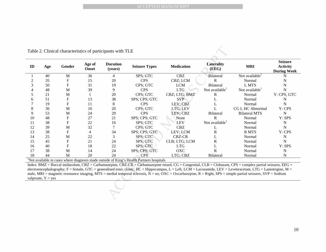
developmental co-morbidities. The clinical characteristics of each patient are shown in
Table 2.
Eighteen age-, gender-, education-, and intelligence-matched neurologically
healthy control participants who met the above eligibility criteria were also recruited via
an email advertisement within King’s College London and poster advertisement in the
community.
The study was approved by the National Health Service National Research
Ethics Service, London – Central and East Research Ethics Committee (13/LO/0399).
All participants gave their written, informed consent in accordance with the Declaration
of Helsinki.
MANUSCRIP
T
ACCEPTED
ACCEPTED MANUSCRIPT
10
Table 2. Clinical characteristics of participants with TLE
ID Age Gender Age of Onset
Duration (years) Seizure Types Medication
Laterality (EEG) MRI
Seizure Activity
During Week 1 40 M 36 4 SPS; GTC CBZ Bilateral Not available1 N 2 35 F 15 20 CPS CBZ; LCM R Normal N 3 50 F 31 19 CPS; GTC LCM Bilateral L MTS N 4 48 M 39 9 CPS LTG Not available1 Not available1 N 5 21 M 1 20 CPS; GTC CBZ; LTG; BMZ R Normal Y: CPS, GTC 6 51 F 13 38 SPS; CPS; GTC SVP L Normal N 7 19 F 11 8 CPS LEV; CBZ L Normal N 8 36 M 16 20 CPS; GTC LTG; LEV L CG L HC Abnormal Y: CPS 9 53 M 24 29 CPS LEV; CBZ Bilateral Bilateral MTS N 10 48 F 27 21 SPS; CPS; GTC None R Normal Y: SPS 11 38 F 22 16 SPS; GTC LEV Not available1 Normal N 12 39 M 32 7 CPS; GTC CBZ L Normal N 13 38 F 4 34 SPS; CPS; GTC LEV; LCM R R MTS Y: CPS 14 25 M 22 3 SPS; GTC CBZ-CR L Normal N 15 45 F 21 24 SPS; GTC CLB; LTG; LCM R Normal N 16 40 F 18 22 SPS; GTC LTG L Normal Y: SPS 17 38 M 14 24 SPS; CPS; GTC OXC R Normal N 18 44 M 20 24 CPS LTG; CBZ Bilateral Normal N
1Not available in cases where diagnosis made outside of King’s Health Partners hospitals Index: BMZ = Buccal midazolam, CBZ = Carbamazepine, CBZ-CR = Carbamazepine retard, CG = Congenital, CLB = Clobazam, CPS = complex partial seizures, EEG = electroencephalography; F = female, GTC = generalised tonic clonic, HC = Hippocampus, L = Left, LCM = Lacosamide, LEV = Levetiracetam, LTG = Lamotrigine, M = male, MRI = magnetic resonance imaging, MTS = medial temporal sclerosis, N = no, OXC = Oxcarbazepine, R = Right, SPS = simple partial seizures, SVP = Sodium valproate, Y = yes
MANUSCRIP
T
ACCEPTED
ACCEPTED MANUSCRIPT
11
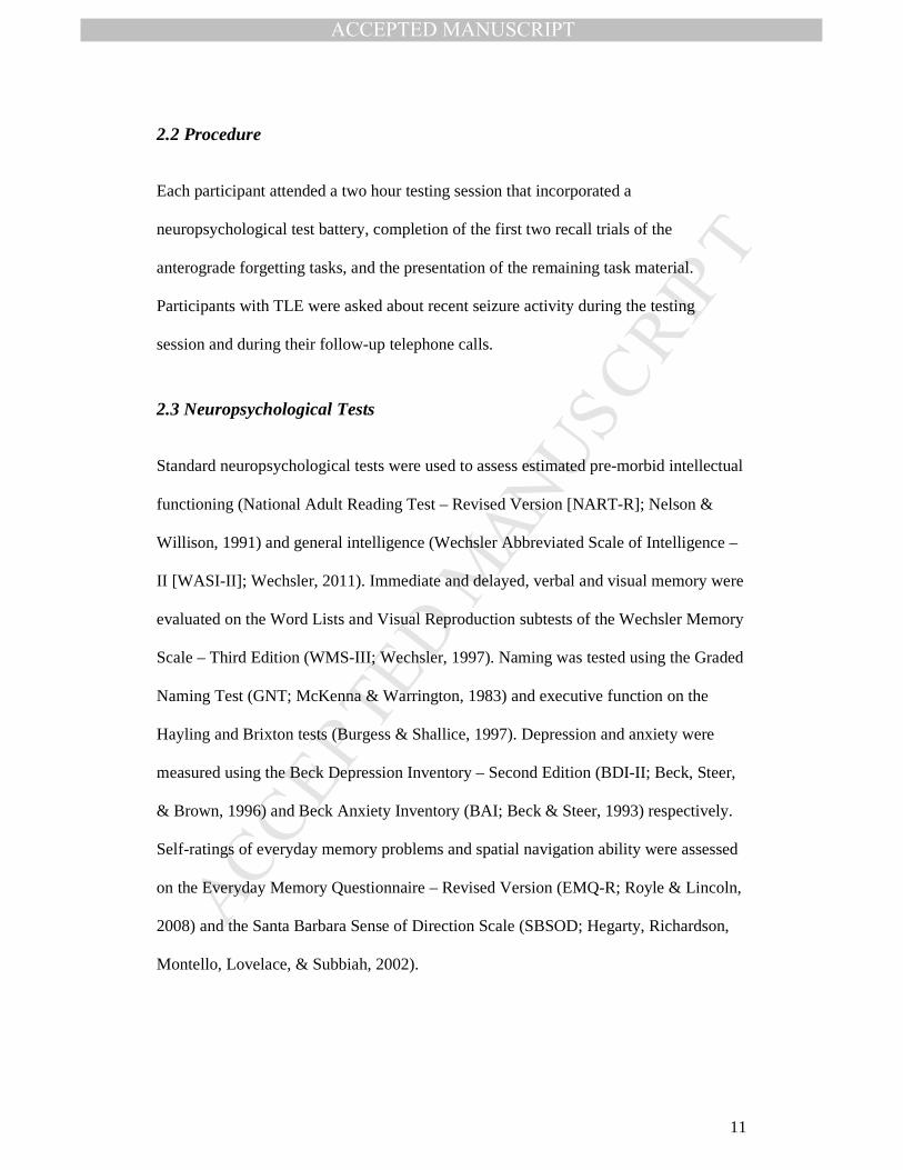
2.2 Procedure
Each participant attended a two hour testing session that incorporated a
neuropsychological test battery, completion of the first two recall trials of the
anterograde forgetting tasks, and the presentation of the remaining task material.
Participants with TLE were asked about recent seizure activity during the testing
session and during their follow-up telephone calls.
2.3 Neuropsychological Tests
Standard neuropsychological tests were used to assess estimated pre-morbid intellectual
functioning (National Adult Reading Test – Revised Version [NART-R]; Nelson &
Willison, 1991) and general intelligence (Wechsler Abbreviated Scale of Intelligence –
II [WASI-II]; Wechsler, 2011). Immediate and delayed, verbal and visual memory were
evaluated on the Word Lists and Visual Reproduction subtests of the Wechsler Memory
Scale – Third Edition (WMS-III; Wechsler, 1997). Naming was tested using the Graded
Naming Test (GNT; McKenna & Warrington, 1983) and executive function on the
Hayling and Brixton tests (Burgess & Shallice, 1997). Depression and anxiety were
measured using the Beck Depression Inventory – Second Edition (BDI-II; Beck, Steer,
& Brown, 1996) and Beck Anxiety Inventory (BAI; Beck & Steer, 1993) respectively.
Self-ratings of everyday memory problems and spatial navigation ability were assessed
on the Everyday Memory Questionnaire – Revised Version (EMQ-R; Royle & Lincoln,
2008) and the Santa Barbara Sense of Direction Scale (SBSOD; Hegarty, Richardson,
Montello, Lovelace, & Subbiah, 2002).
MANUSCRIP
T
ACCEPTED
ACCEPTED MANUSCRIPT
12
2.4 Forgetting Tests
2.4.1 Task Characteristics
2.4.1.1 Nature of Material
Two novel verbal and visuospatial measures were developed to assess
anterograde forgetting. A story task was used to assess verbal forgetting, because prose
tasks have greater ecological validity than word list tasks (Baddeley, Rawlings, &
Hayes, 2013; Butler & Zeman, 2008b). Similarly, we developed a route video task to
assess visuospatial memory, because similar tasks have been shown to have greater
ecological validity than pen-and-paper visual memory measures (Barbeau et al., 2006;
Tramoni et al., 2011). It has also been suggested that route-task performance is
correlated with patients’ subjective memory complaints, and thus may be useful in
clinical assessment (Plancher, Tirard, Gyselinck, Nicolas, & Piolino, 2012).
2.4.1.2 Nature of Retrieval
We assessed the story task by cued recall because this enables a greater degree
of control over responses than traditional free recall. It has also been shown to offer
greater sensitivity than recognition memory tasks (Baddeley et al., 2013). We did not
test both recall and recognition conditions because of the difficulty in measuring both
these facets on a single task whilst simultaneously avoiding ceiling and floor effects.
However, because making a spatial decision typically involves a forced choice decision
from a number of options, we assessed recall on the route task both by a series of two-
option forced-choice spatial decisions and, in addition, by cued recall of landmarks
passed in the video (after each spatial decision).
2.4.1.3 Timeframe of Assessment
MANUSCRIP
T
ACCEPTED
ACCEPTED MANUSCRIPT
13
In order to make inferences about the timeframe of forgetting, we assessed recall
at four intervals. Initial learning was assessed after a 30-second interval, during which a
distractor task was performed, in order to eliminate any short-term memory effects
(Cowan, 1993; Green & Kopelman, 2002; Isaac & Mayes, 1999a, 1999b; Kopelman &
Stanhope, 1997). A 10-minute delay was chosen as the next delay interval because this
interval has been shown to be sensitive in detecting differences in initial retention rates
(Christensen, Kopelman, Stanhope, Lorentz, & Owen, 1998; Isaac & Mayes, 1999a,
1999b; Kopelman & Stanhope, 1997). We then assessed longer-term recall after one
day and then after a week, because these are delay periods commonly used in long-term
forgetting research in epilepsy whilst minimising the potential for floor performance
(Kemp et al., 2012; Manes et al., 2005).
2.4.2 Story Task
2.4.2.1 Story Task Development
Four story forms were created to assess recall at each of the four delay intervals.
Parallel forms were created to avoid repeated recall and subsequent potential re-
encoding of material (Karpicke & Roediger, 2008; Roediger & Karpicke, 2006). In a
first phase of piloting, stories containing 13 units of information and 10 cued recall
questions were developed (see Supplementary Material for an example of a story trial).
These were to be presented in chronological order, and designed so that earlier answers
did not cue later responses within the sequence.
In a second phase of piloting, story trials were matched for difficulty at the 30-
second delay, and ceiling effects were avoided. A learning criterion of 60% accuracy
was selected for the study on the basis that this represented one standard deviation
below the healthy participants’ mean performance.
MANUSCRIP
T
ACCEPTED
ACCEPTED MANUSCRIPT
14
2.4.2.2 Story Task Procedure
Participants heard each story on a laptop computer recording. After presentation
of the first story, participants completed a distractor task for 30 seconds (subtracting
serial 3s from 100) before being asked the 10 cued-recall questions related to that story
trial. Using the 60% criterion, learning was matched on a case-by-case basis: if a
participant did not reach this criterion at the 30-second delay interval, the story was re-
presented, and cued recall tested again, until this criterion was reached. Having
determined the number of presentations needed to reach the 30-second criterion, this
number of presentations was used for the remaining stories, which were tested at 10
minutes, one day, and one week after learning. Story allocation to interval condition
was counterbalanced using a Latin square design. During the 10-minute delay period,
participants completed background neuropsychological tests and questionnaires
(NART, and/or EMQ-R, SBSOD). At the one-day and one-week tests, participants
received pre-arranged telephone calls, and were then asked cued recall questions about
the respective stories. Participants were asked not to rehearse the story during these
intervals.
2.4.3 Route Task
2.4.3.1 Route Task Development
The visuospatial task comprised four routes filmed from the front of a moving
car using a GoPro fish-eye camera. Modifications were made to the video clips such
that the film was paused at spatial decision points and at salient landmarks in the
environment during presentation. The landmarks followed immediately after the
decision points. Each trial consisted of five spatial decision and five landmark points.
MANUSCRIP
T
ACCEPTED
ACCEPTED MANUSCRIPT
15
At testing, ‘stills’ of each of the five spatial decision points were shown in
sequential order. Each still had two numbers superimposed on the picture indicating the
possible directions the car might drive from that point. This gave a two-option forced
choice recognition test. For the ‘landmark’ task, another still was shown of the same
image but without the superimposed numbers. A cued recall question was then asked
about the landmark (Figure 1).
Piloting ensured this approach was feasible, that each trial was equivalent in
difficulty at the 30-second delay interval, and that ceiling effects were avoided. An 80%
learning criterion was selected on the basis that this cut-off represented one standard
deviation below healthy participants’ mean performance in the pilot study.
****Insert Figure 1 around here****
2.4.3.2 Route Task Procedure
During presentation, participants were told that a video of a car driving through
a town would be shown, played on a laptop computer. They were asked to imagine
being a passenger in the car, and to pay attention to where they went and landmarks
passed. The video was paused at different points and their attention was drawn to
specific items to remember. Immediately after the first trial, participants completed a
distractor task for 30 seconds (separating two steel links in a puzzle), before being asked
recall questions corresponding to that trial. If a participant did not reach 80% accuracy,
presentation and recall questioning was repeated until this criterion was reached or until
they received two presentations of material. Having established the number of
presentations needed, this number was used for presentation of the other three film-
clips, for 10-minute, one-day, and one-week recall, which were counter-balanced for
allocation to the test delays according to a Latin square design. During the 10-minute
MANUSCRIP
T
ACCEPTED
ACCEPTED MANUSCRIPT
16
recall interval, background neuropsychological assessment measures were completed
(GNT, and/or EMQ-R, SBSOD). For one-day and one-week recall tests, participants
were told not to visualise or rehearse the route. Participants were emailed a password-
protected file containing the stills for these trials, which they accessed on their home
computer during testing over the telephone.
2.5 Test Scoring
Each trial for both the story and route tasks was scored out of 20 (see Supplementary
Materials for an example of how these tasks were scored). Percentage total recall scores
were calculated at each delay interval. These were used to determine forgetting rates in
terms of group by delay interaction analyses. For secondary analyses, forgetting rate
difference scores were calculated using the formula: (recall at first delay score [i.e. 30-
second] – recall score at later delay [e.g. one-week]) / (recall at first delay score [i.e. 30-
second]) x 100.
2.6 Statistical Analysis
All statistical analyses were carried out using SPSS. Data was checked for normality
(using box plots, Q-Q plots, and the Shapiro-Wilks test) and homogeneity of variance
(using Levene’s test and Mauchley’s test of sphericity as appropriate). Background test
scores were compared using t-tests or Mann-Whitney-U as appropriate. Overall
analyses and interaction effects were examined using mixed ANOVAs with significance
levels set at alpha ≤ .05. Planned comparisons were corrected using Benjamini and
Hochberg’s (1995) False Discovery Rate. Effect sizes were calculated using Cohen’s
(1992) d. In the comparison of subgroups, forgetting rate difference scores (calculated
as above) were checked for normality and then compared using one-way ANOVAs with
MANUSCRIP
T
ACCEPTED
ACCEPTED MANUSCRIPT
17
t-tests of significant results corrected for multiple comparisons as above. Where the
assumption for homogeneity of variance was not met, Welch’s F ratio was used and t-
tests analysed on the assumption of unequal variance (Field, 2013).
3. Results
3.1 Neuropsychological Profile
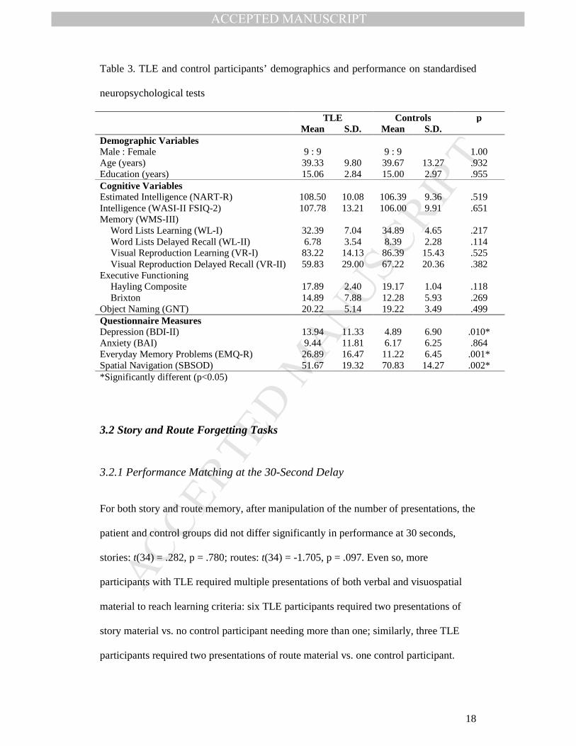
The patient and control groups were matched for gender, age and educational level
(Table 3). There were no differences between groups concerning intellectual
functioning, memory, executive and language functioning (all p > .05). The TLE group
reported more symptoms of depression (BDI-II: U = 81.50, p = .010), greater subjective
everyday memory problems (EMQ-R: t[34] = 3.76, p = .001), and worse spatial
navigation abilities (SBSOD: t[34] = -3.39, p = .002).
MANUSCRIP
T
ACCEPTED
ACCEPTED MANUSCRIPT
18
Table 3. TLE and control participants’ demographics and performance on standardised
neuropsychological tests
TLE Controls p Mean S.D. Mean S.D. Demographic Variables Male : Female 9 : 9 9 : 9 1.00 Age (years) 39.33 9.80 39.67 13.27 .932 Education (years) 15.06 2.84 15.00 2.97 .955 Cognitive Variables Estimated Intelligence (NART-R) 108.50 10.08 106.39 9.36 .519 Intelligence (WASI-II FSIQ-2) 107.78 13.21 106.00 9.91 .651 Memory (WMS-III) Word Lists Learning (WL-I) 32.39 7.04 34.89 4.65 .217 Word Lists Delayed Recall (WL-II) 6.78 3.54 8.39 2.28 .114 Visual Reproduction Learning (VR-I) 83.22 14.13 86.39 15.43 .525 Visual Reproduction Delayed Recall (VR-II) 59.83 29.00 67.22 20.36 .382 Executive Functioning Hayling Composite 17.89 2.40 19.17 1.04 .118 Brixton 14.89 7.88 12.28 5.93 .269 Object Naming (GNT) 20.22 5.14 19.22 3.49 .499 Questionnaire Measures Depression (BDI-II) 13.94 11.33 4.89 6.90 .010* Anxiety (BAI) 9.44 11.81 6.17 6.25 .864 Everyday Memory Problems (EMQ-R) 26.89 16.47 11.22 6.45 .001* Spatial Navigation (SBSOD) 51.67 19.32 70.83 14.27 .002* *Significantly different (p<0.05)
3.2 Story and Route Forgetting Tasks
3.2.1 Performance Matching at the 30-Second Delay
For both story and route memory, after manipulation of the number of presentations, the
patient and control groups did not differ significantly in performance at 30 seconds,
stories: t(34) = .282, p = .780; routes: t(34) = -1.705, p = .097. Even so, more
participants with TLE required multiple presentations of both verbal and visuospatial
material to reach learning criteria: six TLE participants required two presentations of
story material vs. no control participant needing more than one; similarly, three TLE
participants required two presentations of route material vs. one control participant.
MANUSCRIP
T
ACCEPTED
ACCEPTED MANUSCRIPT
19
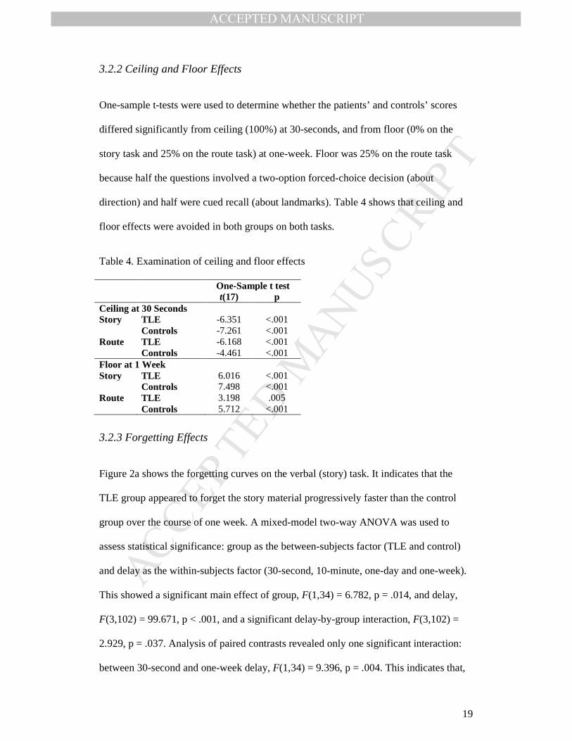
3.2.2 Ceiling and Floor Effects
One-sample t-tests were used to determine whether the patients’ and controls’ scores
differed significantly from ceiling (100%) at 30-seconds, and from floor (0% on the
story task and 25% on the route task) at one-week. Floor was 25% on the route task
because half the questions involved a two-option forced-choice decision (about
direction) and half were cued recall (about landmarks). Table 4 shows that ceiling and
floor effects were avoided in both groups on both tasks.
Table 4. Examination of ceiling and floor effects
One-Sample t test t(17) p Ceiling at 30 Seconds Story TLE -6.351 <.001 Controls -7.261 <.001 Route TLE -6.168 <.001 Controls -4.461 <.001 Floor at 1 Week Story TLE 6.016 <.001 Controls 7.498 <.001 Route TLE 3.198 .005 Controls 5.712 <.001
3.2.3 Forgetting Effects
Figure 2a shows the forgetting curves on the verbal (story) task. It indicates that the
TLE group appeared to forget the story material progressively faster than the control
group over the course of one week. A mixed-model two-way ANOVA was used to
assess statistical significance: group as the between-subjects factor (TLE and control)
and delay as the within-subjects factor (30-second, 10-minute, one-day and one-week).
This showed a significant main effect of group, F(1,34) = 6.782, p = .014, and delay,
F(3,102) = 99.671, p < .001, and a significant delay-by-group interaction, F(3,102) =
2.929, p = .037. Analysis of paired contrasts revealed only one significant interaction:
between 30-second and one-week delay, F(1,34) = 9.396, p = .004. This indicates that,
MANUSCRIP
T
ACCEPTED
ACCEPTED MANUSCRIPT
20
although participants with TLE appeared to forget the stories progressively faster than
control participants, this deviation only became statistically significant at one week
post-learning.
Figure 2b shows forgetting curves for the visuospatial (route) task. On this,
participants with TLE appeared to forget faster than controls between 30 seconds and 10
minutes, with comparable forgetting rates beyond this delay. An overall ANOVA
indicated a significant main effect of group, F(1,34) = 18.374, p < .001 and delay,
F(3,102) = 68.601, p < .001, but no significant delay-by-group interaction, F(3,102) =
1.655, p = .182. Although this interaction was not significant, planned comparisons of
paired contrasts after learning revealed a significant interaction between 30-second and
10-minute recall, F(1,34) = 7.253, p = .011. The paired contrast interaction from 10-
minute to one-week recall was not significant, F(1,34) = .001, p = .973. This suggests
that participants with TLE forgot route material at an accelerated rate only between 30
seconds and 10 minutes after learning, with comparable forgetting rates thereafter.
****Insert Figure 2 around here****
3.3 Analyses of Epilepsy-Related Variables and Forgetting
Participants with TLE were categorised into sub-groups based on (1) EEG laterality of
seizure focus, (2) the presence of medial temporal lobe sclerosis (MTS) on MRI, (3)
seizure activity during the week of the experiment, and (4) dosage of anti-epileptic
medication. Figure 3 shows performance on the story and route tasks in these subgroups
expressed in terms of difference scores.
MANUSCRIP
T
ACCEPTED
ACCEPTED MANUSCRIPT
21
3.3.1 Laterality of Seizure Focus
Six participants with left TLE and six participants with right TLE were compared with
control participants. These subgroups were comparable on all demographic and
cognitive variables (all p > .05).
For story memory, performance was not statistically different between groups at
30-second recall, F(2,27) = .341, p = .741. Figure 3a shows both TLE groups forgot
more than controls over the course of a week. Laterality of seizure focus was not
statistically associated with differences in forgetting rates between 30-second recall and
any later delays (all p > .05).
Likewise, for route memory, performance was not statistically different between
groups at 30-second recall, F(2,27) = .734, p = .489. Figure 3b shows that the two TLE
subgroups forgot at a similar rate between 30 seconds and one day, at a faster rate than
controls, but thereafter the right TLE group appeared to forget faster than the left-sided
group. Statistically significant differences in forgetting rates were observed only
between the 30-second and one-week delays, F(2,27) = 6.424, p = .005 (all other
comparisons p > .05). Right TLE participants showed faster forgetting compared with
left TLE, t(10) = -2.539, p = .029, and control participants, t(22) = 3.254, p = .004.
Forgetting rates of participants with left TLE did not differ significantly from controls,
t(22) = 1.528, p = .141. In summary, participants with right-hemisphere seizures
showed faster forgetting of route material over the course of one week, compared with
controls and participants with left-hemisphere seizures.
3.3.2 MRI Identified Medial Temporal Lobe Sclerosis
Four participants with TLE showed MTS on MRI scan. These participants were
compared with 12 participants with TLE who had ‘normal’ MRI scans (according to
MANUSCRIP
T
ACCEPTED
ACCEPTED MANUSCRIPT
22
radiological reports) and controls. These subgroups were comparable on all
demographic and cognitive variables (all p > .05).
For story memory, performance was comparable between groups at 30-second
delay, F(2,31) = .568, p = .572. Figure 3c shows that participants with MTS exhibited
faster forgetting 10 minutes after learning, compared with the other patient group and
controls. There was a significant between-group difference in forgetting rate from 30-
second to 10-minute recall, F(2,31) = 5.354, p = .010. The participants with sclerosis
showed faster forgetting compared with participants without sclerosis, t(14) = 2.697, p
= .017, and controls, t(20) = 3.350, p = .003. Forgetting rates of participants without
sclerosis did not differ from controls, t(28) = .622, p = .539. Over the course of a week
(Figure 3c), there was a significant between-group difference in forgetting rate, Welch’s
F(2,16.84) = 14.214, p < .001. Those with MRI-detectable MTS demonstrated faster
forgetting between 30 seconds and a week, compared with those who had ‘normal’
MRI scans, t(12,05) = 2.954, p = .012, and controls, t(19.28) = 5.221, p < .001
respectively. However, the participants with ‘normal’ MRI scans also forgot faster than
controls over the course of a week, t(27.30) = 2.789, p = .01. In summary, participants
with MRI-detectable MTS demonstrated faster forgetting of story material during the
first 10 minutes after learning, compared with participants without sclerosis and
controls. By one week after learning, both patient groups had forgotten story material at
rates in excess of controls and those with MTS continued to forget at a rate faster than
those without sclerosis.
For route memory, performance was not statistically different between groups at
30-second delay, F(2,31) = 2.671, p = .085. Figure 3d shows that participants with MTS
appeared to forget route material faster than the other two groups between 30 seconds

MANUSCRIP
T
ACCEPTED
ACCEPTED MANUSCRIPT
23
and 10 minutes. However, this was not reflected in statistical analyses, where no
forgetting rate comparison reached statistical significant (all p > .05).
3.3.3 Seizure Activity During Participation Week
Five participants with TLE experienced at least one seizure during the week of
participation. Two had experienced at least one seizure by one-day delay and all five
experienced at least one seizure between the one-day and one-week delays. These five
participants were compared with 13 participants with TLE who did not experience a
seizure during their participation week, and with controls. Groups were matched on all
demographic and cognitive variables (all p > .05).
For story memory, performance was not statistically different between groups at
30-seconds, F(2,33) = .106, p = .900. Figure 3e shows that both patient groups forgot
story material faster than the control group across all delay intervals, but that the rate of
forgetting between the two patient groups did not differ. On statistical analyses, the only
significant between-group effect across the three groups was in forgetting rates from the
30-second delay to recall at one-week, Welch’s F(2,11.27) = 6.273, p = .015. As
reflected in Figure 3e, the two patient groups’ rates of forgetting did not differ, t(5.64) =
.307, p = .770, but both subgroups forgot story material faster than controls, t(8.89) =
2.681, p = .025 and t(26.58) = 3.489, p = .002, respectively.
For route memory, performance was comparable between groups at 30-second
recall, F(2,33) = 1.684, p = .201. Figure 3f shows that those who had a seizure during
the experiment week forgot faster than the controls by 10 minutes, which continued at
an accelerated rate through the week. The patient group who did not experience seizures
during the participation week also forgot faster than controls over these time-periods,
albeit to a lesser extent. However, it was only between 30-second and 10-minute recall
MANUSCRIP
T
ACCEPTED
ACCEPTED MANUSCRIPT
24
that groups differed, F(2,33) = 4.622, p = .017, and no planned comparisons reached
statistical significance (all p > .05).
3.3.4 Anti-Epileptic Medication
Nine participants with TLE were undergoing monotherapy treatment for their epilepsy
and eight were prescribed polytherapy. These patient sub-groups were compared with
control participants. Groups were matched on demographic variables (all p > .05) but
differed on a number of cognitive variables including intelligence, verbal memory, and
executive functioning (p < .05). Participants on polytherapy performed worse than
controls on measures of verbal memory, t(24) = -2.902, p = .008 and t(24) = -2.960, p =
.007 (WMS-III WL-I and WL-II respectively), and worse than those on monotherapy on
a measure of intelligence (WASI-II FSIQ-2), t(15) = 3.586, p = .003, and the Brixton
test, t(15) = -2.410, p = .029.
Despite these differences, story recall at 30-seconds was comparable between
groups, F(2,32) = .502, p = .610. Figure 3g shows that participants on polytherapy
started forgetting faster than those on monotherapy and controls by 10 minutes post-
learning, although both patient groups forgot at similar accelerated rates by one-week.
This pattern was confirmed on statistical analyses: significant between-group
differences were observed in the first 10 minutes after learning, F(2,32) = 4.319, p =
.022, and between 30-seconds and one-week, Welch’s F(2,18.11) = 6.800, p = .006. In
the first 10 minutes after learning, participants on polytherapy forgot story material
faster than those on monotherapy, t(15) = -2.420, p = .029, and controls, t(24) = 2.911, p
= .008. By one-week, the patient groups were not statistically different from each other,
t(12.12) = .218, p = .831, but both the monotherapy and polytherapy groups differed
from controls, t(24.98) = 3.643, p = .001 and t(19.15) = 2.844, p = .01 respectively.
MANUSCRIP
T
ACCEPTED
ACCEPTED MANUSCRIPT
25
Similarly, for route memory, recall was not statistically different between groups
at 30-seconds, F(2,32) = 3.195, p = .054. Figure 3h shows that those on polytherapy
forgot route material faster by 10 minutes after learning, and those on monotherapy
exhibited a more progressive rate of forgetting compared with controls. Even so, only
the difference between 30 seconds and 10-minutes was significant across groups,
Welch’s F(2,12.76) = 4.027, p = .044 but no planned comparisons reached significance
after correcting for multiple testing (all p > .03).
****Insert Figure 3 around here****
4. Discussion
This study examined: (1) whether patients with TLE demonstrated a faster rate of
forgetting compared with matched controls on two novel measures; (2) whether the
severity of epilepsy-related variables was associated with forgetting rates; and (3)
whether any differences in forgetting rate commenced soon after initial learning, or
much later. Our study was designed to follow a number of principles (Elliott et al.,
2014; Kopelman & Bright, 2012), which would allow us to explore possible causes of
any accelerated forgetting and determine whether our data implicated ‘early’ or ‘late’
memory consolidation disruption.
4.1 Did we find evidence of accelerated forgetting in our TLE sample?
We found that participants with TLE showed faster forgetting of story material by one
week after initial learning. This forgetting was progressive from 30 seconds onwards,
although differences in forgetting rate only became statistically significant after one
week. In previous studies of verbal forgetting, most found that statistically significant
MANUSCRIP
T
ACCEPTED
ACCEPTED MANUSCRIPT
26
accelerated forgetting was observed before one week (i.e. Hoefeijzers et al., 2014;
Jansari et al., 2010; Mulhert et al., 2010; O'Connor et al., 1997). Even so, Lah et al.
(2014) found a pattern of forgetting similar to ours in their sample of TLE participants
with ‘normal’ hippocampi on MRI: there was some initial forgetting in their patient
group which became progressively accelerated, and statistically significant, over one
week.
With regard to route memory, the overall group by time interaction effect was
not statistically significant across the four delay intervals. However, visual inspection
and planned comparisons indicated that the patient sample forgot visuospatial material
faster by 10 minutes. This suggests that participants with TLE forgot route material
more rapidly over this early delay, with comparable forgetting rates thereafter.
Wilkinson et al. (2012) found a non-significant trend for faster forgetting in the first
hour after learning in patients with TLE and right hippocampal sclerosis, whilst Kemp
et al. (2012) found evidence of accelerated forgetting in the first 20 minutes after
learning in a patient with TLE. Moreover, the pattern of our findings on visuospatial
forgetting are consistent with findings in non-epileptic amnesic patients (including those
with temporal lobe pathology), which have shown accelerated forgetting within 10 or 20
minutes, after matching for initial memory performance (Christensen et al., 1998; Green
& Kopelman, 2002; Isaac & Mayes, 1999a, 1999b; Kopelman & Stanhope, 1997).
We also note that in other forgetting studies in epilepsy, some did not find
statistically significant accelerated forgetting (Davidson, Dorris, O'Regan, & Zuberi,
2007; Mulhert et al., 2011; Narayanan et al., 2012) and others only observed accelerated
forgetting after longer delays (Evans et al., 2014; Tramoni et al., 2011). Whilst
heterogeneity of method and materials are likely to have contributed to the variability of
MANUSCRIP
T
ACCEPTED
ACCEPTED MANUSCRIPT
27
these findings, there still appeared to be a pattern of progressively faster forgetting in
the epilepsy group that either did or did not become significant over time.
4.2 Why was there a different pattern between verbal and visuospatial
forgetting?
As noted above, faster forgetting on the visuospatial task appeared to occur within the
first 10 minutes, but, on the verbal task, differences in forgetting only became
statistically significant at one week. Other authors have obtained related findings in
TLE patients and Amlerova et al. (2012) noted that this patient group can be at risk of
spatial memory impairments. Dewar et al. (2015) reported that transient epileptic
amnesia patients demonstrated impaired picture recognition five minutes after learning,
despite this sample not exhibiting accelerated forgetting on a verbal task until hours
after learning (Hoefeijzers et al., 2014). Additionally, Mulhert et al. (2011) showed that
TLE patients were impaired on a spatial recall task at 40-second recall, despite normal
performance on all other verbal and visual measures.
In the present study, our route task relied heavily on specific processes
associated with three-dimensional spatial navigational skills (Morris & Mayes, 2004),
and was selected for its everyday (‘ecological’) validity. Such spatial navigational
processes are known to depend on bilateral interaction between medial temporal lobe
structures (Canovas, Leon, Serrano, Roldan, & Cimadevilla, 2011; Glikmann-Johnston
et al., 2008), known to be important for ‘early’ memory consolidation processes (Dudai,
2004). Our patient group also reported significantly poorer spatial navigational abilities,
lending further support to the possibility that our visuospatial task may have had greater
sensitivity to detect accelerated forgetting within a relatively shorter timeframe
compared with our verbal task. Differing task demands and retrieval memory processes
MANUSCRIP
T
ACCEPTED
ACCEPTED MANUSCRIPT
28
between each measure may have also contributed to differences observed in forgetting
rates.
4.3 What variables were associated with accelerating rates of forgetting?
Various pathophysiological variables were also associated with different patterns of
forgetting. This was particularly evident on the story task, where both the presence of
MTS and anti-epileptic polypharmacy were associated with accelerated forgetting being
detectable earlier, i.e., after 10 minutes. Even so, those patients without MTS, and those
on monotherapy treatment, still exhibited accelerated forgetting compared to controls
but differences in forgetting rate only became significant after one week. The forgetting
curve pattern was progressive for those patients without MTS. For those on
monotherapy, it appeared to become more divergent (after 10 minutes). Regarding the
route task, although the forgetting curves observed were largely similar to those evident
on the story task, comparisons did not reach statistical significance.
Others have also found that greater use of anti-epileptic medication may
influence forgetting rates at early delays (Butler et al., 2009; Jokeit et al., 2005; Lee,
2010; Motamedi & Meador, 2003; Wilkinson et al., 2012). Similarly, hippocampal
sclerosis has been found to influence earlier forgetting (Lah et al., 2014; Wilkinson et
al., 2012). Importantly, Lah et al. (2014) found that, of participants with an ‘abnormal’
hippocampus, most forgetting occurred in the first 24 hours (although they
acknowledged that ceiling effects may have masked any forgetting over even earlier
delays), whilst those without hippocampal sclerosis exhibited a slower rate of forgetting
that only became statistically significant at a week. Further, Wilkinson et al. (2012)
compared left- versus right hippocampal sclerosis in TLE patients: participants with left
hippocampal sclerosis forgot verbal material faster over a one-hour delay than those
MANUSCRIP
T
ACCEPTED
ACCEPTED MANUSCRIPT
29
with right hippocampal sclerosis or controls, but both patient groups went on to exhibit
faster forgetting by six weeks. The pattern of findings in these studies could, therefore,
be seen as broadly consistent with our findings: although MTS may accelerate early
forgetting, those patients without sclerosis observable on MRI, still exhibit a slower,
more progressive, form of accelerated forgetting.
In the present study, we did not find evidence that seizure activity during the
week of testing was associated with accelerating rates of forgetting. This lack of
association is similar to some previous research (Blake et al., 2000; Mulhert et al.,
2011), but not others (Fitzgerald, Thayer, Mohamed, & Miller, 2013; Mameniskiene et
al., 2006; O'Connor et al., 1997; Ricci et al., 2015; Wilkinson et al., 2012). Reasons for
this may be related to our measure of seizure activity, which relied on self-report and
included any reported manifest epileptiform activity. We were not able to record
subclinical activity, timing, or duration of seizures, all of which might contribute to
forgetting (Butler et al., 2010). It is possible these variables were influencing the
accelerated forgetting rates found in both patient subgroups on our tasks.
Interestingly, the only laterality effect we found was on our visuospatial task:
patients with a right-hemisphere origin to their seizures forgot route material over a
week more rapidly than left-hemisphere cases (despite the patient subgroups’ forgetting
curves appearing similar up until one day after learning). There is little other research
finding a similar association; the exception being Narayanan et al. (2012), who found a
similar trend for faster long-term visual forgetting in those with right-hemisphere TLE
by four weeks. However, they did not measure long-term recall at any earlier delay, thus
we cannot elucidate whether accelerated forgetting could have been detectable earlier.
In summary, for the story task, it appears that indicators of greater epilepsy
severity (i.e. MTS, polypharmacy) resulted in accelerated forgetting that was detectable
MANUSCRIP
T
ACCEPTED
ACCEPTED MANUSCRIPT
30
earlier, after 10 minutes. The other patient subgroups (i.e. those with ‘normal’ MRI
scans, monopharmacy) still exhibited accelerated forgetting compared to controls, but
this was only detectable after a week. Manifest experience of seizures during the
participation week did not differentially accelerate forgetting compared to those without
seizures. There were similar visual trends on the route task to this effect, but these did
not reach statistical significance. Nonetheless, our interpretation must be somewhat
tentative, given that the relatively small size of our sample did not permit more rigorous
statistical techniques, such as regression analysis, and may also have increased the risk
of Type 1 errors. In a sample of 21 patients with TLE, Ricci et al. (2015) argued that
only the presence of a hippocampal lesion in TLE patients was predictive of accelerated
forgetting (over 24 hours) when all variables were taken into account (such as seizure
activity, right-hemisphere involvement, longer duration of epilepsy, greater depression,
and hippocampal sclerosis). It will be important for future research in larger series to
elucidate further which factors lead to faster or slower memory decay.
4.4 What are the implications of our findings for memory consolidation
processes?
Our findings implicate ‘early’ memory consolidation disruption in the phenomenon of
accelerated forgetting. Whilst this was particularly evident on the visuospatial task,
where forgetting was accelerated in the first 10 minutes after matched learning, the
effect of this disruption was more graduated on the verbal task. We therefore posit that
faster forgetting in TLE may operate over a continuum of severity (Blake et al., 2000).
At one extreme, these ‘early’ retention deficits are evident soon after learning and could
feasibly be detected using adequately sensitive, or ‘standard’, memory assessment tools.
At the other extreme, the deficit is subtler: the rate of faster forgetting is slower, more
MANUSCRIP
T
ACCEPTED
ACCEPTED MANUSCRIPT
31
progressive, and only becomes statistically detectable after a longer length of time has
passed. Various factors, such as task characteristics and greater epilepsy severity (e.g.
MTS, polypharmacy), can result in faster forgetting being detected earlier.
This interpretation challenges the position others have made in the field: where
statistically significant accelerated forgetting has only been observed after long delays,
it has often been concluded these findings result from a disruption to ‘late’ memory
consolidation (Butler et al., 2010; Butler & Zeman, 2008b). However, we found very
little evidence to suggest forgetting occurred at ‘normal’ rates until a later disruption
(with only a visual trend of divergent story and route forgetting observed for the
monotherapy and right TLE subgroups respectively). Moreover, of the studies listed in
Table 1, ceiling effects confounded many of the divergent forgetting curves observed,
and approximately half demonstrated a progressively faster forgetting rate in their
patient samples, similar to the pattern found on our story task. In summary, our findings
challenge the view that memory stabilisation is not disrupted until later delays.
5. Conclusions
We have shown that people with TLE exhibit faster forgetting for both verbal and
visuospatial material. This was detectable within 10 minutes of learning on the
visuospatial task. On the verbal task, forgetting was slower and more progressive. The
difference in this pattern might be related to material sensitivity, and to the particular
role of the medial temporal structures in spatial navigation tasks, but might also have
reflected other factors as mentioned above.
We have also provided preliminary findings concerning the role of different
pathophysiological variables on the timeframe of forgetting. Markers of the severity of
epilepsy (the presence of MTS and use of multiple anti-epileptic agents) were
MANUSCRIP
T
ACCEPTED
ACCEPTED MANUSCRIPT
32
associated with earlier forgetting, at least on our verbal task. Future research will
require a larger sample size to examine the relative contribution of these factors to
forgetting.
We have argued that our findings implicate the disruption of ‘early’ memory
consolidation processes. The effects of this early disruption can be conceptualised as a
‘continuum’ of forgetting severity: either apparent immediately or one that becomes
more pronounced over time. Whilst we cannot rule out the possibility that memory
traces could be disrupted during ‘late’ consolidation, our data are more consistent with
an early retention deficit.
It remains to be demonstrated in patients with temporal lobe lesions whether
there is a definite difference between those with or without epilepsy or, for that matter,
between TLE and the subgroup with transient epileptic amnesia. Improved
understanding of what factors cause and influence rates of forgetting in this population
will not only advance our theoretical understanding of memory consolidation, but also
aid in the clinical assessment and management of TLE patients reporting concerns with
their memory.
Acknowledgements
We thank all the participants who participated in this study. We also thank Dr Maria
Stefanatou who aided with recruitment and to Daniel Stahl for his advice about
statistical analysis.
Professors Michael Kopelman and Robin Morris belong to the NIHR
Biomedical Research Centre at King’s College London, Institute of Psychiatry,
Psychology and Neuroscience. None of the authors have any conflicts of interest to
disclose.
MANUSCRIP
T
ACCEPTED
ACCEPTED MANUSCRIPT
33
References
Alvarez, P., & Squire, L. R. (1994). Memory consolidation and the medial temporal lobe: a simple network model. Proceedings of the National Academy of Sciences of the United States of America, 91, 7041-7045.
Amlerova, J., Laczo, J., Vlcek, K., Javurkova, A., Andel, R., & Marusic, P. (2012). Risk factors for spatial memory impairment in patients with temporal lobe epilepsy. Epilepsy & Behavior, 26, 57-60.
Atherton, K. E., Nobre, A. C., Zeman, A. Z., & Butler, C. R. (2014). Sleep-dependent memory consolidation and accelerated forgetting. Cortex, 54, 92-105.
Baddeley, A., Rawlings, B., & Hayes, A. (2013). Constrained prose recall and the assessment of long-term forgetting: the case of ageing and the Crimes Test. Memory. doi: 10.1080/09658211.2013.865753
Barbeau, E. J., Didic, M., Felician, O., Tramoni, E., Guedj, E., Ceccaldi, M., & Poncet, M. (2006). Pure progressive amnesia: an atypical amnestic syndrome? Cognitive Neuropsychology, 23(8), 1230-1247.
Beck, A. T., & Steer, R. A. (1993). Beck Anxiety Inventory Manual. San Antonio, TX: Harcourt Brace and Company.
Beck, A. T., Steer, R. A., & Brown, G. K. (1996). Manual for the Beck Depression Inventory-II. San Antonio, TX: Psychological Corporation.
Bell, B. D. (2006). WMS-III logical memory performance after a two-week delay in temporal lobe epilepsy and control groups. Journal of Clinical and Experimental Neuropsychology, 28(8), 1435-1443.
Bell, B. D., Fine, J., Dow, C., Seidenberg, M., & Hermann, B. P. (2005). Temporal lobe epilepsy and the selective reminding test: the conventional 30-minute delay suffices. Psychological Assessment, 17(1), 103-109.
Bengner, T., Malina, T., Lindenau, M., Voges, B., Goebell, E., & Stodieck, S. (2006). Face memory in MRI-positive and MRI-negative temporal lobe epilepsy. Epilepsia, 47(11), 1904-1914.
Benjamini, Y., & Hochberg, Y. (1995). Controlling the false discovery rate: a practical and powerful approach to multiple testing. Journal of the Royal Statistical Society, Series B (Methodological), 57(1), 289-300.
Blake, R. V., Wroe, S. J., Breen, E. K., & McCarthy, R. A. (2000). Accelerated forgetting in patients with epilepsy. Evidence for an impairment in memory consolidation. Brain, 123, 472-483.
Brooks, D. N., & Baddeley, A. D. (1976). What can amnesic patients learn? Neuropsychologia, 14, 111-122.
Burgess, P., & Shallice, T. (1997). The Hayling and Brixton Tests. Bury St Edmunds: Thames Valley Test Company.
Butler, C. R., Bhaduri, A., Acosta-Cabronero, J., Nestor, P. J., Kapur, N., Graham, K. S., . . . Zeman, A. Z. (2009). Transient epileptic amnesia: regional brain atrophy and its relationship to memory deficits. Brain, 132, 357-368.
Butler, C. R., Graham, K. S., Hodges, J. R., Kapur, N., Wardlaw, J. M., & Zeman, A. Z. (2007). The syndrome of transient epileptic amnesia. Annals of Neurology, 61, 587-598.
Butler, C. R., Kapur, N., Zeman, A., Weller, R., & Connelly, A. (2012). Epilepsy-related long-term amnesia: anatomical perspectives. Neuropsychologia, 50, 2973-2980.
Butler, C. R., Mulhert, N., & Zeman, A. Z. (2010). Accelerated long-term forgetting. In S. Della Sala (Ed.), Forgetting (pp. 211-237). Hove: Psychology Press.
MANUSCRIP
T
ACCEPTED
ACCEPTED MANUSCRIPT
34
Butler, C. R., & Zeman, A. Z. (2008a). A case of transient epileptic amnesia with radiological localisation. Nature Clinical Practice: Neurology, 49(9), 516-521.
Butler, C. R., & Zeman, A. Z. (2008b). Recent insights into the impairment of memory in epilepsy: transient epileptic amnesia, accelerated long-term forgetting and remote memory impairment. Brain, 131(9), 2243-2263.
Canovas, R., Leon, I., Serrano, P., Roldan, M. D., & Cimadevilla, J. M. (2011). Spatial navigation impairment in patients with refractory temporal lobe epilepsy: evidence from a new virtual reality-based task. Epilepsy & Behavior, 22, 364-369.
Christensen, H., Kopelman, M. D., Stanhope, N., Lorentz, L., & Owen, P. (1998). Rates of forgetting in Alzheimer dementia. Neuropsychologia, 36, 547-557.
Cohen, J. (1992). A power primer. Psychological Bulletin, 112(1), 155-159. Cowan, N. (1993). Activation, attention, and short-term memory. Memory & Cognition,
21(2), 162-167. Cronel-Ohayon, S., Zesiger, P., Davidoff, V., Boni, A., Roulet, E., & Deonna, T.
(2006). Deficit in memory consolidation (abnormal forgetting rate) in childhood temporal lobe epilepsy. Pre and postoperative long-term observation. Neuropediatrics, 37, 317-324.
Davidson, M., Dorris, L., O'Regan, M., & Zuberi, S. M. (2007). Memory consolidation and accelerated forgetting in children with idiopathic generalised epilepsy. Epilepsy & Behavior, 11, 394-400.
Deak, M. C., Stickgold, R., Pietras, A. C., Nelson, A. P., & Bubrick, E. J. (2011). The role of sleep in forgetting in temporal lobe epilepsy: a pilot study. Epilepsy & Behavior, 21, 462-466.
Dewar, M., Hoefeijzers, S., Zeman, A., Butler, C., & Della Sala, S. (2015). Impaired picture recognition in transient epileptic amnesia. Epilepsy & Behavior, 42, 107-116.
Dudai, Y. (2004). The neurobiology of consolidation, or, how stable is the engram? Annual Review of Psychology, 55, 51-86.
Elliott, G., Isaac, C. L., & Mulhert, N. (2014). Measuring forgetting: a critical review of accelerated long-term forgetting studies. Cortex, 54, 16-32.
Evans, S. J., Elliott, G., Reynders, H., & Isaac, C. L. (2014). Can temporal lobe epilepsy surgery ameliorate accelerated long-term forgetting? Neuropsychologia, 53, 64-74.
Field, A. (2013). Discovering Statistics Using IBM SPSS Statistics (4th ed.). London: Sage Publications Ltd.
Fitzgerald, Z., Mohamed, A., Ricci, M., Thayer, Z., & Miller, L. (2013). Accelerated long-term forgetting: a newly identified memory impairment in epilepsy. Journal of Clinical Neuroscience, 20, 1486-1491.
Fitzgerald, Z., Thayer, Z., Mohamed, A., & Miller, L. A. (2013). Examining factors related to accelerated long-term forgetting in epilepsy using ambulatory EEG monitoring. Epilepsia, 54(5), 819-827.
Gallassi, R., Sambati, L., Poda, R., Maserati, M. S., Oppi, F., Giulioni, M., & Tinuper, P. (2011). Accelerated long-term forgetting in temporal lobe epilepsy: evidence of improvement after left temporal pole lobectomy. Epilepsy & Behavior, 22, 793-795.
Gil-Nagal, A., & Risinger, M. W. (1997). Ictal semiology in hippocampal versus extrahippocampal temporal lobe epilepsy. Brain, 120, 183-192.
MANUSCRIP
T
ACCEPTED
ACCEPTED MANUSCRIPT
35
Giovagnoli, A. R., Casazza, M., & Avanzini, G. (1995). Visual learning on a selective reminding procedure and delayed recall in patients with temporal lobe epilepsy. Epilepsia, 37(7), 704-711.
Glikmann-Johnston, Y., Saling, M. M., Chen, J., Cooper, K. A., Beare, R. J., & Reutens, D. C. (2008). Structural and functional correlates of unilateral mesial temporal lobe spatial memory impairment. Brain, 131(11), 3006-3018.
Green, R. E. A., & Kopelman, M. D. (2002). Contribution of recollection and familiarity judgements to rate of forgetting in organic amnesia. Cortex, 38, 161-178.
Hegarty, M., Richardson, A. E., Montello, D. R., Lovelace, K., & Subbiah, I. (2002). Development of a self-report measure of environmental spatial ability. Intelligence, 30, 425-447.
Hoefeijzers, S., Dewar, M., Dalla Sala, S., Butler, C., & Zeman, A. (2014). Accelerated long-term forgetting can become apparent within 3-8 hours of wakefulness in patients with transient epileptic amnesia. Neuropsychology. doi: 10.1037/neu0000114
Hoefeijzers, S., Dewar, M., Della Sala, S., Zeman, A., & Butler, C. (2013). Accelerated long-term forgetting in transient epileptic amnesia: an acquisition or consolidation deficit? Neuropsychologia, 51, 1549-1555.
Holdstock, J. S., Mayes, A. R., Isaac, C. L., Gong, Q., & Roberts, N. (2002). Differential involvement of the hippocampus and temporal lobe cortices in rapid and slow learning of new semantic information. Neuropsychologia, 40, 748-768.
Huppert, F. A., & Piercy, M. (1977). Recognition memory in amnesic patients: a defect of acquisition? Neuropsychologia, 15, 643-652.
Huppert, F. A., & Piercy, M. (1978). Dissociation between learning and remembering in organic amnesia. Nature, 275(28), 317-318.
Isaac, C. L., & Mayes, A. R. (1999a). Rate of forgetting in amnesia: I. Recall and recognition of prose. Journal of Experimental Psychology: Learning, Memory, and Cognition, 25(4), 942-962.
Isaac, C. L., & Mayes, A. R. (1999b). Rate of forgetting in amnesia: II. Recall and recognition of word lists at different levels of organisation. Journal of Experimental Psychology: Learning, Memory, and Cognition, 25(4), 963-977.
Jansari, A. S., Davis, K., McGibbon, T., Firminger, S., & Kapur, N. (2010). When "long-term memory" no longer means "forever": analysis of accelerated long-term forgetting in a patient with temporal lobe epilepsy. Neuropsychologia, 48, 1707-1715.
Jokeit, H., Daamen, M., Zang, H., Janszky, J., & Ebner, A. (2001). Seizures accelerate forgetting in patients with left-sided temporal lobe epilepsy. Neurology, 57, 125-126.
Jokeit, H., Krämer, G., & Ebner, A. (2005). Do antiepileptic drugs accelerate forgetting? Epilepsy & Behavior, 6, 430-432.
Kapur, N., Millar, J., Colbourn, C., Abbott, P., Kennedy, P., & Docherty, T. (1997). Very long-term amnesia in association with temporal lobe epilepsy: evidence for multiple-stage consolidation processes. Brain and Cognition, 35, 58-70.
Karpicke, J. D., & Roediger, H. L. (2008). The critical importance of retrieval for learning. Science, 319, 966-968.
Kemp, S., Illman, N. A., Moulin, C. J. A., & Baddeley, A. (2012). Accelerated long-term forgetting (ALF) and transient epileptic amnesia (TEA): two cases of epilepsy-related memory disorder. Epilepsy & Behavior, 24, 382-388.
MANUSCRIP
T
ACCEPTED
ACCEPTED MANUSCRIPT
36
Kopelman, M. D. (1985). Rates of forgetting in Alzheimer-type dementia and Korsakoff's syndrome. Neuropsychologia, 23(5), 623-638.
Kopelman, M. D. (1997). Comments on Mayes and Downes: "What do theories of the functional deficit(s) underlying amnesia have to explain?". Memory, 5, 105-114.
Kopelman, M. D. (2000a). Comments on focal retrograde amnesia and the attribution of causality: an exceptionally benign commentary by Narinder Kapur. Cognitive Neuropsychology, 17(7), 639-640.
Kopelman, M. D. (2000b). Focal retrograde amnesia and the attribution of causality: an exceptionally critical review. Cognitive Neuropsychology, 17, 585-621.
Kopelman, M. D. (2002). Disorders of memory. Brain, 125, 2152-2190. Kopelman, M. D., & Bright, P. (2012). On remembering and forgetting our
autobiographical pasts: retrograde amnesia and Andrew Mayes's contribution to neuropsychological method. Neuropsychologia, 50, 2961-2972.
Kopelman, M. D., & Stanhope, N. (1997). Rates of forgetting in organic amnesia following temporal lobe, diencephalic, or frontal lobe lesions. Neuropsychology, 11(3), 343-356.
Koutroumanidis, M., Martin-Miguel, C., Hennessy, M. J., Akanuma, N., Valentin, A., Alarcón, G., . . . Polkey, C. E. (2004). Interictal temporal delta activity in temporal lobe epilepsy: correlations with pathology and outcome. Epilepsia, 45, 1351-1367.
Kwan, P., & Brodie, M. J. (2001). Neuropsychological effects of epilepsy and antiepileptic drugs. Lancet, 357, 216-222.
Lah, S., Mohamed, A., Thayer, Z., Miller, L., & Diamond, K. (2014). Accelerated long-term forgetting of verbal information in unilateral temporal lobe epilepsy: is it related to structural hippocampal abnormalities and/or incomplete learning? Journal of Clinical and Experimental Neuropsychology, 36(2), 158-169.
Lee, G. (2010). Neuropsychology of Epilepsy and Epilepsy Surgery. New York, NY: Oxford University Press.
Lucchelli, F., & Spinnler, H. (1998). Ephemeral new traces and evaporated remote engrams: a form of neocortical temporal lobe amnesia? A preliminary case report. Neurocase, 4(6), 447-459.
Mameniskiene, R., Jatuzis, D., Kaubrys, G., & Budrys, V. (2006). The decay of memory between delayed and long-term recall in patients with temporal lobe epilepsy. Epilepsy & Behavior, 8, 278-288.
Manes, F., Graham, K. S., Zeman, A., de Luján Calcagno, M., & Hodges, J. R. (2005). Autobiographical amnesia and accelerated forgetting in transient epileptic amnesia. Journal of Neurology, Neurosurgery and Psychiatry, 76, 1387-1391.
Martin, R. C., Loring, D. W., Meador, K. J., Lee, G. P., Thrash, N., & Arena, J. G. (1991). Impaired long-term retention despite normal verbal learning in patients with temporal lobe dysfunction. Neuropsychology, 5(1), 3-12.
Mayes, A. R. (1988). Human organic memory disorders. Cambridge: Cambridge University Press.
Mayes, A. R., & Downes, J. J. (1997). What do theories of the functional deficit(s) underlying amnesia have to explain? Memory, 5, 3-36.
Mayes, A. R., Isaac, C. L., Holdstock, J. S., Cariga, P., Gummer, A., & Roberts, N. (2003). Long-term amnesia: a review and detailed illustrative case study. Cortex, 39, 567-603.
McGibbon, T., & Jansari, A. S. (2013). Detecting the onset of accelerated long-term forgetting: evidence from temporal lobe epilepsy. Neuropsychologia, 51, 114-122.
MANUSCRIP
T
ACCEPTED
ACCEPTED MANUSCRIPT
37
McKee, R. D., & Squire, L. R. (1992). Equivalent forgetting rates in long-term memory for diencephalic and medial temporal amnesia. Journal of Neuroscience, 12(10), 3765-3772.
McKenna, P., & Warrington, E. K. (1983). The Graded Naming Test. Windsor: NFER-Nelson.
Morris, R. G., & Mayes, A. R. (2004). Long-term spatial memory: introduction and guide to the Special Section. Neuropsychology, 18, 403-404.
Motamedi, G., & Meador, K. (2003). Epilepsy and cognition. Epilepsy & Behavior, 4, S25-S38.
Mulhert, N., Grünewald, R. A., Hunkin, N. M., Reuber, M., Howell, S., Reynders, H., & Isaac, C. L. (2011). Accelerated long-term forgetting in temporal lobe but not idiopathic generalised epilepsy. Neuropsychologia, 49, 2417-2426.
Mulhert, N., Milton, F., Butler, C. R., Kapur, N., & Zeman, A. Z. (2010). Accelerated forgetting of real-life events in transient epileptic amnesia. Neuropsychologia, 48, 3235-3244.
Nadal, L., & Moscovitch, M. (1997). Memory consolidation, retrograde amnesia and the hippocampal complex. Current Opinion in Neurobiology, 7, 217-227.
Narayanan, J., Duncan, R., Greene, J., Leach, J. P., Razvi, S., McLean, J., & Evans, J. J. (2012). Accelerated long-term forgetting in temporal lobe epilepsy: verbal, nonverbal and autobiographical memory. Epilepsy & Behavior, 25, 622-630.
Nelson, H. E., & Willison, J. R. (1991). National Adult Reading Test (NART): Test Manual (2nd ed.). Windsor: NFER-Nelson.
O'Connor, M., Sieggreen, M. A., Ahern, G., Schomer, D., & Mesulam, M. (1997). Accelerated forgetting in association with temporal lobe epilepsy and paraneoplastic encephalitis. Brain and Cognition, 35, 71-84.
Plancher, G., Tirard, A., Gyselinck, V., Nicolas, S., & Piolino, P. (2012). Using virtual reality to characterise episodic memory profiles in amnestic mild cognitive impairment and Alzheimer's disease: influence of active and passive encoding. Neuropsychologia, 50, 592-602.
Ricci, M., Mohamed, A., Savage, G., Boserio, J., & Miller, L. A. (2015). The impact of epileptiform abnormalities and hippocampal lesions on retention of recent autobiographical experiences: adding insult to injury? Neuropsychologia, 66, 259-266.
Roediger, H. L., & Karpicke, J. D. (2006). Test-enhanced learning: taking memory tests improves long-term retention. Psychological Science, 17(3), 249-255.
Royle, J., & Lincoln, N. B. (2008). The Everyday Memory Questionnaire - Revised: development of a 13-item scale. Disability and Rehabilitation, 30(2), 114-121.
Slamecka, N., & McElree, B. (1983). Normal forgetting of verbal lists as a function of their degree of learning. Journal of Experimental Psychology: Learning, Memory, and Cognition, 9, 384-397.
Tramoni, E., Felician, O., Barbeau, E. J., Guedj, E., Guye, M., Barolomei, F., & Ceccaldi, M. (2011). Long-term consolidation of declarative memory: insight from temporal lobe epilepsy. Brain, 134, 816-831.
Wechsler, D. (1997). Wechsler Memory Scale - Third Edition. San Antonio, TX: The Psychological Corporation.
Wechsler, D. (2011). Wechsler Abbreviated Scale of Intelligence - Second Edition (WASI-II). San Antonio, TX: NCS Pearson.
Wilkinson, H., Holdstock, J. S., Baker, G., Herbert, A., Clague, F., & Downes, J. J. (2012). Long-term accelerated forgetting of verbal and non-verbal information in temporal lobe epilepsy. Cortex, 48, 317-332.
MANUSCRIP
T
ACCEPTED
ACCEPTED MANUSCRIPT
38
Winocur, G., & Moscovitch, M. (2011). Memory transformation and systems consolidation. Journal of the International Neuropsychological Society, 17, 766-780.
MANUSCRIP
T
ACCEPTED
ACCEPTED MANUSCRIPT
39
Figure 1. Example of decision-point stills used during route recall trials
Figure 2. Long-term forgetting performance on: (a) story task; (b) route task
Figure 3. Post-learning forgetting rates on: (a) laterality of seizure focus on story task;
(b) laterality of seizure focus on route task; (c) medial temporal lobe sclerosis on story
task; (d) medial temporal lobe sclerosis on route task; (e) seizure activity during
participation week on story task; (f) seizure activity during participation week on route
task; (g) dosage of anti-epileptic medication on story task; (h) dosage of anti-epileptic
medication on route task
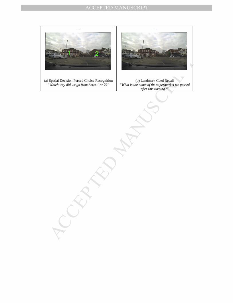
MANUSCRIP
T
ACCEPTED
ACCEPTED MANUSCRIPT
(a) Spatial Decision Forced Choice Recognition (b) Landmark Cued Recall “Which way did we go from here: 1 or 2?” “What is the name of the supermarket we passed
after this turning?”
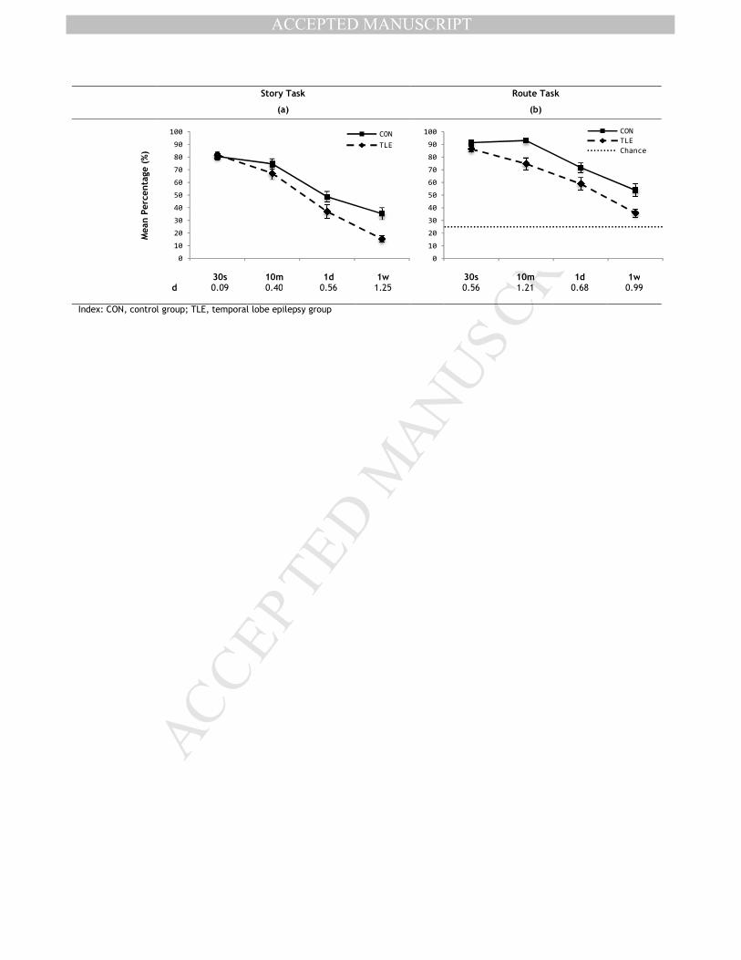
MANUSCRIP
T
ACCEPTED
ACCEPTED MANUSCRIPT
Story Task Route Task
(a) (b)
Mean Percentage (%)
30s 10m 1d 1w 30s 10m 1d 1w d 0.09 0.40 0.56 1.25 0.56 1.21 0.68 0.99
Index: CON, control group; TLE, temporal lobe epilepsy group
0
10
20
30
40
50
60
70
80
90
100 CON
TLE
0
10
20
30
40
50
60
70
80
90
100 CON
TLE
Chance
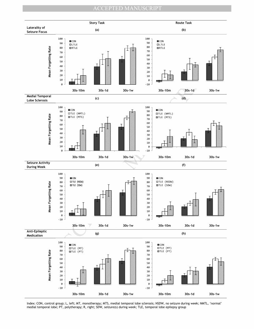
MANUSCRIP
T
ACCEPTED
ACCEPTED MANUSCRIPT
Story Task Route Task
Laterality of
Seizure Focus (a) (b)
Mean Forgetting Rate
30s-10m 30s-1d 30s-1w 30s-10m 30s-1d 30s-1w
Medial Temporal
Lobe Sclerosis (c) (d)
Mean Forgetting Rate
30s-10m 30s-1d 30s-1w 30s-10m 30s-1d 30s-1w
Seizure Activity
During Week (e) (f)
Mean Forgetting Rate
30s-10m 30s-1d 30s-1w 30s-10m 30s-1d 30s-1w
Anti-Epileptic
Medication (g) (h)
Mean Forgetting Rate
30s-10m 30s-1d 30s-1w 30s-10m 30s-1d 30s-1w
Index: CON, control group; L, left; MT, monotherapy; MTS, medial temporal lobe sclerosis; NSDW, no seizure during week; NMTL, ‘normal’ medial temporal lobe; PT, polytherapy; R, right; SDW, seizure(s) during week; TLE, temporal lobe epilepsy group
0
10
20
30
40
50
60
70
80
90
100CON
LTLE
RTLE
-10
0
10
20
30
40
50
60
70
80
90
100CON
LTLE
RTLE
0
10
20
30
40
50
60
70
80
90
100CON
TLE (NMTL)
TLE (MTS)
-10
0
10
20
30
40
50
60
70
80
90
100CON
TLE (NMTL)
TLE (MTS)
-10
0
10
20
30
40
50
60
70
80
90
100
CON
TLE (NSDW)
TLE (SDW)
-10
0
10
20
30
40
50
60
70
80
90
100
CON
TLE (NSDW)
TLE (SDW)
-10
0
10
20
30
40
50
60
70
80
90
100CON
TLE (MT)
TLE (PT)
-10
0
10
20
30
40
50
60
70
80
90
100CON
TLE (MT)
TLE (PT)