Embed Size (px)
Citation preview
Journal of Medical Genetics, 1982, 19, 332-336
Keratoconus posticus circumscriptus, cleft lip andpalate, genitourinary abnormalities, short stature, andmental retardation in sibsI D YOUNG, W G MACRAE, H E HUGHES, AND J S CRAWFORD
From the Departments of Genetics and Ophthalmology, The Hospital for Sick Children,Toronto, Ontario, Canada
SUMMARY This paper describes two sibs in each of whom keratoconus posticus circumscriptus isassociated with multiple abnormalities. These include short stature, mental retardation, cleft lipand palate, and vertebral anomalies. The authors have been unable to trace any former reportsof an identical condition and suggest that the findings in these children may represent a previouslyunrecognised malformation syndrome showing probable autosomal recessive inheritance.
Keratoconus posticus circumscriptus (KPC) is a rarecongenital abnormality in which the cornea showsan area of increased curvature centrally localisedon its posterior surface in association with opacifica-tion of the overlying stroma.1 On slit lamp examina-tion the cornea appears as if its posterior surfacehas been indented by a spherical object of a sizesmaller than that of the cornea itself. In this reportwe describe two sibs, one boy and one girl, in eachof whom KPC is associated with multiple abnor-malities.
Case reports
The affected sibs, who have been under the care ofone of us (JSC) since infancy, were the products ofthe first and fourth pregnancies of healthy unrelatedCaucasian parents. The second and fifth pregnanciesresulted in spontaneous first trimester abortions,and the third in a healthy male infant. There is nohistory of congenital abnormality on either side ofthe family.
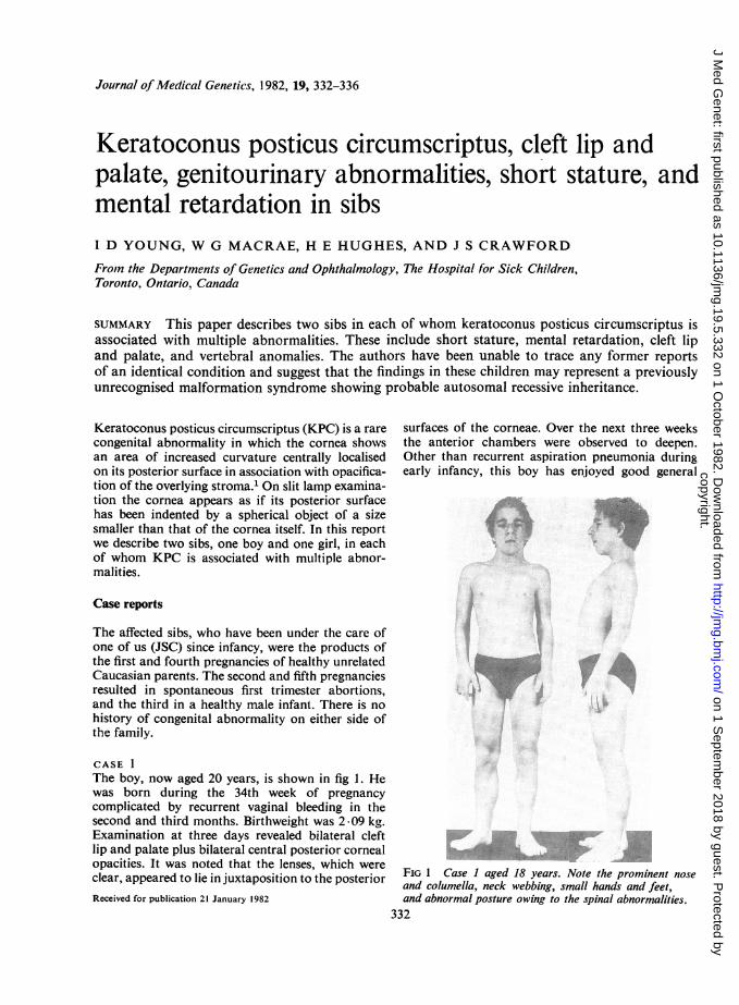
CASE 1The boy, now aged 20 years, is shown in fig 1. Hewas born during the 34th week of pregnancycomplicated by recurrent vaginal bleeding in thesecond and third months. Birthweight was 2.09 kg.Examination at three days revealed bilateral cleftlip and palate plus bilateral central posterior cornealopacities. It was noted that the lenses, which wereclear, appeared to lie in juxtaposition to the posteriorReceived for publication 21 January 1982
surfaces of the corneae. Over the next three weeksthe anterior chambers were observed to deepen.Other than recurrent aspiration pneumonia duringearly infancy, this boy has enjoyed good general
.:^..l .
=^ s &,.,,. '..,> | B B
.t g F.. q >'
sg5 l *V0
FIG 1 Case I aged 18 years. Note the prominent noseand columella, neck webbing, small hands and feet,and abnormal posture owing to the spinal abnormalities.
332
M,
copyright. on 1 S
eptember 2018 by guest. P
rotected byhttp://jm
g.bmj.com
/J M
ed Genet: first published as 10.1136/jm
g.19.5.332 on 1 October 1982. D
ownloaded from
Keratoconus posticis circumscriptuis and multiple abnornmalities in sibs
health. He has undergone bilateral optical iridec-tomies, repair of the cleft lip and palate, bilateralorchidopexy and inguinal herniorrhaphies, andbilateral heel cord lengthening. He has had no seizuresand has normal hearing.Developmental progress has been consistently
slow. He first walked alone at 3 years and utteredmeaningful single words at 4 years. Formal intel-lectual assessment at the age of 13 years using theWechsler intelligence scale for children yielded bothverbal and performance scale IQ values of 51. Hehas always had a happy disposition and has notposed any behaviour problems.At recent examination growth parameters were:
height 146 cm (16 cm below the 3rd centile), span137 cm, upper to lower segment ratio 1.07, weight45-5 kg (5 kg below the 3rd centile), and head cir-cumference 54.5 cm. He was noted to have mildmaxillary hypoplasia with a prominent nose andcolumella, neck webbing with a low posterior hairline, a scoliosis convex to the right involving upperthoracic vertebrae, limitation of extension andsupination at both elbows, bilateral fifth fingerclinodactyly, and short broad feet with bilateral pescavus. Sexual development has been normal.Examination with the slit lamp revealed bilateral
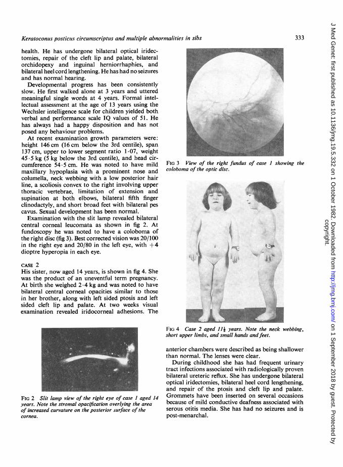
central corneal leucomata as shown in fig 2. Atfundoscopy he was noted to have a coloboma ofthe right disc (fig 3). Best corrected vision was 20/100in the right eye and 20/80 in the left eye, with +4dioptre hyperopia in each eye.
CASE 2His sister, now aged 14 years, is shown in fig 4. Shewas the product of an uneventful term pregnancy.At birth she weighed 2.4 kg and was noted to havebilateral central corneal opacities similar to thosein her brother, along with left sided ptosis and leftsided cleft lip and palate. At two weeks visualexamination revealed iridocorneal adhesions. The
FIG 2 Slit lamp view of the right eye of case I aged 14years. Note the stromal opacification overlying the area
of increased curvature on the posterior surface of thecornea.
r
FIG 3 View of the right fundus of case I showing thecoloboma of the optic disc.
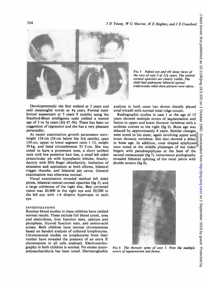
FIG 4 Case 2 aged Ili years. Note the neck webbing,short upper limbs, and small hands andfeet.
anterior chambers were described as being shallowerthan normal. The lenses were clear.During childhood she has had frequent urinary
tract infections associated with radiologically provenbilateral ureteric reflux. She has undergone bilateraloptical iridectomies, bilateral heel cord lengthening,and repair of the ptosis and cleft lip and palate.Grommets have been inserted on several occasionsbecause of mild conductive deafness associated withserous otitis media. She has had no seizures and ispost-menarchal.
333
t.Aow**.i
copyright. on 1 S
eptember 2018 by guest. P
rotected byhttp://jm
g.bmj.com
/J M
ed Genet: first published as 10.1136/jm
g.19.5.332 on 1 October 1982. D
ownloaded from
334 ~~~~~~~~~ID Young, W G Macrae, H E Hulghes, and J S Crawford
Developmentally she first walked at 3 years andsaid meaningful words at 4k years. Formal intel-lectual assessment at 5 years 9 months using theStanford-Binet intelligence scale yielded a mentalage of 3 to 31 years (IQ 47-56). There has been nosuggestion of regression and she has a very pleasantpersonality.At recent examination growth parameters were:
height 124 cm (24 cm below the 3rd centile), span110 cm, upper to lower segment ratio 1 .12, weight39 kg, and head circumference 52.5 cm. She wasnoted to have a prominent nose, a short webbedneck with low posterior hair line, a small left sidedpreauricular pit with hypoplastic lobules, brachy-dactyly with fifth finger clinodactyly, limitation ofextension and supination at both elbows, bilateraltrigger thumbs, and bilateral pes cavus. Generalexamination was otherwise normal.
Visual examination revealed residual left sidedptosis, bilateral central corneal opacities (fig 5), anda large coloboma of the right disc. Best correctedvision was 20/400 in the right eye and 20/200 inthe left eye, with +6 dioptre hyperopia in eacheye.
INVESTIGATIONSRoutine blood studies in these children have yieldednormal results. These include full blood count, ureaand electrolytes, liver function tests, calcium andphosphate, thyroid function tests, and amino-acidscreen. Both children have normal chromosomesbased on banded analysis of cultured lymphocytes.Chromosomal studies on lymphocytes from theirmother have revealed the presence of an extra Xchromosome in all cells analysed. Electrocardio-graphy in both children is normal. No excess muco-polysacchariduria has been noted. Dermatoglyphic
FIG 5 Naked eye and slit lamp views ofthe eyes of case 2 at JJj years. The centralcorneal opacities are clearly visible. Thechild had undergone bilateral opticaliridectomies when these pictures were taken.
analysis in both cases has shown distally placedaxial triradii with normal total ridge counts.
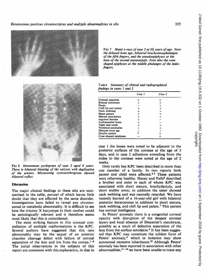
Radiographic studies in case 1 at the age of 15years showed multiple errors of segmentation andfusion in upper and lower thoracic vertebrae with ascoliosis convex to the right (fig 6). Bone age wasdelayed by approximately 4 years. Similar changeswere noted in his sister, again involving upper andlower thoracic vertebrae. She also showed a delayin bone age. In addition, cone shaped epiphyseswere noted at the middle phalanges of her indexfingers with pseudoepiphyses at the base of thesecond metacarpals (fig 7). Intravenous pyelographyrevealed bilateral splitting of the renal pelvis withdouble ureters (fig 8).
FIG 6 The thoracic spine of case 1. Note the multipleerrors ofsegmentation andfusion.
334
copyright. on 1 S
eptember 2018 by guest. P
rotected byhttp://jm
g.bmj.com
/J M
ed Genet: first published as 10.1136/jm
g.19.5.332 on 1 October 1982. D
ownloaded from
Keratoconuis posticus circumscriptus and multiple abnormalities in sibs
FIG 7 Hand x-rays ofcase 2 at 6j years ofage. Notethe delayed bone age, bilateral brachymesophalangiaof the fifth fingers, and the pseudoepiphyses at thebase of the second metacarpals. Note also the coneshaped epiphyses at the middle phalanges of the indexfingers.
TABLE Summary of clinical and radiographicalfindings in cases I and 2
Case I Case 2
Corneal opacities + +Retinal coloboma + +Ptosis - +Cleft lip and palate + +Neck webbing + +Short stature + +Mental retardation + +Inguinal herniae +Undescended testes +Tight heel cords + +Vertebral anomalies + +Delayed bone age + +Double ureters ? +Cone shaped epiphyses - +
FIG 8 Intravenous pyelogram of case 2 aged 9 years.There is bilateral blunting of the calyces with duplicationof the ureters. Micturating cystourethrogram showedbilateral reflux.
Discussion
The major clinical findings in these sibs are sum-marised in the table, perusal of which leaves littledoubt that they are affected by the same disorder.Investigations have failed to reveal any chromo-somal or metabolic abnormality. It is difficult to seehow the trisomy X karyotype in their mother couldbe aetiologically relevant and it therefore seemsmost likely that this is coincidental.The most striking feature in this unusual con-
stellation of multiple malformations is the KPC.Several authors have suggested that this rare
abnormality may be the sequel of an anteriorchamber cleavage defect with failure of normalseparation of the lens and iris from the cornea.1-3The initial observations in the subjects of thisreport are consistent with this explanation, in that in
case 1 the lenses were noted to be adjacent to theposterior surfaces of the corneae at the age of 3days, and in case 2 adhesions extending from theirides to the corneae were noted at the age of 2weeks.Only rarely has KPC been described in more than
one member of a family. In two reports bothparent and child were affected.45 These patientswere otherwise healthy. Haney and Falls6 describeda brother and sister in each of whom KPC wasassociated with short stature, brachydactyly, andshort stubby arms; in addition the sister showedneck webbing and was mentally retarded. We haverecently learned of a 16-year-old girl with bilateralposterior keratoconus in addition to short stature,neck webbing, and cleft lip and palate.7 This patienthas normal intelligence.
In Peters' anomaly there is a congenital cornealopacity with disruption of the deepest stromallayers and local absence of Descemet's membrane,possibly as a result of defective separation of thelens from the surface ectoderm.8 It has been sugges-ted that KPC may constitute the mildest form ofPeters' anomaly,9 which in isolation may showautosomal recessive inheritance.10 Although Peters'anomaly has been reported in association with otherabnormalities,11-13 we have been unable to trace any
335
.4. ,s.0
copyright. on 1 S
eptember 2018 by guest. P
rotected byhttp://jm
g.bmj.com
/J M
ed Genet: first published as 10.1136/jm
g.19.5.332 on 1 October 1982. D
ownloaded from

3 D Young, W G Macrae, H E Hughes, and J S Crawford
report of its presence in a pattern of malformationssimilar to that described here. Similarly, cornealclouding may be a feature of several malformationsyndromes,'4-20 but in none of these do the findingscorrespond closely with those in the subjects of thisreport.
Thus, although the findings in our patients dobear some resemblance to those previously des-cribed, particularly the cases of Haney and Falls6and Streeten et al,7 in none of these cases do theclinical features overlap sufficiently to suggest anidentical disorder. In conclusion, therefore, it isproposed that the multiple abnormalities in thesesibs constitute a previously undescribed pleiotropicsingle gene disorder. Clearly the family history ismost consistent with autosomal recessive inheritance.
We are most grateful to Professor B W Streeten andher colleagues for providing us with clinical detailsof their patient, and to Miss Allison Chiti for herinvaluable secretarial assistance.
This paper was presented at the Clinical GeneticsSociety meeting in November 1981.
References1 Krachmer JH. Posterior keratoconus. Arch Ophthalmol
1978 ;96:1867-73.2 Hagedoorn A, Velzeboer CMJ. Postnatal partial spon-taneous correction of a severe congenital anomaly of theanterior segment of an eye. Arch Opthalmol 1959;62:685-93.
3 Charan H. Keratoconus posticus circumscriptus withindentation of the lens. Br J Ophthalmol 1967;51 :486-8.
4 Jacobs HB. Posterior conical cornea. Br J Ophthalmol1957 ;41 :31-9.
5 Collier M. Le k6ratoc6ne posterieur. Arch Ophthalmol(Paris) 1962;22:376-91.
6 Haney WP, Falls HF. The occurrence of congenitalkeratoconus posticus circumscriptus in two siblingspresenting a previously unrecognised syndrome. Am JOphthalmol 1961 ;52:53-7.
7 Streeten BW, Karpik AG, Spitzer KH. Posterior kerato-conus associated with systemic abnormalities. ArchOphthalmol (in press).
8 Townsend WM. Congenital corneal leukomas. 1. Central
defect in Descemet's membrane. Am J Ophthalmol 1974;77:80-6.
9 Alkemade PPH. Dysgenesis mesodermalis of the iris andthe cornea. Assen: Van Gorcum, 1969.
10 Cross HE. Penetrance and variability in anterior chambermalformations. Birth Defects 1979;XV,No 5B:131-44.
1 Pagon RA, Chandler JW, Collie WR, et al. Hydroce-phalus, agyria, retinal dysplasia, encephalocele (HARD±E) syndrome: an autosomal recessive condition. BirthDefects 1978;XIV,No 6B:233-41.
12 Ide CH, Matta C, Holt JE, Felker GV. Dysgenesismesodermalis of the cornea (Peters' anomaly) associatedwith cleft lip and palate. Ann Ophthalmol 1975;7:841-2.
13 Ruprecht KW, Majewski F. Familiare arhinie mitPetersscher anomalie und kiefermissbildungen, ein neufsfehlbildungssyndrom? Klin Monatsbl Augenheilkd 1978;172:708-15.
14 Pinsky, L, DiGeorge AM, Harley D, Baird HW. Micro-ophthalmos, corneal opacity, mental retardation, andspastic cerebral palsy. An oculocerebral syndrome.J Pediatr 1965;67:387-98.
15 Mietens C, Weber H. A syndrome characterized bycorneal opacity, nystagmus, flexion contracture of theelbows, growth failure, and mental retardation. J Pediatr1966;69;624-9.
16 Cross HE, McKusick VA, Breen W. A new oculocerebralsyndrome with hypopigmentation. J Pediatr 1967;70:398-406.
17 Herrmann J, Feingold M, Tuffli GA, Opitz JM. Afamilial dysmorphogenetic syndrome of limb deformities,characteristic facial appearance and associated anomalies:the "pseudothalidomide" or "SC-syndrome". BirthDefects 1969;V,No 3:81-9.
18 Balci S, Say B, Firat T. Corneal opacity, microphthalmia,mental retardation, microcephaly and generalised mus-cular spasticity associated with hyperglycinemia. ClinGenet 1974;5:36-9.
19 Fryns JP, Moerman F, Goddeeris P, Bossuyt C, Van denBerghe H. A new lethal syndrome with cloudy corneae,diaphragmatic defects and distal limb deformities. HumGenet 1979;50:65-70.
20 Fryns JP, Van den Berghe H. Corneal clouding, sub-valvular aortic stenosis, and midfacial hypoplasiaassociated with mental deficiency and growth retardation-a new syndrome? Eur J Pediatr 1979;131:179-83.
Requests for reprints to Dr W G Macrae,Department of Ophthalmology, The Hospital forSick Children, 555 University Avenue, Toronto,Ontario, Canada M5G IX8.
336
copyright. on 1 S
eptember 2018 by guest. P
rotected byhttp://jm
g.bmj.com
/J M
ed Genet: first published as 10.1136/jm
g.19.5.332 on 1 October 1982. D
ownloaded from