Embed Size (px)
DESCRIPTION
sistem respirasi
Citation preview
ANATOMY OF RESPIRATORY SYSTEM
Dr. Mega Sari Sitorus, Mkes.
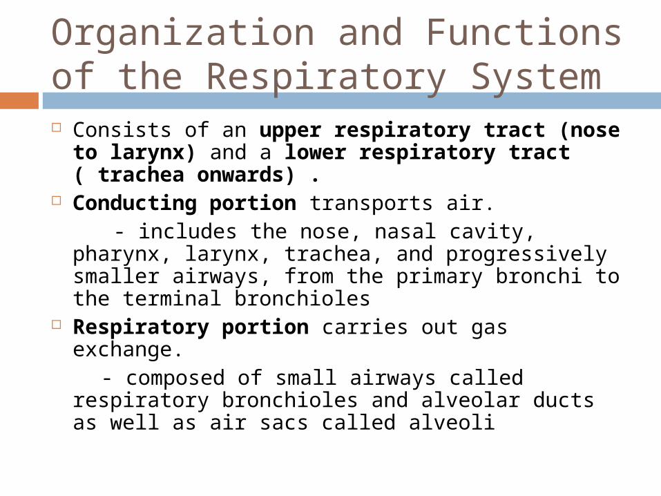
Organization and Functions of the Respiratory System Consists of an upper respiratory tract (nose to
larynx) and a lower respiratory tract ( trachea onwards) .
Conducting portion transports air. - includes the nose, nasal cavity, pharynx, larynx,
trachea, and progressively smaller airways, from the primary bronchi to the terminal bronchioles
Respiratory portion carries out gas exchange. - composed of small airways called respiratory
bronchioles and alveolar ducts as well as air sacs called alveoli
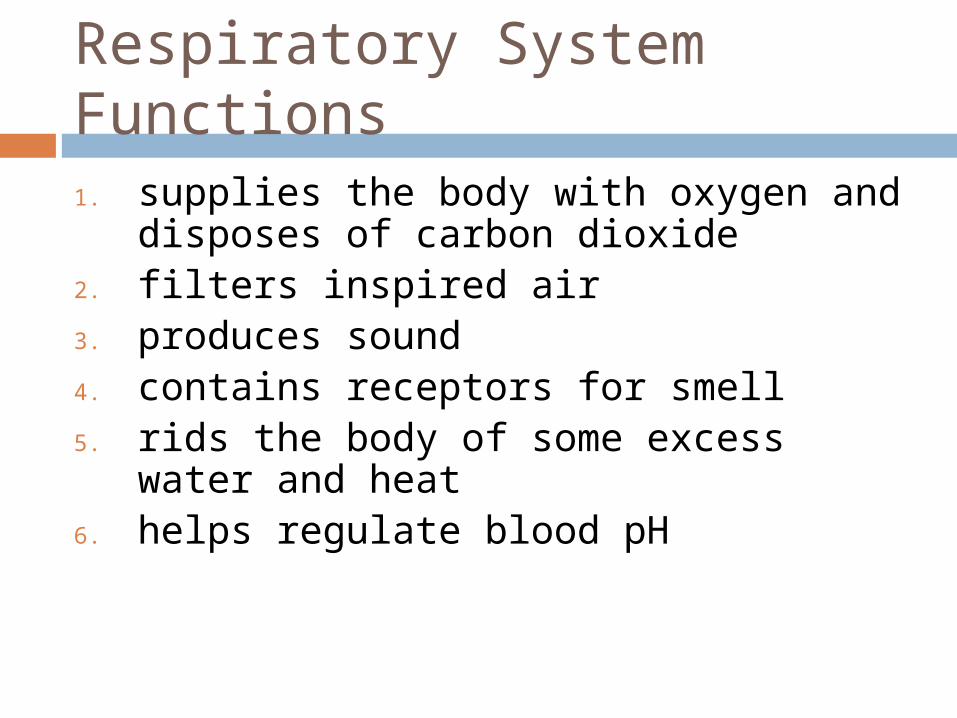
Respiratory System Functions 1. supplies the body with oxygen and disposes of
carbon dioxide2. filters inspired air3. produces sound4. contains receptors for smell5. rids the body of some excess water and heat6. helps regulate blood pH
Breathing
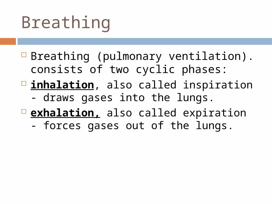
Breathing (pulmonary ventilation). consists of two cyclic phases:
inhalation, also called inspiration - draws gases into the lungs.
exhalation, also called expiration - forces gases out of the lungs.
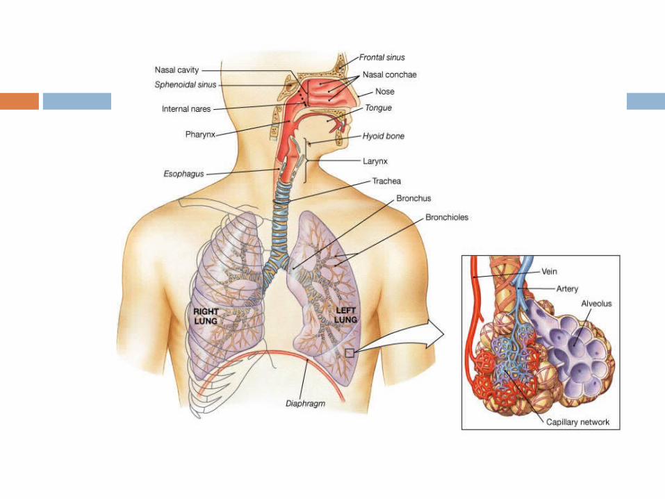
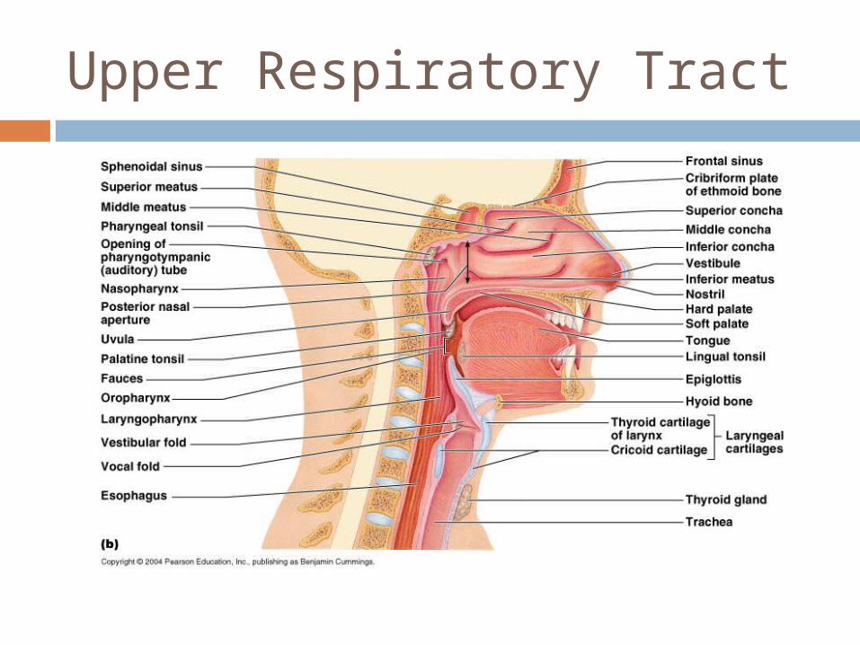
Upper Respiratory Tract
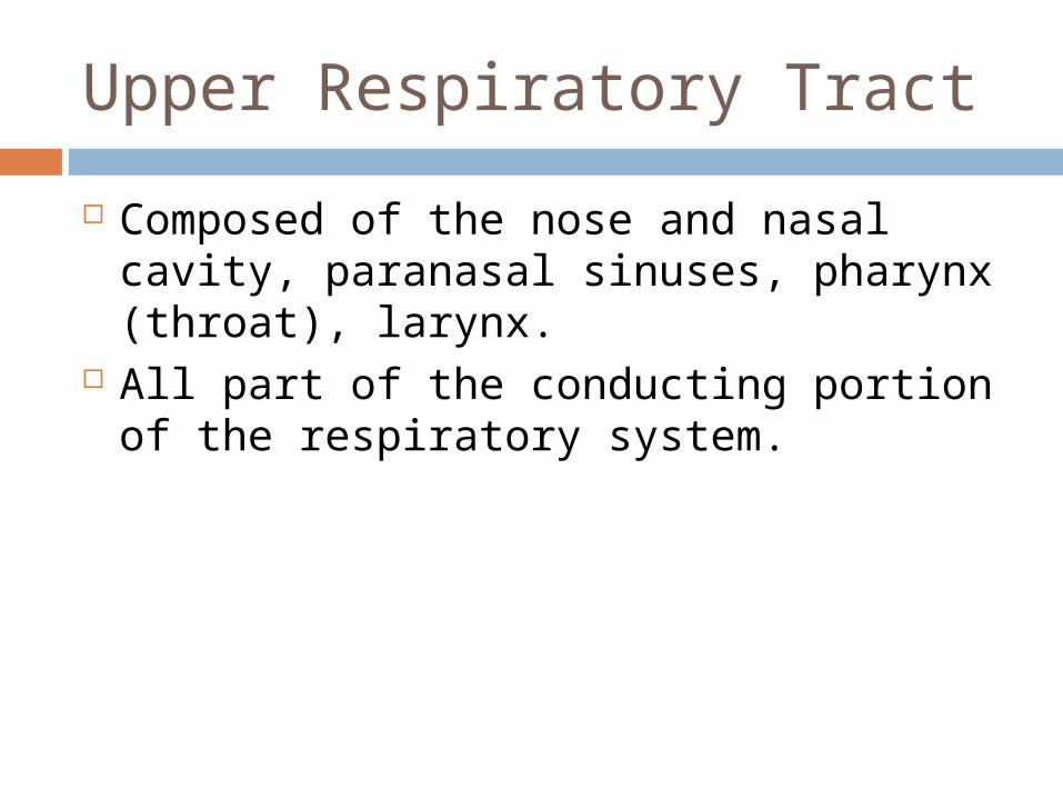
Composed of the nose and nasal cavity, paranasal sinuses, pharynx (throat), larynx.
All part of the conducting portion of the respiratory system.
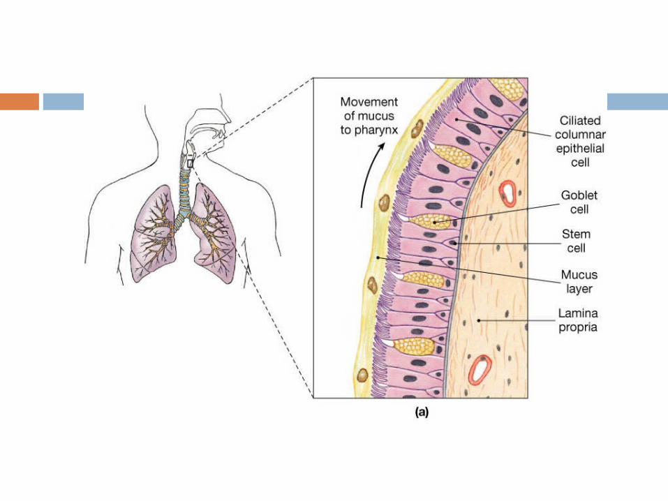
Respiratory mucosa
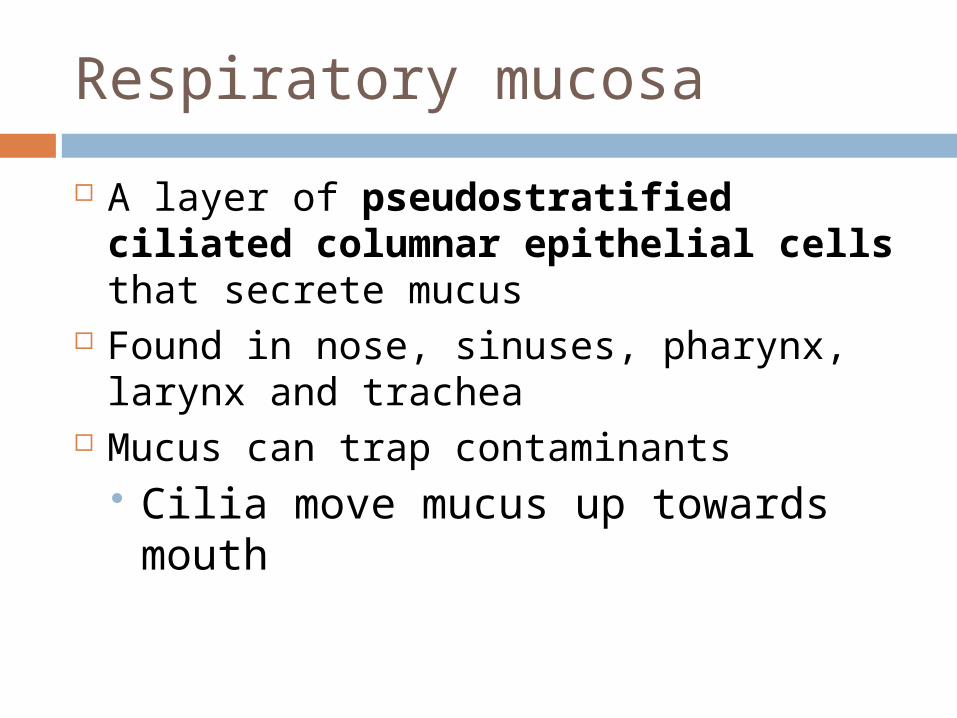
A layer of pseudostratified ciliated columnar epithelial cells that secrete mucus
Found in nose, sinuses, pharynx, larynx and trachea
Mucus can trap contaminants Cilia move mucus up towards
mouth
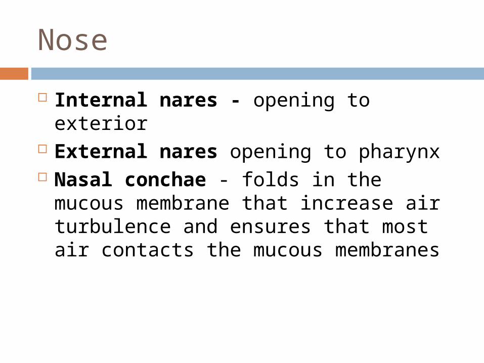
Nose
Internal nares - opening to exterior External nares opening to pharynx Nasal conchae - folds in the mucous
membrane that increase air turbulence and ensures that most air contacts the mucous membranes
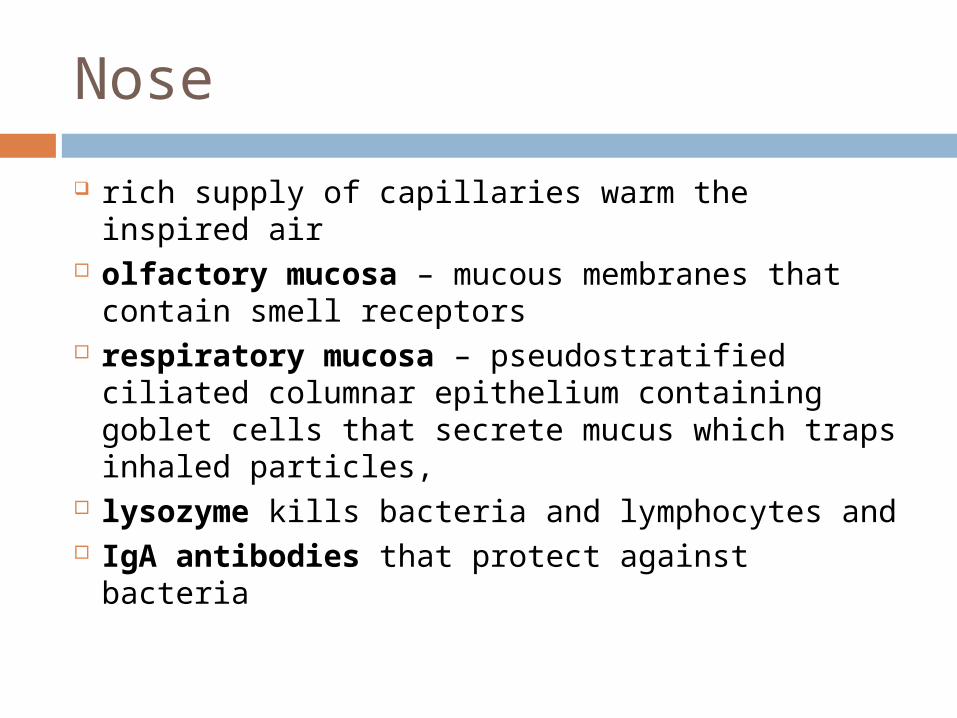
Nose
rich supply of capillaries warm the inspired air olfactory mucosa – mucous membranes that contain
smell receptors respiratory mucosa – pseudostratified ciliated
columnar epithelium containing goblet cells that secrete mucus which traps inhaled particles,
lysozyme kills bacteria and lymphocytes and IgA antibodies that protect against bacteria
Upper Respiratory Tract
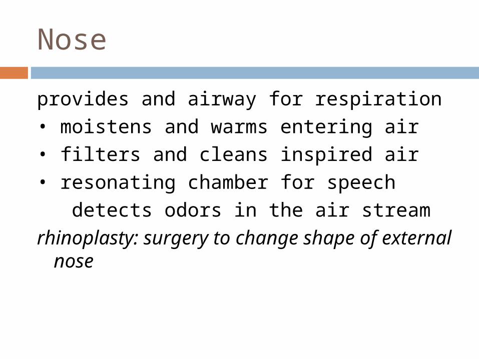
Nose
provides and airway for respiration
• moistens and warms entering air
• filters and cleans inspired air
• resonating chamber for speech
detects odors in the air stream
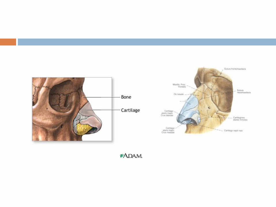
rhinoplasty: surgery to change shape of external nose
Paranasal Sinuses
Four bones of the skull contain paired air spaces called the paranasal sinuses - frontal, ethmoidal, sphenoidal, maxillary
Decrease skull bone weight Warm, moisten and filter incoming air Add resonance to voice. Communicate with the nasal cavity by
ducts. Lined by pseudostratified ciliated
columnar epithelium.
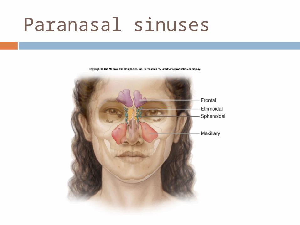
Paranasal sinuses
Pharynx
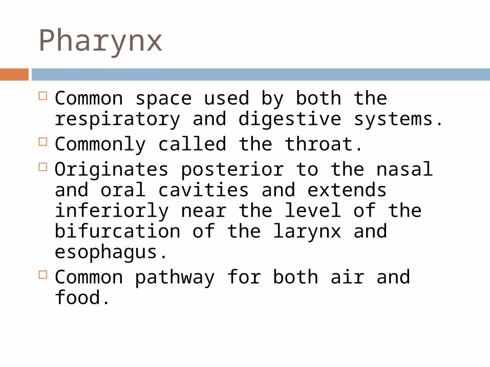
Common space used by both the respiratory and digestive systems.
Commonly called the throat. Originates posterior to the nasal and oral
cavities and extends inferiorly near the level of the bifurcation of the larynx and esophagus.
Common pathway for both air and food.
Pharynx
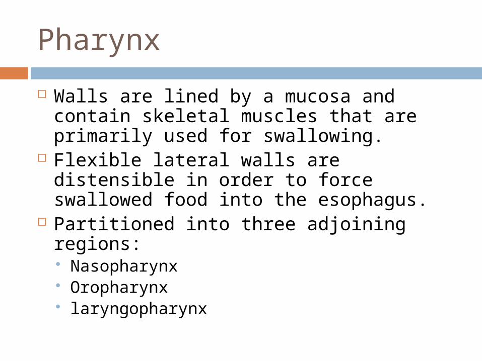
Walls are lined by a mucosa and contain skeletal muscles that are primarily used for swallowing.
Flexible lateral walls are distensible in order to force swallowed food into the esophagus.
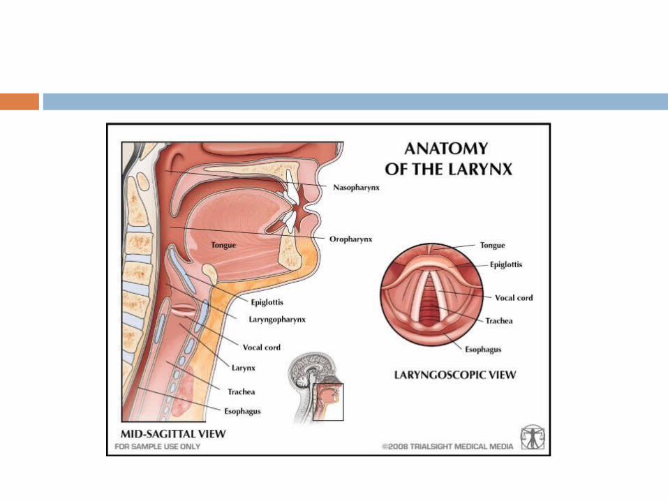
Partitioned into three adjoining regions: Nasopharynx Oropharynx laryngopharynx
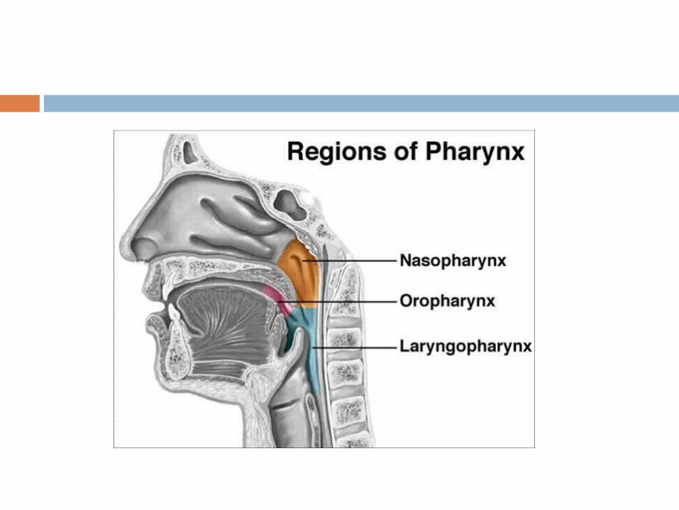
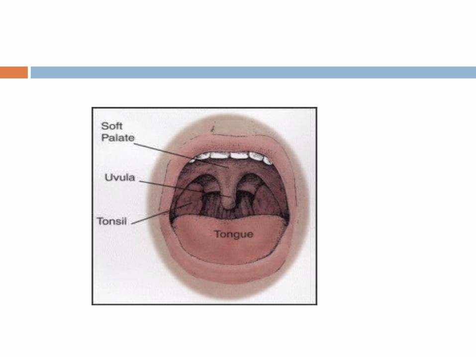
Nasopharynx
Superior-most region of the pharynx. Covered with pseudostratified ciliated columnar epithelium.
Located directly posterior to the nasal cavity and superior to the soft palate, which separates the oral cavity.
Normally, only air passes through. Material from the oral cavity and oropharynx is
typically blocked from entering the nasopharynx by the uvula of soft palate, which elevates when we swallow.
In the lateral walls of the nasopharynx, paired auditory/eustachian tubes connect the nasopharynx to the middle ear.
Posterior nasopharynx wall also houses a single pharyngeal tonsil (commonly called the adenoids).
Oropharynx
The middle pharyngeal region. Immediately posterior to the oral cavity. Bounded by the edge of the soft palate superiorly and
the hyoid bone inferiorly. Common respiratory and digestive pathway through
which both air and swallowed food and drink pass. Contains nonkeratinized stratified squamous
epithelim. Lymphatic organs here provide the first line of defense
against ingested or inhaled foreign materials. Palatine tonsils are on the lateral wall between the arches, and the lingual tonsils are at the base of the tongue.
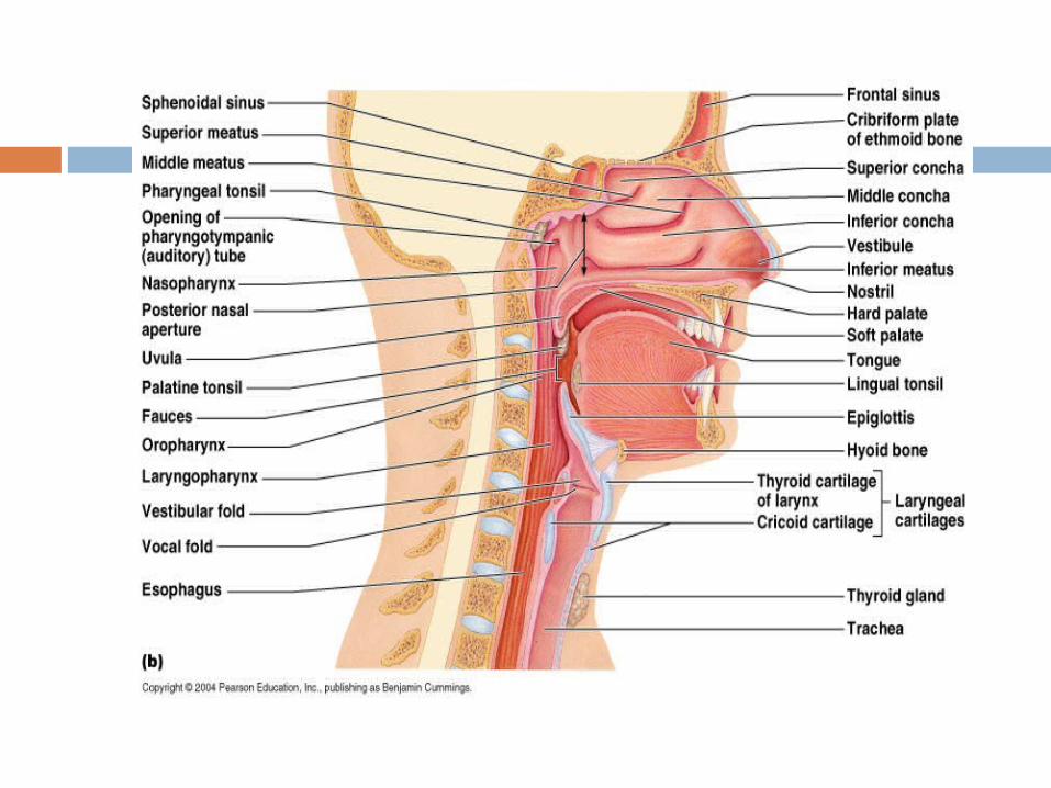
Laryngopharynx
Inferior, narrowed region of the pharynx. Extends inferiorly from the hyoid bone to
the larynx and esophagus. Terminates at the superior border of the
esophagus and the epiglottis of the larynx. Lined with a nonkeratinized stratified
squamous epithelium. Permits passage of both food and air.
Lower Respiratory Tract
Conducting airways (trachea, bronchi, up to terminal bronchioles).
Respiratory portion of the respiratory system (respiratory bronchioles, alveolar ducts, and alveoli).
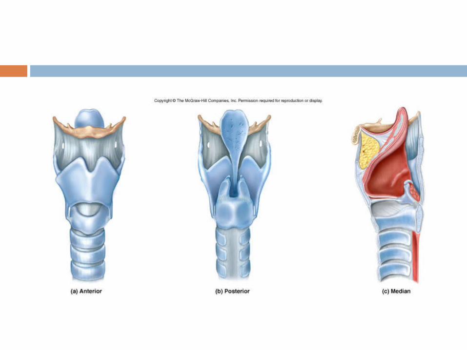
Larynx
Voice box is a short, somewhat cylindrical airway ends in the trachea.
Prevents swallowed materials from entering the lower respiratory tract.
Conducts air into the lower respiratory tract.
Produces sounds. Supported by a framework of nine pieces
of cartilage (three individual pieces and three cartilage pairs) that are held in place by ligaments and muscles.
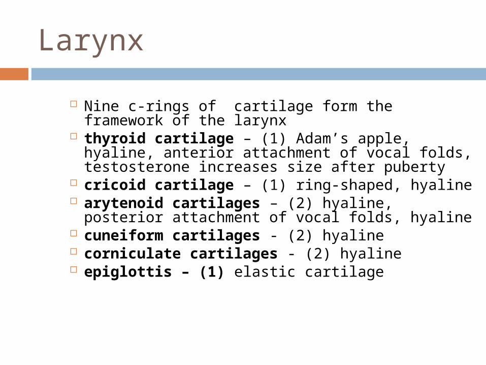
Larynx
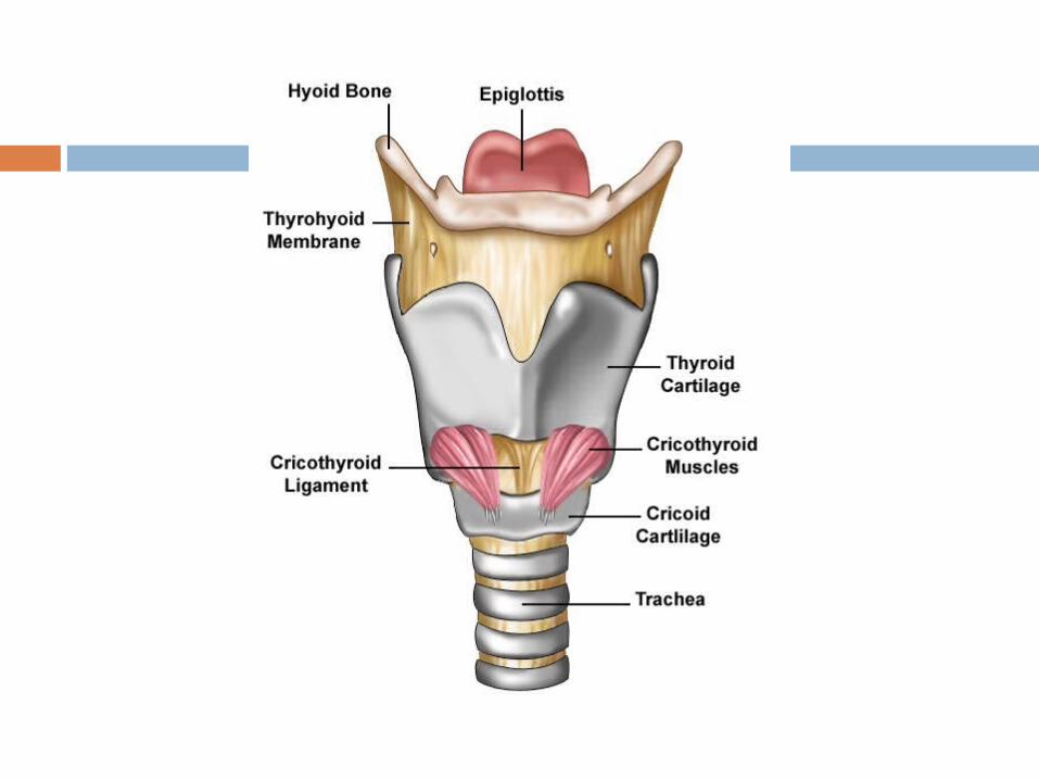
Nine c-rings of cartilage form the framework of the larynx
thyroid cartilage – (1) Adam’s apple, hyaline, anterior attachment of vocal folds, testosterone increases size after puberty
cricoid cartilage – (1) ring-shaped, hyaline arytenoid cartilages – (2) hyaline, posterior
attachment of vocal folds, hyaline cuneiform cartilages - (2) hyaline corniculate cartilages - (2) hyaline epiglottis – (1) elastic cartilage
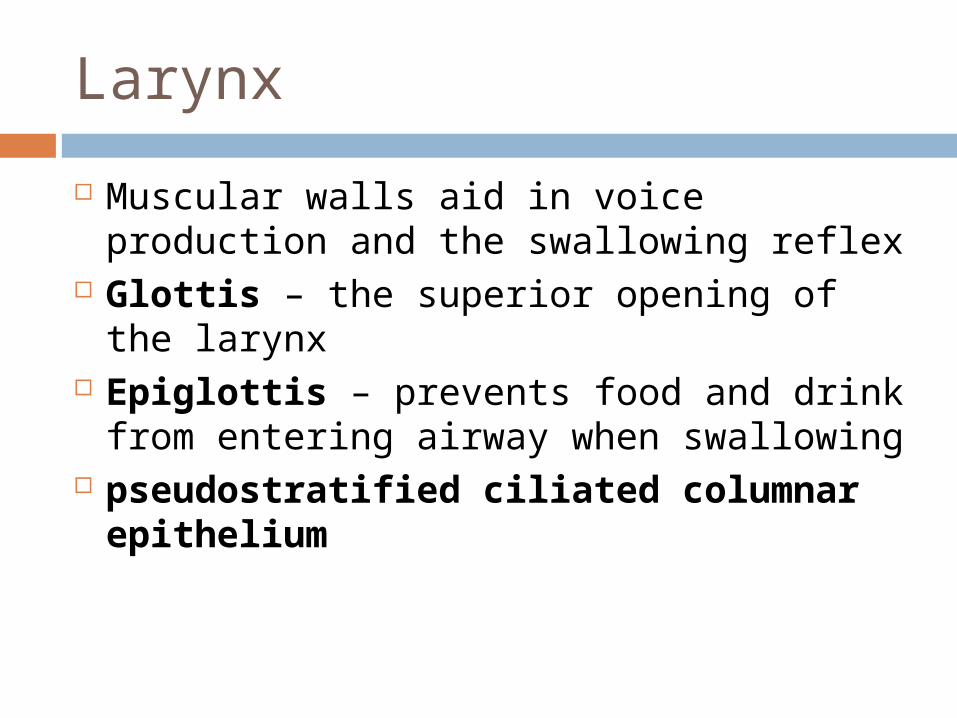
Larynx
Muscular walls aid in voice production and the swallowing reflex
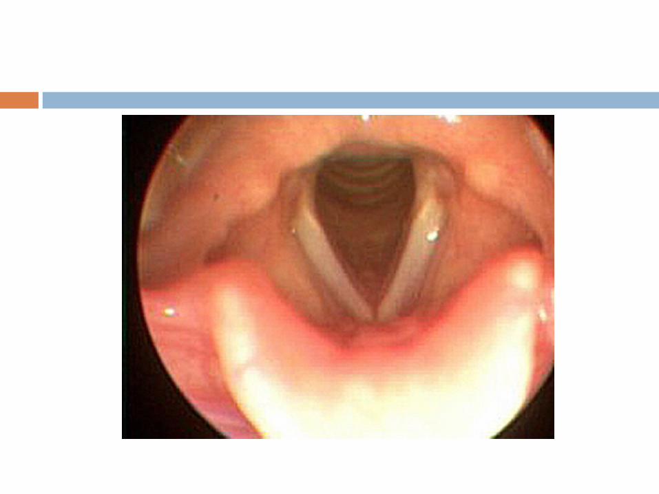
Glottis – the superior opening of the larynx Epiglottis – prevents food and drink from entering
airway when swallowing pseudostratified ciliated columnar
epithelium
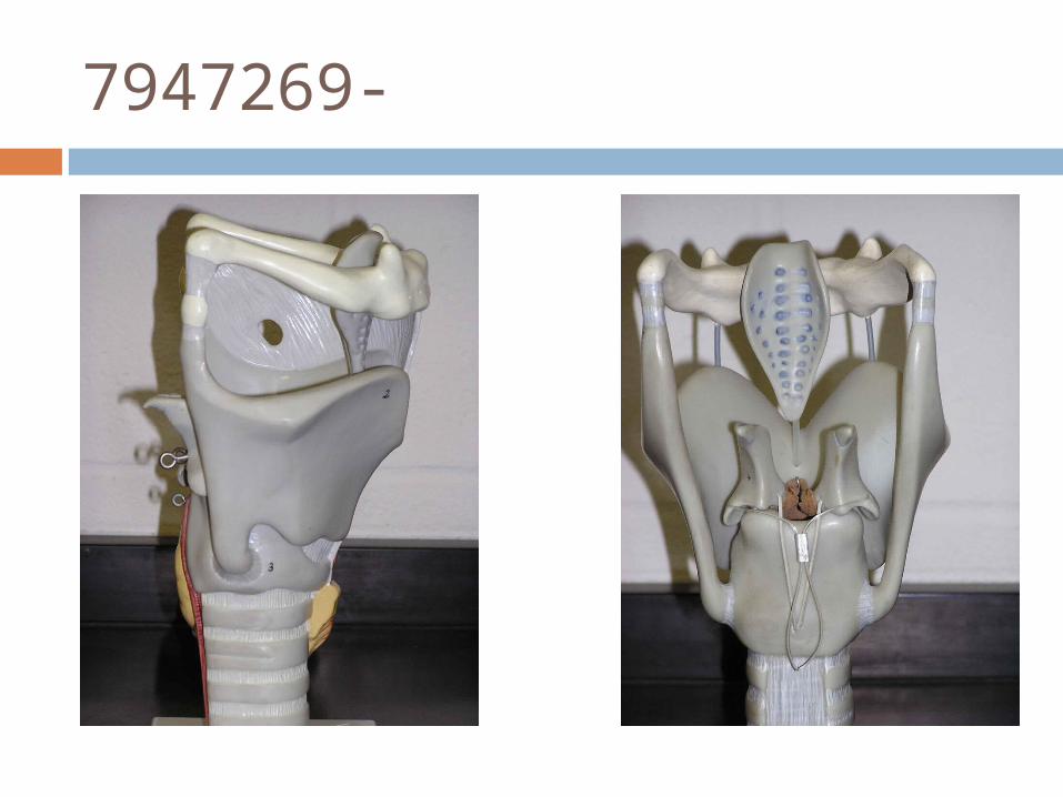
7947269-
Sound Production
Inferior ligaments are called the vocal folds. - are true vocal cords because they produce
sound when air passes between them Superior ligaments are called the vestibular
folds. - are false vocal cords because they have no
function in sound production, but protect the vocal folds.
The tension, length, and position of the vocal folds determine the quality of the sound.
Sound production
Intermittent release of exhaled air through the vocal folds
Loudness – depends on the force with which air is exhaled through the cords
Pharynx, oral cavity, nasal cavity, paranasal sinuses act as resonating chambers that add quality to the sound
Muscles of the face, tongue, and lips help with enunciation of words
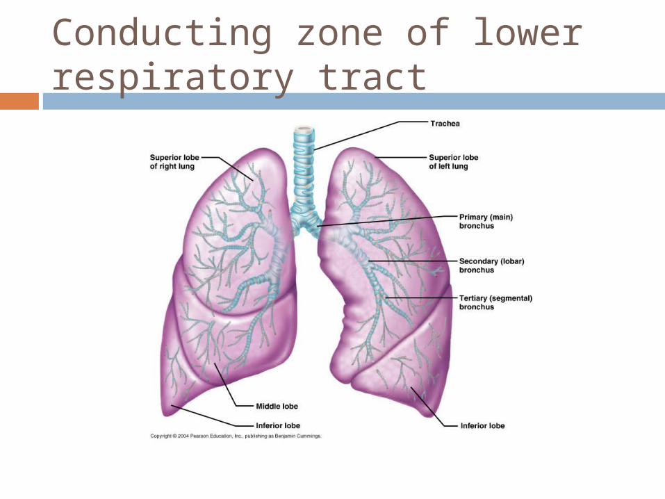
Conducting zone of lower respiratory tract
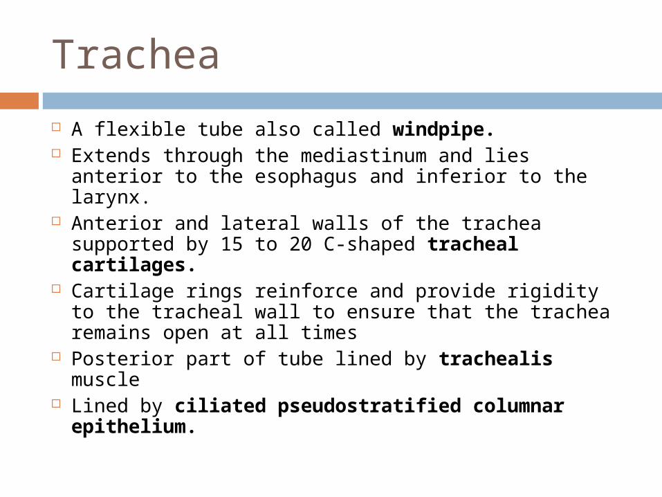
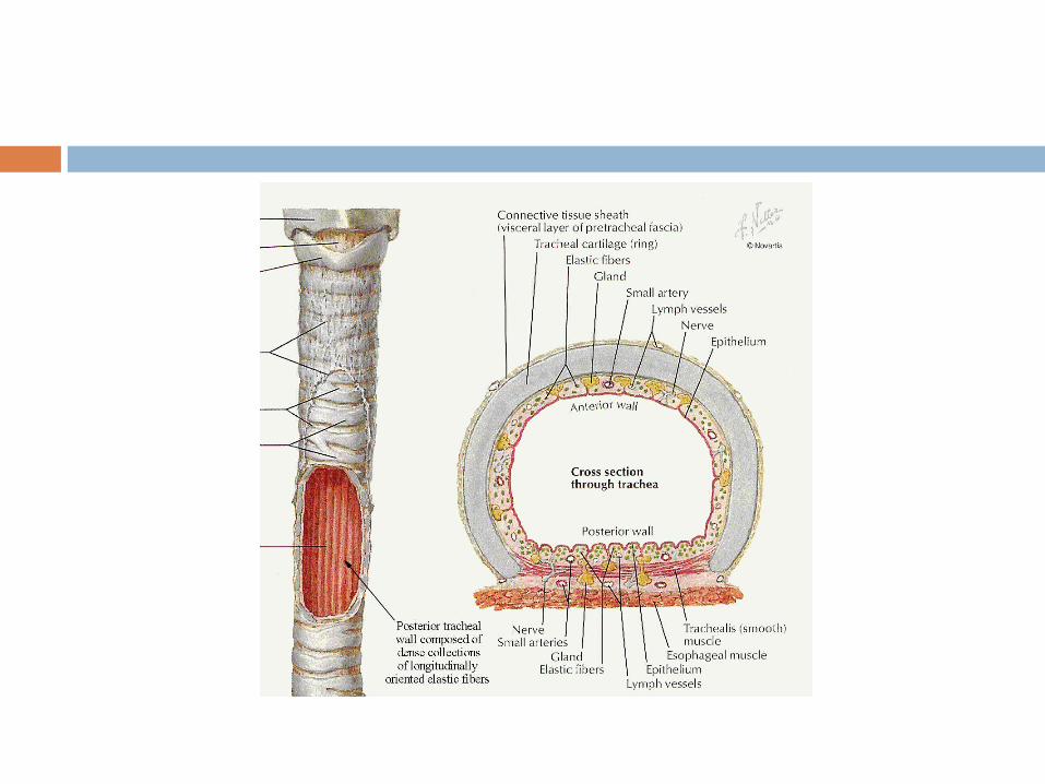
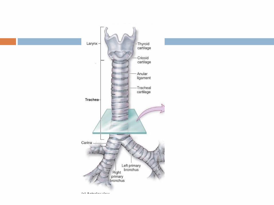
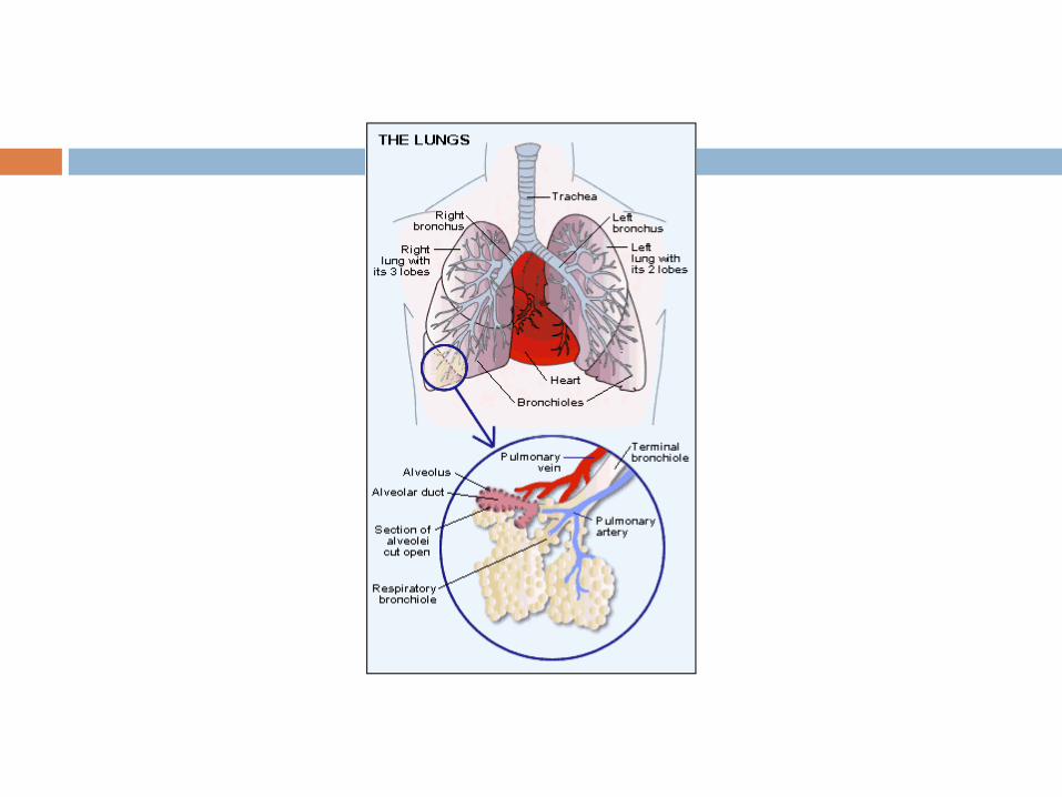
Trachea
A flexible tube also called windpipe. Extends through the mediastinum and lies anterior
to the esophagus and inferior to the larynx. Anterior and lateral walls of the trachea supported
by 15 to 20 C-shaped tracheal cartilages. Cartilage rings reinforce and provide rigidity to the
tracheal wall to ensure that the trachea remains open at all times
Posterior part of tube lined by trachealis muscle Lined by ciliated pseudostratified columnar
epithelium.
Trachea
At the level of the sternal angle, the trachea bifurcates into two smaller tubes, called the right and left primary bronchi.
Each primary bronchus projects laterally toward each lung.
The most inferior tracheal cartilage separates the primary bronchi at their origin and forms an internal ridge called the carina.
Bronchial tree
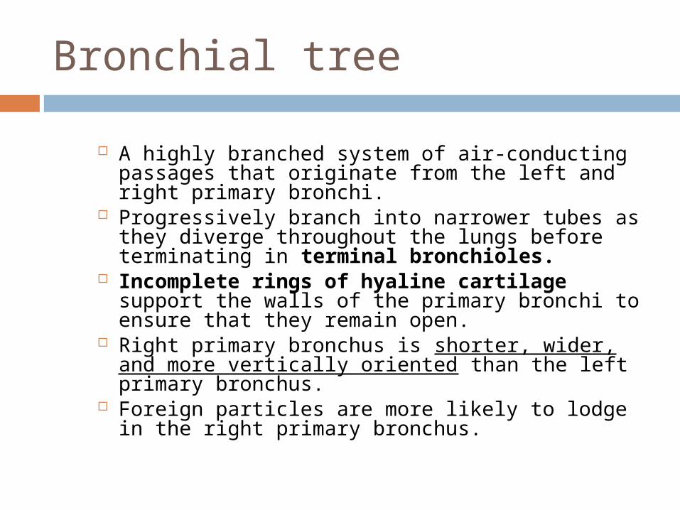
A highly branched system of air-conducting passages that originate from the left and right primary bronchi.
Progressively branch into narrower tubes as they diverge throughout the lungs before terminating in terminal bronchioles.
Incomplete rings of hyaline cartilage support the walls of the primary bronchi to ensure that they remain open.
Right primary bronchus is shorter, wider, and more vertically oriented than the left primary bronchus.
Foreign particles are more likely to lodge in the right primary bronchus.
Bronchial tree
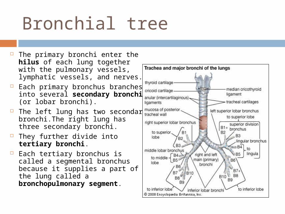
The primary bronchi enter the hilus of each lung together with the pulmonary vessels, lymphatic vessels, and nerves.
Each primary bronchus branches into several secondary bronchi (or lobar bronchi).
The left lung has two secondary bronchi.The right lung has three secondary bronchi.
They further divide into tertiary bronchi.
Each tertiary bronchus is called a segmental bronchus because it supplies a part of the lung called a bronchopulmonary segment.
Bronchial Tree
Secondary bronchi tertiary bronchi bronchioles terminal bronchioles
with successive branching amount of cartilage decreases and amount of smooth muscle increases, this allows for variation in airway diameter
during exertion and when sympathetic division active bronchodilation
mediators of allergic reactions like histamine bronchoconstriction
epithelium gradually changes from ciliated pseudostratified columnar epithelium to simple cuboidal epithelium in terminal bronchioles
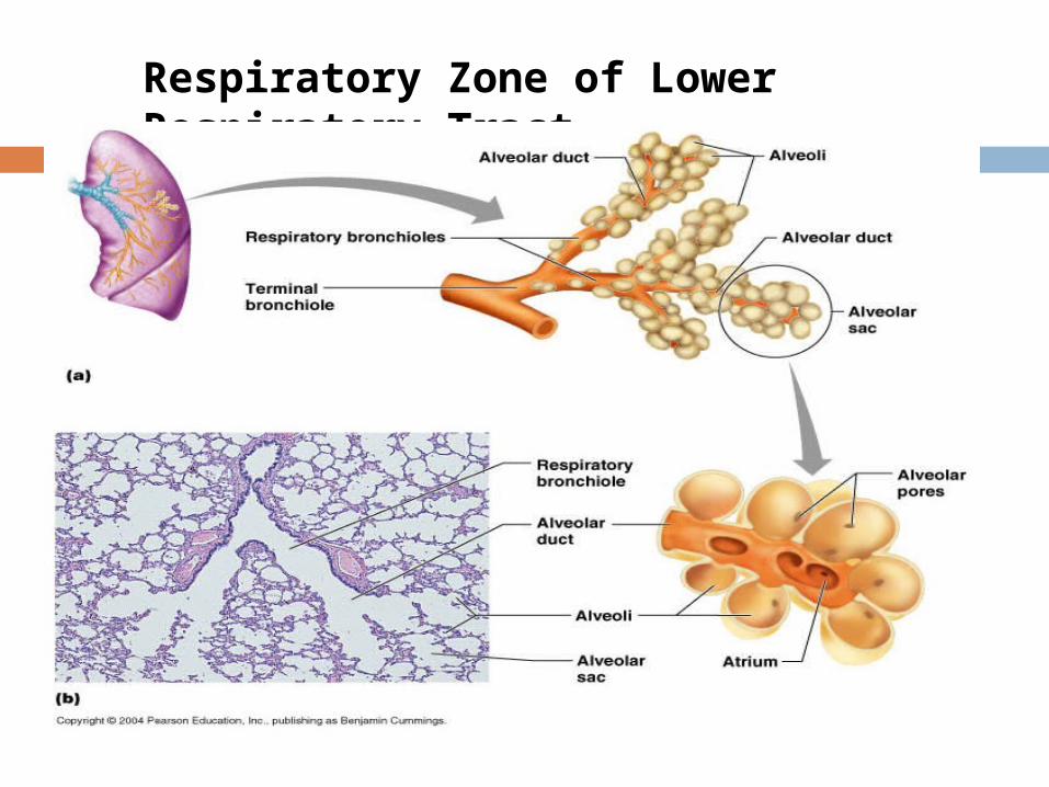
Respiratory Zone of Lower Respiratory Tract
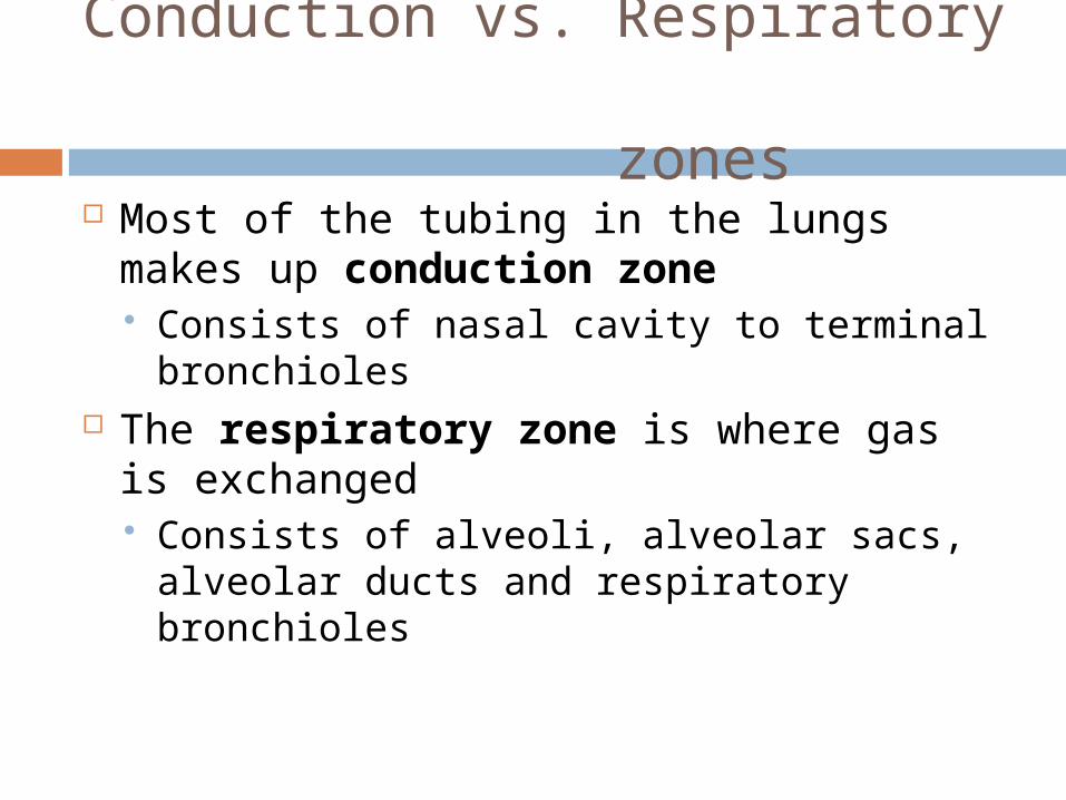
Conduction vs. Respiratory zones Most of the tubing in the lungs makes up
conduction zone Consists of nasal cavity to terminal
bronchioles The respiratory zone is where gas is
exchanged Consists of alveoli, alveolar sacs, alveolar
ducts and respiratory bronchioles
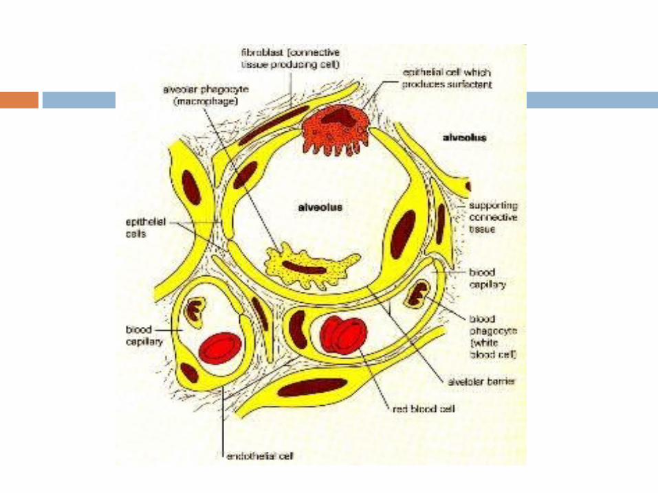
Respiratory Bronchioles, Alveolar Ducts, and Alveoli Lungs contain small saccular outpocketings
called alveoli. They have a thin wall specialized to promote
diffusion of gases between the alveolus and the blood in the pulmonary capillaries.
Gas exchange can take place in the respiratory bronchioles and alveolar ducts as well as in the alveoli, each lung contains approximately 300 to 400 million alveoli.
The spongy nature of the lung is due to the packing of millions of alveoli together.
Respiratory Membrane
squamous cells of alveoli . basement membrane of alveoli. basement membrane of capillaries simple squamous cells of capillaries about .5 μ in thickness
Cells in Alveolus
Type I cells : simple squamous cells forming lining
Type II cells : or septal cells secrete surfactant
Alveolar macrophages
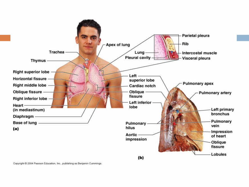
Gross Anatomy of the Lungs
Each lung has a conical shape. Its wide, concave base rests upon the muscular diaphragm.
Its superior region called the apex projects superiorly to a point that is slightly superior and posterior to the clavicle.
Both lungs are bordered by the thoracic wall anteriorly, laterally, and posteriorly, and supported by the rib cage.
Toward the midline, the lungs are separated from each other by the mediastinum.
The relatively broad, rounded surface in contact with the thoracic wall is called the costal surface of the lung.
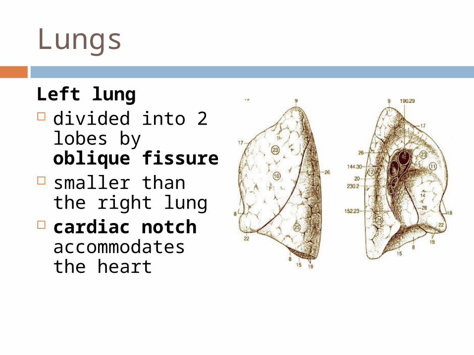
Lungs
Left lung divided into 2 lobes by
oblique fissure smaller than the right
lung cardiac notch
accommodates the heart
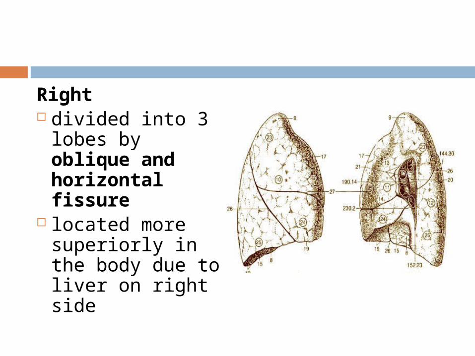
Right divided into 3 lobes
by oblique and horizontal fissure
located more superiorly in the body due to liver on right side
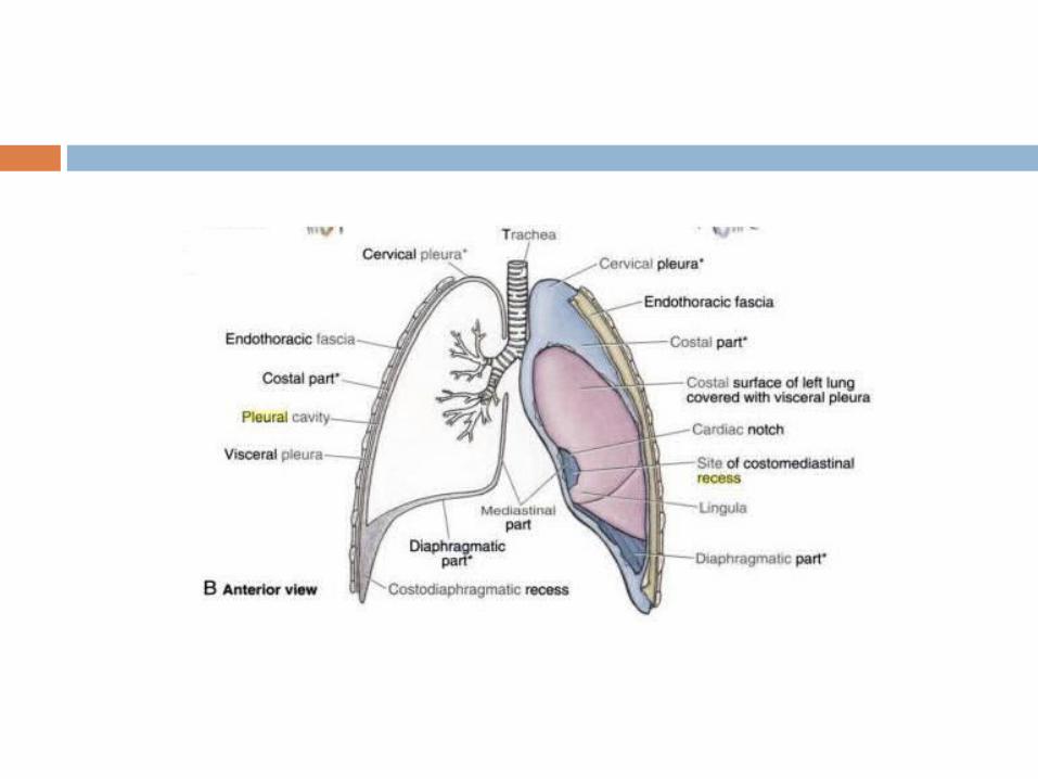
Pleura and Pleural Cavities
The outer surface of each lung and the adjacent internal thoracic wall are lined by a serous membrane called pleura.
The outer surface of each lung is tightly covered by the visceral pleura.
while the internal thoracic walls, the lateral surfaces of the mediastinum, and the superior surface of the diaphragm are lined by the parietal pleura.
The parietal and visceral pleural layers are continuous at the hilus of each lung.
Pleural Cavities
The potential space between the serous membrane layers is a pleural cavity.
The pleural membranes produce a thin, serous pleural fluid that circulates in the pleural cavity and acts as a lubricant, ensuring minimal friction during breathing.
Pleural effusion – pleuritis with too much fluid
Blood supply of Lungs
pulmonary circulation - bronchial circulation – bronchial arteries supply
oxygenated blood to lungs, bronchial veins carry away deoxygenated blood from lung tissue superior vena cava
Response of two systems to hypoxia – pulmonary vessels undergo vasoconstriction bronchial vessels like all other systemic vessels
undergo vasodilation
Respiratory events
Pulmonary ventilation = exchange of gases between lungs and atmosphere
External respiration = exchange of gases between alveoli and pulmonary capillaries
Internal respiration = exchange of gases between systemic capillaries and tissue cells
Two phases of pulmonary ventilation Inspiration, or inhalation - a very
active process that requires input of energy.The diaphragm, contracts, moving downward and flattening, when stimulated by phrenic nerves.
Expiration, or exhalation - a passive process that takes advantage of the recoil properties of elastic fiber. ・ The diaphragm relaxes.The elasticity of the lungs and the thoracic cage allows them to return to their normal size and shape.
Muscles that ASSIST with respiration The scalenes help increase thoracic cavity
dimensions by elevating the first and second ribs during forced inhalation.
The ribs elevate upon contraction of the external intercostals, thereby increasing the transverse dimensions of the thoracic cavity during inhalation.
Contraction of the internal intercostals depresses the ribs, but this only occurs during forced exhalation.
Normal exhalation requires no active muscular effort.
Muscles that ASSIST with respiration Other accessory muscles assist with
respiratory activities. The pectoralis minor, serratus
anterior, and sternocleidomastoid help with forced inhalation,
while the abdominal muscles(external and internal obliques, transversus abdominis, and rectus abdominis) assist in active exhalation.
Ventilation Control by Respiratory Centers of the Brain The trachea, bronchial tree, and lungs are
innervated by the autonomic nervous system.
The autonomic nerve fibers that innervate the heart also send branches to the respiratory structures.
The involuntary, rhythmic activities that deliver and remove respiratory gases are regulated in the brainstem within the reticular formation through both the medulla oblongata and pons.
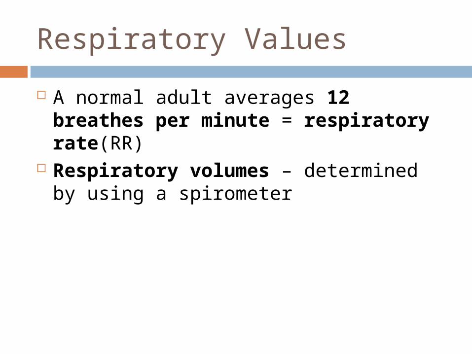
Respiratory Values
A normal adult averages 12 breathes per minute = respiratory rate(RR)
Respiratory volumes – determined by using a spirometer