Embed Size (px)
DESCRIPTION
Lesi Rongga Mulut
Citation preview
ABSTRACT In the United States, cancers of the oral cavity and oropharynx represent
approximately three percent of all malignancies in men and two percent of all malignancies in
women. The American Cancer Society estimates that 28,900 new cases of oral cancer will be
diagnosed in 2002, and nearly 7,400 people will die from this disease. Over 90 percent of
these tumors are squamous cell carcinomas, which arise from the oral mucosal lining. In spite
of the ready accessibility of the oral cavity to direct examination, these malignancies still are
often not detected until a late stage, and the survival rate for oral cancer has remained
essentially unchanged over the past three decades. The purpose of this article is to review the
clinical features of oral cancer and premalignant oral lesions, with an emphasis on early
detection. (CA Cancer J Clin 2002;52:195-215.)
INTRODUCTION
Cancers of the oral cavity and oropharynx represent approximately threepercent of all malignancies in men and two percent of all malignancies in womenin the United States. It is estimated that these tumors will account for 28,900 newcases and 7,400 deaths in 2002 in the United States.1 Squamous cell carcinoma,which arises from the oral mucosal lining, accounts for over 90 percent of thesetumors.2-4 This article will review the epidemiology and clinical features of oraland oropharyngeal squamous cell carcinoma, with a special emphasis on therecognition of early cancer and premalignant oral lesions.
EPIDEMIOLOGY
Oral cancer most commonly occurs in middle-aged and older individuals,although a disturbing number of these malignancies is also being documented inyounger adults in recent years.5-7 From an epidemiological and clinicopathologicalperspective, “oral cancer” can be divided into three categories: carcinomas of theoral cavity proper, carcinomas of the lip vermilion, and carcinomas arising in theoropharynx. Intraoral and oropharyngeal tumors are more common among menthan women, with a male:female ratio of over 2:1.2,8-9 However, the disparity in themale:female ratio has become less pronounced over the past half century, probablybecause women have been more equally exposing themselves to known oralcarcinogens such as tobacco and alcohol.4,5 The annual incidence of oral and
Oral Cancer and PrecancerousLesionsBrad W. Neville, DDS;Terry A. Day, MD, FACS
Dr. Neville is Professor andDirector, Division of Oral andMaxillofacial Pathology, Departmentof Stomatology, College of DentalMedicine, Medical University ofSouth Carolina, Charleston, SC.
Dr. Day is Associate Professor andDirector, Division of Head and NeckOncologic Surgery, Department ofOtolaryngology, Head and NeckSurgery, College of Medicine,Medical University of South Carol-ina, Charleston, SC.
This article is also available atwww.cancer.org.
CA Cancer J Clin 2002;52:195-215
Volume 52 • Number 4 • July/August 2002 195
pharyngeal cancer in African Americans (12.4cases per 100,000 population) is higher thanamong whites (9.7 cases per 100,000); the highestincidence rate is among African-Americanmales (20.5 cases per 100,000 population).3,9
In contrast to intraoral and oropharyngealcarcinomas, cancers of the lip vermilion aremore akin epidemiologically to squamous cellcarcinoma of the skin and occur primarily inwhite men.2 These lip tumors are most stronglyassociated with chronic sun exposure, althoughsometimes they have been related to the sitewhere cigarettes or pipestems have habitually
been held.10 These malignancies are muchmore common in men, probably because menare more likely to have vocations and/oravocations that result in greater cumulative sunexposure. At one time, the lip was the mostcommon site for oral cancer; however, theincidence of cancer in this location hasdecreased significantly over the past halfcentury because fewer men hold outdooroccupations.2,4
Despite advances in surgery, radiation, andchemotherapy, the five-year survival rate fororal cancer has not improved significantly over
196 CA A Cancer Journal for Clinicians
Oral Cancer and Precancerous Lesions
1950 1955 1960 1965 1970 1975 1980 1985 1990 19950
2
4
6
8
10
12
Rate
per
100
,000
Year of Death
White Women
African-American Men
All Men
White Men
African-American Women
All Women
FIGURE 1
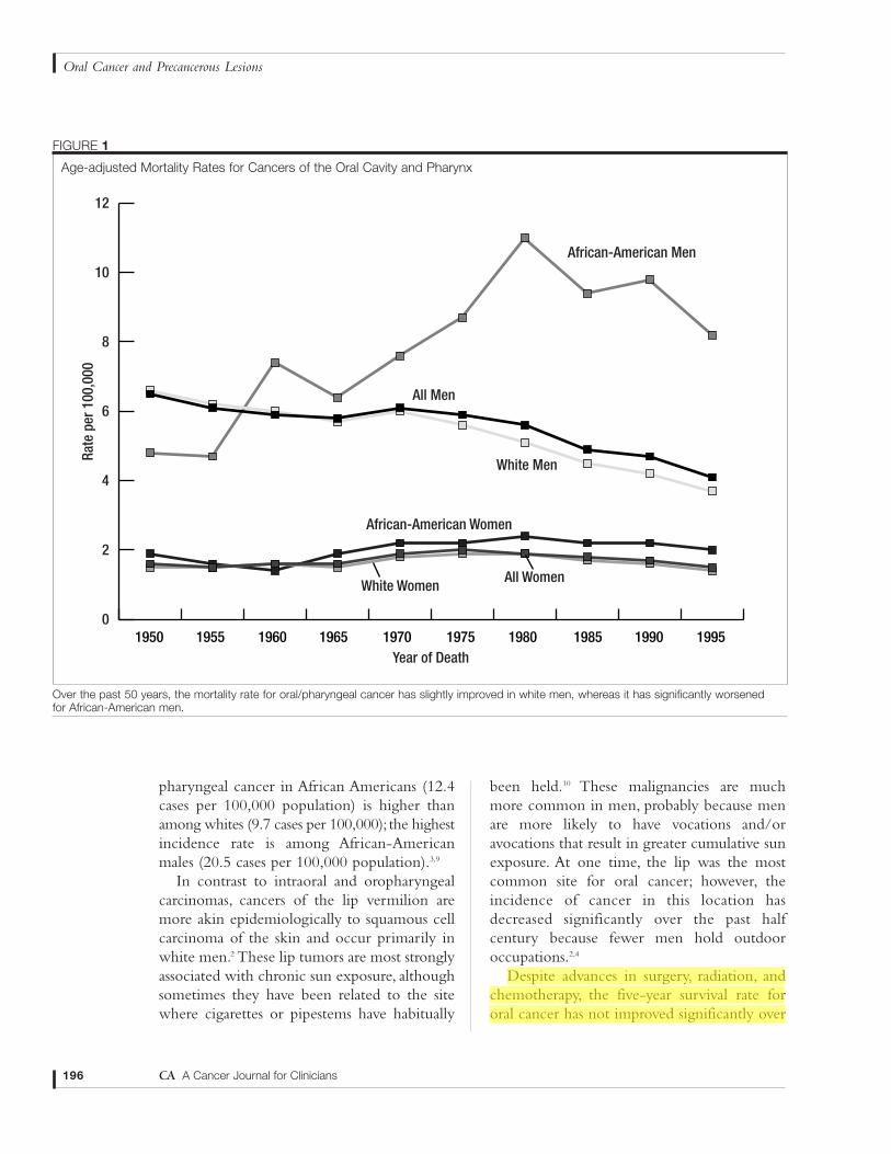
Age-adjusted Mortality Rates for Cancers of the Oral Cavity and Pharynx
Over the past 50 years, the mortality rate for oral/pharyngeal cancer has slightly improved in white men, whereas it has significantly worsened for African-American men.
the past several decades and it remains at about50 to 55 percent.3,9 Unfortunately, AfricanAmericans have a significantly higher mortalityrate when compared with whites (4.4 versus2.4 per 100,000 population), partly becauseamong African Americans, tumors are moreoften discovered at an advanced stage (Figure1).3,9,11,12 From 1985 to 1996, the five-yearsurvival rate for carcinoma of the tongue inAfrican-American men was 27 percent,compared with a 47 percent five-year survivalrate among white men.3 For floor of mouthcancers, the survival rate was 52 percent inwhites, compared with only 33 percent amongAfrican Americans. When compared withintraoral carcinoma, the prognosis for lipcancer is quite good, with a five-year survivalrate of 95 percent.2,3
RISK FACTORS
The strong association between cancers ofthe oral cavity and pharynx with tobacco use iswell established. Epidemiological studies showthat the risk of developing oral cancer is five tonine times greater for smokers than fornonsmokers, and this risk may increase to asmuch as 17 times greater for extremely heavysmokers of 80 or more cigarettes per day.2,13-17
The percentage of oral cancer patients whosmoke (approximately 80 percent) is two tothree times greater than that of the generalpopulation. In addition, treated oral cancerpatients who continue to smoke have a two tosix times greater risk of developing a secondmalignancy of the upper aerodigestive tractthan those who stop smoking.10,18 Marijuanause is also considered to be a potential riskfactor and may be partly responsible for therise in oral cancers seen among youngadults.3,7,19 However, further epidemiologicalstudies are necessary to confirm the purportedassociation of marijuana and oral cancer inyounger patients.
Snuff and chewing tobacco have also beenassociated with an increased risk for oralcancer.20 In one study of women in thesouthern United States, chronic users of snuffwere estimated to have a four times greater riskof developing oral cancer.21 In addition, asignificant number of oral cancers in smokelesstobacco users develop at the site of tobaccoplacement. However, the use of smokelesstobacco appears to be associated with a muchlower cancer risk than that associated withsmoked tobacco.The incidence of oral cancerin West Virginia is below the national average,even though this state has the highestconsumption of chewing tobacco in theUnited States.22 Recent studies fromScandinavia have suggested that the use ofSwedish snuff (which is nonfermented and haslower nitrosamine levels) is not associated withan increased risk for oral cancer.17,23
Alcohol use has been identified as a majorrisk factor for cancers of the upperaerodigestive tract. In studies controlled forsmoking, moderate-to-heavy drinkers havebeen shown to have a three to nine timesgreater risk of developing oral cancer.13,14,16,17
One study from France showed that extremelyheavy drinkers (greater than 100 grams ofalcohol per day) had a 30 times greater risk ofdeveloping oral and oropharyngeal cancer (atypical serving of beer, wine, or liquor containsten to 15 grams of alcohol).15 Of even greatersignificance is the synergistic effect of alcoholand smoking; some subsets of patients who areboth heavy smokers and heavy drinkers canhave over one hundred times greater risk fordeveloping a malignancy.15,16
In India and Southeast Asia, the chronic useof betel quid (paan) in the mouth has beenstrongly associated with an increased risk fororal cancer.24-26 The quid typically consists of a betel leaf that is wrapped around a mixture of areca nut and slaked lime, usually withtobacco and sometimes with sweeteners andcondiments. The slaked lime results in the
Volume 52 • Number 4 • July/August 2002 197
CA Cancer J Clin 2002;52:195-215

release of an alkaloid from the areca nut, whichproduces a feeling of euphoria and well-beingin the user. Betel quid chewing often results ina progressive, scarring precancerous conditionof the mouth known as oral submucousfibrosis. In India, one study showed a malignanttransformation rate of 7.6 percent for oralsubmucous fibrosis.25
Recent evidence suggests that humanpapillomavirus (HPV) may be associated withsome oral and oropharyngeal cancers.27-31
HPV-16 has been detected in up to 22 percentof oral cancers, and HPV-18 has been found inup to 14 percent of cases.28 Dietary factors,such as a low intake of fruits and vegetables,may also be related to an increased cancerrisk.32,33 As previously indicated, chronic actinicexposure is associated with the development ofcarcinomas of the lip vermilion.
A number of studies have suggested that orallichen planus, especially the erosive form, maybe associated with an increased cancer risk,although other investigators have questionedthe strength of this association.34-36 Irondeficiency anemia in combination withdysphagia and esophageal webs (known asPlummer-Vinson or Paterson-Kelly syndrome)is associated with an elevated risk for devel-opment of carcinoma of the oral cavity, oro-pharynx, and esophagus.37,38 Immunosuppressionappears to predispose some individuals to anincreased risk for oral cancer. Carcinomas of thelip have been reported in a number of kidneytransplant patients receiving immunosuppressivemedications, and oral carcinomas have beendocumented in young AIDS patients.39-42
EARLY DIAGNOSIS
Despite the great strides that have been
made in recent decades to improve theprognosis for a number of cancers throughoutthe body, the prognosis for oral cancer has notexperienced a similar improvement.3,8,11
Because five-year survival is directly related tostage at diagnosis, prevention and earlydetection efforts have the potential not onlyfor decreasing the incidence, but also forimproving the survival of those who developthis disease. Early diagnosis depends upon anastute clinician or patient who may identify asuspicious lesion or symptom while it is still atan early stage. However, it is apparent thatmany clinicians, including dentists andphysicians, may not be knowledgeable aboutthe risk factors, diagnosis, and early detectionof these cancers and/or are not performingroutine oral cancer examinations.43-49
The Centers for Disease Control andPrevention’s 1998 National Health InterviewSurvey (NHIS) Adult Prevention Supplementincluded questions regarding examinations fororal cancer. Participants were asked “Have youever had a test for oral cancer in which thedoctor or dentist pulls on your tongue,sometimes with gauze wrapped around it, andfeels under the tongue and inside the cheeks?”Only 16 percent of respondents reported thatthey ever had such an exam. This reportedcumulative prevalence of oral cancer examswas higher in whites (18 percent) than inAfrican Americans (10 percent), AmericanIndians/Alaska Natives (8 percent), orAsian/Pacific Islanders (11 percent). Formersmokers (21 percent) were more likely thancurrent smokers (13 percent) or people whohad never smoked (16 percent) to recall havingever had this examination. Among allindividuals who reported having had an oralcancer exam, 70 percent reported that their lastexam was within the past year.*
198 CA A Cancer Journal for Clinicians
Oral Cancer and Precancerous Lesions
*Vilma Cokkinides, PhD, (personal communication, May 2002), based on an analysis of the NHIS 1998 Adult PreventionSupplement Public Use Data Release accessed at www.ccdc.gov/nchs/nhis.htm.
Early oral cancers and precancerous lesionsare often subtle and asymptomatic.Therefore, itis important for the clinician to maintain ahigh index of suspicion, especially if riskfactors such as tobacco use or alcohol abuse arepresent. Invasive oral squamous cell carcinomais often preceded by the presence of clinicallyidentifiable premalignant changes of the oralmucosa. These lesions often present as eitherwhite or red patches, known as leukoplakia anderythroplakia. As the cancer develops, thepatient may notice the presence of anonhealing ulcer. Later-stage symptomsinclude bleeding, loosening of teeth, difficultywearing dentures, dysphagia, dysarthria,odynophagia, and development of a neck mass.
The American Cancer Society recommendsa cancer-related check-up annually for allindividuals aged 40 and older, and every threeyears for those between the ages of 20 and 39,which “should include health counseling and,depending on a person’s age, might includeexaminations for cancers of the thyroid, oralcavity, skin, lymph nodes, testes, and ovaries.”50
According to the US Preventive HealthServices Task Force (USPHSTF), “there isinsufficient evidence to recommend for oragainst routine screening of asymptomaticpersons for oral cancer by primary careclinicians … clinicians may wish to include anexamination for cancerous and precancerouslesions of the oral cavity in the periodic healthexamination of persons who chew or smoketobacco (or did so previously), older personswho drink regularly, and anyone withsuspicious symptoms or lesions detectedthrough self-examination. … Appropriatecounseling should be offered to those personswho smoke cigarettes, pipes, or cigars, thosewho use chewing tobacco or snuff, and thosewho demonstrate evidence of alcohol abuse.”51
The USPHSTF document also notes that“…both the National Cancer Institute and theNational Institute of Dental Research(subsequently renamed the National Institute
of Dental and Craniofacial Research) supportefforts to promote the early detection of oralcancers during routine dental examinations.”
Clearly, the low prevalence of oral cancerscreening reported in the NHIS indicates thatmost clinicians are not following ACSrecommendations, and are not even followingthe USPHSTF suggestion for examinations intobacco users and other high-risk individuals.
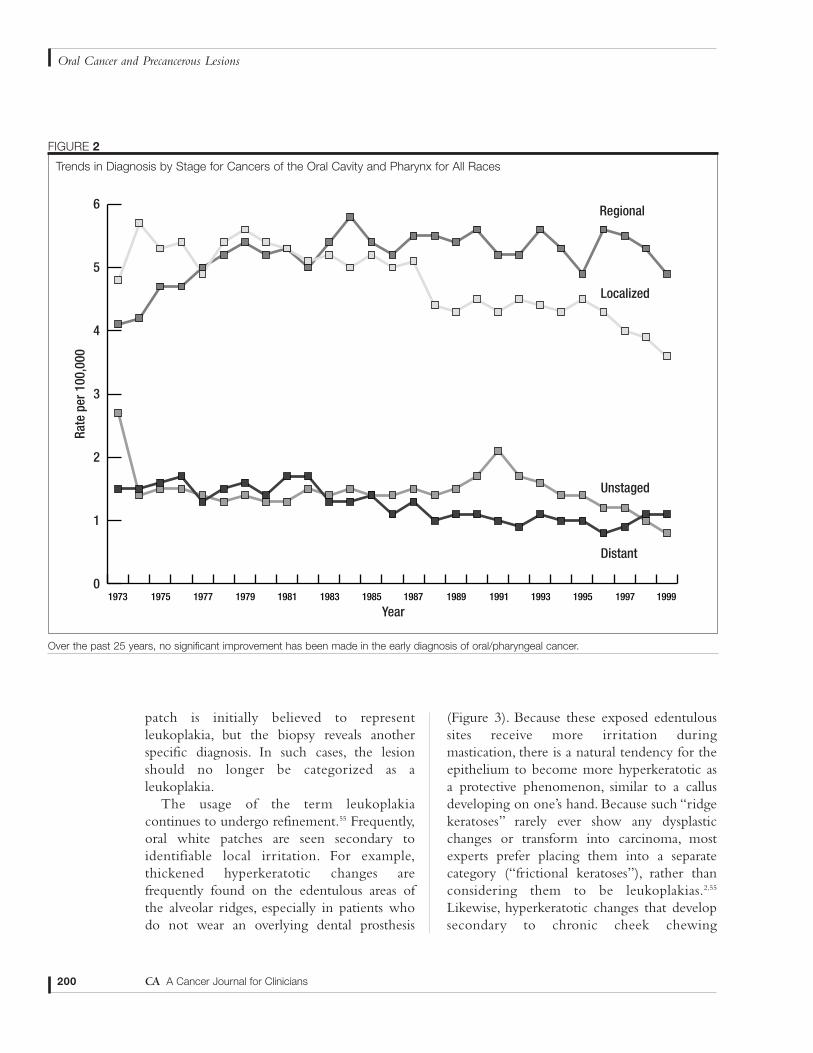
Unfortunately, there has been littleimprovement in the early detection of oralcancer because many patients do not presentfor diagnosis and treatment until they haveStage III or Stage IV disease (Figure 2).Therefore, in order to improve oral cancersurvival, public education efforts are alsonecessary to encourage patients to avoid high-risk behaviors and to ask their health careproviders about regular oral cancer screeningexaminations.
LEUKOPLAKIA
The term leukoplakia was first used bySchwimmer in 1877 to describe a white lesionof the tongue, which probably represented asyphilitic glossitis.52 The definition ofleukoplakia has often been confusing andcontroversial—so much so, that some cliniciansnow avoid using this term in their lexicon. Asdefined by the World Health Organization,leukoplakia is “a white patch or plaque thatcannot be characterized clinically orpathologically as any other disease.”53 As such,leukoplakia should be used only as a clinicalterm; it has no specific histopathologicalconnotation and should never be used as amicroscopic diagnosis.54 In the evaluation ofthe patient, leukoplakia is a clinical diagnosis ofexclusion. If an oral white patch can bediagnosed as some other condition (e.g.,candidiasis, lichen planus, leukoedema, etc.),then the lesion should not be considered to bean example of leukoplakia. Sometimes a white
Volume 52 • Number 4 • July/August 2002 199
CA Cancer J Clin 2002;52:195-215
patch is initially believed to representleukoplakia, but the biopsy reveals anotherspecific diagnosis. In such cases, the lesionshould no longer be categorized as aleukoplakia.
The usage of the term leukoplakiacontinues to undergo refinement.55 Frequently,oral white patches are seen secondary toidentifiable local irritation. For example,thickened hyperkeratotic changes arefrequently found on the edentulous areas ofthe alveolar ridges, especially in patients whodo not wear an overlying dental prosthesis
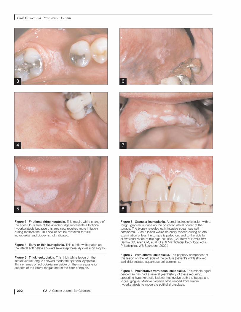
(Figure 3). Because these exposed edentuloussites receive more irritation duringmastication, there is a natural tendency for theepithelium to become more hyperkeratotic asa protective phenomenon, similar to a callusdeveloping on one’s hand. Because such “ridgekeratoses” rarely ever show any dysplasticchanges or transform into carcinoma, mostexperts prefer placing them into a separatecategory (“frictional keratoses”), rather thanconsidering them to be leukoplakias.2,55
Likewise, hyperkeratotic changes that developsecondary to chronic cheek chewing
200 CA A Cancer Journal for Clinicians
Oral Cancer and Precancerous Lesions
1973 1975 1977 1979 1981 1983 1985 1987 1989 1991 1993 1995 1997 1999 0
1
2
3
4
5
6
Rate
per
100
,000
Year
Regional
Localized
Unstaged
Distant
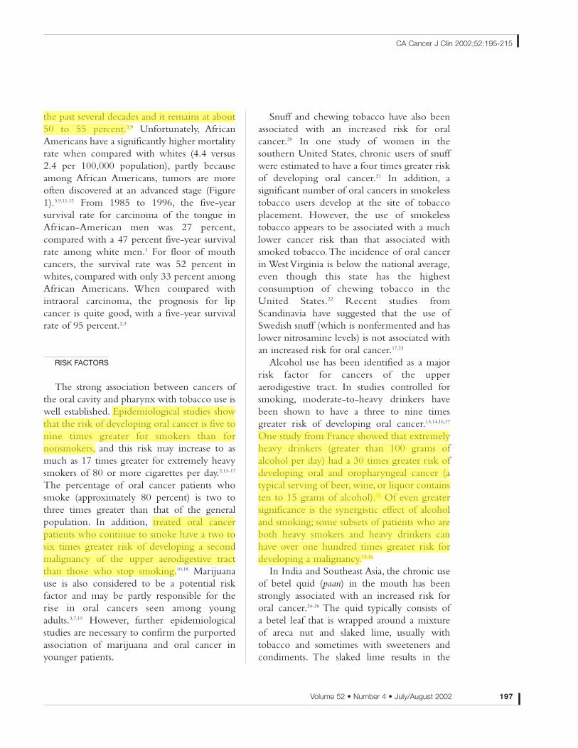
FIGURE 2
Trends in Diagnosis by Stage for Cancers of the Oral Cavity and Pharynx for All Races
Over the past 25 years, no significant improvement has been made in the early diagnosis of oral/pharyngeal cancer.
(“morsicatio buccarum”) or tongue chewing(“morsicatio linguarum”) should not beclassified as leukoplakia; such lesions are notpremalignant and they are readily reversible ifthe irritation is avoided.
Two specific tobacco-related lesions of theoral mucosa, nicotine stomatitis and tobaccopouch keratosis, have often been includedunder the broad umbrella of leukoplakia.However, because these lesions have a specificknown cause and prognosis, we prefer toclassify them separately from leukoplakia.
Leukoplakia is seen most frequently inmiddle-aged and older men, with an increasingprevalence with age.2,56 Fewer than one percentof men below the age of 30 have leukoplakia,but the prevalence increases to an alarmingeight percent in men over the age of 70.56 Theprevalence in women past the age of 70 isapproximately two percent.The most commonsites are the buccal mucosa, alveolar mucosa,and lower lip; however, lesions in the floor ofmouth, lateral tongue, and lower lip are mostlikely to show dysplastic or malignantchanges.57
Early or thin leukoplakia appears as a slightlyelevated grayish-white plaque that may beeither well defined or may gradually blend intothe surrounding normal mucosa (Figure 4).2,58
As the lesion progresses, it becomes thicker andwhiter, sometimes developing a leatheryappearance with surface fissures (homogeneousor thick leukoplakia) (Figure 5). Someleukoplakias develop surface irregularities andare referred to as granular or nodularleukoplakias (Figure 6). Other lesions develop apapillary surface and are known as verrucous orverruciform leukoplakia (Figure 7).
One uncommon variant, known asproliferative verrucous leukoplakia (PVL), ischaracterized by widespread, multifocal sites ofinvolvement, often in patients without knownrisk factors.59-63 The condition begins withconventional flat white patches that, over time,
tend to become much thicker and papillary innature (Figure 8). This papillary proliferationmay progress to the point where the lesion canbe categorized microscopically as a verrucouscarcinoma. However, in spite of treatment, thelesions have a high recurrence rate and ofteneventually transform into more aggressivesquamous cell carcinoma.
In recent years, a number of oral whitepatches have been identified that appear to berelated to the use of toothpastes or mouthrinses containing the herbal extract,sanguinaria.64-66 Such lesions most frequentlyhave been identified on the maxillary alveolarmucosa and buccal vestibule, although somepatients have developed lesions on themandibular alveolar mucosa. Microscopically,these lesions usually show hyperkeratosis andepithelial atrophy, sometimes in associationwith true dysplasia, although the potential forthe development of cancer is uncertain.
Volume 52 • Number 4 • July/August 2002 201
CA Cancer J Clin 2002;52:195-215
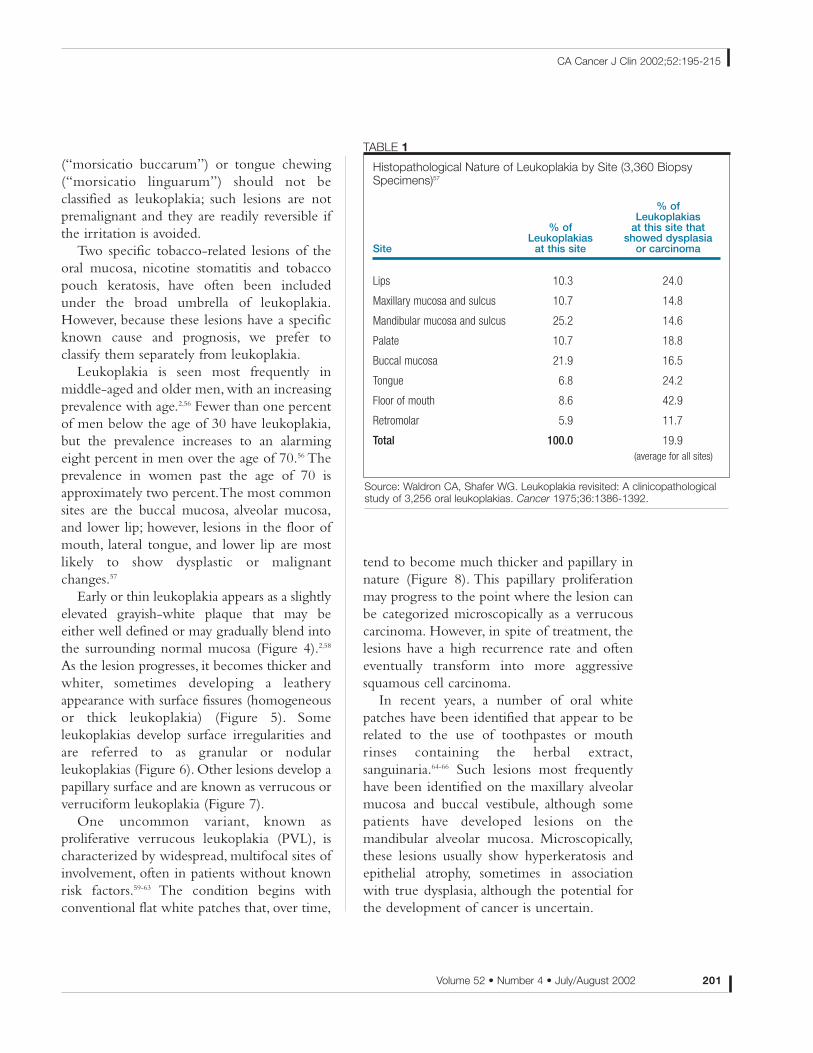
Histopathological Nature of Leukoplakia by Site (3,360 BiopsySpecimens)57
% ofLeukoplakias
% of at this site thatLeukoplakias showed dysplasia
Site at this site or carcinoma
Lips 10.3 24.0
Maxillary mucosa and sulcus 10.7 14.8
Mandibular mucosa and sulcus 25.2 14.6
Palate 10.7 18.8
Buccal mucosa 21.9 16.5
Tongue 6.8 24.2
Floor of mouth 8.6 42.9
Retromolar 5.9 11.7
Total 100.0 19.9(average for all sites)
TABLE 1
Source: Waldron CA, Shafer WG. Leukoplakia revisited: A clinicopathologicalstudy of 3,256 oral leukoplakias. Cancer 1975;36:1386-1392.
202 CA A Cancer Journal for Clinicians
Oral Cancer and Precancerous Lesions
Figure 3 Frictional ridge keratosis. This rough, white change ofthe edentulous area of the alveolar ridge represents a frictionalhyperkeratosis because this area now receives more irritationduring mastication. This should not be mistaken for trueleukoplakia, and biopsy is not indicated.
Figure 4 Early or thin leukoplakia. This subtle white patch onthe lateral soft palate showed severe epithelial dysplasia on biopsy.
Figure 5 Thick leukoplakia. This thick white lesion on thelateral/ventral tongue showed moderate epithelial dysplasia.Thinner areas of leukoplakia are visible on the more posterioraspects of the lateral tongue and in the floor of mouth.
Figure 6 Granular leukoplakia. A small leukoplakic lesion with arough, granular surface on the posterior lateral border of thetongue. The biopsy revealed early invasive squamous cellcarcinoma. Such a lesion would be easily missed during an oralexamination unless the tongue is pulled out and to the side toallow visualization of this high-risk site. (Courtesy of Neville BW,Damm DD, Allen CM, et al. Oral & Maxillofacial Pathology, ed 2,Philadelphia, WB Saunders, 2002.)
Figure 7 Verruciform leukoplakia. The papillary component ofthis lesion on the left side of the picture (patient’s right) showedwell-differentiated squamous cell carcinoma.
Figure 8 Proliferative verrucous leukoplakia. This middle-agedgentleman has had a several year history of these recurring,spreading hyperkeratotic lesions that involve both the buccal andlingual gingiva. Multiple biopsies have ranged from simplehyperkeratosis to moderate epithelial dysplasia.
3
4
5
6
7
8
Volume 52 • Number 4 • July/August 2002 203
CA Cancer J Clin 2002;52:195-215
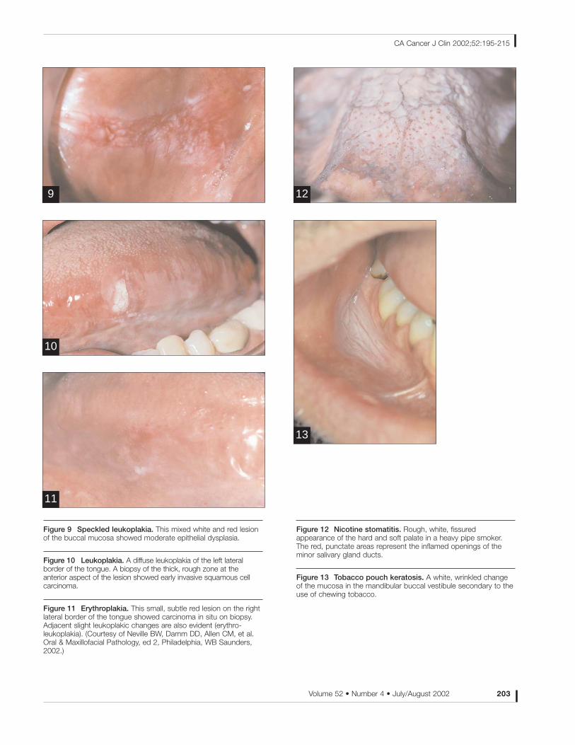
Figure 9 Speckled leukoplakia. This mixed white and red lesionof the buccal mucosa showed moderate epithelial dysplasia.
Figure 10 Leukoplakia. A diffuse leukoplakia of the left lateralborder of the tongue. A biopsy of the thick, rough zone at theanterior aspect of the lesion showed early invasive squamous cellcarcinoma.
Figure 11 Erythroplakia. This small, subtle red lesion on the rightlateral border of the tongue showed carcinoma in situ on biopsy.Adjacent slight leukoplakic changes are also evident (erythro-leukoplakia). (Courtesy of Neville BW, Damm DD, Allen CM, et al.Oral & Maxillofacial Pathology, ed 2, Philadelphia, WB Saunders,2002.)
Figure 12 Nicotine stomatitis. Rough, white, fissuredappearance of the hard and soft palate in a heavy pipe smoker.The red, punctate areas represent the inflamed openings of theminor salivary gland ducts.
Figure 13 Tobacco pouch keratosis. A white, wrinkled changeof the mucosa in the mandibular buccal vestibule secondary to theuse of chewing tobacco.
9
10
11
12
13
Because sanguinaria-associated keratoses canbe extensive or multifocal, sometimes they aremisinterpreted as early proliferative verrucousleukoplakia.
Some leukoplakias occur in combinationwith adjacent red patches or erythroplakia. Ifthe red and white areas are intermixed, thelesion is called a speckled leukoplakia orspeckled erythroplakia (Figure 9).
The frequency of dysplastic or malignantalterations in oral leukoplakia has ranged from15.6 to 39.2 percent in several studies.54,57,67-69 Inone large, well known retrospective study thatlooked at approximately 3,300 biopsies of oralwhite lesions,Waldron and Shafer determinedthat 19.9 percent of leukoplakias showed somedegree of epithelial dysplasia (Table 1).57 In thisgroup, 3.1 percent were unsuspected squamouscell carcinoma, 4.6 percent showed severedysplasia or carcinoma in situ, and 12.2 percentshowed mild-to-moderate epithelial dysplasia.Differences in the frequency of dysplasticchanges in leukoplakia studies may reflectselection bias or differences in the clinicaldefinition of oral leukoplakia. If white lesionssuch as frictional ridge keratoses and nicotine
stomatitis are not included as examples ofclinical leukoplakia, the percentage of casesshowing dysplastic changes will be higher.
The location of oral leukoplakia has asignificant correlation with the frequency offinding dysplastic or malignant changes atbiopsy. In the study by Waldron and Shafer, thefloor of mouth was the highest-risk site, with42.9 percent of leukoplakias showing somedegree of epithelial dysplasia, carcinoma in situ,or unsuspected invasive squamous cellcarcinoma.57 The tongue and lip were alsoidentified as high-risk sites, with dysplasia orcarcinoma present in 24.2 percent and 24.0percent of these cases, respectively.
The clinical appearance of leukoplakia mayalso indicate some correlation with thelikelihood that the lesion will show dysplasticor malignant features. In general, the thickerthe leukoplakia, the greater the chance offinding dysplastic changes; therefore, averrucous leukoplakia is more likely to showdysplasia than is a thick homogeneousleukoplakia, which, in turn, is more likely toshow dysplasia than is a thin leukoplakia(Figure 10).58 Leukoplakias with an intermixed
204 CA A Cancer Journal for Clinicians
Oral Cancer and Precancerous Lesions
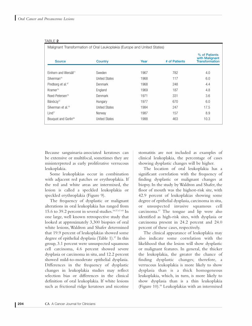
Malignant Transformation of Oral Leukoplakia (Europe and United States)
% of Patientswith Malignant
Source Country Year # of Patients Transformation
Einhorn and Wersäll71 Sweden 1967 782 4.0
Silverman70 United States 1968 117 6.0
Pindborg et al.67 Denmark 1968 248 4.4
Kramer74 England 1969 187 4.8
Roed-Petersen75 Denmark 1971 331 3.6
Bánóczy72 Hungary 1977 670 6.0
Silverman et al.76 United States 1984 247 17.5
Lind77 Norway 1987 157 8.9
Bouquot and Gorlin56 United States 1988 463 10.3
TABLE 2
red component (speckled leukoplakia or mixedleukoplakia/erythroplakia) are at greatest riskfor showing dysplasia or carcinoma. Pindborgand associates found 14 percent of speckledleukoplakias to show carcinoma, whereasanother 51 percent showed epithelial dys-plasia.67 However, all leukoplakias should beviewed with suspicion because even small,subtle lesions can manifest significant dysplasiaor unsuspected carcinoma.57,70 Therefore,directed conventional biopsy is recommendedfor any true oral leukoplakia.
In addition to a small percentage ofleukoplakias that will show invasive carcinomawhen they are first sampled for biopsy, it is alsorecognized that currently non-carcinomatousleukoplakias are at risk for future malignanttransformation. Several clinical studies havebeen conducted in Europe and the UnitedStates to assess the potential for malignanttransformation of oral leukoplakia (Table2).58,70-77 Most of the earlier studies showed arisk of malignant transformation in the rangeof 3.6 to 6.0 percent. However, several of themore recent studies have shown more alarmingmalignant transformation rates ranging from8.9 to 17.5 percent.58,76,77 Although the reasonfor these results is unclear, it may be due to amore restrictive definition of what isconsidered clinical leukoplakia and furtherunderscores the seriousness of “trueleukoplakia.” The study by Silverman andcolleagues showed an overall malignanttransformation of 17.5 percent.76 In this study,only 6.5 percent of homogeneous leukoplakiasunderwent malignant change; however, 23.4percent of speckled leukoplakias and 36.4percent of leukoplakias with microscopicevidence of dysplastic changes transformedinto cancer.
When compared with “conventionalleukoplakia,” proliferative verrucous leuko-plakia is a particularly high-risk condition. In a follow-up study of 54 cases of proliferativeverrucous leukoplakia, Silverman and Gorsky
found that 70.3 percent of the patientssubsequently developed squamous cellcarcinoma.62
Although leukoplakia is more common inmen than women, several studies have shownthat women with leukoplakia have a higher riskof developing oral carcinoma.70,72,75 Anotherdisturbing finding is that leukoplakias innonsmokers are more likely to undergomalignant transformation than leukoplakias inpatients who do smoke.71,72,75,76 This should not be interpreted to detract from the well-established role of tobacco in oralcarcinogenesis, but may indicate that non-smokers who develop leukoplakia do so as aresult of other more potent carcinogenic factors.
ERYTHROPLAKIA
The term erythroplasia was originally used byQueyrat to describe a red, precancerous lesionof the penis.78 The term erythroplakia is used fora clinically and histopathologically similarprocess that occurs on the oral mucosa. Similarto the definition for leukoplakia, erythroplakiais a clinical term that refers to a red patch thatcannot be defined clinically or pathologicallyas any other condition.53 This definitionexcludes inflammatory conditions that mayresult in a red clinical appearance.
Oral erythroplakia occurs most frequentlyin older men and appears as a red macule orplaque with a soft, velvety texture (Figure 11).2
The floor of mouth, lateral tongue, retromolarpad, and soft palate are the most common sitesof involvement. Often the lesion is welldemarcated, but some examples may graduallyblend into the surrounding mucosa. Somelesions may be intermixed with white areas(erythroleukoplakia). Erythroplakia is oftenasymptomatic, although some patients maycomplain of a sore, burning sensation.
Although erythroplakia is not nearly ascommon as leukoplakia, it is much more likely
Volume 52 • Number 4 • July/August 2002 205
CA Cancer J Clin 2002;52:195-215
to show dysplasia or carcinoma. In a sister studyto their large series of leukoplakia cases, Shaferand Waldron also analyzed their biopsyexperience with 65 cases of erythroplakia.79 Allerythroplakia cases showed some degree ofepithelial dysplasia; 51 percent showed invasivesquamous cell carcinoma, 40 percent werecarcinoma in situ or severe epithelial dysplasia,and the remaining 9 percent demonstratedmild-to-moderate dysplasia. Therefore, trueclinical erythroplakia is a much moreworrisome lesion than leukoplakia.80 Likewise,in a mixed erythroleukoplakia, the redcomponent is more likely to demonstratedysplastic changes than is the white component;when selecting an appropriate biopsy site in amixed lesion, the clinician should make sure thatthe specimen includes the red component.
NICOTINE STOMATITIS
Nicotine stomatitis is a thickened,hyperkeratotic alteration of the palatal mucosathat is most frequently related to pipe smoking,but milder examples can also developsecondary to cigar smoking or, rarely, fromcigarette smoking.2,53 The palatal mucosabecomes thickened and hyperkeratotic,sometimes developing a fissured surface(Figure 12).The surface often develops papularelevations with red centers, which representthe inflamed openings of the minor salivarygland ducts.
The term nicotine stomatitis is actually amisnomer because it isn’t the nicotine thatcauses the changes; the changes are caused bythe intense heat generated from the smoking.Nicotine stomatitis is seen more often in pipesmokers because of the great amount of heatthat is generated from the pipestem. (Similarlesions have even been reported in patientswho drink extremely hot beverages.)81
Although nicotine stomatitis is a tobacco-related pathosis, it is not considered to be
premalignant and it is readily reversible withdiscontinuation of the tobacco habit.
However, in some Southeast Asian andSouth American countries, individuals practicea habit known as reverse smoking in which thelit end of the cigarette or cigar is placed in themouth. This habit creates a more severe heat-related alteration of the palatal mucosa knownas reverse smoker’s palate, which has beenassociated with a significant risk of malignanttransformation.10,82,83
TOBACCO POUCH KERATOSIS
Another specific tobacco-related oralmucosal alteration occurs in association withsmokeless tobacco use, either from snuff orchewing tobacco.2,84-87 Such lesions typicallyoccur in the buccal or labial vestibule wherethe tobacco is held, but they can also extendonto the adjacent gingiva and buccal mucosa.Early lesions may show slight wrinkling thatdisappears when the tissues are stretched.Other lesions may appear as hyperkeratotic,granular patches. Advanced lesions exhibitgreatly thickened zones of grayish whitemucosa with well-developed folds and fissures(Figure 13). The degree of clinical alterationdepends on the type and quantity of tobacco,the duration of tobacco usage, and hostsusceptibility.
Tobacco pouch keratoses can occur at anyage, even in children and adolescents. InWestern cultures, these lesions currently areseen most frequently in young men and menolder than 65 years of age; such lesions are lesscommon among middle-aged men because thehabit of using smokeless tobacco has not beenas popular in this generation.2 In some ruralSouthern populations, smokeless tobaccokeratoses are seen with some degree offrequency in older women, who may havestarted their snuff-dipping habit in earlychildhood.84 Overall, it is estimated that 15
206 CA A Cancer Journal for Clinicians
Oral Cancer and Precancerous Lesions
percent of chewing tobacco users and 60percent of snuff users will develop clinicallesions, if mild examples are included.2
Microscopically, smokeless tobacco keratosesshow hyperkeratosis and acanthosis of themucosal epithelium.True epithelial dysplasia isuncommon; when dysplasia is found, it isusually mild in degree.84 However, significantdysplasia or squamous cell carcinomaoccasionally may be discovered.
Most tobacco pouch keratoses are readilyreversible within two to six weeks aftercessation of the tobacco habit.88 If the lesiondoes not resolve after the habit is stopped, thenan incisional biopsy of the area should beperformed and the patient managedaccordingly. Some clinicians also recommendbiopsy for lesions in patients who will notdiscontinue their tobacco habit.
SQUAMOUS CELL CARCINOMA
Early squamous cell carcinoma oftenpresents as a white patch (leukoplakia), redpatch (erythroplakia), or a mixed red and whitelesion (erythroleukoplakia). With time,superficial ulceration of the mucosal surfacemay develop (Figure 14). As the lesion grows,it may become an exophytic mass with afungating or papillary surface (Figure 15); othertumors have an endophytic growth pattern thatis characterized by a depressed, ulceratedsurface with a raised, rolled border (Figure16).2,89 Pain is not a reliable indicator as towhether a particular lesion may be malignant;larger, advanced carcinomas will often bepainful, but many early oral cancers will betotally asymptomatic or may be associated withonly minor discomfort.
The most common site for intraoralcarcinoma is the tongue, which accounts foraround 40 percent of all cases in the oral cavityproper.These tumors most frequently occur onthe posterior lateral border and ventral surfaces
of the tongue. The floor of the mouth is thesecond most common intraoral location. Less-common sites include the gingiva, buccalmucosa, labial mucosa, and hard palate.2,4
The lateral tongue and floor of mouth (withextension back to the lateral soft palate andtonsillar area) combine to form a horseshoe-shaped region of the oral mucosa, which is atgreatest risk for cancer development.There aretwo major factors that may explain why thisregion is at high risk: first, any carcinogens willmix with saliva, pool in the bottom of themouth, and constantly bathe these sites;secondly, these regions of the mouth arecovered by a thinner, nonkeratinized mucosa,which provides less protection againstcarcinogens.14
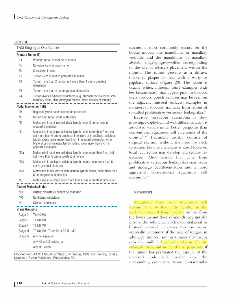
It is important for the clinician to be aware ofthis high-risk region when examining the oralcavity. During an examination, if a tongue bladeor other instrument is used simply to depress thetongue in order to see the rest of the mouth,then the two most common sites for intraoralcancer will be hidden. It is recommended that acotton gauze be used to grasp the tip of thetongue, allowing it to be pulled upward and toeach side so that the lateral tongue and oral floorcan be adequately seen.90
In addition to the oral cavity proper,squamous cell carcinomas also often developon the lip vermilion and the oropharynx.Vermilion carcinomas show a strikingpredilection for the lower lip, and usually occurin light-skinned individuals with a long historyof actinic damage. The lesion usually arises inan actinic cheilosis, a premalignant conditionthat is akin to actinic keratosis of the skin.Actinic cheilosis is characterized by atrophy ofthe vermilion border, which may develop dry,scaly changes. As the condition progresses,ulcerated sites may appear which partially heal,only to recur at a later date (Figure 17). (Thepatient often mistakes these recurring ulceratedlesions for “fever blisters.”) The evolving cancerslowly becomes a crusted, nontender, indurated
Volume 52 • Number 4 • July/August 2002 207
CA Cancer J Clin 2002;52:195-215

208 CA A Cancer Journal for Clinicians
Oral Cancer and Precancerous Lesions
15
Figure 14 Squamous cell carcinoma. Ulcerated lesion of theventral tongue/floor of mouth.
Figure 15 Squamous cell carcinoma. Exophytic, papillary massof the buccal mucosa.
Figure 16 Squamous cell carcinoma. Deeply invasive andcrater-like ulcer of the anterior floor of mouth and alveolar ridge.The lesion had eroded into the underlying mandible.
Figure 17 Actinic cheilosis. Atrophic and ulcerated changes ofthe lower lip vermilion. Biopsy revealed early invasive squamouscell carcinoma.
Figure 18 Squamous cell carcinoma. Crusted, ulcerated massof the lower lip vermilion.
Figure 19 Squamous cell carcinoma. Red, granular lesion of theleft lateral soft palate and tonsillar region.
14
16
17
18
19
ulcer or mass (Figure 18).2,89
Oropharyngeal carcinomas have a clinicalappearance that is similar to cancers found inthe oral cavity proper (Figure 19). Such tumorsoften arise on the lateral soft palate andtonsillar region, but also may originate fromthe base of the tongue. Unfortunately, suchtumors are typically larger and more advancedat the time of discovery than are more anteriorcancers of the oral cavity.2,3 Presentingsymptoms often include difficulty inswallowing (dysphagia), pain during swal-lowing (odynophagia), and pain referred to theear (otalgia).
VERRUCOUS CARCINOMA
Verrucous carcinoma is a low-grade variantof oral squamous cell carcinoma and comprisesapproximately three percent of all primaryinvasive carcinomas of the oral mucosa.91 It isoften associated with long-term use ofsmokeless tobacco, although examples alsooccur among nonusers.92,93 This tumor occursmore often in older men, although manyexamples have also been documented in olderwomen in areas of the country, such as therural South, where the habit of snuff dippinghas been popular among women.20,93 Verrucous
Volume 52 • Number 4 • July/August 2002 209
CA Cancer J Clin 2002;52:195-215
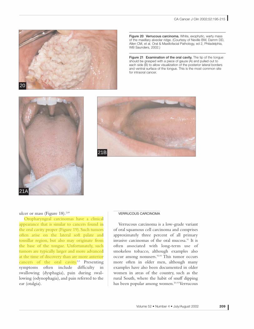
Figure 20 Verrucous carcinoma. White, exophytic, warty massof the maxillary alveolar ridge. (Courtesy of Neville BW, Damm DD,Allen CM, et al. Oral & Maxillofacial Pathology, ed 2, Philadelphia,WB Saunders, 2002.)
Figure 21 Examination of the oral cavity. The tip of the tongueshould be grasped with a piece of gauze (A) and pulled out toeach side (B) to allow visualization of the posterior lateral bordersand ventral surface of the tongue. This is the most common sitefor intraoral cancer.
20
21B
21A
carcinoma most commonly occurs on thebuccal mucosa, the mandibular or maxillaryvestibule, and the mandibular or maxillaryalveolar ridge/gingiva—often correspondingto the site of tobacco placement within themouth. The tumor presents as a diffuse,thickened plaque or mass with a warty orpapillary surface (Figure 20). The lesion isusually white, although some examples withless keratinization may appear pink. In tobaccousers, tobacco pouch keratosis may be seen onthe adjacent mucosal surfaces; examples innonusers of tobacco may arise from lesions ofso-called proliferative verrucous leukoplakia.59
Because verrucous carcinoma is slowgrowing, exophytic, and well differentiated, it isassociated with a much better prognosis thanconventional squamous cell carcinoma of themouth.20,92 Treatment usually consists ofsurgical excision without the need for neckdissection because metastasis is rare. However,local recurrences may develop and require re-excision. Also, lesions that arise fromproliferative verrucous leukoplakia may recurand undergo dedifferentiation into a moreaggressive conventional squamous cellcarcinoma.59
METASTASIS
Metastases from oral squamous cellcarcinomas most frequently develop in theipsilateral cervical lymph nodes. Tumors fromthe lower lip and floor of mouth may initiallyinvolve the submental nodes. Contralateral orbilateral cervical metastases also can occur,especially in tumors of the base of tongue, inadvanced tumors, and in tumors that occurnear the midline. Involved nodes usually areenlarged, firm, and nontender to palpation. Ifthe tumor has perforated the capsule of theinvolved node and invaded into thesurrounding connective tissue (extracapsular
210 CA A Cancer Journal for Clinicians
Oral Cancer and Precancerous Lesions
TNM Staging of Oral Cancer
Primary Tumor (T)
TX Primary tumor cannot be assessed
T0 No evidence of primary tumor
Tis Carcinoma in situ
T1 Tumor 2 cm or less in greatest dimension
T2 Tumor more than 2 cm but not more than 4 cm in greatestdimension
T3 Tumor more than 4 cm in greatest dimension
T4 Tumor invades adjacent structures (e.g., through cortical bone, intomaxillary sinus, skin, pterygoid muscle, deep muscle of tongue)
Nodal Involvement (N)
NX Regional lymph nodes cannot be assessed
N0 No regional lymph node metastasis
N1 Metastasis in a single ipsilateral lymph node, 3 cm or less in greatest dimension
N2 Metastasis in a single ipsilateral lymph node, more than 3 cm butnot more than 6 cm in greatest dimension; or in multiple ipsilaterallymph nodes, none more than 6 cm in greatest dimension; or inbilateral or contralateral lymph nodes, none more than 6 cm ingreatest dimension
N2a Metastasis in a single ipsilateral lymph node, more than 3 cm butnot more than 6 cm in greatest dimension
N2b Metastasis in multiple ipsilateral lymph nodes, none more than 6cm in greatest dimension
N2c Metastasis in bilateral or contralateral lymph nodes, none more than6 cm in greatest dimension
N3 Metastasis in a lymph node more than 6 cm in greatest dimension
Distant Metastasis (M)
MX Distant metastasis cannot be assessed
M0 No distant metastasis
M1 Distant metastasis
Stage Grouping
Stage 0 Tis N0 M0
Stage I T1 N0 M0
Stage II T2 N0 M0
Stage III T3 N0 M0; T1 or T2 or T3 N1 M0
Stage IV Any T4 lesion, or
Any N2 or N3 lesions, or
Any M1 lesion
TABLE 3
Modified from AJCC Manual for Staging of Cancer, 1997, Ed: Fleming ID, et al.Lippincott-Raven Publishers, Philadelphia, PA.
spread), the node will feel fixed andimmovable. As many as 30 percent of oralcancers have cervical metastases, either palpableor occult, at the time of initial evaluation.94 Inparticular, the tongue has a rich blood supplyand lymphatic drainage, which accounts for thefact that up to 66 percent of patients withprimary tongue lesions have neck disease at thetime of diagnosis.95 Distant metastases are mostcommon in the lungs, but any part of the bodymay be affected.
Staging
Staging of oral cancer is important forestablishing proper treatment and determiningprognosis. Tumors are staged using the TNMsystem, where T represents the size of theprimary tumor, N indicates the status of theregional lymph nodes, and M indicates thepresence or absence of distant metastases.Thissystem is outlined in Table 3.
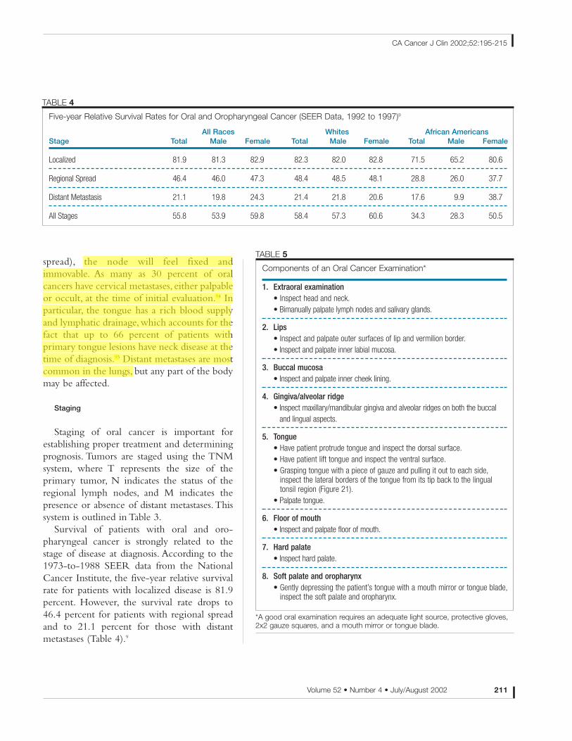
Survival of patients with oral and oro-pharyngeal cancer is strongly related to thestage of disease at diagnosis. According to the1973-to-1988 SEER data from the NationalCancer Institute, the five-year relative survivalrate for patients with localized disease is 81.9percent. However, the survival rate drops to46.4 percent for patients with regional spreadand to 21.1 percent for those with distantmetastases (Table 4).9
Volume 52 • Number 4 • July/August 2002 211
CA Cancer J Clin 2002;52:195-215
Five-year Relative Survival Rates for Oral and Oropharyngeal Cancer (SEER Data, 1992 to 1997)9
All Races Whites African AmericansStage Total Male Female Total Male Female Total Male Female
Localized 81.9 81.3 82.9 82.3 82.0 82.8 71.5 65.2 80.6
Regional Spread 46.4 46.0 47.3 48.4 48.5 48.1 28.8 26.0 37.7
Distant Metastasis 21.1 19.8 24.3 21.4 21.8 20.6 17.6 9.9 38.7
All Stages 55.8 53.9 59.8 58.4 57.3 60.6 34.3 28.3 50.5
TABLE 4
Components of an Oral Cancer Examination*
1. Extraoral examination• Inspect head and neck.• Bimanually palpate lymph nodes and salivary glands.
2. Lips• Inspect and palpate outer surfaces of lip and vermilion border.• Inspect and palpate inner labial mucosa.
3. Buccal mucosa• Inspect and palpate inner cheek lining.
4. Gingiva/alveolar ridge• Inspect maxillary/mandibular gingiva and alveolar ridges on both the buccal
and lingual aspects.
5. Tongue• Have patient protrude tongue and inspect the dorsal surface.• Have patient lift tongue and inspect the ventral surface.• Grasping tongue with a piece of gauze and pulling it out to each side,
inspect the lateral borders of the tongue from its tip back to the lingual tonsil region (Figure 21).
• Palpate tongue.
6. Floor of mouth• Inspect and palpate floor of mouth.
7. Hard palate• Inspect hard palate.
8. Soft palate and oropharynx• Gently depressing the patient’s tongue with a mouth mirror or tongue blade,
inspect the soft palate and oropharynx.
TABLE 5
*A good oral examination requires an adequate light source, protective gloves,2x2 gauze squares, and a mouth mirror or tongue blade.
DIAGNOSIS AND TREATMENT
Because most individuals are seen morecommonly by primary care physicians andgeneral dentists than by specialists, it isimportant for these clinicians to performscreening examinations to identify potentialoral and pharyngeal cancers. Table 5summarizes the recommended components ofan oral cancer examination (Figure 21).90
When a suspicious lesion is identified, aconventional biopsy using a scalpel or smallbiopsy forceps remains the best and mostaccurate means of assessing it. As stated byAlexander et al., “Noninvasive screeningtechniques such as cytologic testing (includingbrush biopsy)… have many pitfalls and shouldnot be considered as substitutes for biopsywhen there is concern about malignancy.”96
The biopsy can be obtained by the primarycaregiver or by referral to a head and neckspecialist (e.g., otolaryngologist/head and necksurgeon, oral and maxillofacial surgeon, etc.).
In addition to the need for improved earlydetection by clinicians, it is also important thatthe patient and general public areknowledgeable about the disease.43,97 Delays inidentification and recognition of suspiciouslesions contribute to advanced stage atdiagnosis and lower survival statistics.98-105
A complete, detailed discussion about themanagement of oral cancer and precancerouslesions is beyond the scope of this article.Generally speaking, it has been recommendedthat leukoplakias that show moderate epithelialdysplasia or worse be removed or destroyed ifpossible.2 The management of lesions showingmild dysplasia depends on the size, location,and apparent cause of the lesion. Sometimesearly dysplastic lesions may be reversible if thesource of irritation (e.g., smoking) can beeliminated. Molecular markers, such as DNAcontent and loss of heterozygosity, hold thepromise of becoming important tools forpredicting the risk of malignant transformationfor oral leukoplakias.106-108
The patient with invasive oral cancer is bestmanaged by a coordinated, multidisciplinaryteam of health care professionals, which mayinclude a head and neck surgeon, oral andmaxillofacial pathologist, general pathologist,radiation oncologist, neuroradiologist, recon-structive surgeon, medical oncologist, generaldentist, oral and maxillofacial surgeon, maxil-lofacial prosthodontist, dental hygienist, nursespecialist, speech pathologist, nutritionist, andtobacco cessation counselor.109
Up to 15 percent of individuals with oralcancer have been identified to harbor a secondprimary cancer; therefore, it is important that acomplete head and neck examination,including the larynx, is performed.110 Manyclinicians perform an endoscopic examinationto include the larynx, esophagus, trachea, andlungs in order to identify other potentiallesions in the high-risk patient. For patientswho present with a neck mass but no obviousprimary site (or if the neck mass is moreamenable to biopsy than the primary tumor), afine needle aspiration remains the diagnosticmethod of choice rather than an open biopsy,because open biopsy has been reported to berelated to a lower survival rate when notaccompanied by a simultaneous neckdissection.111,112
Imaging studies are now routinelyperformed to evaluate the primary tumor and neck disease. Both contrast-enhancedcomputed tomographic (CT) scans andmagnetic resonance imaging (MRI) may beutilized in determining the extent of theprimary tumor, invasion, regional lymph node status, and distant metastatic disease,thereby providing important staging infor-mation.113,114 Positron emission tomography(PET) scans are also becoming an increasinglypopular tool for the identification of primary,recurrent, and metastatic disease.
Treatment options are variable and depend on the size and location of the primary tumor,lymph node status, presence or absence of distantmetastases, the patient’s ability to tolerate
212 CA A Cancer Journal for Clinicians
Oral Cancer and Precancerous Lesions
treatment, and the patient’s desires. Surgeryand/or radiation therapy remain the goldstandards for treatment of cancers of the lip andoral cavity.Oropharyngeal cancer may be treatedwith surgery and/or radiation therapy for early-stage disease. For advanced-stage disease, surgerywith adjuvant radiation therapy may beindicated, whereas recent evidence suggests thatthe addition of chemotherapy to radiationtherapy may provide a survival advantage overradiation therapy alone in this population.115,116 Itis important to take into account disease statusand prevalence of occult disease in the neckwhen evaluating primary cancers of the lip, oralcavity, and oropharynx.117 Regardless of thetreatment modality used, many patients willrequire consideration of problems related toairway protection, enteral feedings, xerostomia,mucositis, dysphagia, and voice change.
CONCLUSIONS
The ability to control oral and oro-pharyngeal cancer will depend on twocornerstones: prevention and early diagnosis.Continuing educational campaigns are neededon the local, state, and national level in order toeducate the public about the risk factors andearly signs/symptoms associated with thisdisease. Individuals also need to be encouragedto seek regular professional oral examinationsby a dentist and/or physician. Finally, healthcare workers must be encouraged to performoral cancer examinations as part of their patientcare regime, and to be knowledgeable aboutearly signs of oral carcinoma.118,119 CA
Volume 52 • Number 4 • July/August 2002 213
CA Cancer J Clin 2002;52:195-215
REFERENCES
1.American Cancer Society, Cancer facts and fig-ures 2002.Atlanta, GA:American Cancer Society;2002.
2. Neville BW, Damm DD,Allen CM, et al. Oral& maxillofacial pathology. 2nd ed. Phila., PA:Saunders; 2002;337-369.
3. Silverman S Jr. Demographics and occurrenceof oral and pharyngeal cancers. The outcomes,the trends, the challenge. J Am Dent Assoc2001;132:7S-11S.
4. Silverman S Jr. Epidemiology. In: Silverman SJr ed. Oral Cancer. 4th ed. Hamilton, Ontario,Canada: BC Decker Inc;1998;1-6.
5. Chen JK, Katz RV, Krutchkoff DJ. Intraoralsquamous cell carcinoma. Epidemiologic patternsin Connecticut from 1935 to 1985. Cancer1990;66:1288-1296.
6. Llewellyn CD, Johnson NW, WarnakulasuriyaKA. Risk factors for squamous cell carcinoma ofthe oral cavity in young people—a comprehen-sive literature review. Oral Oncol 2001;37:401-418.
7. Schantz SP,Yu GP. Head and neck cancer inci-dence trends in young Americans, 1973-1997,with a special analysis for tongue cancer. ArchOtolaryngol Head Neck Surg 2002;128:268-274.
8. Swango PA. Cancers of the oral cavity andpharynx in the United States: An epidemiologicoverview. J Public Health Dent 1996;56:309-318.
9. Ries LAG, Hankey BF, Miller BA, et al. Cancer
Statistics Review 1973-1988. National CancerInstitute, NIH Publication No. 91-2789, 1991.
10. Silverman S Jr, Shillitoe EF. Etiology andPredisposing Factors. In: Silverman S Jr ed. OralCancer, 4th ed. Hamilton, Ontario, Canada: BCDecker Inc;1998, 7-24.
11. Goldberg HI, Lockwood SA,Wyatt SW, et al.Trends and differentials in mortality from cancersof the oral cavity and pharynx in the UnitedStates, 1973-1987. Cancer 1994;74:565-572.
12. Caplan DJ, Hertz-Picciotto I. Racial differ-ences in survival of oral and pharyngeal cancerpatients in North Carolina. J Public Health Dent1998;58:36-43.
13. Mashberg A, Boffetta P,Winkelman R, et al.Tobacco smoking, alcohol drinking, and cancerof the oral cavity and oropharynx among U.S.veterans. Cancer 1993;72:1369-1375.
14. Jovanovic A, Schulten EA, Kostense PJ, et al.Tobacco and alcohol related to the anatomicalsite of oral squamous cell carcinoma. J OralPathol Med 1993;22:459-462.
15.Andre K, Schraub S, Mercier M, et al. Role ofalcohol and tobacco in the aetiology of head andneck cancer: A case-control study in the Doubsregion of France. Oral Oncol, Eur J Cancer1995;31B:301-309.
16. Blot WJ, McLaughlin JK, Winn DM, et al.Smoking and drinking in relation to oral andpharyngeal cancer. Cancer Res 1988;48:3282-3287.
17. Lewin F, Norell SE, Johansson H, et al.Smoking tobacco, oral snuff, and alcohol in the
etiology of squamous cell carcinoma of the headand neck.A population-based case-referent studyin Sweden. Cancer 1998;82:1367-1375.
18. Silverman S Jr, Griffith M. Smoking charac-teristics of patients with oral carcinoma and therisk for second oral primary carcinoma. J AmDent Assoc 1972;85:637-640.
19. Zhang ZF, Morgenstern H, Spitz MR, et al.Marijuana use and increased risk of squamous cellcarcinoma of the head and neck. CancerEpidemiol Biomarkers Prev 1999;8:1071-1078.
20. Brown RL, Suh JM, Scarborough JE, et al.Snuff dippers’ intraoral cancer: Clinical character-istics and response to therapy. Cancer 1965;18:2-13.
21.Winn DM, Blot WJ, Shy CM, et al. Snuff dip-ping and oral cancer among women in the south-ern United States. N Engl J Med 1981;304:745-749.
22. Bouquot JE, Meckstroth RL. Oral cancer in atobacco-chewing U.S. population – no apparentincreased incidence or mortality. Oral Surg OralMed Oral Pathol Oral Radiol Endod 1998;86:697-706.
23. Johnson N. Tobacco use and oral cancer: Aglobal perspective. J Dent Educ 2001;65:328-339.
24. Pindborg JJ, Murti PR, Bhonsle RB, et al.Oral submucous fibrosis as a precancerous condi-tion. Scand J Dent Res 1984;92:224-229.
25. Murti PR, Bhonsle RB, Pindborg JJ, et al.Malignant transformation rate in oral submucousfibrosis over a 17-year period. Community Dent
214 CA A Cancer Journal for Clinicians
Oral Cancer and Precancerous Lesions
Oral Epidemiol 1985;13:340-341.
26. Murti PR, Bhonsle RB, Gupta PC, et al.Etiology of oral submucous fibrosis with specialreference to the role of areca nut chewing. J OralPathol Med 1995;24:145-152.
27. Miller CS,White DK. Human papillomavirusexpression in oral mucosa, premalignant condi-tions, and squamous cell carcinoma. A retrospec-tive review of the literature. Oral Surg Oral MedOral Pathol Oral Radiol Endod 1996;82:57-68.
28. Sugerman PB, Shillitoe EJ. The high riskhuman papillomaviruses and oral cancer:Evidence for and against a causal relationship.Oral Dis 1997;3:130-147.
29. Lindel K, Beer KT, Laissue J, et al. Humanpapillomavirus positive squamous cell carcinomaof the oropharynx: A radiosensitive subgroup ofhead and neck carcinoma. Cancer 2001;92:805-813.
30. Gillison ML, Shah KV. Human papillo-mavirus-associated head and neck squamous cellcarcinoma: Mounting evidence for an etiologicrole for human papillomavirus in a subset of headand neck cancers. Curr Opin Oncol2001;13:183-188.
31. Mork J, Lie AK, Glattre E, et al. Human papil-lomavirus infection as a risk factor for squamous-cell carcinoma of the head and neck. N Engl JMed 2001;344:1125-1131.
32.Winn DM, Ziegler RG, Pickle LW, et al. Dietin the etiology of oral and pharyngeal canceramong women from the southern United States.Cancer Res 1984;44:1216-1222.
33.Winn DM. Diet and nutrition in the etiologyof oral cancer. Am J Clin Nutr 1995;61:437S-445S.
34. Silverman S Jr,Gorsky M,Lozada-Nur F, et al.A prospective study of findings and managementin 214 patients with oral lichen planus. Oral SurgOral Med Oral Pathol 1991;72:665-670.
35. Barnard NA, Scully C, Eveson JW, et al. Oralcancer development in patients with oral lichenplanus. J Oral Pathol Med 1993;22:421-424.
36. Eisenberg E. Oral lichen planus: A benignlesion. J Oral Maxillofac Surg 2000;58:1278-1285.
37. Watts JM. The importance of the Plummer-Vinson syndrome in the etiology of carcinoma ofthe upper gastrointestinal tract. Postgrad Med J1961;37:523-533.
38. Larsson LG, Sandström A, Westling P.Relationship of Plummer-Vinson disease to can-cer of the upper alimentary tract in Sweden.Cancer Res 1975;35:3308-3316.
39. de Visscher JG, Bouwes Bavinck JN, van derWaal I. Squamous cell carcinoma of the lower lipin renal-transplant recipients. Report of six cases.Int J Oral Maxillofac Surg 1997;26:120-123.
40. van Zuuren EJ, de Visscher JGAM, BouwesBavinck JN.Carcinoma of the lip in kidney trans-plant recipients. J Amer Acad Dermatol1988;38:497-499.
41. Flaitz CM, Nichols CM, Adler-Storthz K, etal. Intraoral squamous cell carcinoma in humanimmunodeficiency syndrome virus infection. A
clinicopathologic study. Oral Surg Oral Med OralPathol Oral Radiol Endod 1995;80:55-62.
42. Flaitz CM, Silverman S Jr. HumanImmunodeficiency Virus (HIV)-AssociatedMalignancies. In Silverman S Jr (ed). OralCancer, 4th ed. Hamilton, Ontario, Canada: BCDecker, Inc; 1998:165-170.
43.Yellowitz JA, Goodman HS. Assessing physi-cians’ and dentists’ oral cancer knowledge, opin-ions and practices. J Am Dent Assoc 1995;126:53-59.
44.Arvidson-Bufano UB, Blank LW,Yellowitz JA.Nurses’ oral health assessments of nursing homeresidents pre- and post-training: A pilot study.Spec Care Dentist 1996;16:58-64.
45. Blank LW,Arvidson-Bufano UB,Yellowitz JA.The effect of nurses’ background on performanceof nursing home resident oral health assessmentspre- and post-training. Spec Care Dentist1996;16:65-70.
46.Yellowitz J,Horowitz AM,Goodman HS, et al.Knowledge, opinions and practices of generaldentists regarding oral cancer:A pilot survey. J AmDent Assoc 1998;129:579-583.
47.Yellowitz JA, Horowitz AM, Drury TF, et al.Survey of U.S. dentists’ knowledge and opinionsabout oral pharyngeal cancer. J Am Dent Assoc2000;131:653-661.
48. Horowitz AM, Drury TF, Goodman HS, et al.Oral pharyngeal cancer prevention and earlydetection. Dentists’ opinions and practices. J AmDent Assoc 2000;131:453-462.
49. Horowitz AM, Siriphant P, Sheikh A, et al.Perspectives of Maryland dentists on oral cancer.J Am Dent Assoc 2001;131:65-72.
50. Smith RA, Cokkinides V, von EschenbachAC, et al.American Cancer Society guidelines forthe early detection of cancer. CA Cancer J Clin2002;52:8-22.
51. United States Preventive Services Task Force(USPSTF). In: Guide to clinical preventive serv-ices: Report of the United States PreventiveServices Task Force 2d ed. Baltimore, MD:Williams & Wilkins, 1996. Available at:http://www.ahcpr.gov/clinic/cpsix.htm.Accessed May 17, 2002.
52. Schwimmer E. Die idiopathischen Schleim-hautplaques der Mundhöhle (Leukoplakia buc-calis).Arch Dermat Syph 1877;9:570-611.
53. Kramer IR, Lucas RB, Pindborg, JJ, et al.WHO Collaborating Centre for OralPrecancerous Lesions. Definition of leukoplakiaand related lesions:An aid to studies on oral pre-cancer. Oral Surg Oral Med Oral Pathol1978;46:518-539.
54. Shafer WB, Waldron CA. A clinical andhistopathologic study of oral leukoplakia. SurgGynecol Obstet 1961;112:411-420.
55. Axéll T, Pindborg JJ, Smith CJ, et al. Oralwhite lesions with special reference to precancer-ous and tobacco-related lesions: Conclusions ofan international symposium held in Uppsala,Sweden, May 18-21 1994. InternationalCollaborative Group on Oral White Lesions JOral Pathol Med 1996;25:49-54.
56. Bouquot JE, Gorlin RJ. Leukoplakia, lichenplanus, and other oral keratoses in 23,616 whiteAmericans over the age of 35 years. Oral SurgOral Med Oral Pathol 1986;61:373-381.
57.Waldron CA, Shafer WG. Leukoplakia revisit-ed:A clinicopathologic study of 3256 oral leuko-plakias. Cancer 1975;36:1386-1392.
58. Bouquot JE,Whitaker SB. Oral leukoplakia—Rationale for diagnosis and prognosis of its clini-cal subtypes or “phases.” Quintessence Int1994:25:133-140.
59. Hansen LS, Olson JA, Silverman S Jr.Proliferative verrucous leukoplakia. A long-termstudy of thirty patients. Oral Surg Oral Med OralPathol 1985;60:285-298.
60. Kahn MA, Dockter ME, Hermann-PetrinJM. Proliferative verrucous leukoplakia. Fourcases with flow cytometric analysis. Oral SurgOral Med Oral Pathol 1994;78:469-475.
61. Zakrzewska JM, Lopes V, Speight P, et al.Proliferative verrucous leukoplakia: A report often cases. Oral Surg Oral Med Oral Pathol OralRadiol Endod 1996;82:396-401.
62. Silverman S Jr, Gorsky M. Proliferative verru-cous leukoplakia. A follow-up study of 54 cases.Oral Surg Oral Med Oral Pathol Oral RadiolEndod 1997;84:154-157.
63. Fettig A, Pogrel MA, Silverman S Jr, et al.Proliferative verrucous leukoplakia of the gingi-va. Oral Surg Oral Med Oral Pathol Oral RadiolEndod 2000;90:723-730.
64. Damm DD, Curran A, White DK, et al.Leukoplakia of the maxillary vestibule—an asso-ciation with Viadent? Oral Surg Oral Med OralPathol Oral Radiol Endod 1999;87:61-66.
65. Eversole LR, Eversole GM, Kopcik J.Sanguinaria-associated oral leukoplakia: Com-parison with other benign and dysplastic leuko-plakic lesions. Oral Surg Oral Med Oral PatholOral Radiol Endod 2000;89:455-464.
66. Mascarenhas AK, Allen CM, Loudon J. Theassociation between Viadent use and oral leuko-plakia. Epidemiology 2001;12:741-743.
67. Pindborg JJ, Renstrup G, Poulsen HE, et al.Studies in oral leukoplakia.V. Clinical and histo-logic signs of malignancy. Acta Odont Scand1963;21:407-414.
68. Bánóczy J, Csiba A. Occurrence of epithelialdysplasia in oral leukoplakia.Analysis and follow-up study of 12 cases. Oral Surg Oral Med OralPathol 1976;42:766-774.
69. Feller L, Altini M, Slabbert H. Pre-malignantlesions of the oral mucosa in a South Africansample: A clinicopathologic study. J Dent AssocSouth Africa 1991;46:261-265.
70. Silverman S Jr. Observations on the clinicalcharacteristics and natural history of oral leuko-plakia. J Am Dent Assoc 1968;76:772-777.
71. Einhorn J, Wersäll J. Incidence of oral carci-noma in patients with leukoplakia of the oralmucosa. Cancer 1967;20:2189-2193.
72. Bánóczy J. Follow-up studies in oral leuko-plakia. J Maxillofac Surg 1977;5:69-75.
73. Pindborg JJ, Jφlst O, Renstrup G, et al. Studieson oral leukoplakia: A preliminary report on the
Volume 52 • Number 4 • July/August 2002 215
CA Cancer J Clin 2002;52:195-215
period prevalence of malignant transformation inleukoplakia based on a follow-up study of 248patients. J Am Dent Assoc 1968;76:767-771.
74. Kramer IRH. Precancerous conditions of theoral mucosa: A computer-aided study. Ann RColl Surg Eng 1969;45:340-356.
75. Roed-Petersen B. Cancer development inoral leukoplakia: Follow-up of 331 patients. JDent Res 1971;50:711.
76. Silverman S Jr, Gorsky M, Lozada F. Oralleukoplakia and malignant transformation: A fol-low-up study of 257 patients. Cancer1984;53:563-568.
77. Lind PO. Malignant transformation in oralleukoplakia. Scand J Dent Res 1987;95:449-455.
78. Queyrat L. Erythroplasie de gland. Bull SocFranc Derm Syph 1911;22:378-382.
79. Shafer WG,Waldron CA. Erythroplakia of theoral cavity. Cancer 1975;36:1021-1028.
80. Mashberg A, Samit A. Early diagnosis ofasymptomatic oral and oropharyngeal squamouscancers. CA Cancer J Clin 1995;45:328-351.
81. Rossie KM, Guggenheimer J. Thermallyinduced “nicotine” stomatitis.A case report. OralSurg Oral Med Oral Pathol 1990;70:597-599.
82. Pindborg JJ, Mehta FS, Gupta PC, et al.Reverse smoking in Andhra Pradesh, India: Astudy of palatal lesions among 10,169 villagers. BrJ Cancer 1971;25:10-20.
83. Ortiz GM, Pierce AM, Wilson DF. Palatalchanges associated with reverse smoking inFilipino women. Oral Dis 1996;2:232-237.
84. Smith JF, Mincer HA, Hopkins KP, et al.Snuff-dipper’s lesion.A cytological and patholog-ical study in a large population.Arch Otolaryngol1970;92:450-456.
85. Greer RO Jr, Poulson TC. Oral tissue alter-ations associated with the use of smokeless tobac-co by teen-agers. Part I. Clinical findings. OralSurg Oral Med Oral Pathol 1983;56:275-284.
86. Grady D, Greene J, Daniels TE, et al. Oralmucosal lesions found in smokeless tobacco users.J Am Dent Assoc 1990;121:117-123.
87. Kaugars GE, Riley WT, Brandt RB, et al.Theprevalence of oral lesions in smokeless tobaccousers and an evaluation of risk factors. Cancer1992;70:2579-2585.
88. Martin GC, Brown JP, Eifler CW, et al. Oralleukoplakia status six weeks after cessation ofsmokeless tobacco use. J Am Dent Assoc1999;130:945-954.
89. Silverman S Jr, Dillon WP, Fischbein NJ.Diagnosis In: Silverman S Jr ed. Oral Cancer. 4thed. Hamilton, Ontario, Canada: BC Decker Inc;1998;41-66.
90. National Institute of Dental and CraniofacialResearch. Perform a death-defying act. The 90-second oral cancer examination. J Am Dent Assoc2001;132:36S-40S.
91. Bouquot JE. Oral verrucous carcinoma.Incidence in two US populations. Oral Surg OralMed Oral Pathol Oral Radiol Endod 1998;86:318-324.
92.Ackerman LV.Verrucous carcinoma of the oralcavity. Surgery 1948;23:670-678.
93. McCoy JM,Waldron CA.Verrucous carcino-ma of the oral cavity.A review of forty-nine cases.Oral Surg Oral Med Oral Pathol 1981;52:623-629.
94. Shah JP, Candela FC, Poddar AK.The patternsof cervical lymph node metastasis from squamouscarcinoma of the oral cavity. Cancer1990;66:109-113.
95. Ho CM, Lam KH, Wei WI, et al. Occultlymph node metastasis in small oral tongue can-cers. Head Neck 1992;14:359-363.
96. Alexander RE, Wright JM, Thiebaud S.Evaluating, documenting and following up oralpathological conditions. A suggested protocol. JAm Dent Assoc 2001;132:329-335.
97. Centers for Disease Control and Prevention.Preventing and controlling oral and pharyngealcancer. Recommendations from a national strate-gic planning conference. MMWR 1998;47RR-14.
98. Shafer WG. Initial mismanagement and delayin diagnosis of oral cancer. J Am Dent Assoc1975;90:1262-1264.
99. Elwood JM, Gallagher RP. Factors influencingearly diagnosis of cancers of the oral cavity. CanMed Assoc J 1985;133:651-656.
100. Guggenheimer J,Verbin RS, Johnson JT, etal. Factors delaying the diagnosis of oral andoropharyngeal carcinomas. Cancer 1989;64:932-935.
101. Prout MN, Hereen TC, Barber CE, et al. Useof health services before diagnosis of head andneck cancer among Boston residents. Am J PrevMed 1990;6:77-83.
102. Schnetler JF. Oral cancer diagnosis and delaysin referral. Br J Oral Maxillofac Surg1992;30:210-213.
103. Smart CR. Screening for cancer of theaerodigestive tract. Cancer 1993;72:1061-1065.
104. Carpenter RD,Yellowitz JA, Goodman HS.Oral cancer mortality in Maryland. MarylandMed J 1993;42:1105-1109.
105. Hollows P, McAndrew PG, Perini MG.Delays in the referral and treatment of oral squa-mous cell carcinoma. Br Dent J 2000;188:262-265.
106. Rosin MP, Cheng X, Poh C, et al. Use ofallelic loss to predict malignant risk for low-gradeoral epithelial dysplasia. Clin Cancer Res2000;6:357-362.
107. Sudbø J, Kildal W, Risberg B, et al. DNAcontent as a prognostic marker in patients withoral leukoplakia. N Engl J Med 2001;344:1270-1278.
108. Lippman SM, Hong WK. Molecular markersof the risk of oral cancer. N Engl J Med2001;34:1323-1326.
109. Ord RA, Blanchaert RH Jr. Current man-agement of oral cancer. A multidisciplinaryapproach. J Am Dent Assoc 2001;132:19S-23S.
110. Lippman SM, Hong WK. Second primary
tumors in head and neck squamous cell carcino-ma: The overshadowing threat for patients withearly-stage disease. Int J Radiat Oncol Biol Phys1989;17:691-694.
111. Lefebvre JL, Coche-Dequeant B,Van JT, etal. Cervical lymph nodes from an unknown pri-mary tumor in 190 patients. Am J Surg1990;160:443-446.
112. Kleid S, Millar HS. The case against openneck biopsy.Aust N Z J Surg 1993;63:678-681.
113. Som PM, Curtin HD, Mancuso AA. Animaging-based classification for the cervicalnodes designed as an adjunct to recent clinicallybased nodal classifications. Arch OtolaryngolHead Neck Surg 1999;125:388-396.
114. Robbins KT. Integrating radiological criteriainto the classification of cervical lymph node dis-ease. Arch Otolaryngol Head Neck Surg1999;125:385-387.
115. Forastiere A, Goepfert H, Goffinet D, et al.NCCN practice guidelines for head and neckcancer. National Comprehensive CancerNetwork Proceedings. Oncology 1998;12:39-247.
116. Calais G, Alfonsi M, Bardet E, et al.Randomized trial of radiation therapy versusconcomitant chemotherapy and radiation therapyfor advanced-stage oropharynx carcinoma. J NatlCancer Inst 1999;91:2081-2086.
117. Robbins KT, Atkinson JL, Byers RM, et al.The use and misuse of neck dissection for headand neck cancer. J Am Coll Surg 2001;193:91-102.
118. Horowitz AM, Goodman HS,Yellowitz JA,et al.The need for health promotion in oral can-cer prevention and early detection. J PublicHealth Dent 1996;56:319-330.
119. Goodman HS,Yellowitz JA, Horowitz AM.Oral cancer prevention.The role of family prac-titioners.Arch Fam Med 1995;4:628-636.


![[09-10] (IPD) Kelainan Rongga Mulut Dan Esofagus](https://img.pdfslide.us/doc/110x75/577cd7881a28ab9e789f3716/09-10-ipd-kelainan-rongga-mulut-dan-esofagus.jpg)