Embed Size (px)
Citation preview
Contents lists available at ScienceDirect
Journal of the Mechanical Behavior of Biomedical Materials
journal homepage: www.elsevier.com/locate/jmbbm
Tribocorrosion behavior of bio-functionalized highly porous titanium
F. Toptana,b,⁎, A.C. Alvesa, A.M.P. Pintoa,b, P. Ponthiauxc
a CMEMS-UMinho – Center for MicroElectroMechanical Systems – Universidade do Minho, Azurém, 4800-058 Guimarães, Portugalb Universidade do Minho, Dept. Eng. Mecânica, Azurém, 4800-058 Guimarães, Portugalc Laboratoire de Génie des Procédés et Matériaux-LGPM, École Centrale de Paris, Grande Voie des Vignes, 92290 Chatenây-Malabry, France
A R T I C L E I N F O
Keywords:Anodic treatmentPowder metallurgyPorous TiTribocorrosion
A B S T R A C T
Titanium and its alloys are widely used in orthopedic and dental implants, however, some major clinicalconcerns such as poor wear resistance, lack of bioactivity, and bone resorption due to stress shielding are yet tobe overcome. In order to improve these drawbacks, highly porous Ti samples having functionalized surfaceswere developed by powder metallurgy with space holder technique followed by anodic treatment.Tribocorrosion tests were performed in 9 g/L NaCl solution using a unidirectional pin-on-disc tribometerunder 3 N normal load, 1 Hz frequency and 4 mm track diameter. Open circuit potential (OCP) was measuredbefore, during and after sliding. Worn surfaces investigated by field emission gun scanning electron microscope(FEG-SEM) equipped with energy dispersive X-ray spectroscopy (EDS). Results suggested bio-functionalizedhighly porous samples presented lower tendency to corrosion under sliding against zirconia pin, mainly due tothe load carrying effect given by the hard protruded oxide surfaces formed by the anodic treatment.
1. Introduction
Titanium and its alloys are widely used in orthopedic and dentalimplants due to their excellent corrosion resistance, high yieldstrength, good ductility and better biocompatibility (Chen et al.,2009; Guo et al., 2013). However, some major clinical concerns arestill valid, namely bone resorption due to stress shielding, lack ofbioactivity, and low wear resistance.
Most commercially used Ti-based implant materials exhibit muchhigher Young's modulus (Commercially pure Ti: 105 GPa, Ti-6Al-4V:110 GPa) as compared to human bones (varies in a range of 4–30 GPa)(Guo et al., 2013; De Viteri and Fuentes, 2013; Nag and Banerjee,2012). Studies have shown that implants do not adequately strain thebone that can result in disuse atrophy and bone resorption due to thedifference on Young's modulus between implant and bone. Thisphenomenon known as stress shielding that is one of the major causeof orthopedic implant failures by resulting in bone resorption (Leeet al., 2012).
In order to avoid stress shielding, on one hand, β and near β alloyswith lower Young's modulus such as Ti-Nb-Ta-Zr, Ti-Nb-Si, Ti-Mo-Nb,Ti-Zr-Mo are being developed (Guo et al., 2013; Martins and Grandini,2014; Correa et al., 2014), but on the other hand, porous Ti implantshaving open-cellular structure are also being developed not just toreduce the biomechanical mismatch, but as well for the possibilities ofthe new-bone tissue in-growth, or even the transport of the body fluids
and their potential use on drug delivery systems (Goriainov et al., 2014;Amin Yavari et al., 2014; Lee et al., 2014; Jha et al., 2013).
Ti is the most biocompatible metallic material, however, it cannotcreate a direct bond with bone to promote new bone formation at theearly stage after implantation, which is required for the rapid fixationof bone to implant. This leads to the early implant failure particularlyfor the patient groups with the diseases such as diabetes, osteoporosis,and chronic inflammation (Gosavi et al., 2013; Tanigawa et al., 2013;Hu et al., 2011).
When an implant material attached to the bone, during the cyclicloads, relative movements cause wear (Thomann and Uggowitzer,2000; Ganesh et al., 2012). Furthermore, implants are surrounded bycorrosive body fluids, thus they suffer not only wear, but the simulta-neous action of corrosion and wear that is defined as tribocorrosion,being an irreversible process that occurs on the surface causing thedeterioration of the material (Marino and Mascaro, 2004; Landoltet al., 2004). It is known that under this simultaneous action, totalmaterial loss may be significantly higher than that of the mechanicalwear or corrosion, individually (Mischler, 2008).
In addition to hard coatings, surface modifications are also beingapplied in order to improve the wear resistance of Ti (Hu and Lim,2010; Wood, 2007; Alves et al., 2013). Among these surface modifica-tion techniques, anodic treatment not only leads to better corrosionand tribocorrosion resistance through the formation of porous titaniumoxide layers, but also improves the interaction of the implant surface
http://dx.doi.org/10.1016/j.jmbbm.2017.01.006Received 21 July 2016; Received in revised form 14 December 2016; Accepted 3 January 2017
⁎ Corresponding author at: Universidade do Minho, Dept. Eng. Mecânica, Azurém, 4800-058 Guimarães, Portugal.E-mail address: [email protected] (F. Toptan).
Journal of the mechanical behavior of biomedical materials 69 (2017) 144–152
Available online 04 January 20171751-6161/ © 2017 Elsevier Ltd. All rights reserved.
MARK
with host tissue by tailoring Ti surfaces in terms of topography,porosity, and composition leading to a better osteointegration (Alveset al., 2013; Ishizawa and Ogino, 1995; Szesz et al., 2013; Teixeiraet al., 2015; Fazel et al., 2015). The technique also allows toincorporate Ca and P with a similar Ca/P ratio to hydroxyapatite(HA) which afterwards can be precipitated as crystals by hydrothermaltreatment that leads to increase the bioactivity (Benea et al., 2014).There are some studies in the literature showing that it is also possibleto modify highly porous Ti surfaces by anodic treatment (Amin Yavariet al., 2014; Teixeira et al., 2015; Fan et al., 2011).
Most of the studies on highly porous Ti were focused on themechanical behavior, while there is very limited information on thecorrosion or wear behavior. Electrochemical studies showed thatporosity can alter the corrosion behavior of materials either bylocalized corrosion due to the trapping of electrolyte species and theexhaustion of oxygen in the small, isolated pores, or by the differencesbetween the nature and the structure of the oxide film formed on thetop surfaces and on the inner pore surfaces (Seah and Chen, 1993;Seah et al., 1998; Li et al., 2002; Menini et al., 2006; Xie et al., 2013;Alves et al., 2016).
The limited number of studies performed on the wear behavior ofporous metals showed that porosity can have positive or negativeeffects on the wear behavior of metallic materials. First of all, poresresult with decreased real contact area between the sliding surfacesthus increase the contact pressure on the metallic surface where highereffective stress and higher possibility of crack nucleation and propaga-tion can increase the material degradation (Mondal et al., 2009; Hamidet al., 2008; Jha et al., 2011). However, during sliding, depending ontheir fraction and size, the pores can be filled up by the wear debris,furthermore, the debris can be compacted in the pores under theinfluence of the applied load. In this case, the real contact area will beincreased, consequently, the contact pressure will be decreased thatmay result in increased wear resistance (Mondal et al., 2009; Jha et al.,2011). On the other hand, under lubricated conditions such as bodyconditions where body fluids can act as lubricant, pores can act asreservoirs for the body fluids and as a result, may lower the coefficientof friction and increase the wear resistance (Salahinejad et al., 2010).
Although these first attempts made valuable insights to the corro-sion and wear behavior of the highly porous metals, it is clear thatfurther studies are needed in order to have a better understanding ontheir degradation mechanisms. Moreover, the combined action ofcorrosion and wear, and their synergistic interactions are also neededto be evaluated. However, to the best of our knowledge, there is nostudy available in the literature on the tribocorrosion behavior of thesematerials. Thus, the present study aimed to be the first insight to thetribocorrosion behavior of as-processed and anodized Ti having closedand open pores.
2. Materials and methods
2.1. Processing
Highly porous cylindrical Ti samples 12 mm in diameter and 3 mmin height were processed by powder metallurgy with space holdertechnique using angular shaped Ti powders having average size of25 μm (Grade 2, Alfa Aesar), angular shaped urea space holderparticles under 500 μm (Scharlau), and PVA as binder (SigmaAldrich Chemistry).
Titanium, urea (30 and 50 vol%) and PVA (0.4 vol%) were mixedusing a ball mill rotating with alumina balls at 130 rpm during 4 h.Powder blends were uniaxially pressed in a zinc stearate lubricatednitrided stainless steel die under 350 MPa for 2 min. Green compactswere pre-heated under argon atmosphere at 450 °C during 3 h forbinder and space holder removal. Afterwards, the samples were heatedup to 1100 °C with 5 °C/min heating rate and sintered during 3 h inhorizontal tubular furnace under high vacuum ( < 10−5 mbar). The
binder/space holder removal and sintering temperatures were chosenafter differential thermal analysis and thermal gravimetric analysis(DTA/TG), together with dilatometric analysis that had been presentedelsewhere (Alves et al., 2016).
2.2. Anodic treatment
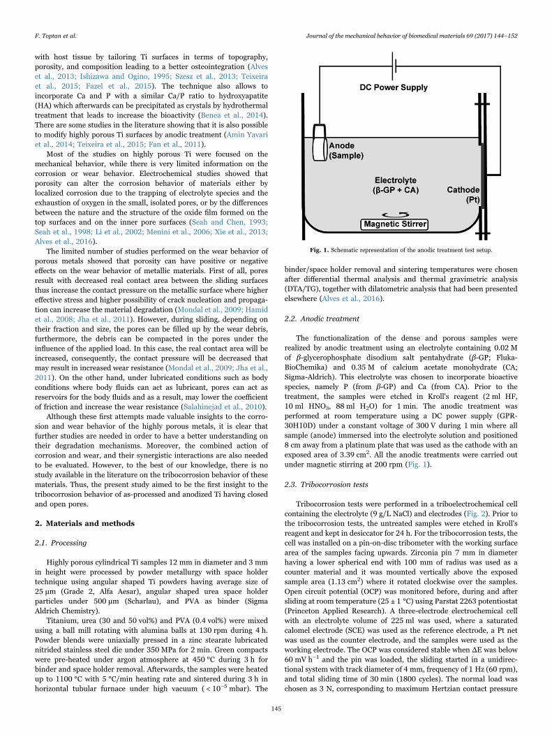
The functionalization of the dense and porous samples wererealized by anodic treatment using an electrolyte containing 0.02 Mof β-glycerophosphate disodium salt pentahydrate (β-GP; Fluka-BioChemika) and 0.35 M of calcium acetate monohydrate (CA;Sigma-Aldrich). This electrolyte was chosen to incorporate bioactivespecies, namely P (from β-GP) and Ca (from CA). Prior to thetreatment, the samples were etched in Kroll's reagent (2 ml HF,10 ml HNO3, 88 ml H2O) for 1 min. The anodic treatment wasperformed at room temperature using a DC power supply (GPR-30H10D) under a constant voltage of 300 V during 1 min where allsample (anode) immersed into the electrolyte solution and positioned8 cm away from a platinum plate that was used as the cathode with anexposed area of 3.39 cm2. All the anodic treatments were carried outunder magnetic stirring at 200 rpm (Fig. 1).
2.3. Tribocorrosion tests
Tribocorrosion tests were performed in a triboelectrochemical cellcontaining the electrolyte (9 g/L NaCl) and electrodes (Fig. 2). Prior tothe tribocorrosion tests, the untreated samples were etched in Kroll'sreagent and kept in desiccator for 24 h. For the tribocorrosion tests, thecell was installed on a pin-on-disc tribometer with the working surfacearea of the samples facing upwards. Zirconia pin 7 mm in diameterhaving a lower spherical end with 100 mm of radius was used as acounter material and it was mounted vertically above the exposedsample area (1.13 cm2) where it rotated clockwise over the samples.Open circuit potential (OCP) was monitored before, during and aftersliding at room temperature (25 ± 1 °C) using Parstat 2263 potentiostat(Princeton Applied Research). A three-electrode electrochemical cellwith an electrolyte volume of 225 ml was used, where a saturatedcalomel electrode (SCE) was used as the reference electrode, a Pt netwas used as the counter electrode, and the samples were used as theworking electrode. The OCP was considered stable when ΔE was below60 mV h−1 and the pin was loaded, the sliding started in a unidirec-tional system with track diameter of 4 mm, frequency of 1 Hz (60 rpm),and total sliding time of 30 min (1800 cycles). The normal load waschosen as 3 N, corresponding to maximum Hertzian contact pressure
Fig. 1. Schematic representation of the anodic treatment test setup.
F. Toptan et al. Journal of the mechanical behavior of biomedical materials 69 (2017) 144–152
145
of 80.8 MPa, in order to not to exceed the yield strength of dense Ti(typically between 275 and 410 MPa for CP Ti grade 2 (ASM Inc,2016)), but as well, to have significantly higher contact pressures ascompared to the ones reported for the hip implants (between approx. 3and 9 MPa (Yoshida et al., 2006)), since it is known that theattachment of a porous material to the bone helps to distribute loadover a larger area resulting in lower stress concentrations (Aly, 2010).After sliding, the pin was unloaded and the OCP values kept onmonitoring during 30 min.
2.4. Characterization
As-processed, as-functionalized samples and worn surfaces werecharacterized by using FEI Nova 200 Field Emission Gun ScanningElectron Microscopes (FEG-SEM), equipped with EDAX-Pegasus en-ergy dispersive X-ray spectroscopy and EDAX Metek New XL30 (EDS),as well X-ray diffraction (XRD, Cu Kα radiation, Bruker D8 Discover).Surface topographies were examined by microtopography (TILMicrotopographe CHR 150).
Porous Ti samples were examined by micro computed tomography(micro-CT) in order to investigate the porosity percentage and porositydistribution by using X-view X 50-CT (North Star Imaging) micro-CTsystem. The acquisition was realized with the accelerating voltage of50 KV and a tube current of 400 μA. 1200 projections were taken foreach sample over 360° (0.3° range) with an exposure time of 3 s perprojection. CMOS image sensor (3888×3072 pixels) coupled to CsIscintillator was used to detect the transmission beam. Each sample wasscanned with a pixel size of 17 μm×17 μm, yielding a field of view ofabout 3 cm. Each sample acquisition generated 2400 TIFF projections,which were used in the reconstruction process resulting in volumetricdata of 20.2 GB. The average scanning time of each sample was 90 min.
3. Results and discussion
3.1. As-processed surfaces
Three-dimensional microtomograhpic reconstructions of the poroussamples are given in Fig. 3 (hereafter the porous samples will be referred asTi22 and Ti37 due to the real porosity values previously measured by imageanalysis (Alves et al., 2016)). As can be seen on the images, Ti22 samplespresented mainly closed pores whereas Ti37 samples exhibited combina-tion of open (interconnected) and closed pores. 3D microtomograhpicreconstructions also revealed relatively homogeneous distribution of thepores in the three-dimensional structure. Average porosity values were alsocalculated from microtomograhpic reconstructions as 21 and 35% for Ti22and Ti37, respectively.
Secondary electron (SE) SEM images of the as-etched and as-functionalized samples are given in Fig. 4. In order to obtain similarroughness values, dense Ti samples were grinded with 180 mesh SiCpaper before etching and the grinding marks were still visible after
etching (Fig. 4a). All etched surfaces exhibited residual porosity (inaddition to the induced porosity on Ti22 and Ti37 samples) that is anatural consequence of the conventional powder metallurgy processingroute (Alves et al., 2016; Bi et al., 2012). As it was previously discussedelsewhere (Alves et al., 2016) for the as-processed samples, theporosity was measured by image analysis as 22 and 37% for the poroussamples processed by adding 30 and 50 vol.% of urea, respectively, as aresult of the shrinkage that had been reported as increased with theincreasing amount of space holder (Alves et al., 2016; Laptev et al.,2004; Tuncer et al., 2011). On the other hand, 1% of difference inporosity between the values obtained by image analysis and micro-CTcan be related to the plastic deformation occurred during grinding andpolishing performed before image analysis.
Anodic treated surfaces exhibited typical volcano-like porous andrough surface structure (Fig. 4d–f). While as-etched samples presentedaverage surface roughness (Sa) of 0.63 µm, anodized samples presentedthe average values of 1.27 µm. As a consequence of the increasedroughness, grinding marks were barely visible on the dense functiona-lized samples (Fig. 4d). Moreover, in addition to the outmost surfaces,inner pores were also successfully functionalized on the porous samples(Fig. 4e and f).
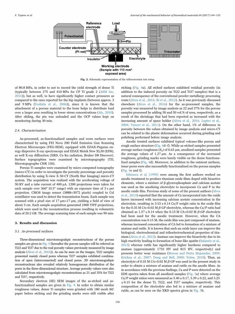
Ishizawa et al. (1995) were among the first authors worked onanodic treatment to produce titanium oxide films doped with bioactiveelements, where a mixture of β-glycerophosphate and calcium acetatewas used as the anodizing electrolyte to incorporate Ca and P in theanodic oxide film. Previous study of some of the present authors (Alveset al., 2013) reported that the amount of Ca incorporated into the oxidelayers increased with increasing calcium acetate concentration in theelectrolyte, resulting in 3.53 ± 0.14 Ca/P weight ratio in the oxide filmfor the 0.35 M CA+0.02 M β-GP electrolyte, whereas the Ca/P ratio hadobtained as 1.57 ± 0.14 when the 0.15 M CA+0.02 M β-GP electrolytehad been used for the anodic treatment. However, when the CAconcentration was 0.15 M, the oxide film was just composed of anatase,whereas increased concentration of CA led to formation of a mixture ofanatase and rutile. It is known that such an oxide layer can improve thebiological, electrochemical and triboelectrochemical properties of tita-nium (Alves et al., 2015). Anatase can improve the bioactivity due to itshigh reactivity leading to formation of bone-like apatite (Salantiu et al.,2015) whereas rutile has significantly higher hardness compared toanatase (approximately 1733 HV and 815 HV, respectively) andpresents better wear resistance (Biswas and Dutta Majumdar, 2009;Krishna et al., 2007; Dong and Bell, 2000; Yetim, 2010). Thus, anelectrolyte of 0.35 M CA+0.02 M β-GP was used in the present study inorder to obtain a mixture of anatase and rutile on the anodic films. Asin accordance with the previous findings, Ca and P were detected on theEDS spectra taken from all anodized samples (Fig. 5a) where averageCa/P weight ratios were measured as 3.40 ± 0.17, 3.39 ± 0.22, and 3.25± 0.15 for the dense Ti, Ti22, and Ti37 samples, respectively. Thiscomposition of the electrolyte also led to a mixture of anatase andrutile, as can be seen on the XRD spectra given in Fig. 5b.
Fig. 2. Schematic representation of the tribocorrosion test setup.
F. Toptan et al. Journal of the mechanical behavior of biomedical materials 69 (2017) 144–152
146

3.2. Worn surfaces
Fig. 6a–f presents the representative lower magnification SEMimages of the worn surfaces both in secondary electron (SE) andbackscattered electron (BSE) modes. Untreated dense Ti samplespresented well-known worn surface features for Ti, namely, parallelsliding grooves due to abrasive wear, together with compacted oxidizedwear debris. While the wear tracks were well distinguishable on thedense samples (both untreated and treated), the tracks were not as
clearly noticeable on the porous samples. Additionally, in all cases,functionalized samples exhibited less visible wear damage as comparedto the untreated samples. On the other hand, formation of compactedwear debris or a tribolayer was more distinguishable on both untreatedand treated dense worn surfaces.
Fig. 6g–j gives the representative EDS spectra taken from the wornsample and counter material (zirconia pin) surfaces. Presence of O onthe untreated samples reveals the formation of the oxidative wear in allcases (Fig. 6g). On the treated samples, presence of Ca and P shows
Fig. 3. Three-dimensional microtomograhpic reconstructions of a) Ti22 and b) Ti37 samples.
Fig. 4. Secondary electron SEM images of the as-etched (a–c) and as-functionalized (d–f) dense, Ti22, and Ti37 samples, respectively.
F. Toptan et al. Journal of the mechanical behavior of biomedical materials 69 (2017) 144–152
147
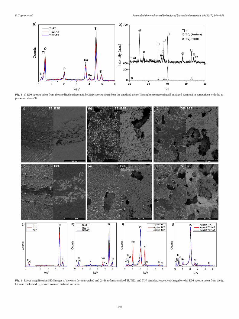
Fig. 5. a) EDS spectra taken from the anodized surfaces and b) XRD spectra taken from the anodized dense Ti samples (representing all anodized surfaces) in comparison with the as-processed dense Ti.
Fig. 6. Lower magnification SEM images of the worn (a–c) as-etched and (d–f) as-functionalized Ti, Ti22, and Ti37 samples, respectively, together with EDS spectra taken from the (g,h) wear tracks and (i, j) worn counter material surfaces.
F. Toptan et al. Journal of the mechanical behavior of biomedical materials 69 (2017) 144–152
148
that the functionalized layer was not totally removed in any of the cases(Fig. 6h). Furthermore, all pin surfaces exhibited Ti indicating thematerial transfer from sample surfaces to the counter material due toadhesive wear. Nevertheless, the transfer of Ti was always compara-tively less for the anodic treated samples as compared to the untreatedsamples Fig. 6i–j.
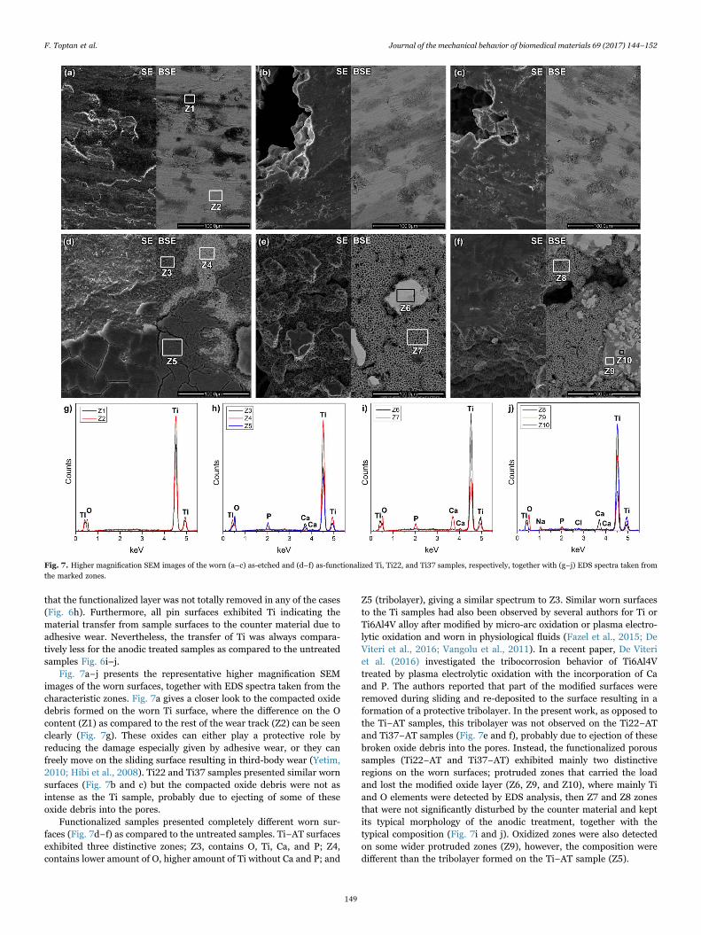
Fig. 7a−j presents the representative higher magnification SEMimages of the worn surfaces, together with EDS spectra taken from thecharacteristic zones. Fig. 7a gives a closer look to the compacted oxidedebris formed on the worn Ti surface, where the difference on the Ocontent (Z1) as compared to the rest of the wear track (Z2) can be seenclearly (Fig. 7g). These oxides can either play a protective role byreducing the damage especially given by adhesive wear, or they canfreely move on the sliding surface resulting in third-body wear (Yetim,2010; Hibi et al., 2008). Ti22 and Ti37 samples presented similar wornsurfaces (Fig. 7b and c) but the compacted oxide debris were not asintense as the Ti sample, probably due to ejecting of some of theseoxide debris into the pores.
Functionalized samples presented completely different worn sur-faces (Fig. 7d−f) as compared to the untreated samples. Ti−AT surfacesexhibited three distinctive zones; Z3, contains O, Ti, Ca, and P; Z4,contains lower amount of O, higher amount of Ti without Ca and P; and
Z5 (tribolayer), giving a similar spectrum to Z3. Similar worn surfacesto the Ti samples had also been observed by several authors for Ti orTi6Al4V alloy after modified by micro-arc oxidation or plasma electro-lytic oxidation and worn in physiological fluids (Fazel et al., 2015; DeViteri et al., 2016; Vangolu et al., 2011). In a recent paper, De Viteriet al. (2016) investigated the tribocorrosion behavior of Ti6Al4Vtreated by plasma electrolytic oxidation with the incorporation of Caand P. The authors reported that part of the modified surfaces wereremoved during sliding and re-deposited to the surface resulting in aformation of a protective tribolayer. In the present work, as opposed tothe Ti−AT samples, this tribolayer was not observed on the Ti22−ATand Ti37−AT samples (Fig. 7e and f), probably due to ejection of thesebroken oxide debris into the pores. Instead, the functionalized poroussamples (Ti22−AT and Ti37−AT) exhibited mainly two distinctiveregions on the worn surfaces; protruded zones that carried the loadand lost the modified oxide layer (Z6, Z9, and Z10), where mainly Tiand O elements were detected by EDS analysis, then Z7 and Z8 zonesthat were not significantly disturbed by the counter material and keptits typical morphology of the anodic treatment, together with thetypical composition (Fig. 7i and j). Oxidized zones were also detectedon some wider protruded zones (Z9), however, the composition weredifferent than the tribolayer formed on the Ti−AT sample (Z5).
Fig. 7. Higher magnification SEM images of the worn (a–c) as-etched and (d–f) as-functionalized Ti, Ti22, and Ti37 samples, respectively, together with (g–j) EDS spectra taken fromthe marked zones.
F. Toptan et al. Journal of the mechanical behavior of biomedical materials 69 (2017) 144–152
149
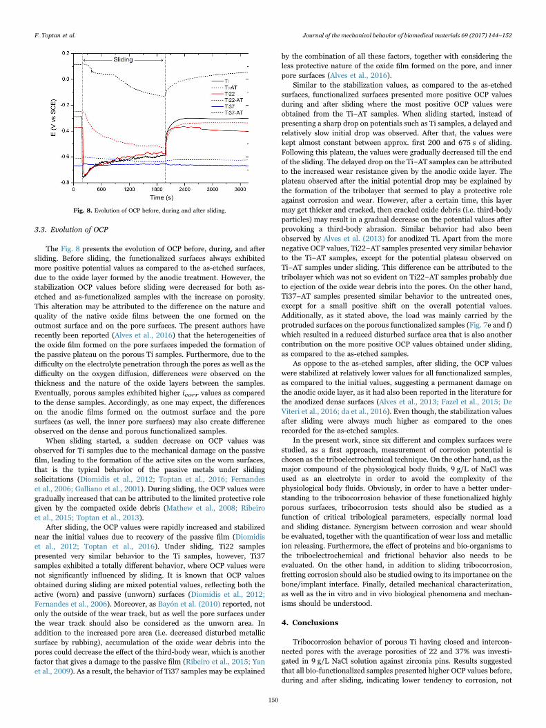
3.3. Evolution of OCP
The Fig. 8 presents the evolution of OCP before, during, and aftersliding. Before sliding, the functionalized surfaces always exhibitedmore positive potential values as compared to the as-etched surfaces,due to the oxide layer formed by the anodic treatment. However, thestabilization OCP values before sliding were decreased for both as-etched and as-functionalized samples with the increase on porosity.This alteration may be attributed to the difference on the nature andquality of the native oxide films between the one formed on theoutmost surface and on the pore surfaces. The present authors haverecently been reported (Alves et al., 2016) that the heterogeneities ofthe oxide film formed on the pore surfaces impeded the formation ofthe passive plateau on the porous Ti samples. Furthermore, due to thedifficulty on the electrolyte penetration through the pores as well as thedifficulty on the oxygen diffusion, differences were observed on thethickness and the nature of the oxide layers between the samples.Eventually, porous samples exhibited higher icorr values as comparedto the dense samples. Accordingly, as one may expect, the differenceson the anodic films formed on the outmost surface and the poresurfaces (as well, the inner pore surfaces) may also create differenceobserved on the dense and porous functionalized samples.
When sliding started, a sudden decrease on OCP values wasobserved for Ti samples due to the mechanical damage on the passivefilm, leading to the formation of the active sites on the worn surfaces,that is the typical behavior of the passive metals under slidingsolicitations (Diomidis et al., 2012; Toptan et al., 2016; Fernandeset al., 2006; Galliano et al., 2001). During sliding, the OCP values weregradually increased that can be attributed to the limited protective rolegiven by the compacted oxide debris (Mathew et al., 2008; Ribeiroet al., 2015; Toptan et al., 2013).
After sliding, the OCP values were rapidly increased and stabilizednear the initial values due to recovery of the passive film (Diomidiset al., 2012; Toptan et al., 2016). Under sliding, Ti22 samplespresented very similar behavior to the Ti samples, however, Ti37samples exhibited a totally different behavior, where OCP values werenot significantly influenced by sliding. It is known that OCP valuesobtained during sliding are mixed potential values, reflecting both theactive (worn) and passive (unworn) surfaces (Diomidis et al., 2012;Fernandes et al., 2006). Moreover, as Bayón et al. (2010) reported, notonly the outside of the wear track, but as well the pore surfaces underthe wear track should also be considered as the unworn area. Inaddition to the increased pore area (i.e. decreased disturbed metallicsurface by rubbing), accumulation of the oxide wear debris into thepores could decrease the effect of the third-body wear, which is anotherfactor that gives a damage to the passive film (Ribeiro et al., 2015; Yanet al., 2009). As a result, the behavior of Ti37 samples may be explained
by the combination of all these factors, together with considering theless protective nature of the oxide film formed on the pore, and innerpore surfaces (Alves et al., 2016).
Similar to the stabilization values, as compared to the as-etchedsurfaces, functionalized surfaces presented more positive OCP valuesduring and after sliding where the most positive OCP values wereobtained from the Ti−AT samples. When sliding started, instead ofpresenting a sharp drop on potentials such as Ti samples, a delayed andrelatively slow initial drop was observed. After that, the values werekept almost constant between approx. first 200 and 675 s of sliding.Following this plateau, the values were gradually decreased till the endof the sliding. The delayed drop on the Ti−AT samples can be attributedto the increased wear resistance given by the anodic oxide layer. Theplateau observed after the initial potential drop may be explained bythe formation of the tribolayer that seemed to play a protective roleagainst corrosion and wear. However, after a certain time, this layermay get thicker and cracked, then cracked oxide debris (i.e. third-bodyparticles) may result in a gradual decrease on the potential values afterprovoking a third-body abrasion. Similar behavior had also beenobserved by Alves et al. (2013) for anodized Ti. Apart from the morenegative OCP values, Ti22−AT samples presented very similar behaviorto the Ti−AT samples, except for the potential plateau observed onTi−AT samples under sliding. This difference can be attributed to thetribolayer which was not so evident on Ti22−AT samples probably dueto ejection of the oxide wear debris into the pores. On the other hand,Ti37−AT samples presented similar behavior to the untreated ones,except for a small positive shift on the overall potential values.Additionally, as it stated above, the load was mainly carried by theprotruded surfaces on the porous functionalized samples (Fig. 7e and f)which resulted in a reduced disturbed surface area that is also anothercontribution on the more positive OCP values obtained under sliding,as compared to the as-etched samples.
As oppose to the as-etched samples, after sliding, the OCP valueswere stabilized at relatively lower values for all functionalized samples,as compared to the initial values, suggesting a permanent damage onthe anodic oxide layer, as it had also been reported in the literature forthe anodized dense surfaces (Alves et al., 2013; Fazel et al., 2015; DeViteri et al., 2016; da et al., 2016). Even though, the stabilization valuesafter sliding were always much higher as compared to the onesrecorded for the as-etched samples.
In the present work, since six different and complex surfaces werestudied, as a first approach, measurement of corrosion potential ischosen as the triboelectrochemical technique. On the other hand, as themajor compound of the physiological body fluids, 9 g/L of NaCl wasused as an electrolyte in order to avoid the complexity of thephysiological body fluids. Obviously, in order to have a better under-standing to the tribocorrosion behavior of these functionalized highlyporous surfaces, tribocorrosion tests should also be studied as afunction of critical tribological parameters, especially normal loadand sliding distance. Synergism between corrosion and wear shouldbe evaluated, together with the quantification of wear loss and metallicion releasing. Furthermore, the effect of proteins and bio-organisms tothe triboelectrochemical and frictional behavior also needs to beevaluated. On the other hand, in addition to sliding tribocorrosion,fretting corrosion should also be studied owing to its importance on thebone/implant interface. Finally, detailed mechanical characterization,as well as the in vitro and in vivo biological phenomena and mechan-isms should be understood.
4. Conclusions
Tribocorrosion behavior of porous Ti having closed and intercon-nected pores with the average porosities of 22 and 37% was investi-gated in 9 g/L NaCl solution against zirconia pins. Results suggestedthat all bio-functionalized samples presented higher OCP values before,during and after sliding, indicating lower tendency to corrosion, not
Fig. 8. Evolution of OCP before, during and after sliding.
F. Toptan et al. Journal of the mechanical behavior of biomedical materials 69 (2017) 144–152
150
just due to the improved corrosion resistance by the oxide layersformed on the outmost surface and pore surfaces, but also due to itshigh hardness and therefore high resistance to wear. As a result, duringtribocorrosion action, the counter material mainly slid over theprotruded anodized surfaces resulting in less mechanical damage onthe functionalized surfaces. Moreover, ejection of the wear debris intothe pores decreased the third-body abrasion that also contributed tothe improved tribocorrosion performance. However, further studies areneeded in order to have a deeper understanding to the tribocorrosionbehavior of porous Ti, namely potentiostatic tribocorrosion tests forunderstanding the corrosion kinetics under sliding, as well, frettingcorrosion tests in order to simulate better the bone/implant interface.
Acknowledgments
This study was supported by FCT with the reference project UID/EEA/04436/2013, by FEDER funds through the COMPETE 2020 –Programa Operacional Competitividade e Internacionalização (POCI)with the reference project POCI-01-0145-FEDER-006941, Programade Acções Universitárias Integradas Luso-Francesas' (PAUILF TC-12_14), and the Calouste Gulbenkian Foundation through “Programade Mobilidade Académica para Professores”. The authors also grate-fully acknowledge the "Investissements d'avenir" programs (nos. ANR-11-IDEX-0003-02 and ANR-10- EQPX-37 MATMECA Grant) forfinancial support.
References
Alves, A.C., Oliveira, F., Wenger, F., Ponthiaux, P., Celis, J.-P., Rocha, L.A., 2013.Tribocorrosion behaviour of anodic treated titanium surfaces intended for dentalimplants. J. Phys. D Appl. Phys. 46, 404001.
Alves, A.C., Sendão, I., Ariza, E., Toptan, F., Ponthiaux, P., Pinto, A.M.P., 2016.Corrosion behaviour of porous Ti intended for biomedical applications. J PorousMater. 23, 1261–1268.
Alves, S.A., Bayón, R., de Viteri, V.S., Garcia, M.P., Igartua, A., Fernandes, M.H., et al.,2015. Tribocorrosion behavior of calcium- and phosphorous-enriched titanium oxidefilms and study of osteoblast interactions for dental implants. J. Biol. Tribol. Corros.1, 23.
Aly, M.S., 2010. Effect of pore size on the tensile behavior of open-cell Ti foams:experimental results. Mater. Lett. 64, 935–937.
Amin Yavari, S., Ahmadi, S.M., van der Stok, J., Wauthle, R., Riemslag, A.C., Janssen, M.,et al., 2014. Effects of bio-functionalizing surface treatments on the mechanicalbehavior of open porous titanium biomaterials. J. Mech. Behav. Biomed. Mater. 36,109–119.
ASM Inc. Webpage, 2016. ⟨http://asm.matweb.com/search/SpecificMaterial.asp?Bassnum=MTP641⟩.
Bayón, R., Nevshupa, R., Zubizarreta, C., Ruiz, U., de Gopegui, Barriga, J., Igartua, A.,2010. Characterisation of tribocorrosion behaviour of multilayer PVD coatings. Anal.Bioanal. Chem. 396, 2855–2862.
Benea, L., Mardare-Danaila, E., Mardare, M., Celis, J.-P., 2014. Preparation of titaniumoxide and hydroxyapatite on Ti–6Al–4V alloy surface and electrochemical behaviourin bio-simulated fluid solution. Corros. Sci. 80, 331–338.
Bi, H.L., Yu, C.Z., Cao, P., He, Y.H., 2012. Porous Ti-6Al-4V alloy prepared by a press-and-sinter process. Key Eng. Mater. 520, 76–81.
Biswas, A., Dutta Majumdar, J., 2009. Surface characterization and mechanical propertyevaluation of thermally oxidized Ti-6Al-4V. Mater. Charact. 60, 513–518.
Chen, X.-B., Li, Y.-C., Du Plessis, J., Hodgson, P.D., Wen, C., 2009. Influence of calciumion deposition on apatite-inducing ability of porous titanium for biomedicalapplications. Acta Biomater. 5, 1808–1820.
Correa, D.R.N., Kuroda, P.A.B., Grandini, C.R., 2014. Structure, microstructure, andselected mechanical properties of Ti-Zr-Mo alloys for biomedical applications. Adv.Mater. Res. 922, 75–80.
De Viteri, V.S., Fuentes, E., 2013. Titanium and titanium alloys as biomaterials. In:Gegner, J. (Ed.), Tribol. Fundam. Adv.. InTech.
De Viteri, V.S., Bayón, R., Igartua, A., Barandika, G., Moreno, J.E., Peremarch, C.P.,et al., 2016. Structure, tribocorrosion and biocide characterization of Ca, P and Icontaining TiO2 coatings developed by plasma electrolytic oxidation. Appl. Surf. Sci.367, 1–10.
Diomidis, N., Mischler, S., More, N.S., Roy, M., 2012. Tribo-electrochemicalcharacterization of metallic biomaterials for total joint replacement. Acta Biomater.8, 852–859.
Dong, H., Bell, T., 2000. Enhanced wear resistance of titanium surfaces by a new thermaloxidation treatment. Wear 238, 131–137.
Fan, X., Feng, B., Weng, J., Wang, J., Lu, X., 2011. Processing and properties of poroustitanium with high porosity coated by bioactive titania nanotubes. Mater. Lett. 65,2899–2901.
Fazel, M., Salimijazi, H.R., Golozar, M.A., Jazi, M.R. Garsivaz, 2015. A comparison of
corrosion, tribocorrosion and electrochemical impedance properties of pure Ti andTi6Al4V alloy treated by micro-arc oxidation process. Appl. Surf. Sci. 324, 751–756.
Fernandes, A.C., Vaz, F., Ariza, E., Rocha, L.A., Ribeiro, A.R.L., Vieira, A.C., et al., 2006.Tribocorrosion behaviour of plasma nitrided and plasma nitrided+oxidised Ti6Al4Valloy. Surf. Coat. Technol. 200, 6218–6224.
Galliano, F., Galvanetto, E., Mischler, S., Landolt, D., 2001. Tribocorrosion behavior ofplasma nitrided Ti-6Al-4V alloy in neutral NaCl solution. Surf. Coat. Technol. 145,121–131.
Ganesh, B.K.C., Ramanaih, N., Chandrasekhar Rao, P.V., 2012. Dry sliding wear behaviorof Ti–6Al–4V implant alloy subjected to various surface treatments. Trans. IndianInst. Met. 65, 425–434.
Goriainov, V., Cook, R., Latham, J.M., Dunlop, D.G., Oreffo, R.O.C., 2014. Bone andmetal: an orthopaedic perspective on osseointegration of metals. Acta Biomater. 10,4043–4057.
Gosavi, S., Gosavi, S., Alla, R., Titanium, 2013. A MIracle Metal in Dentistry. TrendsBiomater. Artif. Organs 27, 42–46.
Guo, Y., Georgarakis, K., Yokoyama, Y., Yavari, A.R., 2013. On the mechanical propertiesof TiNb based alloys. J. Alloy. Compd. 571, 25–30.
Hamid, A.A., Ghosh, P.K., Jain, S.C., Ray, S., 2008. The influence of porosity andparticles content on dry sliding wear of cast in situ Al(Ti)-Al2O3(TiO2) composite.Wear 265, 14–26.
Hibi, Y., Murakami, T., Miyake, K., Sasaki, S., 2008. Influence of microstructure on thewear behavior of sic-reinforced titanium-matrix composites lubricated by water andby ethanol. J. Am. Ceram. Soc. 91, 508–513.
Hu, R.-H., Lim, J.-K., 2010. Hardness and wear resistance improvement of surfacecomposite layer on Ti–6Al–4V substrate fabricated by powder sintering. Mater. Des.31, 2670–2675.
Hu, X., Shen, H., Shuai, K., Zhang, E., Bai, Y., Cheng, Y., et al., 2011. Surface bioactivitymodification of titanium by CO2 plasma treatment and induction of hydroxyapatite:in vitro and in vivo studies. Appl. Surf. Sci. 257, 1813–1823.
Ishizawa, H., Ogino, M., 1995. Characterization of thin hydroxyapatite layers formed onanodic titanium oxide films containing Ca and P by hydrothermal treatment. J.Biomed. Mater. Res. 29, 1071–1079.
Ishizawa, H., Fujino, M., Ogino, M., 1995. Mechanical and histological investigation ofhydrothermally treated and untreated anodic titanium oxide films containing Ca andP. J. Biomed. Mater. Res. 29, 1459–1468.
Jha, N., Badkul, A., Mondal, D.P., Das, S., Singh, M., 2011. Sliding wear behaviour ofaluminum syntactic foam: a comparison with Al–10 wt% SiC composites. Tribol. Int.44, 220–231.
Jha, N., Mondal, D.P., Dutta Majumdar, J., Badkul, A., Jha, A.K., Khare, A.K., 2013.Highly porous open cell Ti-foam using NaCl as temporary space holder throughpowder metallurgy route. Mater. Des. 47, 810–819.
Krishna, D. Siva Rama, Brama, Y.L., Sun, Y., 2007. Thick rutile layer on titanium fortribological applications. Tribol. Int. 40, 329–334.
Landolt, D., Mischler, S., Stemp, M., Barril, S., 2004. Third body effects and materialfluxes in tribocorrosion systems involving a sliding contact. Wear 256, 517–524.
Laptev, A., Bram, M., Buchkremer, H.P., Stöver, D., 2004. Study of production route fortitanium parts combining very high porosity and complex shape. Powder Metall. 47,85–92.
Lee, B., Lee, T., Lee, Y., Lee, D.J., Jeong, J., Yuh, J., et al., 2014. Space-holder effect ondesigning pore structure and determining mechanical properties in porous titanium.Mater. Des. 57, 712–718.
Lee, W.-T., Koak, J.-Y., Lim, Y.-J., Kim, S.-K., Kwon, H.-B., Kim, M.-J., 2012. Stressshielding and fatigue limits of poly-ether-ether-ketone dental implants. J. Biomed.Mater. Res. B Appl. Biomater. 100, 1044–1052.
Li, Y.-H., Rao, G.-B., Rong, L.-J., Li, Y.-Y., 2002. The influence of porosity on corrosioncharacteristics of porous NiTi alloy in simulated body fluid. Mater. Lett. 57,448–451.
Marino, C.E.B., Mascaro, L.H., 2004. EIS characterization of a Ti-dental implant inartificial saliva media: dissolution process of the oxide barrier. J. Electroanal. Chem.568, 115–120.
Marques, I. da S.V., Alfaro, M.F., da Cruz, N.C., Mesquita, M.F., Sukotjo, C., Mathew,M.T., et al., 2016. Tribocorrosion behavior of biofunctional titanium oxide filmsproduced by micro-arc oxidation: synergism and mechanisms. J. Mech. Behav.Biomed. Mater. 60, 8–21.
Martins, J.R.S., Grandini, C.R., 2014. The influence of heat treatment on the structureand microstructure of Ti-15Mo-xNb system alloys for biomedical applications.Mater. Sci. Forum 783–786, 1255–1260.
Mathew, M.T., Ariza, E., Rocha, L.A., Fernandes, A.C., Vaz, F., 2008. TiCxOy thin filmsfor decorative applications: tribocorrosion mechanisms and synergism. Tribol. Int.41, 603–615.
Menini, R., Dion, M.-J., So, S.K.V., Gauthier, M., Lefebvre, L.-P., 2006. Surface andcorrosion electrochemical characterization of titanium foams for implantapplications. J. Electrochem. Soc. 153, B13.
Mischler, S., 2008. Triboelectrochemical techniques and interpretation methods intribocorrosion: a comparative evaluation. Tribol. Int. 41, 573–583.
Mondal, D.P., Das, S., Jha, N., 2009. Dry sliding wear behaviour of aluminum syntacticfoam. Mater. Des. 30, 2563–2568.
Nag, S., Banerjee, R., 2012. Fundamentals of medical implant materials. In: Narayan, R.(Ed.), ASM Handbook, Vol. 23.
Ribeiro, A.M., Alves, A.C., Rocha, L.A., Silva, F.S., Toptan, F., 2015. Synergism betweencorrosion and wear on CoCrMo−Al2O3 biocomposites in a physiological solution.Tribol. Int. 91, 198–205.
Salahinejad, E., Amini, R., Marasi, M., Hadianfard, M.J., 2010. Microstructure and wearbehavior of a porous nanocrystalline nickel-free austenitic stainless steel developedby powder metallurgy. Mater. Des. 31, 2259–2263.
F. Toptan et al. Journal of the mechanical behavior of biomedical materials 69 (2017) 144–152
151
Salantiu, A.-M., Fekete, C., Muresan, L., Pascuta, P., Popa, F., Popa, C., 2015. Anodicoxidation of PM porous titanium for increasing the corrosion resistance ofendosseous implants. Mater. Chem. Phys. 149-150, 453–459.
Seah, K.H.W., Chen, X., 1993. A comparison between the corrosion characteristics of 316stainless steel, solid titanium and porous titanium. Corros. Sci. 34, 1841–1851.
Seah, K.H.W., Thampuran, R., Teoh, S.H., 1998. The influence of pore morphology oncorrosion. Corros. Sci. 40, 547–556.
Szesz, E.M., Pereira, B.L., Kuromoto, N.K., Marino, C.E.B., de Souza, G.B., Soares, P.,2013. Electrochemical and morphological analyses on the titanium surface modifiedby shot blasting and anodic oxidation processes. Thin Solid Films 528, 163–166.
Tanigawa, H., Asoh, H., Ohno, T., Kubota, M., Ono, S., 2013. Electrochemical corrosionand bioactivity of titanium–hydroxyapatite composites prepared by spark plasmasintering. Corros. Sci. 70, 212–220.
Teixeira, M., Alves, A.C., Silva, F.S., Pinto, A.M., Toptan, F., 2015. Microstructuralcharacterization of biofunctionalized titanium foams. Microsc. Microanal. 21, 55–56.
Thomann, U.I., Uggowitzer, P.J., 2000. Wear–corrosion behavior of biocompatibleaustenitic stainless steels. Wear 239, 48–58.
Toptan, F., Rego, A., Alves, A.C., Guedes, A., 2016. Corrosion and tribocorrosionbehavior of Ti–B4C composite intended for orthopaedic implants. J. Mech. Behav.Biomed. Mater. 61, 152–163.
Toptan, F., Alves, A.C., Kerti, I., Ariza, E., Rocha, L.A., 2013. Corrosion andtribocorrosion behaviour of Al-Si-Cu-Mg alloy and its composites reinforced with
B4C particles in 0.05M NaCl solution. Wear 306, 27–35.Tuncer, N., Arslan, G., Maire, E., Salvo, L., 2011. Investigation of spacer size effect on
architecture and mechanical properties of porous titanium. Mater. Sci. Eng. A 530,633–642.
Vangolu, Y., Alsaran, A., Yildirim, O.S., 2011. Wear properties of micro arc oxidized andhydrothermally treated Ti6Al4V alloy in simulated body fluid. Wear 271,2322–2327.
Wood, R.J.K., 2007. Tribo-corrosion of coatings: a review. J. Phys. D Appl. Phys. 40,5502–5521.
Xie, F.X., He, X.B., Cao, S.L., Lu, X., Qu, X.H., 2013. Structural characterization andelectrochemical behaviour of a laser-sintered porous Ti-10Mo alloy. Corros. Sci. 67,217–224.
Yan, Y., Neville, A., Dowson, D., Williams, S., Fisher, J., 2009. Effect of metallicnanoparticles on the biotribocorrosion behaviour of metal-on-metal hip prostheses.Wear 267, 683–688.
Yetim, A.F., 2010. Investigation of wear behavior of titanium oxide films, produced byanodic oxidation, on commercially pure titanium in vacuum conditions. Surf. Coat.Technol. 205, 1757–1763.
Yoshida, H., Faust, A., Wilckens, J., Kitagawa, M., Fetto, J., Chao, E.Y.S., 2006. Three-dimensional dynamic hip contact area and pressure distribution during activities ofdaily living. J. Biomech. 39, 1996–2004.
F. Toptan et al. Journal of the mechanical behavior of biomedical materials 69 (2017) 144–152
152