Embed Size (px)
Citation preview
~ 762 ~
ISSN Print: 2394-7500 ISSN Online: 2394-5869 Impact Factor: 5.2 IJAR 2016; 2(2): 762-765 www.allresearchjournal.com Received: 20-12-2015 Accepted: 22-01-2016 Dr Sharad Agarkhedkar Dr. D. Y. Patil Medical College, Pimpri, Pune, M.S., India. Dr Shailaja Mane Dr. D. Y. Patil Medical College, Pimpri, Pune, M.S., India. Dr Mohd Mohsin Dr. D. Y. Patil Medical College, Pimpri, Pune, M.S., India. Dr Padedam Raghaviah Dr. D. Y. Patil Medical College, Pimpri, Pune, M.S., India. Dr Vardhan Patel Dr. D. Y. Patil Medical College, Pimpri, Pune, M.S., India. Dr Jignesh Thakor Dr. D. Y. Patil Medical College, Pimpri, Pune, M.S., India. Correspondence Mohd. Mohsin Dr. D. Y. Patil Medical College, Pimpri, Pune, M.S., India.
A case of antenatally diagnosed hypoplastic left heart
syndrome
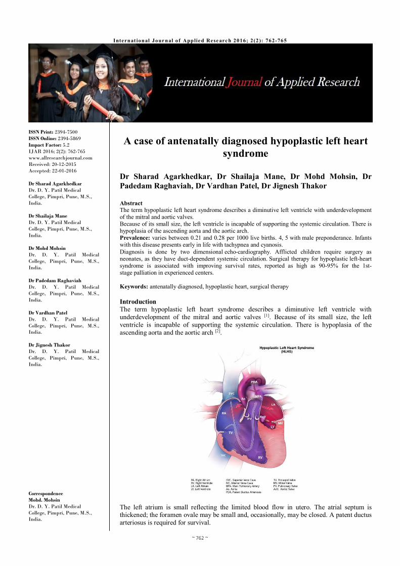
Dr Sharad Agarkhedkar, Dr Shailaja Mane, Dr Mohd Mohsin, Dr Padedam Raghaviah, Dr Vardhan Patel, Dr Jignesh Thakor Abstract The term hypoplastic left heart syndrome describes a diminutive left ventricle with underdevelopment of the mitral and aortic valves. Because of its small size, the left ventricle is incapable of supporting the systemic circulation. There is hypoplasia of the ascending aorta and the aortic arch. Prevalence: varies between 0.21 and 0.28 per 1000 live births. 4, 5 with male preponderance. Infants with this disease presents early in life with tachypnea and cyanosis. Diagnosis is done by two dimensional echo-cardiography. Afflicted children require surgery as neonates, as they have duct-dependent systemic circulation. Surgical therapy for hypoplastic left-heart syndrome is associated with improving survival rates, reported as high as 90-95% for the 1st-stage palliation in experienced centers. Keywords: antenatally diagnosed, hypoplastic heart, surgical therapy Introduction The term hypoplastic left heart syndrome describes a diminutive left ventricle with underdevelopment of the mitral and aortic valves [1]. Because of its small size, the left ventricle is incapable of supporting the systemic circulation. There is hypoplasia of the ascending aorta and the aortic arch [2].
The left atrium is small reflecting the limited blood flow in utero. The atrial septum is thickened; the foramen ovale may be small and, occasionally, may be closed. A patent ductus arteriosus is required for survival.
Internat ional Jour nal of Applie d Rese arc h 2016; 2(2): 762-765
~ 763 ~
International Journal of Applied Research
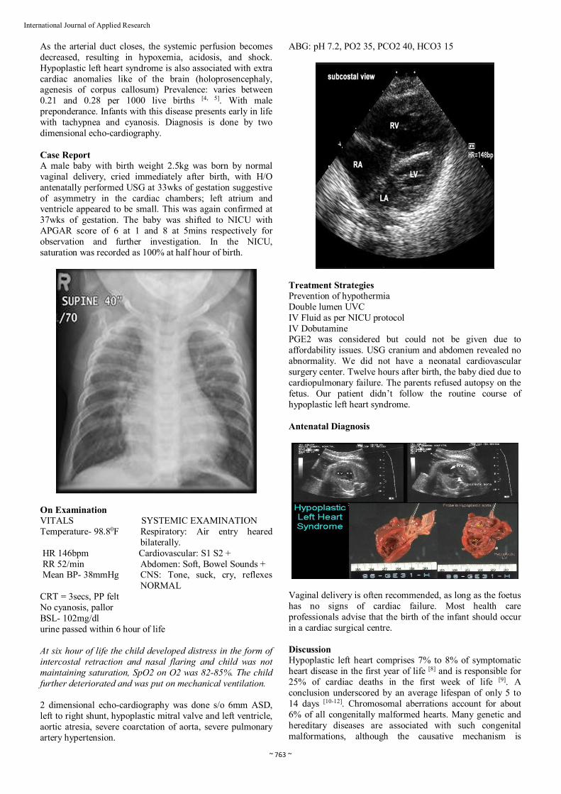
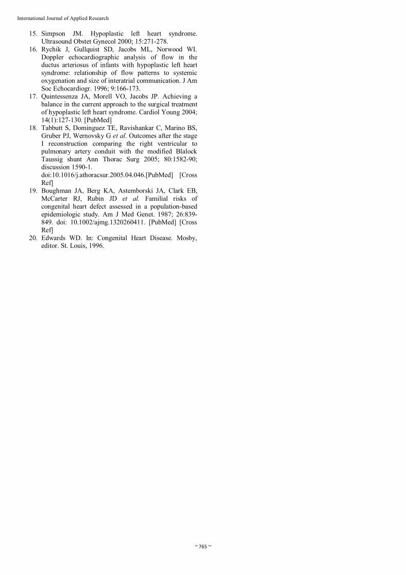
As the arterial duct closes, the systemic perfusion becomes decreased, resulting in hypoxemia, acidosis, and shock. Hypoplastic left heart syndrome is also associated with extra cardiac anomalies like of the brain (holoprosencephaly, agenesis of corpus callosum) Prevalence: varies between 0.21 and 0.28 per 1000 live births [4, 5]. With male preponderance. Infants with this disease presents early in life with tachypnea and cyanosis. Diagnosis is done by two dimensional echo-cardiography. Case Report A male baby with birth weight 2.5kg was born by normal vaginal delivery, cried immediately after birth, with H/O antenatally performed USG at 33wks of gestation suggestive of asymmetry in the cardiac chambers; left atrium and ventricle appeared to be small. This was again confirmed at 37wks of gestation. The baby was shifted to NICU with APGAR score of 6 at 1 and 8 at 5mins respectively for observation and further investigation. In the NICU, saturation was recorded as 100% at half hour of birth.
On Examination VITALS SYSTEMIC EXAMINATION Temperature- 98.80F Respiratory: Air entry heared
bilaterally. HR 146bpm Cardiovascular: S1 S2 + RR 52/min Abdomen: Soft, Bowel Sounds + Mean BP- 38mmHg CNS: Tone, suck, cry, reflexes
NORMAL CRT = 3secs, PP felt No cyanosis, pallor BSL- 102mg/dl urine passed within 6 hour of life At six hour of life the child developed distress in the form of intercostal retraction and nasal flaring and child was not maintaining saturation, SpO2 on O2 was 82-85%. The child further deteriorated and was put on mechanical ventilation. 2 dimensional echo-cardiography was done s/o 6mm ASD, left to right shunt, hypoplastic mitral valve and left ventricle, aortic atresia, severe coarctation of aorta, severe pulmonary artery hypertension.
ABG: pH 7.2, PO2 35, PCO2 40, HCO3 15
Treatment Strategies Prevention of hypothermia Double lumen UVC IV Fluid as per NICU protocol IV Dobutamine PGE2 was considered but could not be given due to affordability issues. USG cranium and abdomen revealed no abnormality. We did not have a neonatal cardiovascular surgery center. Twelve hours after birth, the baby died due to cardiopulmonary failure. The parents refused autopsy on the fetus. Our patient didn’t follow the routine course of hypoplastic left heart syndrome. Antenatal Diagnosis
Vaginal delivery is often recommended, as long as the foetus has no signs of cardiac failure. Most health care professionals advise that the birth of the infant should occur in a cardiac surgical centre. Discussion Hypoplastic left heart comprises 7% to 8% of symptomatic heart disease in the first year of life [8] and is responsible for 25% of cardiac deaths in the first week of life [9]. A conclusion underscored by an average lifespan of only 5 to 14 days [10-12]. Chromosomal aberrations account for about 6% of all congenitally malformed hearts. Many genetic and hereditary diseases are associated with such congenital malformations, although the causative mechanism is

~ 764 ~
International Journal of Applied Research
unknown. Precarious survival depends on three tenuous variables: patency of the ductus arteriosus, pulmonary vascular resistance, and an adequate interatrial communication [12]. Tachypnea, tachycardia, and cyanosis are present during the brief interval of ductal patency [13]. Risk is greatest during the period of normal ductal closure when systemic blood flow and coronary blood flow decrease or cease altogether. A fall in pulmonary vascular resistance diverts blood from the systemic circulation into the pulmonary circulation and augments flow into the obstructed left atrium. A rise in pulmonary vascular resistance improves systemic blood flow, but at the price of hypoxemia. Ninety-five percent of afflicted infants die within the first month of life [9, 14]. Fetal echocardiography permits the diagnosis as early as the 24th week of gestation [16]. Flow patterns in the fetal ductus can be monitored, [17] and the condition of the atrial septum can be determined. Echocardiography with color flow imaging and Doppler interrogation establishes the diagnosis of hypoplastic left heart with aortic atresia and a hypoplastic but perforate mitral valve [15]. A hypoplastic but perforate mitral valve communicates with a small left ventricle that gives rise to an atretic aortic valve and a tubular hypoplastic ascending aorta. The ventricular septum and the free wall of the hypoplastic left ventricle are thick and immobile, and the small cavity is lined with endocardial fibroelastosis. Coarctation is identified as a thin discrete posterior ledge extending across the lumen of the aorta at the level of the ductus arteriosus or as kinking and narrowing at the site of ductal insertion. Doppler interrogation establishes retrograde flow into the hypoplastic ascending aorta and occasionally identifies biphasic flow in the proximal coronary arteries. Direct ventriculo-coronary arterial communications can be identified. Management Afflicted children require surgery as neonates, as they have duct-dependent systemic circulation. Currently, there are two major treatment modalities. These are primary cardiac transplantation, or a series of staged functionally univentricular palliations [18]. Functionally univentricular palliations: The functionally univentricular palliation typically includes three operations. 1. The first stage of palliation, or the Norwood operation,
is performed at birth. 2. The second stage is a bi-directional Glenn operation,
usually undertaken at 6 to 8 months of age. The third, and final, stage is the Fontan operation, which can be performed between the ages of 18 months and 4 years. For patients undergoing functionally univentricular palliation, leading to creation of the Fontan circulation, the highest risk of mortality is following the initial operation, with up to three-tenths of patients dying in some reported series.19 The Norwood operation consists of constructing a new aortic root and arch, disconnecting the pulmonary trunk from the pulmonary circulation, and incorporating it into the systemic outflow tract. A modified Blalock-Taussig shunt, of 3 to 4 milimeters in diameter, is constructed to supply blood to the lungs. Nowadays, the operative mortality for conversion to the Fontan circulation is also less than 5%. Cardiac catheterization is usually undertaken prior to both the second and third stages of palliation to study anatomical and physiological details, and to perform corrective interventions
Prognosis and Outcome Untreated patients most often succumb during the 1st few months of life, usually during the 1st or 2nd wk. Occasionally, unoperated patients may live for months or, rarely, years. Up to 30% of infants with hypoplastic left-heart syndrome have evidence of either a major or minor central nervous system abnormality. Surgical therapy for hypoplastic left-heart syndrome is associated with improving survival rates, reported as high as 90-95% for the 1st-stage palliation in experienced centers. Genetic Counselling Upon diagnosis, both genetic counselling and testing should be offered to both parents. Multiple genetic syndromes have been reported, including Turner's syndrome, Noonan's syndrome, Smith-Lemli-Opitz syndrome, Holt-Oram syndrome, and many others (20, 21). Therefore genetic counselling forms an important part of it. References 1. Noonan JA, Nadas AS. The hypoplastic left heart
syndrome: an analysis of 101 cases. Pediatr Clin North Am Text book of pediatric cardiology Myung K. Park 6th edition 1959; 5:1029.
2. Glauser TA, Rorke LB, Weinberg PW. Congenital brain anomalies associated with the hypoplastic left heart syndrome. Pediatrics 1990; 85:984.
3. Botto LD, Correa A, Erickson JD. Racial and temporal variations in the prevalence of heart defects. Pediatr 2001; 107(3):1.
4. Hoffman JE, Kaplan S. The incidence of congenital heart disease. J Am Coll Cardiol. 2002; 39:1890.
5. Glauser TA, Zackai E, Weinberg P, Clancy R. Holt-Oram syndrome associated with the hypoplastic left heart syndrome. Clin Genet 1989; 36:69-72. [PubMed]
6. Blake DM, Copel JA, Kleinman CS. Hypoplastic left heart syndrome: prenatal diagnosis, clinical profile, and management. Am J Obstet Gynecol. 1991; 165:529-534. [PubMed]
7. Bailey LL, Gundry SR. Hypoplastic left heart syndrome. Pediatr Clin North Am. 1990; 37:137-150.
8. Fyler DC. Report of the New England Regional Infant Cardiac Program. Pediatrics 1980; 65:375-461.
9. Roberts WC, Perry LW, Chandra RS, Myers GE, Shapiro SR, Scott LP. Aortic valve atresia: a new classification based on necropsy study of 73 cases. Am J Cardiol. 1976; 37:753-756.
10. Cohen DM, Allen HD. New developments in the treatment of hypoplastic left heart syndrome. Curr Opin Cardiol. 1997; 12:44-50.
11. Hoshino K, Ogawa K, Hishitani T, Kitazawa R, Uehara R. Hypoplastic left heart syndrome: duration of survival without surgical intervention. Am Heart J. 1999; 137:535-542.
12. Bailey LL, Gundry SR. Hypoplastic left heart syndrome. Pediatr Clin North Am 1990; 37:137-150.
13. Noonan JA, Nadas AS. The hypoplastic left heart syndrome; an analysis of 101 cases. Pediatr Clin North Am 1958; 5:1029-1056.
14. Ludman P, Foale R, Alexander N, Nihoyannopoulos P. Cross sectional echocardiographic identification of hypoplastic left heart syndrome and differentiation from other causes of right ventricular overload. Br Heart J. 1990; 63:355-361.
~ 765 ~
International Journal of Applied Research
15. Simpson JM. Hypoplastic left heart syndrome. Ultrasound Obstet Gynecol 2000; 15:271-278.
16. Rychik J, Gullquist SD, Jacobs ML, Norwood WI. Doppler echocardiographic analysis of flow in the ductus arteriosus of infants with hypoplastic left heart syndrome: relationship of flow patterns to systemic oxygenation and size of interatrial communication. J Am Soc Echocardiogr. 1996; 9:166-173.
17. Quintessenza JA, Morell VO, Jacobs JP. Achieving a balance in the current approach to the surgical treatment of hypoplastic left heart syndrome. Cardiol Young 2004; 14(1):127-130. [PubMed]
18. Tabbutt S, Dominguez TE, Ravishankar C, Marino BS, Gruber PJ, Wernovsky G et al. Outcomes after the stage I reconstruction comparing the right ventricular to pulmonary artery conduit with the modified Blalock Taussig shunt Ann Thorac Surg 2005; 80:1582-90; discussion 1590-1. doi:10.1016/j.athoracsur.2005.04.046.[PubMed] [Cross Ref]
19. Boughman JA, Berg KA, Astemborski JA, Clark EB, McCarter RJ, Rubin JD et al. Familial risks of congenital heart defect assessed in a population-based epidemiologic study. Am J Med Genet. 1987; 26:839-849. doi: 10.1002/ajmg.1320260411. [PubMed] [Cross Ref]
20. Edwards WD. In: Congenital Heart Disease. Mosby, editor. St. Louis, 1996.