Embed Size (px)
Citation preview
LUND UNIVERSITY
PO Box 117221 00 Lund+46 46-222 00 00
Congenital stationary night blindness with hypoplastic discs, negativeelectroretinogram and thinning of the inner nuclear layer
Al Oreany, Abdulaziz Abdulrahman; Al Hadlaq, Abdulaziz; Schatz, Patrik
Published in:Graefe's Archive for Clinical and Experimental Ophthalmology
DOI:10.1007/s00417-016-3346-6
2016
Document Version:Peer reviewed version (aka post-print)
Link to publication
Citation for published version (APA):Al Oreany, A. A., Al Hadlaq, A., & Schatz, P. (2016). Congenital stationary night blindness with hypoplasticdiscs, negative electroretinogram and thinning of the inner nuclear layer. Graefe's Archive for Clinical andExperimental Ophthalmology, 254(10), 1951-1956. https://doi.org/10.1007/s00417-016-3346-6
Total number of authors:3
General rightsUnless other specific re-use rights are stated the following general rights apply:Copyright and moral rights for the publications made accessible in the public portal are retained by the authorsand/or other copyright owners and it is a condition of accessing publications that users recognise and abide by thelegal requirements associated with these rights. • Users may download and print one copy of any publication from the public portal for the purpose of private studyor research. • You may not further distribute the material or use it for any profit-making activity or commercial gain • You may freely distribute the URL identifying the publication in the public portal
Read more about Creative commons licenses: https://creativecommons.org/licenses/Take down policyIf you believe that this document breaches copyright please contact us providing details, and we will removeaccess to the work immediately and investigate your claim.
1
Congenital Stationary Night Blindness with Hypoplastic Discs, Negative Electroretinogram and Thinning of the
Inner Nuclear Layer
Abdulaziz Abdulrahman Al Oreany,1 Abdulaziz Al Hadlaq,1 Patrik Schatz, MD PhD.1,2
1 Vitreoretinal Division, King Khaled Eye Specialist Hospital, Riyadh, Kingdom of Saudi Arabia.
2 Department of Ophthalmology, Clinical Sciences, Scane County University Hospital, University of Lund, Sweden.
Correspondence to: Patrik Schatz, MD PhD, Executive Medical Department
King Khaled Eye Specialist Hospital, Al-Oruba Street, PO Box 7191 Riyadh 11462, Kingdom of Saudi Arabia.
Telephone number: Tel +966 11 482 1234 ext 3773 | Fax +966 11 4821234 ext 3727. E mail: [email protected]
ACKNOWLEDGEMENTS
a. Funding/support: Grants from the Cronqvist Stiftelse of the Swedish Society of Medicine and the Foundation for the
visually impaired in the Skane County.
b. Conflict of interest disclosures: None for each of the authors.
c. Other acknowledgement: We thank Mr Abdulrahman Al Gaeed and Mr Eric Mingoa for skilful technical assistance .
2
ABSTRACT
Purpose: To describe congenital stationary night blindness (CSNB) with a negative electroretinogram, hypoplastic
discs, nystagmus and thinning of the inner nuclear layer (INL).
Methods: Retinal structure was analyzed qualitatively with spectral domain optical coherence tomography and wide
field imaging. Retinal function was evaluated with full-field electroretinography (ffERG). Molecular genetic testing
included next-generation sequencing (NGS) of the known genes involved in CSNB.
Results: Patients presented with CSNB presented with nystagmus, high myopia, hypoplastic discs and negative ffERG
with no measurable rod response. The retinas appeared normal and automated segmentation of retinal layers
demonstrated a relative reduction of thickness of the INL. There was no significant change in the ffERG after
prolonged 2 hour dark adaptation compared to standard 30 minute dark adaptation. Affec ted family members harboured
the homozygous 1-bp deletion c.2394delC in exon 18 of the TRPM1 gene, whereas their unaffected parents were
heterozygous carriers .
Conclusions: This data expands the genotype and phenotype spectrum of CSNB. The lack of improvement of rod
responses after prolonged dark adaptation, together with thinning of the INL, is compatible with postreceptoral
transmission dysfunction in the bipolar cells. Such knowledge may prove useful in future development of treatment for
outer retinal dystrophies, using opsin genes to restore light responses in survivor neurons in the inner retina.
Key words: Congenital stationary night blindness, optical coherence tomography, optic disc hypoplasia, full-field
electroretinography.
3
INTRODUCTION
Congenital stationary night blindness (CSNB) refers to a group of mainly non-progressive retinal disorders
featuring night blindness due to mutations in genes affecting either retinoid metabolism in the retinal pigment
epithelium, photoreceptor transduction or signal transmission through the retinal bipolar cells [1]. CSNB is clinically
and genetically heterogeneous. Some forms are associated with poor visual acuity, myopia, nystagmus, strabismus and
fundus abnormalities [2-4]. CSNB is referred to as “complete” (MIM#613216), when there is no full-field
electroretinography (full-field ERG) rod response combined with an electronegative rod-cone ERG with a normal a-
wave (emanating from the photoreceptors) and severely reduced b-wave [1]. Such findings are consistent with an ON-
bipolar cell dysfunction.
The rods connect with the dendritic tips of ON-bipolar cells (rod bipolar cells). An increase in light intensity
induces a depolarization in the ON-bipolar cells via a reduction of glutamate release from presynaptic rods . The signal
transduction cascade in the dendritic tips of ON-bipolar cells involves the metabotropic Glutamate Receptor, 6
(mGluR6) glutamate receptors signalling to Transient Receptor Potential cation channel, subfamily M, member 1
(TRPM1) proteins that form part of the transduction channel. Mutations in TRPM1 are thought to be the leading cause
for the complete form of autosomal recessive CSNB, also called CSNB1 [1]. Like TRPM1, the other mutations causing
autosomal recessive complete CSNB occur in genes expressed in ON-bipolar cells, whose protein products are thought
to participate in the rod pathway signal transduction [1].
Clinically autosomal recessive complete CSNB is typically associated with high myopia and nystagmus [5].
Here, we describe a consanguineous family with autosomal recessive complete CSNB and high myopia, nystagmus and
in addition optic disc hypoplasia, associated with the novel homozygous frameshift mutation c.2394delC in the TRPM1
gene. We present data that indicate that there is a specific postreceptoral transmission dysfunction in the bipolar cells in
this form of CSNB. Such knowledge may prove useful in future development of treatment for outer retinal dystrophies,
using opsin genes to restore light responses in survivor neurons in the inner retina (Optogenetics).
METHODS
This is a retrospective study of a family with 2 affected twin 20-year old brothers (II:1 and II:2) with CSNB
and their unaffected parents. Informed consent was obtained. Institutional Review Board (IRB)/Ethics Committee
approval was obtained. The research adhered to the tenets of the Declaration of Helsinki.

4
For both affected twin brothers, retinal structure was analyzed qualitatively with transfoveal horizontal spectral
domain optical coherence tomography scans (OCT, Heidelberg Engineering, Inc., Heidelberg, Germany) and wide field
imaging (Optos PLC, Dunfermline, UK). Five myopic age-matched controls, were recruited to analyze differences in
retinal structure between the patients and controls. For this analysis, automated retinal sublayer segmentation using the
Heidelberg software was done, and the following 5 retinal sublayers were isolated and enlarged and compared
qualitatively for differences in thickness, while preserving the proportions within each layer, and between the patient
II:2 and each control: A. Photoreceptor and retinal pigment epithelium (RPE) layer: Between the external limiting
membrane and Bruchs membrane B. Outer nuclear layer: Between the outer plexiform layer line and the external
limiting membrane C. Inner nuclear layer: Between the inner nuclear layer line and the inner plexiform layer line D.
Ganglion cell layer: Between the ganglion cell layer line and the retinal nerve fiber layer line. E. Total retinal and RPE
thickness.
In both affected twin brothers retinal function was evaluated with full-field electroretinography (ffERG,
Nicolet Biomedical Instruments, Madison, Wisconsin, USA), in dark adapted and light adapted state according to
ISCEV standards [6], with a few modifications as follows . Retinal function was evaluated with full-field
electroretinography (ffERG, Nicolet Biomedical Instruments, Madison, Wisconsin, USA), in dark adapted and light
adapted state according to ISCEV standards, with a few modifications as follows [6].Full-field electroretinograms were
recorded in a Nicolet analysis system (Nicolet Biomedical Instruments, Madison, Wisconsin, USA), after dark
adaptation of subjects for 40 min, dilatation of the pupils with topical cyclopentolate 1% and metaoxedrine 2,5% and
topical anaesthesia, with a Burian Allen bipolar contact lens and a ground electrode applied to the forehead. Responses
were obtained stimulating with single full-field flash (30 ms) with blue light light (0.81 cd-s/m2: rod response) and with
white light (10.02 cd-s/m2: combined rod-cone response). Photopic responses (not shown) were obtained with a
background illumination of 3.4 cd-s/m2 in order to saturate the rods.
Molecular genetic testing in both affected twin brothers included next-generation sequencing (NGS, Illumina
HiSeq 1500, performed by Center for Human Genetics Bioscientia, Ingelheim, Germany) of the known genes involved
in CSNB: CABP4, CACNAIF, GNAT1, GPR179, GRK, GRM6, LRIT3, NYX, PDE6B, RHD, SLC24A1, TRPM1 .
Genomic DNA was fragmented and the coding exons of the analyzed genes as well as the corresponding exon-intron

5
boundaries were enriched using the Roche/NimbleGen sequence capture approach, amplified and sequenced using
NGS. The target regions were sequenced with an average coverage of 506-fold. For more than 99% of the regions of
interest a 20-fold coverage was obtained. NGS data analysis was performed using bioinformatic analysis tools as well
as JSI Medical Systems software (version 4.1.2). Identified variants and indels were filtered against external and
internal databases and filtered depending on their allele frequency focusing on rare variants with a minor allele
frequency (MAF) of 1% or less. Nonsense, frameshift and canonical splice site variants were primarily considered
likely pathogenic. Variants that have been annotated as common polymorphisms in databases or in the literature were
not considered further.
Putatively pathogenic differences between the wildtype sequence (human reference genome according to
UCSC Genome Browser: hg19, GRCh37) and the patients sequence mentioned and interpreted in this report were
validated using polymerase chain reaction (PCR) amplification followed by conventional Sanger sequencing. The
resulting sequence data for the TRPM1 gene (OMIM: #603576; locus: chromosome 15q13.3) were compared to the
reference sequence NM_001252020.1.
For the unaffected parents, exon 18 of TRPM1 was apmlified by PCR and directly sequenced. In addition, wide field
fundus photography and autofluorescence imaging was obtained.
We did not perform mutation screening in genes that may be associated with optic nerve hypoplasia, such as HESX1,
SOX2, or ATOH7.
RESULTS
The affected brother (II:1) presented with a corrected visual acuity (CVA) of 20/30 OU (Refraction OD -
7_1.75x175, OS-8.75-2.25x10⁵) and his brother (II:2) with 20/40 OD and 20/50 OS (Refraction OD -8_-1x60, OS -8_-
0.5x150). Both presented with was a small amplitude pendular horizontal nystagmus OU. Fundi were normal except for
myopic changes (such as apparently thin retinas with visible choroidal vessels) and hypoplastic tilted discs (Fig. 1A).
Fundus autofluorescence demonstrated a normal distribution of autofluorescence (Fig. 1B) Optical coherence
tomography showed normal retinal layers that could be automatically segmented using the Heidelberg software in
brother II:2 (Fig. 1C right column). Full field electroretinography demonstrated a non-recordable rod response and an
electronegative pattern, with a reduced b wave amplitude compared to a wave amplitude in the combined rod -cone

6
I
response (Fig. 2A), and light adapted responses were normal to mildly reduced including a mildly reduced b/a ratio in
the single flash cone response (Fig. 2B) There was no significant change in the full field electroretinogram after
prolonged 2 hour dark adaptation compared to after standard 30 minute dark adaptation (Fig. 2B).
Total retinal thickness did not seem to be reduced compared to controls, however there seemed to be a relative
thinning of the inner nuclear layer compared to other retinal layers (Fig. 3A-E).
In both, MRI had been done because of hypoplastic discs, to rule out septo-optic dysplasia, with normal
findings, including normal midline structure and normal appearing optic nerves. Visual fields were normal. The mother
and father had normal fundi, normal fundus autofluorescence imaging (not shown) and were asymptomatic.
In both brothers, next-generation sequencing (NGS, Illumina HiSeq 1500) revealed the homozygous 1-bp
deletion c.2394delC in exon 18 of the TRPM1 gene, which leads to a frameshift and subsequent formation of a
premature stop codon (p.Thr799Profs*110). This frameshift mutation probably results either in mRNA degradation by
nonsense-mediated mRNA decay (NMD) or in the expression of a truncated TRPM1 protein, and can be regarded
pathogenic. The result was confirmed by Sanger sequencing. To the best of our knowledge, this mutation has neither
been annotated in databases nor been described in the literature so far. Specifically, it is not mentioned in the ExAc
browser (http://exac.broadinstitute.org/, access date March 29, 2016). The detection of the homozygous frameshift
deletion in TRPM1 is in accordance with the CSNB phenotype of these patients [7]. Parental consanguinity indicates
that it is highly likely that the alteration is homozygous. In principle, high coverage of NGS data generated by Illumina
HiSeq sequencing enables copy number variation (CNV) analysis. Here, we found no indication for a large deletion or
duplication comprising exon 18 on the other TRPM1 allele. Both parents were heterozygous carriers for the mutation.
DISCUSSION
TRPM1 (MIM #603576) is a member of the transient receptor potential (TRP) channel family of proteins
which permit Ca2+ entry into hyperpolarized cells, leading to depolarization. The mechanism may involve
phosphatidylinositol and protein kinase C signaling [1,8]. TRPM1 is believed to be the cation channel in the ON bipolar
cells that is responsible for the depolarization of these cells during light stimulation as follows. In the dark, glutamate is
released from photoreceptors, binds to the metabotropic gluatamate receptor mGluR6 (encoded by GRM6) on the rod
bipolar cells [9], which in turn leads, by an unidentified mechanism which probably involves nyctalopin (NYX) [10],
G-Protein coupled Receptor 179 (GPR179) [11], and Leucine-rich Repeat, Immunoglobulin-like, and Transmembrane

7
domains-containing protein 3 (LRIT3) [12], to the closure of the cation channel TRPM1 [1]. Upon light exposure, the
cessation of glutamate release from the photoreceptors leads to the opening of TRPM1, in turn leading to ON bipolar
cell depolarization, giving rise to the b-wave. Mutations in GRM6 lead to the loss of mGluR6 at the cell surface of the
ON bipolar cells, resulting in the failure of depolarization of these cells and thus a severely reduced b-wave and
complete CSNB [2]. Likewise, mutations in NYX, GPR179, and LRIT3, may cause complete CSNB [1]. These
proteins, including TRPM1, are all localized to the dendritic tips of ON-bipolar cells, and are all implicated in
autosomal recessive complete CSNB with negative electroretinogram [9-13].
Our report adds to the complexity of CSNB genotypes and phenotypes. We demonstrated preservation of all
retinal layers, to a degree that enabled automated layer segmentation, in spite of nystagmus , in 1 of the 2 affected twins.
Godara et al. examined 3 patients with GRM6 mutations with spectral domain optical coherence tomography, report ing
retinal thinning outside the foveal region due to changes in the inner retina, including the ganglion cell layer, with
preservation of the outer retina [14]. These data were compared to 93 controls which however were probably not
matched for myopia, which may have a significant impact on retinal thickness measurements . Here, we compared
qualitatively the thickness of several automatically segmented retinal sublayers in the right eye of 1 of the 2 affected
twins with those from 5 healthy myopic control eyes, indicating that there may be a selective thinning of the inner
nuclear layer in this form of CSNB. However, we also note that among our myopic normal controls, there was
variability in the thickness of the various retinal sublayers, including total retinal thickness.
A lack of improvement of rod responses after prolonged dark adaptation and a negative electroretinogram
indicates a postreceptoral transmission dysfunction in the bipolar cells in this form of CSNB. This was also supported
by the finding of a novel homozygous mutation 1-bp deletion c.2394delC in exon 18 of the TRPM1 gene, known to
encode a calcium channel in the ON-bipolar cells, with a prominent role in the propagation of the light driven neural
signaling in the retina. The associated myopia and nystagmus have been reported previously,5 but optic disc hypoplasia
have not been described as far as we know.
The reason why autosomal recessive CSNB may be associated with high myopia, nystagmus and (as in this
study) optic disc hypoplasia is not known. One possibility is thatTRPM1 may act as a trophic factor during embryonic
development, considering its role in synaptic activity and the fact that proper synaptic activity is required for synapse
formation and development. The associated optic disc hypoplasia could possibly be due failure of proper optic nerve
axon formation due to disturbed synaptogenesis, for example between the bipolar cells and the ganglion cells. The latter
8
is the last synaptic element during embryogenesis to link photoreceptors in the outer retina and RGCs in the inner retina
[15,16]. This could possibly also account for the observed reduction in thickness of the inner nuclear layer.
In summary, we describe clinical and molecular genetic data that indicate a specific involvement of the ON
bipolar cells in this form of CSNB. Such knowledge may prove useful in future development of treatment for outer
retinal dystrophies, using opsin genes to restore light responses in survivor neurons in the inner retina (Optogenetics).
For example, a successful TRPM1 gene transfer using an appropriate viral vector to target the bipolar cells in this form
of CSNB, could be assessed by demonstrating a reversal of the negative electroretinogram. Such a viral vector could
subsequently be manipulated to carry an appropriate opsin gene, instead of TRPM1,to treat outer retinal dystrophies.
Funding: Cronqvist Stiftelse of the Swedish Society of Medicine and the Foundation for the visually impaired in the
Skane County provided financial support in the form of research funding to Patrik Schatz. The sponsor had no role in
the design or conduct of this research.
Conflict of Interest: All authors certify that they have no affiliations with or involvement in any organization or entity
with any financial interest (such as honoraria; educational grants; participation in speakers' bureaus; membership,
employment, consultancies, stock ownership, or other equity interest; and expert testimony or patent-licensing
arrangements), or non-financial interest (such as personal or professional relationships, affiliations, knowledge or
beliefs) in the subject matter or materials discussed in this manuscript.
Ethical approval: All procedures performed in studies involving human participants were in accordance with the ethical
standards of the institutional and/or national research committee and with the 1964 Helsinki declaration and its later
amendments or comparable ethical standards.
Informed consent: Informed consent was obtained from all individual participants included in the study.

9
REFERENCES
1. Zeitz C, Robson AG, Audo I (2015) Congenital stationary night blindness: an analysis and update of genotype-
phenotype correlations and pathogenic mechanisms. Prog Retin Eye Res 45:58-110.
2. Zeitz C, Forster U, Neidhardt J, et al (2007) Night blindness-associated mutations in the ligand-binding,
cysteine-rich, and intracellular domains of the metabotropic glutamate receptor 6 abolish protein trafficking. Hum
Mutat 28:771-780.
3. Schatz P, Preising M, Lorenz B, et al (2010) Lack of autofluorescence in fundus albipunctatus associated with
mutations in RDH5. Retina 30:1704-1713.
4. Skorczyk-Werner A, Kocięcki J, Wawrocka A, et al (2015) The first case of Oguchi disease, type 2 in a Polish
patient with confirmed GRK1 gene mutation. Klin Oczna. 117:27-30.
5. Bijveld MM, Florijn RJ, Bergen AA, et al (2013) Genotype and phenotype of 101 Dutch patients with
congenital stationary night blindness. Ophthalmology 120:2072-2081.
6. Marmor MF, Zrenner E (1999) Standard for clinical electroretinography. Doc Ophthalmol 97: 143–156.
7. Audo I, Kohl S, Leroy BP, et al (2009) TRPM1 is Mutated in Patients with Autosomai-Recessive Complete
Congenital Stationary Night Blindness. Am J Hum Genet 85:720-729.
8. Clapham DE, Runnels LW, Strubing, C (2001) The TRP ion channel family. Nat Rev Neurosci 2:387–396.
9. Dryja TP, McGee TL, Berson EL, et al (2005) Night blindness and abnormal coneelectroretinogram ON
responses in patients with mutations in the GRM6 gene encoding mGluR6. Proc Natl Acad Sci U S A 102:4884–4889.
10. Bech-Hansen NT, Naylor MJ, Maybaum TA, et al (2000) Mutations in NYX, encoding theleucine-rich
proteoglycan nyctalopin, cause X-linked complete congenitalstationary night blindness. Nat Genet 26:319–323.
11. Audo I, Bujakowska K, Orhan E, et al (2012) Whole-exome sequencing identifies mutations in GPR179
leading toautosomal-recessive complete congenital stationary night blindness. Am J Hum Genet 90:321–330.
12. Zeitz C, Jacobson SG, Hamel CP, et al (2013) Whole-exome sequencing identifies LRIT3 mutations as a
cause of autosomal-recessive complete congenitalstationary night blindness. Am J Hum Genet 92:67–75.
13. Morgans CW, Ren G, and Akileswaran L (2006). Localization of nyctalopin in the mammalian retina. Eur J
Neurosci 23:1163–1171.
14 Godara P, Cooper RF, Sergouniotis PI, et al (2012). Assessing retinal structure in complete congenital
stationary night blindness and Oguchi disease. Am J Ophthalmol 154:987-1001.

10
15. Stone J, Maslim J, Rapaport D (1984). The development of the topographical organization of the cat’s retina.
In: Development of Visual Pathways in Mammals (J. Stone, B. Breher, D.H. Rapaport, eds ) New York, Alan R. Liss,
16. Nishimura Y, Rakic P (1987). Synaptogenesis in the primate retina proceeds from the ganglion cells towards
the photoreceptors. Neurosci Res Suppl 6:253-268 .
11
Figure legends
.
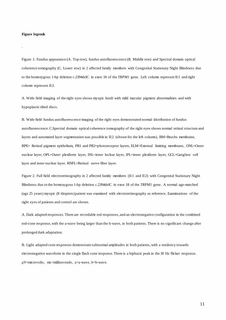
Figure 1: Fundus appearance (A. Top row), fundus autofluorescence (B. Middle row) and Spectral domain optical
coherence tomography (C. Lower row) in 2 affected family members with Congenital Stationary Night Blindness due
to the homozygous 1-bp deletion c.2394delC in exon 18 of the TRPM1 gene. Left column represent II:1 and right
column represent II:2.
A. Wide field imaging of the right eyes shows myopic fundi with mild macular pigment abnormalities and with
hypoplastic tilted discs.
B. Wide field fundus autofluorescence imaging of the right eyes demonstrated normal distribution of fundus
autofluorescence. C.Spectral domain optical coherence tomography of the right eyes shows normal retinal structure and
layers and automated layer segmentation was possible in II:2 (shown for the left column), BM=Bruchs membrane,
RPE= Retinal pigment epithelium, PR1 and PR2=photoreceptor layers, ELM=External limiting membrane, ONL=Outer
nuclear layer, OPL=Outer plexiform layer, INL=inner luclear layer, IPL=inner plexiform layer, GCL=Ganglion cell
layer and inner nuclear layer. RNFL=Retinal nerve fiber layer.
Figure 2. Full field electroretinography in 2 affected family members (II:1 and II:2) with Congenital Stationary Night
Blindness due to the homozygous 1-bp deletion c.2394delC in exon 18 of the TRPM1 gene. A normal age-matched
(age 25 years) myopic (8 diopters) patient was examined with electroretinography as reference. Examinations of the
right eyes of patients and control are shown.
A. Dark adapted responses. There are recordable rod responses, and an electronegative configuration in the combined
rod-cone response, with the a-wave being larger than the b-wave, in both patients. There is no significant change after
prolonged dark adaptation.
B. Light adapted cone responses demonstrate subnormal amplitudes in both patients, with a tendency towards
electronegative waveform in the single flash cone response. There is a biphasic peak in the 30 Hz flicker response.
µV=microvolts, ms=milliseconds, a=a-wave, b=b-wave.

12
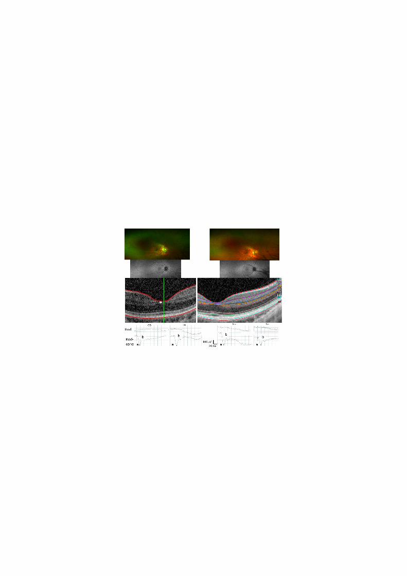
Figure 3: Retinal sublayer thickness profiles in patient II:2 with Congenital Stationary Night Blindness due to the
homozygous 1-bp deletion c.2394delC in exon 18 of the TRPM1 gene. These were obtained from transfoveal horizontal
spectral domain optical coherence tomography line scans. Qualitative comparison with 5 age matched (range 20-30,
median 22 years) eyes (C1-C5) from 5 unrelated healthy myopic (myopia range 6-9, median 7.5 diopters) controls.
These layers were obtained by automated segmentation with the Heidelberg software with subsequent isolation of
retinal sublayers. The scaling within each layer is preserved and equal between patient and controls. There seemed to be
a relative and selective reduction of thickness of the inner nuclear layer in II:2, however some degree of generalized
retinal thinning, including the inner nuclear layer, seems to be present in controls 3-5.
A. Photoreceptor and retinal pigment epithelium (RPE) layer: Between the external limiting membrane and Bruchs
membrane.
B. Outer nuclear layer (ONL): Between the outer plexiform layer line and the external limiting membrane.
C. Inner nuclear layer (INL): Between the inner nuclear layer line and the inner plexiform layer line.
D. Ganglion cell layer (GCL): Between the ganglion cell layer line and the retinal nerve fiber layer line.
E. Total retinal and RPE thickness profile.

13


![Index [link.springer.com]978-0-387-69069-8/1.pdf · Blindness, in infancy congenital causes optic nerve disorders, 10 retinal blindness, 4–5 stationary night blindness, 10 cortical](https://img.pdfslide.us/doc/110x75/606fb63edccffa252f3531cc/index-link-978-0-387-69069-81pdf-blindness-in-infancy-congenital-causes.jpg)