Embed Size (px)
Citation preview
14 The Diabetic Foot Journal Vol 18 No 1 2015
Article
Is septic arthritis arising from a diabetic foot wound a diagnosis that we should consider more often?
Rachel Murchison, Catherine Gooday, Ketan Dhatariya
Citation: Murchison R, Gooday C, Dhatariya K (2015) Is septic arthritis arising from a diabetic foot wound a diagnosis that we should consider more often? The Diabetic Foot Journal 18: 14–8
Article points
1. Septic arthritis is a medical emergency.
2. Diabetes is a risk factor for the development of septic arthritis.
3. It should be suspected in patients with a short history of a hot, swollen joint with restricted movement.
Key words
- Accurate diagnosis- Infection- Septic arthritis
Authors
Rachel Murchison is Podiatrist in Diabetes; Catherine Gooday is Principal Podiatrist; Ketan Dhatariya is Consultant Endocrinologist. All are based at the Diabetic Foot Clinic, Elsie Bertram Diabetes Centre, Norwich
Septic arthritis is a difficult to diagnose condition where a joint — usually the hip or knee — becomes inflamed due to infection with bacteria, usually Gram-positive cocci. It is a medical emergency that can lead to high morbidity and mortality, and diabetes is a risk factor. British Society for Rheumatology guidelines (2006) suggest patients with a short history of a hot, swollen joint with restricted movement should be regarded as having septic arthritis until proven otherwise.
Foot ulcers are a common complication of diabetes most frequently associated with neuropathy and ischaemia (Abbott et al,
2002); 2.5% of the diabetic population will have a foot ulcer at any one time (Kerr et al, 2014). Most ulcers are relatively straightforward, requiring standard treatment with off-loading, antibiotics and revascularisation, where necessary (National Institution for Health and Care Excellence [NICE], 2004). However, complications may arise that the heathcare professional may not immediately think of as being directly associated with diabetes-related foot disorder.
This article describes two patients, both long-standing attendees of a multidisciplinary specialist diabetic foot clinic, who presented with acute onset episodes of septic arthritis of the knee.
Septic arthritisThe incidence of septic arthritis in the general population has been infrequently reported, but has been estimated to be between 2.0 and 13.5 per 100,000 patient years in the Middle East, Western Europe and South-East Asia (Al Arfaj, 2008; Mathews et al, 2010; Mahakkanukrauh et al, 2013), but has been reported to be as high as 29 per 100,000 among indigenous Australians (Garcia-Arias et al, 2011).
Septic arthritis is described as joint infection that occurs by pathogenic inoculation directly into the joint or by haematogenous routes (Shirtliff and
Mader, 2002). It is a medical emergency and can lead to high morbidity and mortality (Coakley et al, 2006). Septic arthritis is difficult to diagnose, and the 2006 British Society for Rheumatology guidelines suggest patients with a short history of a hot, swollen joint with restricted movement should be regarded as having septic arthritis until proven otherwise (Coakley et al, 2006).
Synovial fluid is very vascular and, therefore, highly susceptible to pathogens. The management is difficult, but if the condition is not diagnosed expeditiously, it can quickly lead to irreversible joint damage. It is described as — and should be treated as — an orthopaedic emergency (Shirtliff and Mader, 2002). It has been reported to be fatal in about 11% of cases (Coakley et al, 2006).
The large joints are most commonly affected; usually the hip or the knee. Gram-positive cocci,
Figure 1. Evidence of Mrs X’s previous toe amputations
and long-standing plantar ulcers on her left foot.
Is septic arthritis arising from a diabetic foot wound a diagnosis that we should consider more often?
such as Staphylococcus (S.) sp, is found in 60% of cases (Dubost et al, 2000) and Streptococcus sp is the cause in a further 30% of cases (Ryan et al, 1997). Risk factors include osteoarthritis or rheumatoid arthritis, prosthetic joints, intravenous drug abuse, alcoholism, diabetes, previous intra-articular corticosteroid injections and cutaneous ulceration (Dubost, 2000).
When septic arthritis is suspected, the joint must be
aspirated under aseptic conditions, and blood cultures taken before antibiotics are given. Because the most common causative organisms are Staphylococcus sp and Streptococcus, the British Society of Rheumatology guidelines recommend flucloxacillin 2 g four times a day intravenously (Coakley et al, 2006). The joints should be aspirated as often as required. Due to the potential long-term damage to joints, it is
“Septic arthritis is described as joint infection that occurs by pathogenic inoculation directly into the joint or by haematogenous routes.”
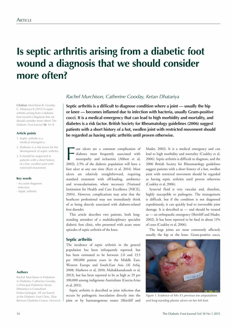
Figure 2. Plain X-ray of Mrs X’s painful right knee showing minor degenerative changes only.
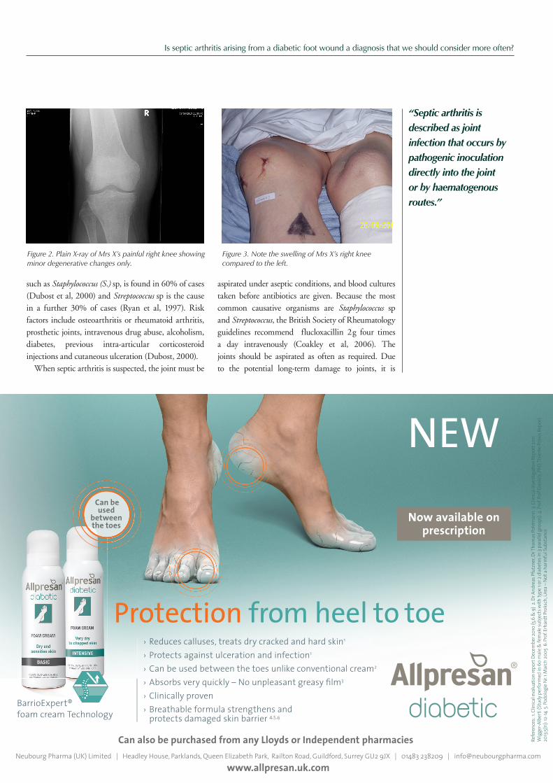
Figure 3. Note the swelling of Mrs X’s right knee compared to the left.
› Reduces calluses, treats dry cracked and hard skin1
› Protects against ulceration and infection1
› Can be used between the toes unlike conventional cream2
› Absorbs very quickly – No unpleasant greasy fi lm3
› Clinically proven› Breathable formula strengthens and protects damaged skin barrier 4,5,6
Refer
ence
s: 1. C
linica
l eva
luatio
n rep
ort D
ecem
ber 2
010
(5,6 &
9) 2
. Dr A
ndre
as Pf
utzn
er, D
r Tho
mas
Pohlm
ann
3. Clin
ical In
vesti
gatio
n Rep
ort 2
011
Wigg
er-Al
berti
(Stu
dy pe
rform
ed in
60 m
ale &
fem
ale su
bjects
with
type
1 or 2
diab
etes
in 3
para
llel g
roup
s) 4.
Prof
. Rolf
Dan
iels, P
hD, T
hiem
e Pra
xis Re
port
2013;
5(11):
12-14
5. P
odolo
gie N
r. 1 M
arch
2005
6. P
rof. E
rhar
dt Pr
oksc
h, Ur
ea –
Not a
harm
ful S
ubsta
nce
Neubourg Pharma (UK) Limited | Headley House, Parklands, Queen Elizabeth Park, Railton Road, Guildford, Surrey GU2 9JX | 01483 238209 | [email protected] www.allpresan.uk.com
Can also be purchased from any Lloyds or Independent pharmacies
BarrioExpert®foam cream Technology
Protection from heel to toe
Now available onprescription
NEW Can be
usedbetween the toes
AZ_gb_diabetic_Porzellan_A5_01.indd 1 04.02.15 17:19
Is septic arthritis arising from a diabetic foot wound a diagnosis that we should consider more often?
16 The Diabetic Foot Journal Vol 18 No 1 2015
recommended that antibiotics should be given for 6 weeks, initially 2 weeks intravenously, followed by 4 weeks orally (Coakley et al, 2006).
Case 1Mrs X was a 77-year-old woman with a 12-year history of type 2 diabetes treated with insulin and oral hypoglycaemic agents. She presented in September 2013 with a painful swollen knee. Her most recent HbA
1c, taken within 3 months of
the current episode, was 61 mmol/mol. All of her foot pulses were palpable, she was insensate to a 10g monofilament, and had vibration perception threshold of greater than 25 volts in both feet when tested with the neurothesiometer. In 2005, Mrs X’s left 1–3 toes were amputated at the metatarsal heads due to infection and she had long-standing ulceration over several of her plantar left metatarsal heads (Figure 1). Her only other relevant medical history was diabetic retinopathy and coeliac disease.
For the 4 months prior to presentation, there was an ulcer over her left first metatarsal head, caused by blistering from new shoes. For the preceding 2 months, Mrs X had been receiving the gold standard treatment for diabetic neuropathic foot ulceration — a total contact cast (TCC) of the left limb along with usual podiatric care of sharp debridement of the wound and regular dressing changes. The wound had been progressing well and remained uninfected, therefore, no antibiotics had been prescribed.
In September 2013, after 2 months of casting, she began to complain of pain and swelling in her right knee. Examination of the knee showed signs of acute inflammation with swelling, heat and redness.
An urgent X-ray was arranged, but this showed only minor degenerative changes in the knee (Figure 2).
It was thought that she could be developing a Charcot of the knee or had suffered mechanical strain on the joint as a result of the TCC on the contralateral limb. It was decided to remove the TCC and review again in 2 weeks to see if this had improved her symptoms.
However, she was unable to attend the follow-up appointment because her knee pain was worsening. Her GP had reviewed her and given her simple analgesia and referred to physiotherapy. She was in regular telephone contact with the specialist diabetes foot clinic where she described increasing pain and immobility. As a result, she was not able to be reviewed in foot clinic.
Four weeks after the initial podiatry clinic review, the GP admitted Mrs X to hospital with a swollen knee (Figure 3). A further X-ray of her knee was clear and DVT was ruled out by an ultrasound scan. By this time, she had developed a further ulcer on her left third metatarsal head. Her C-reactive protein (CRP) measurement on admission was 95 mg/L (NR 0–10), suggesting an inflammatory response, but her white cell count and neutrophil count were normal, suggesting no infection was present.
Her knee was reviewed urgently by the orthopaedic team and aspirated under aseptic technique. Microscopy showed Gram-positive cocci, white blood cells+++, red blood cells+, with no crystals seen. A fully sensitive S. aureus was cultured from the aspirate. A deep tissue swab of her foot wound had grown coliforms. Septic arthritis of the knee was diagnosed and, after microbiological advice, Mrs X was put onto
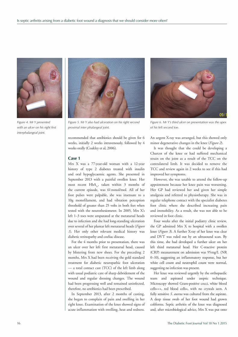
Figure 5. Mr Y also had ulceration on his right second
proximal inter phalangeal joint.
Figure 6. Mr Y’s third ulcer on presentation was the apex
of his left second toe.
Figure 4. Mr Y presented
with an ulcer on his right first
interphalangeal joint.
Is septic arthritis arising from a diabetic foot wound a diagnosis that we should consider more often?
4 weeks’ treatment with intravenous flucloxacillin 1 g four times a day and oral fusidic acid 500 mg three times a day. Her knee was washed out three times over the next few weeks and she had intensive physiotherapy.
Her contralateral foot remained a challenge because this was still ulcerated and was being heavily relied on for weight-bearing during this period. She was in hospital for a total of 6 weeks and then had a further 6 weeks in rehabilitation. Her right knee eventually resolved with no residual joint problems. Her foot ulcers healed after a further year of treatment, but she remains high-risk.
Case 2Mr Y was a 53-year-old male with type 1 diabetes since 1984, treated with a basal bolus insulin regimen. His HbA
1c 6 months prior to this admission had
been 95 mmol/mol. He had no palpable foot pulses, suggesting ischaemia, and was insensate to a 10g monofilament, with vibration perception threshold
of greater than 25 volts bilaterally when tested with the neurothesiometer. In 2010, he had undergone a left leg femoral angioplasty and iliac stent and, in 2011, bilateral iliac angioplasty and right femoral endartectomy due to atherosclerotic disease.
His other relevant medical history was rheumatoid arthritis, for which he took long-term high dose steroids, ischaemic heart disease, with a myocardial infarction in 2005 and subsequent congestive cardiac failure, interstitial lung disease, and chronic obstructive pulmonary disease. Two years previously, a deep tissue swab from a foot ulcer showed methicillin-resistant S aureus (MRSA). Mr Y smoked and had a 10-year history of foot pathology — mainly ulceration of the dorsum, plantar and apex of the left first toe. In 2012, he required an amputation of his left hallux and first ray due to osteomyelitis.
In October 2013, he was admitted as an emergency by his GP. Mr Y was feeling generally unwell, but also had acute onset bilateral knee pain.
“These joints initially need aspirating quickly by an orthopaedic or rheumatology specialist and, as in our two cases, may well need further surgical treatment.”
Cuxson Gerrard & Co. Ltd.,Broadwell Road, Oldbury, West Midlands B69 4BF, UK.
Hapla Gold fights bacteria and fungi andrelieves pressureand frictionIntroducing our most advanced Hapla felt yet... Hapla Gold is an all wool felt which is enhancedwith an antibacterial treatment, making it idealfor use on the at risk foot.
The Hapla 3D System has been successfullyused for over 10 years.
The “Hapla 3D System” offers 3 steps to total pressurerelief using 3 layers of Hapla 10mm Semi Compressed Feltand 5cm Chirofix retention tape.1st Layer10mm Hapla Felt cut as 'D' filler to fill spaceunder medial longitudinalarch. Bevelled with piecrust cut and securedwith Chirofix retentiontape.
2nd Layer10mm Hapla Felt cut asplantar slab/valgus pad.Aperture added to exposewound and woundmargin. Pad bevelled withpie crust and secured withChirofix.
3rd Layer10mm Hapla Feltcut as plantar coverand secured inplace with Chirofix.
For your FREE DVD demonstrating the Hapla 3D System call0800 018 7117
Pressure relief for foot ulcers
14523 CG Hapla Gold 3D Advert free DVD 24/08/2012 10:25 Page 1
Is septic arthritis arising from a diabetic foot wound a diagnosis that we should consider more often?
18 The Diabetic Foot Journal Vol 18 No 1 2015
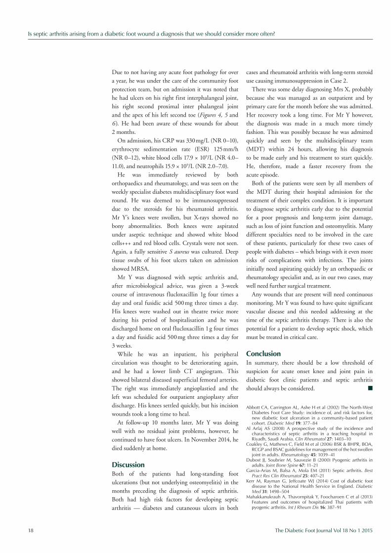
Due to not having any acute foot pathology for over a year, he was under the care of the community foot protection team, but on admission it was noted that he had ulcers on his right first interphalangeal joint, his right second proximal inter phalangeal joint and the apex of his left second toe (Figures 4, 5 and 6 ). He had been aware of these wounds for about 2 months.
On admission, his CRP was 330 mg/L (NR 0–10), erythrocyte sedimentation rate (ESR) 125 mm/h (NR 0–12), white blood cells 17.9 × 10⁹/L (NR 4.0–11.0), and neutrophils 15.9 × 10⁹/L (NR 2.0–7.0).
He was immediately reviewed by both orthopaedics and rheumatology, and was seen on the weekly specialist diabetes multidisciplinary foot ward round. He was deemed to be immunosuppressed due to the steroids for his rheumatoid arthritis. Mr Y’s knees were swollen, but X-rays showed no bony abnormalities. Both knees were aspirated under aseptic technique and showed white blood cells+++ and red blood cells. Crystals were not seen. Again, a fully sensitive S aureus was cultured. Deep tissue swabs of his foot ulcers taken on admission showed MRSA.
Mr Y was diagnosed with septic arthritis and, after microbiological advice, was given a 3-week course of intravenous flucloxacillin 1g four times a day and oral fusidic acid 500 mg three times a day. His knees were washed out in theatre twice more during his period of hospitalisation and he was discharged home on oral flucloxacillin 1 g four times a day and fusidic acid 500 mg three times a day for 3 weeks.
While he was an inpatient, his peripheral circulation was thought to be deteriorating again, and he had a lower limb CT angiogram. This showed bilateral diseased superficial femoral arteries. The right was immediately angioplastied and the left was scheduled for outpatient angioplasty after discharge. His knees settled quickly, but his incision wounds took a long time to heal.
At follow-up 10 months later, Mr Y was doing well with no residual joint problems, however, he continued to have foot ulcers. In November 2014, he died suddenly at home.
DiscussionBoth of the patients had long-standing foot ulcerations (but not underlying osteomyelitis) in the months preceding the diagnosis of septic arthritis. Both had high risk factors for developing septic arthritis — diabetes and cutaneous ulcers in both
cases and rheumatoid arthritis with long-term steroid use causing immunosuppression in Case 2.
There was some delay diagnosing Mrs X, probably because she was managed as an outpatient and by primary care for the month before she was admitted. Her recovery took a long time. For Mr Y however, the diagnosis was made in a much more timely fashion. This was possibly because he was admitted quickly and seen by the multidisciplinary team (MDT) within 24 hours, allowing his diagnosis to be made early and his treatment to start quickly. He, therefore, made a faster recovery from the acute episode.
Both of the patients were seen by all members of the MDT during their hospital admission for the treatment of their complex condition. It is important to diagnose septic arthritis early due to the potential for a poor prognosis and long-term joint damage, such as loss of joint function and osteomyelitis. Many different specialties need to be involved in the care of these patients, particularly for these two cases of people with diabetes – which brings with it even more risks of complications with infections. The joints initially need aspirating quickly by an orthopaedic or rheumatology specialist and, as in our two cases, may well need further surgical treatment.
Any wounds that are present will need continuous monitoring. Mr Y was found to have quite significant vascular disease and this needed addressing at the time of the septic arthritis therapy. There is also the potential for a patient to develop septic shock, which must be treated in critical care.
ConclusionIn summary, there should be a low threshold of suspicion for acute onset knee and joint pain in diabetic foot clinic patients and septic arthritis should always be considered. n
Abbott CA, Carrington AL, Ashe H et al (2002) The North-West Diabetes Foot Care Study: incidence of, and risk factors for, new diabetic foot ulceration in a community-based patient cohort. Diabetic Med 19: 377–84
Al Arfaj AS (2008) A prospective study of the incidence and characteristics of septic arthritis in a teaching hospital in Riyadh, Saudi Arabia. Clin Rheumatol 27: 1403–10
Coakley G, Mathews C, Field M et al (2006) BSR & BHPR, BOA, RCGP and BSAC guidelines for management of the hot swollen joint in adults. Rheumatology 45: 1039–41
Dubost JJ, Soubrier M, Sauvezie B (2000) Pyogenic arthritis in adults. Joint Bone Spine 67: 11–21
Garcia-Arias M, Balsa A, Mola EM (2011) Septic arthritis. Best Pract Res Clin Rheumatol 25: 407–21
Kerr M, Rayman G, Jeffcoate WJ (2014) Cost of diabetic foot disease to the National Health Service in England. Diabetic Med 31: 1498–504
Mahakkanukrauh A, Thavornpitak Y, Foocharoen C et al (2013) Features and outcomes of hospitalized Thai patients with pyogenic arthritis. Int J Rheum Dis 16: 387–91