Embed Size (px)
Citation preview

Seminar on Septic Arthritis
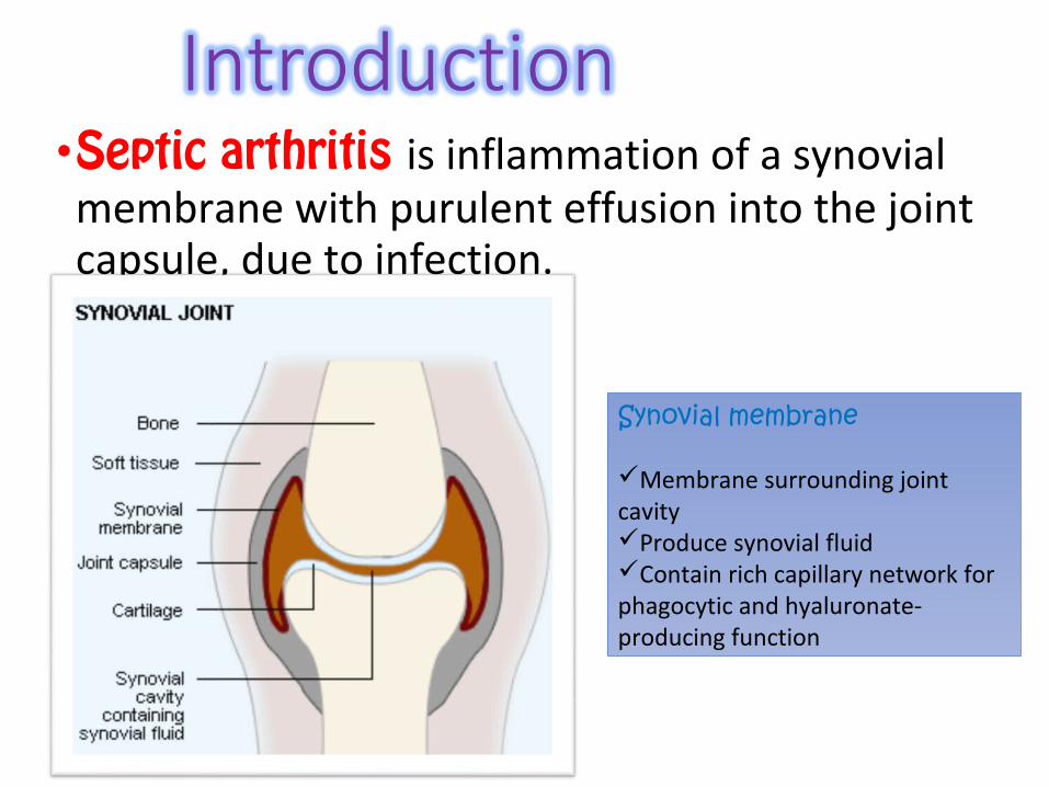
•Septic arthritis is inflammation of a synovial membrane with purulent effusion into the joint capsule, due to infection.
Synovial membrane
Membrane surrounding joint cavityProduce synovial fluidContain rich capillary network for phagocytic and hyaluronate-producing function

Causes of septic arthritis
Bacterial•Non gonococcal arthritis•Gonococcal arthritis
others•Virus•Fungi•Mycobacteria etc.
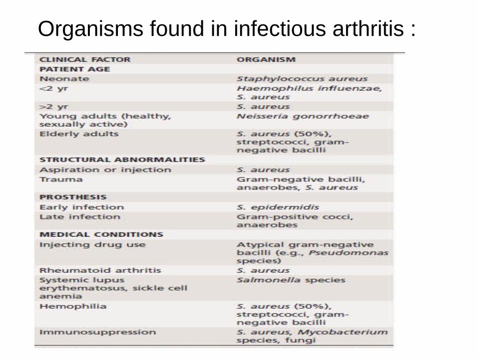
Organisms found in infectious arthritis :

•Infectious agents can gain entrance to a joint via 3 routes:
Haematogenous
Direct inoculation
Direct spread from adjacent focal infection

Most common form of spreadUsually affect people with underlying medical problem
May result from penetrating traumaIntroduction of organisms during diagnostic and surgical procedures. For eg arthroscopy and intra-articular injection
More common in children.Osteomyelitis usually begin in the metaphyseal region, from which it breaks through the periosteum into the joint.

Synovial membrane is highly vascularised.↓
Bacteria can easily enter synovial joint via blood stream.↓
There will be inflammatory reaction with seropurulent exudate and increase in synovial fluid.
↓As pus appear in the joint, the articular cartilage is eroded and destroyed.Partly by the bacterial enzyme, and partly by the enzyme released from
synovium, inflammatory cell and pus
Infant
Destroy the epiphysis, which is still largely
cartilaginous.
Children
Vascular occlusion lead to necrosis of
epiphyseal bone
Adult
Effect confined on articular cartilage
Extensive erosion can occur due to synovial
proliferation and ingrowth

a) In the early stage, there is an acute synovitis with a purulent joint effusion
b) Soon the articular cartilage is attacked by bacterial and cellular enzyme.
c) If infection is not arrested , the cartilage may be completely destroyed
d) Healing then leads to ankylosis

...
•Within 24-48 hrs of bacterial invasion:• Infiltration by neutrophils •Vascular congestion•Synovial proliferation•Within 1 week following bacterial invasion:•Continual purulent effusion•Continual synovial proliferation• Infiltration by mononuclear cells•Granulation tissue •Abscess development•Within 10 days after abscess formation:•Cytokine induced protelytic enzymes are released•End result is joint destruction and or systemic sepsis

If left untreated, it will spread to the underlying bone and out of joint to form abscess and sinus.
Healing with:1.Complete resolution2.Partial loss of articular cartilage and fibrosis of joint3.Loss of articular cartilage and bony ankylosis4.Bony destruction and permanent deformity

ACUTE BACTERIAL ARTHRITIS
•Staphylococcus Aureus – 50%•Streptococcus species, such as Streptococcus
viridans, S Pneumoniae & group B streptocci•Gram negative bacilli – 10% - E.coli & pseudomonas
– More common•Sites : Monoarticular involvement - -85% , knee –
most common•Other – hip , wrist, shoulder & ankle•Sternoclavicular and sacroiliac joint - IVDA

PREDISPOSING FACTORS:
•Artificial joint implants•Bacterial infection elsewhere in body•Chronic illness or disease (such as diabetes, rheumatoid arthritis, and sickle cell disease)•Intravenous (IV) or injection drug use•Medications that suppress immune system•Recent joint trauma•Recent joint arthroscopy or other surgery

Differ according to age
In new born infants
More on septicaemia Rather than joint pain
Baby is irritable & refuse to feed
Tachycardia with fever
Joints are warmth, tenderness, resistance to movement
Umbilical cord and inflamed IV site should be suspicious of source of
infection
In childreno acute pain in single large joint(esp hip)
o Pseudoparesis
o Child is ill,rapid pulse and swinging fever
o Overlying skin looks red & superficial joint swelling may be obvious
o Local warmth and marked tenderness
o All movements are restricted by pain or spasm.
o Look for source of infection from septic toe or discharge ear
In adults
Often in the superficial joint(knee, wrist or ankle )
Joints painful, swollen & inflamed.
Warmth and marked local tenderness & movement restricted.
look for gonococcal infection or drug abuse.
Patient with rheumatoid arthritis and especially those on corticosteroid may develop “silent” joint infection.

Physical examination:•Lower limb antalgic limp / cannot walk•Upper limb affected part is closedly guarded•Marked tenderness, active and passive range of motion are limited•Examine for synovial effusion, erythema, heat and tenderness.•Spasm of muscles around the joint may be marked.•Patient may hold the joint in a position to reduce the intra-articular pressure to minimize pain.

Investigations Explaination
Full blood count Elevated white blood cell count
ESR > 40 mm/hr
CRP > 20 mg/dL
Blood culture May be positive

Synovial fluid analysis
Aseptic technique is used during aspiration of synovial fluid.Avoid taken from infected site of skin.The fluid is then analyzed by gross and microscopic examination and culture.
Gross examinations include appearance, volume, viscosity, Microscopic examinations include leucocyte count, staining of smears,, protein.
Finally, culture and sensitivity for definitive diagnosis and treatment.

Suspected condition
Appearance
Viscosity White cells
Crystals Biochemistry Bacteriology
Normal Clear yellow
High Few - As for plasma -
Septic arthritis
Purulent Low + - Glucose low +
Tuberculous arthritis
Turbid Low + - Glucose low +
Rheumatoid arthritis
Cloudy Low + + - - -
Gout Cloudy Normal ++ Urate - -
Pseudogout Cloudy Normal + Pyrophosphate
- -
Osteoarthritis
Clear yellow
High few Often + - -

X ray Early Stage – Normal
Look for soft tissue swelling, loss of tissue planes, widening of joint space and slight subluxation due to fluid in joint. Gas may be seen with E. coli infection
Late stage – Narrowing and irregularity of joint space
Plain film findings of superimposed osteomyelitis may develop (periosteal reaction, bone destruction, sequestrum formation).

Narrowing of joint space and irregularity of subchondral bone.
Joint space losssubchondral erosions and
sclerosis of the femoral head
osteonecrosis and complete collapse of
the femoral head

Ultrasonography-•More reliable in revealing a joint effusion in early cases.•Widening of space between capsule and bone of > 2mm indicates effusion.•Echo-free transient synovitis•Positively echogenic septic arthritis

CT scans, MRI, and bone scans
•CT scans – soft tissue swelling, joint effusions, abscess formation, guide joint aspiration, monitor therapy and planning operative approaches.•MRI – extent of infection, diagnosing infections that are difficult to access, better anatomical detail.•Bone scans- detect localized areas of inflammation.

Treatment :• The first priority is to aspirate the joint and examine the fluid,
treatment is then started without further delay.• General measures:- analgesics- iv fluids• Splintage--the joint must be rested either on a splint or in a widely split
plaster-in neonates and infants, with hip infection the joint is held
abducted and 30 degree flexed, on traction to prevent dislocation.• Antibiotics –- treatment is started once blood and samples are obtained.-empirical treatment is started depending on most likely organism.

Empirical antibiotic therapy

Pathogen directed antimicrobial therapy :

Surgical DrainageIndications:-Joints that don’t respond to antimicrobial therapy and daily arthrocentesis-Any joint with limited accessibility, including the sternoclavicular or the hip joint-Patient with underlying disease( DM,RA, immunosuppression etc) need more aggressive treatment with earlier surgical intervention
Arthroscopic debridement and copious irrigation with normal saline – more frequently in knee joint septic arthritis

- Bone destruction and dislocation of the joint (esp Hip)
•Cartilage destruction -may lead to either fibrosis or bony ankylosis- in adult partial destruction of the joint will result in secondary osteoarthritis
•Growth disturbance - presenting as either localised deformity or shortening of the bone

Gonococcal arthritis
- results from gonococcal infection (colonization of urethra, cervix, pharynx)
-Sexually active healthy persons
-More common in women than men
-Congenital Complement component deficiency

Clinical features-
•Disseminated gonococcal infection- fever, chills, rash, small no. of papules that progresses to haemorrhagic pustules present on trunk and extensor surfaces of distal extremities.•Migratory arthritis and tenosynovitis of the knees,
hand, wrists, feet and ankles.•Cultures of synovial fluid are negative, blood
cultures positive < 45%, synovial fluid may be difficult to obtain , usually contains 10000 – 20000 leucocuytes/micro L.

True gonococcal arthritis-
• Less common than DGI•A single joint such as hip, knee, ankle or wrist is
usually involved.•Synovial fluid contains > 50000leucocytes/micro L,
obtained at ease, cultures of synovial fluid are positive <40%, blood cultures negative.

Treatment-
• Initiallly, ceftriaxone ( 1 g IV every 24 h)• Local and systemic signs resolve, oral antibiotic
(ciprofloxacin 500mg BD) should be started for 7 days. •Penicillin susceptible- amoxiciilin 500 mg TDS .•Suppurative arthritis usually respond to needle
aspiration and antibiotic treatment for 7-10 days.

Mycobacterial arthritis
• 1% of all cases of TB and 10% of extrapulmonary cases• Pathology –• Enters the body via lung(droplet infection) or the gut( swallowing
infected milk priducts), rarely through skin• It causes granulomatous infection associated with tissue necrosis and
caseation.• Primary complex – initial lesion in lungs , phayrnx or gut with
lymphatic spread to regional lymph nodes.• Secondary spread- widespread dissemination via blood stream
giving rise to extrapulmonary lesions.• Tertiary lesion- foci developing to destructive lesions.• Once they get foothold they elicit a chronic inflammation.

synovium involved
becomes thick and oedematous, marked effusion
pannus of granulation tissue develops, articular cartilage slowly destroyed, increased vascularity causes osteopenia
if unchecked, caseation and infection extend into surrounding soft tissues and produce cold abscess
may burst forming sinus or tuberculous ulcer

Clinical features
•Previous history of infection or recent contact with TB•A long history of pain and swelling•Marked synovial thickening• Involvement of only one joint•Severe muscle wasting•Enlarged and matted regional lymph nodes•Night cries, fever, night sweats, loss of weight.

investigations
•X- ray – soft tissue swelling , periarticular osteoporosis, articular space narrowing, epiphyseal enlargement in children, erosion of subarticular bone, little or no periosteal reaction.•ESR elevated, Mantoux test positive•Synovial fluid contains average cell count of
20000/micro L with 50% neutrophils. AFB staining may be positive•Culture of synovial tissue taken at biopsy is more
reliable.•NAA assays can shorten the time to diagnose

treatment
•Rest
•Chemotherapy : two months course of isoniazide(600mg), rifampicin(450mg), pyrazinamide(1500mg), ethambutol(1200mg) thrice weekly and then 4 months course of isoniazide,ethambutol rifampicin thrice weekly.

Fungal arthritis
• Infection causes granulomatous reaction, often leading to abscess formation, tissue destruction and ulcer formation•Superficial and deep infections.•Superficial mycoses- primarily infections of skin and
mucous membrane. Eg, madurmycoses, sporothrix , candida, actinomycoses.•Deep mycoses- blastomyces, histoplasma ,
Cryptococcus, coccidioides, aspergillus. Gain entry through lungs.

Madurmycosis- cut in foot
spread through subcutaneous tissues and along the tendon sheaths
Bones and joints are infected by direct invasion, local abscesses form and break through the skin as multiple sinuses.

Clinical features
Subcutanoeus nodule, tenderSwollen foot, induratedDischarging sinuses and ulcersX- rays : multiple bone cavities , progressive bone destructionOrganism can be identified in sinus discharge or biopsy

treatment
• Intravenous amphotericin B advocated
•Necrotic tissue –excised
•Amputation- may be necessary

Candidiasis
•Normal commensal organisms• Immunosuppression predisposing factor•Gain entry through direct contamination during
surgery or other invasive procedures.•Diagnosis is usually made by tissue sampling and
culture•Treament – joint irrigation, curettage, IV
amphotericin B

Viral arthritis
• Infects synovial tissue during systemic infections or by provoking an immunologic reaction that involves joints.•Rubella- arthralgia , frank arthritis within 3 days of
rash following natural infection with rubella•Parvovirus B 19- arthritis, arthropathy , stiffness of
joints•Acute Hep B- arthralgia, fever, urticarial 2 weeks
before onset of jaundice.

summary
•Acute monoarthritis should be evaluated emergently to out possibility of septic arthritis
•Untreated septic arthritis can lead to rapid joint space destruction and systemic sepsis, so early diagnosis is imperative.
- Aspiration of the involved joint is critical to identifying the orgsnism
•Consider septic arthritis in patients with underlying inflammatory arthritis if one joint is more acutely inflamed than others
-Therapy with empirical antibiotic should immediately follow aspiration, with subsequent narrower coverage only after culture results are obtained
•Treatment includes appropriate joint drainage and surgical options depends on the joint involved.