Embed Size (px)
Citation preview
HIT Betriebs GmbH amUniversitätsklinikum Heidelbergmit beschränkter Haftung
www.med.uni-heidelberg.de/hit
Ion- and proton-beams: Experience with Monte Carlo Simulation
Katia Parodi, Ph.D.
Heidelberg Ion Therapy Centre, Heidelberg, Germany(Previously: Massachusetts General Hospital, Boston, USA)
Workshop on Monte Carlo usage in the medical fieldRome, Italy,07.12.2007
Massachusetts General Hospitaland Harvard Medical School
The advantages of ion therapy
Physical validation of the FLUKA MC tool
Examples of clinical applications (from dose calculations to PET monitoring)
Conclusion and outlook
Overview
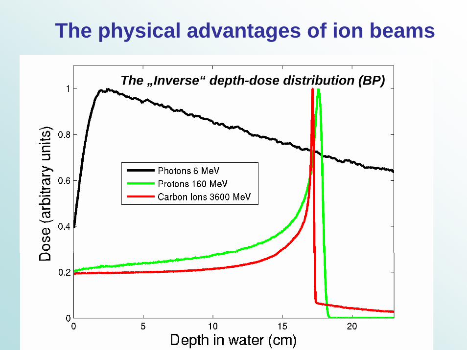
The physical advantages of ion beams
The „Inverse“ depth-dose distribution (BP)
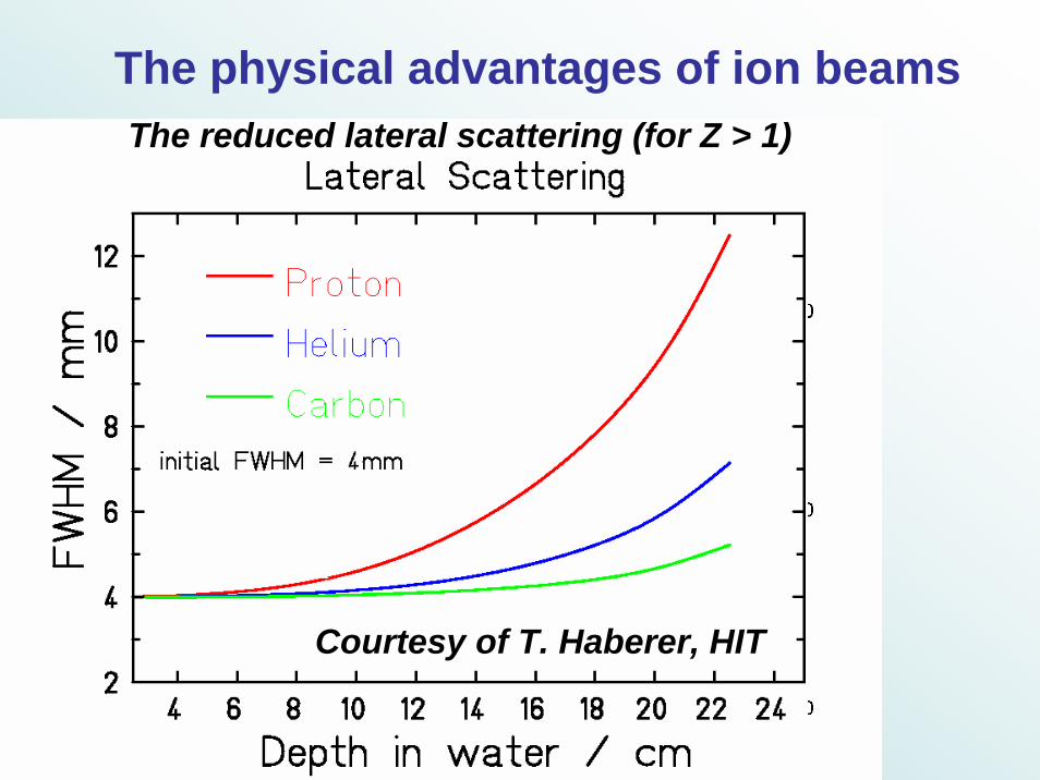
The physical advantages of ion beamsThe reduced lateral scattering (for Z > 1)
Courtesy of T. Haberer, HIT
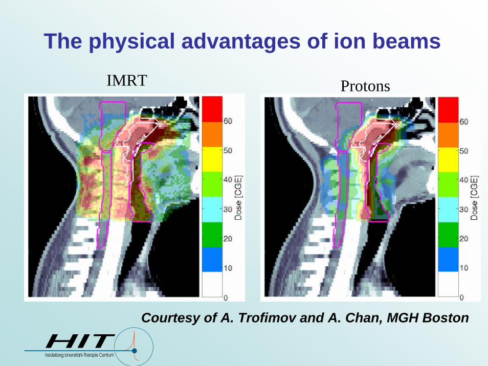
The physical advantages of ion beamsIMRT Protons
Courtesy of A. Trofimov and A. Chan, MGH Boston
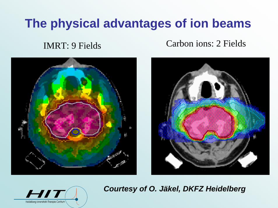
The physical advantages of ion beamsIMRT: 9 Fields Carbon ions: 2 Fields
Courtesy of O. Jäkel, DKFZ Heidelberg

The role of MC in ion therapy
In clinical practice: validation of critical TPS dose calculations (e.g., inhomogeneous tissue, metallic implants)
In commissioning of new facilities: specification of the beam parameters and generation of TPS input data ( meas. time)
In dedicated applications: powerful tools for nuclear reaction related issues, like PET monitoring of ion treatment
Long computational time Realistic representation of physical interactions in complextargets, e.g., patient ☺

The FLUKA MC code(http://www.fluka.org)
Reliable nuclear models
Already applied to proton therapy for dosimetricand radiobiological studies (Biaggi et al NIM B 159, 1999)
Import of raw CT scans with optimized algorithms for efficient transport in voxel geometries(Andersen et al Radiat. Prot. Dosimetry 116, 2005)
Huge efforts for ion transport in connection with NASAgrant since 2000
Promising results from initial studies of ion beam fragmentation in water (Sommerer et al PMB 51, 2006)
Recent improvements for therapeutic ion applications:BME generator for low energy nucleus-nucleus reactions (Cerutti et al Proc. VII LASNPA Conf., Cusco, Peru, 2007)
The
GO
LE
M p
hant
omPe
tous
si-H
enss
etal
, 200
2
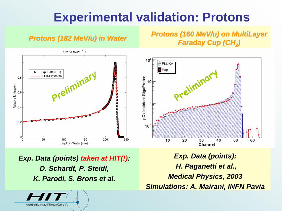
Experimental validation: Protons Protons (160 Protons (160 MeV/uMeV/u) on ) on MultiLayerMultiLayer
Faraday Cup (CHFaraday Cup (CH22))
Exp. Data (points):H. Paganetti et al.,
Medical Physics, 2003 Simulations: A. Mairani, INFN Pavia
Prelim
inary
Exp. Data (points) taken at HIT(!): D. Schardt, P. Steidl,
K. Parodi, S. Brons et al.
Preliminary
Protons (182 Protons (182 MeV/uMeV/u) in Water) in Water
Exp. Data (points): Haettner et al, Rad. Prot. Dos. 2006Simulations: A. Mairani, Ph.D. Thesis, 2007
FLUKA
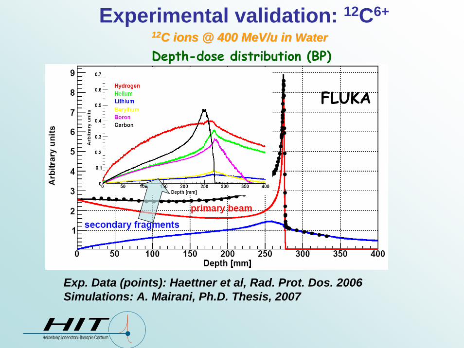
Depth-dose distribution (BP)
1212C ions @ 400 C ions @ 400 MeV/uMeV/u in in WaterWaterExperimental validation: 12C6+
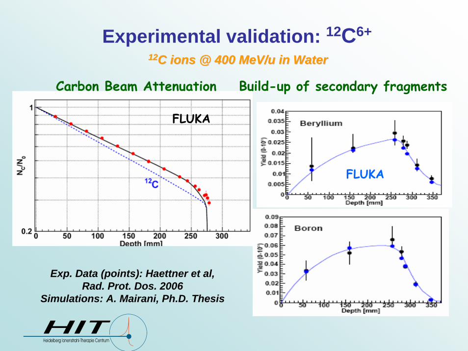
1212C ions @ 400 C ions @ 400 MeV/uMeV/u in Water in Water
Exp. Data (points): Haettner et al, Rad. Prot. Dos. 2006
Simulations: A. Mairani, Ph.D. Thesis
Carbon Beam Attenuation
FLUKA
Build-up of secondary fragments
FLUKA
Experimental validation: 12C6+
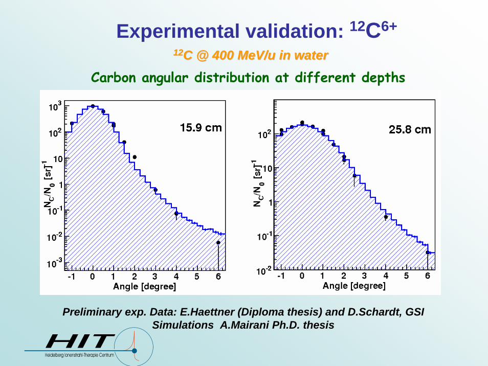
Experimental validation: 12C6+
Preliminary exp. Data: E.Haettner (Diploma thesis) and D.Schardt, GSI Simulations A.Mairani Ph.D. thesis
1212C @ 400 C @ 400 MeV/uMeV/u in waterin water
Carbon angular distribution at different depths

Experimental validation: 12C6+
Heavy Fragment angular distribution at 31.2 cm
1212C @ 400 C @ 400 MeV/uMeV/u in waterin water
Preliminary exp. Data: E.Haettner (Diploma thesis) and D.Schardt, GSI Simulations A.Mairani Ph.D. thesis

Two examples of clinical dose calculations
I. For protons @ MGH Boston II. For 12C @ GSI Darmstadt

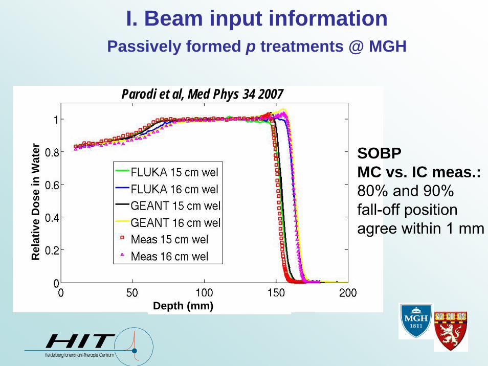
I. Beam input informationPassively formed p treatments @ MGH
At MGH: FLUKA coupled with beam phase-space from a Geant4 MC-calculation of nozzle and beam modifiers
Paganetti et al,Med. Phys. 2004
Example of set-up(Tsukuba)
The general principles of Passive Modulation
I. Beam input informationPassively formed p treatments @ MGHR
elat
ive
Dos
e in
Wat
er
Depth (mm)
Parodi et al, Med Phys 34 2007
SOBPMC vs. IC meas.:80% and 90% fall-off positionagree within 1 mm

II. Beam input informationActive rasterscan system for 12C treatments @ GSI
Haberer et al, NIMA 1993
Intensity- andPosition- controlledmagnetic scanning
Active variationof beam energy, focus and intensityfrom accelerator

II. Beam input informationActive rasterscan system for 12C treatments @ GSI
Experimental data (points) from S.Brons (HIT)Simulations: A. Mairani, Ph.D. Thesis
SOBP MC vs IC meas
FLUKA coupled with control file of raster scanning system and modeling ridge filter(F. Sommerer presented at MC Workshop, Ghent 2006, http://www.ewg-mctp.ugent.be )
FLUKAData
Lateral Profiles

CT segmentation into 27 materials (Schneider et al PMB 45, 2000, extended to include Ti in Parodi et al, Med. Phys. 34, 2007)
Soft tissue
Air, Lung,Adipose tissue
Skeletal tissue
I-II. Using the information from the patient CT
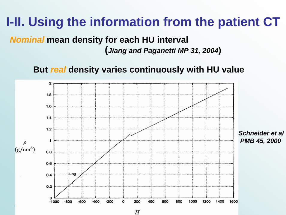
Nominal mean density for each HU interval(Jiang and Paganetti MP 31, 2004)
I-II. Using the information from the patient CT
Schneider et alPMB 45, 2000
But real density varies continuously with HU value

I. Adaptation of FLUKA to follow the TPS (XiO/CMS) calibration curve for p @ MGH
HU dependent adjustment of nuclear and electromagnetic processes, reproducing same calibration curve as TPS
(similar to Jiang and Paganetti MP 31, 2004)
Parodi et al MP 34, 2007, Parodi et PMB 52, 2007
1000
Ti

II. Adaptation of FLUKA to follow the TPS (TRiP) calibration curve for 12C @ GSI
O. Geiß et al, GSI Scientific Report 1997O. Jäkel et al, Med. Phys. 28 2001
A. Mairani Ph.D. thesis
FLUKATRiP
12C (270 MeV/u) on CT phantoms

I. Proton therapy @ MGH: MC vs XiO/CMS
Clivus Chordoma PatientXiO/CMS FLUKA
Prescribed dose: 1 GyEMC : ~ 5.5 106 protons in 10 independent runs
(11h each on Linux Cluster mostly using 2.2GHz Athlon processors)
Parodi et al, JPCS 74, 2007
mGy mGy

XiO/CMS FLUKAmGy mGy
I. Proton therapy @ MGH: MC vs XiO/CMS
FLUKAXiO/CMS
mGy mGy

FLUKA mGy
Prescribed dose: 2 GyEMC : ~ 7.4 107p in 12 independent runs (~ 130h each on 2.2 GHz Linux cluster)
XiO/CMS
metallic implants
K. Parodi et al, IJROBP 2007
mGy I. Proton therapy @ MGH: MC vs XiO/CMS
Paraspinal tumor with metallic implants
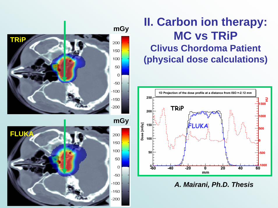
II. Carbon ion therapy: MC vs TRiP
Clivus Chordoma Patient(physical dose calculations)
A. Mairani, Ph.D. Thesis
TRiPmGy
FLUKA
mGyFLUKA
TRiP
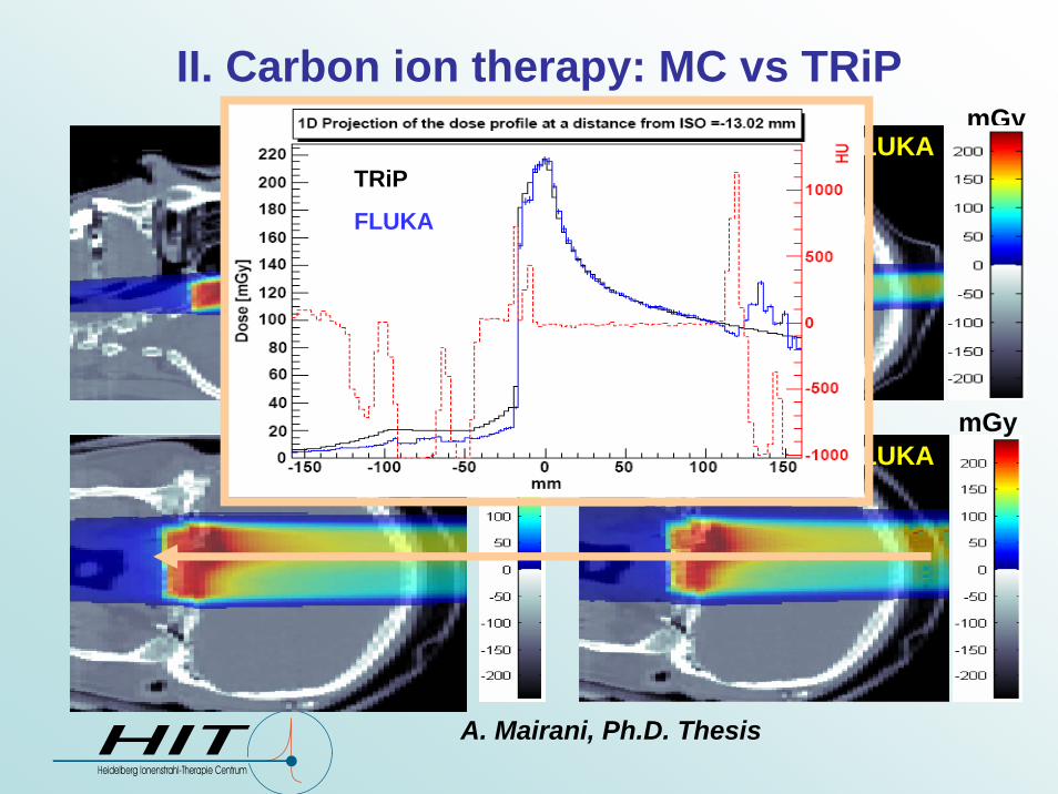
A. Mairani, Ph.D. Thesis
II. Carbon ion therapy: MC vs TRiP
TRiP FLUKAmGymGy
mGyFLUKA
mGyTRiP
TRiP
FLUKA

Two examples of application at the upcomingHeidelberg Ion Therapy Center (HIT)
Ion species• low-LET: Protons
(later He)• high-LET: Carbon
(Oxygen)Beam delivery• Rasterscanning with
active energy variation(like GSI)
• Required parameters:255 Energy steps4 (6) Foci10 IntensitiesFLUKA simulations
HIT, Heidelberg, Germany
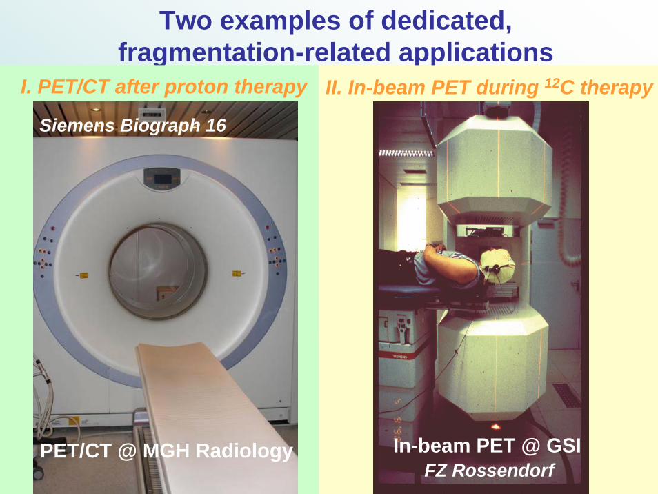
Two examples of dedicated, fragmentation-related applications
PET/CT @ MGH Radiology
Siemens Biograph 16
I. PET/CT after proton therapy II. In-beam PET during 12C therapy
In-beam PET @ GSIFZ Rossendorf

The principle of PET monitoring in ion therapy
Production of positron emitters (15O, 11C, 13N..., T1/2 ~ 2, 20 and 10min...) as a by-product of irradiation
Before collision After collisionProton
Target fragment
Proton
Atomic nucleusof tissue
16O 15O Neutron
Before collision After collisionProjectilefragment
Target fragment
Projectile12C ion
Atomic nucleusof tissue
16O 15ONeutrons
C12 11C
15O 15N + e+ + νeT1/2
180°Δt = 0
Eγ = 511 keV
InIn--vivo, nonvivo, non--invasive detection invasive detection of irradiation induced of irradiation induced ββ++--activityactivityby means of PETby means of PET
Courtesy of W. Enghardt, FZ Rossendorf
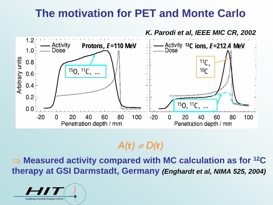
The motivation for PET and Monte Carlo
⇒⇒ Measured activity compared with MC calculation as for 12C therapy at GSI Darmstadt, Germany (Enghardt et al, NIMA 525, 2004)
K. Parodi et al, IEEE MIC CR, 200212C ions, E=212 A MeVProtons, E=110 MeV
11C,10C
15O, 11C, ...
15O, 11C, ...
A(A(rr) ) ≠≠ D(D(rr))
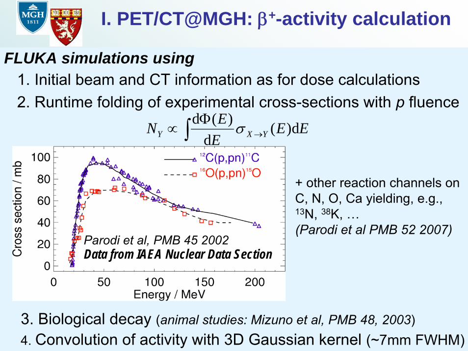
I. PET/CT@MGH: β+-activity calculation
FLUKA simulations using1. Initial beam and CT information as for dose calculations 2. Runtime folding of experimental cross-sections with p fluence
3. Biological decay (animal studies: Mizuno et al, PMB 48, 2003)4. Convolution of activity with 3D Gaussian kernel (~7mm FWHM)
∫ →Φ
∝ EEEEN YXY d)(
d)(d σ
+ other reaction channels on C, N, O, Ca yielding, e.g., 13N, 38K, …(Parodi et al PMB 52 2007)
Data from IAEA Nuclear Data SectionParodi et al, PMB 45 2002
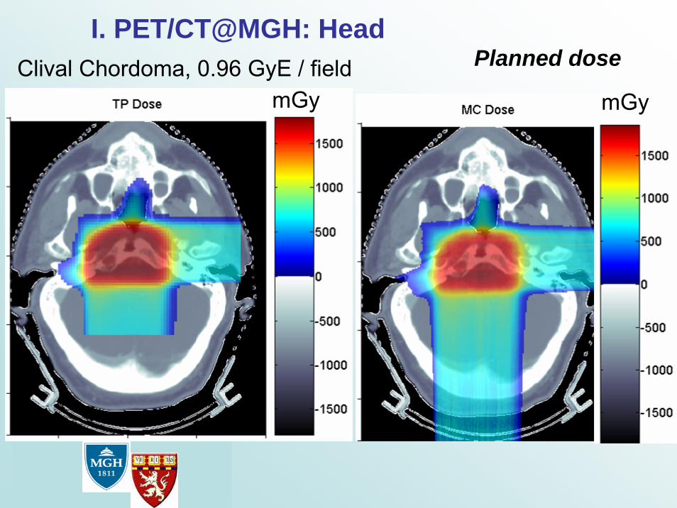
I. PET/CT@MGH: Head
mGy mGyClival Chordoma, 0.96 GyE / field Planned dose
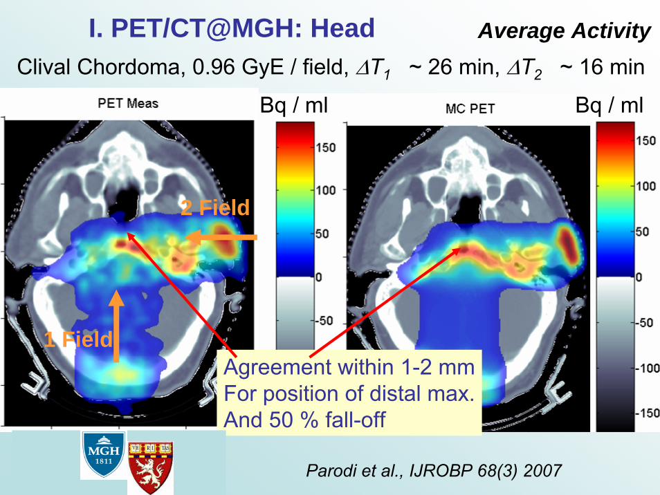
I. PET/CT@MGH: HeadClival Chordoma, 0.96 GyE / field, ΔT1 ~ 26 min, ΔT2 ~ 16 min
Bq / ml Bq / ml
Parodi et al., IJROBP 68(3) 2007
Agreement within 1-2 mmFor position of distal max.And 50 % fall-off
Average Activity
1 Field
2 Field
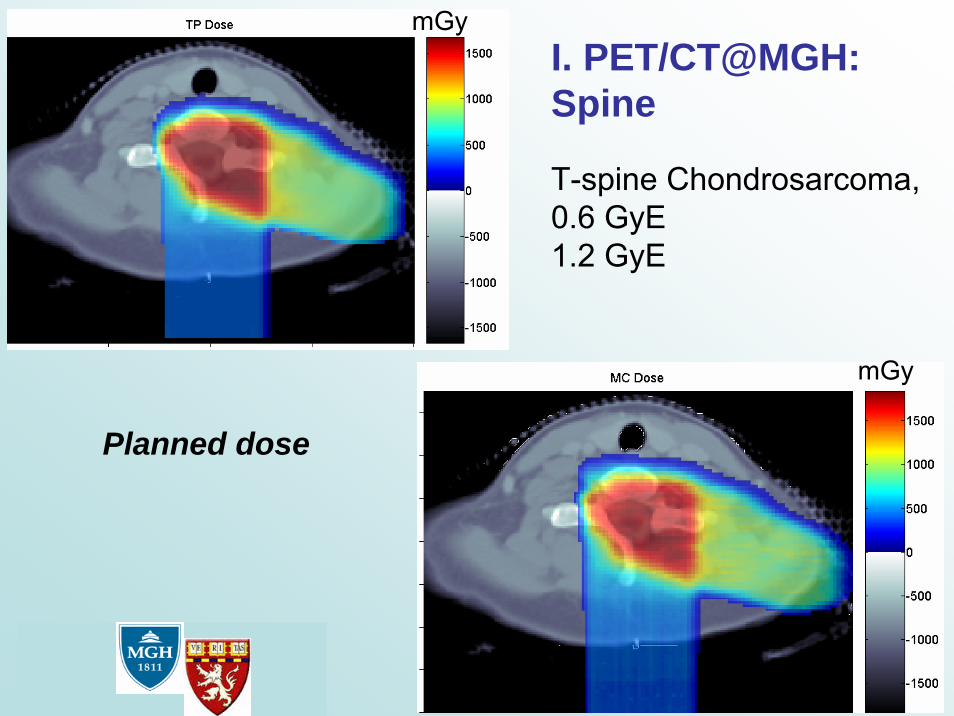
I. PET/CT@MGH: Spine
T-spine Chondrosarcoma, 0.6 GyE1.2 GyE
mGy
mGy
Planned dose

I. PET/CT@MGH: Spine
Average Activity
Bq / ml
Bq / ml
Parodi et al., IJROBP 68(3) 2007
T-spine Chondrosarcoma, 0.6 GyE DT1 ~ 22 min,1.2 GyE DT2 ~ 16 min
1 Field
2 Field
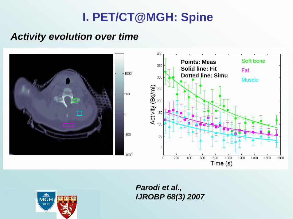
Points: MeasSolid line: FitDotted line: Simu
Activity evolution over time
I. PET/CT@MGH: Spine
Parodi et al., IJROBP 68(3) 2007

FLUKAMeasurementF. Sommerer et al PMB 51 2006, PhD Thesis, Wien 2007
260 MeV/u 12C ion on Graphite, backprojections
II. In-beam PET imaging of heavier ionsOngoing work on:
Application of FLUKA to PET monitoring of ions (e.g. 12C, 16O) based on internal nuclear modelsSimulation of imaging process (β+-decay, propagation of e+ and annihilation photons, detection) same as for measured data
Exact replica of the experimental setup, PET heads includedFLUKA irradiation+decay features exploitedMC γ’s detection converted to list-mode data by modified PETSIM1
Backprojection with same routines as in experiment 1Pönisch et al. PMB 49 2004
In-beam PET phantom experiments @ GSI
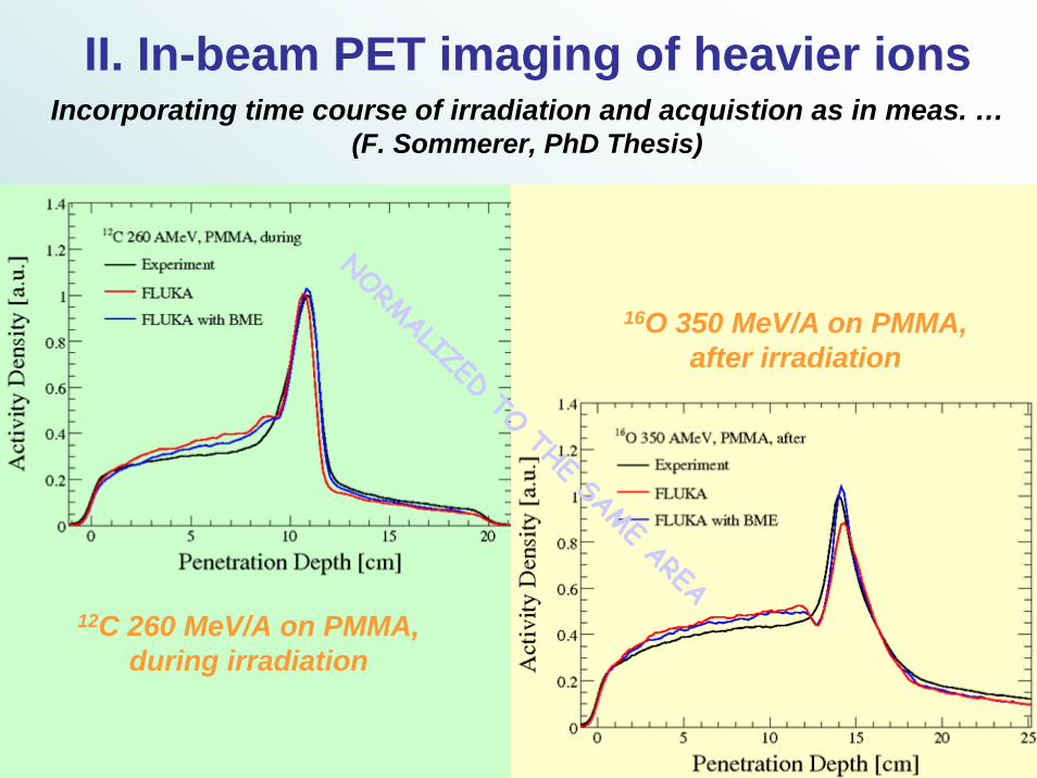
II. In-beam PET imaging of heavier ions
12C 260 MeV/A on PMMA, during irradiation
16O 350 MeV/A on PMMAafter irradiation
NORMALIZED TO THE SAME AREA
,
Incorporating time course of irradiation and acquistion as in meas. …(F. Sommerer, PhD Thesis)
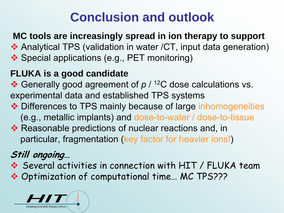
Conclusion and outlookMC tools are increasingly spread in ion therapy to support
Analytical TPS (validation in water /CT, input data generation)Special applications (e.g., PET monitoring)
FLUKA is a good candidateGenerally good agreement of p / 12C dose calculations vs.
experimental data and established TPS systemsDifferences to TPS mainly because of large inhomogeneities(e.g., metallic implants) and dose-to-water / dose-to-tissue Reasonable predictions of nuclear reactions and, inparticular, fragmentation (key factor for heavier ions!)
Still ongoing…Several activities in connection with HIT / FLUKA team Optimization of computational time… MC TPS???

Acknowledgement
CERN Geneva:A. Ferrari, F. Sommerer, F. Cerutti
HIT Heidelberg:S. Brons, T. Haberer, P. Heeg, J. Naumann
INFN Pavia:A. Mairani
MGH Boston:H. Paganetti, T. Bortfeld, H. Shih
FZ Rossendorf:W. Enghardt