Embed Size (px)
Citation preview
Iodothyronine Levels in the Human Developing Brain:Major Regulatory Roles of Iodothyronine Deiodinases inDifferent Areas
MONIQUE H. A. KESTER, RAQUEL MARTINEZ DE MENA, MARIA JESUS OBREGON,DANIJELA MARINKOVIC, ALLAN HOWATSON, THEO J. VISSER, ROBERT HUME, AND
GABRIELLA MORREALE DE ESCOBAR
Department of Internal Medicine (M.H.A.K., D.M., T.J.V.), Erasmus University Medical Center, 3000 DR Rotterdam, TheNetherlands; Departmento de Endocrinologıa (R.M.d.M., M.J.O., G.M.d.E.), Instituto de Investigaciones Biomedicas AlbertSols, Consejo Superior de Investigaciones Cientificas-UAM, 28029 Madrid, Spain; Pathology Department (A.H.), RoyalHospital for Sick Children, Yorkhill National Health Service Trust, Glasgow G3 8SJ, Scotland, United Kingdom; andMaternal and Child Health Sciences (R.H.), University of Dundee, Dundee DD1 9SY, Scotland, United Kingdom
Thyroid hormones are required for human brain develop-ment, but data on local regulation are limited. We describe theontogenic changes in T4, T3, and rT3 and in the activities of thetypes I, II, and III iodothyronine deiodinases (D1, D2, and D3)in different brain regions in normal fetuses (13–20 wk post-menstrual age) and premature infants (24–42 wk postmen-strual age). D1 activity was undetectable.
The developmental changes in the concentrations of theiodothyronines and D2 and D3 activities showed spatial andtemporal specificity but with divergence in the cerebral cor-tex and cerebellum. T3 increased in the cortex between 13 and20 wk to levels higher than adults, unexpected given the lowcirculating T3. Considerable D2 activity was found in the cor-tex, which correlated positively with T4 (r � 0.65). Cortex D3activity was very low, as was D3 activity in germinal eminence
and choroid plexus. In contrast, cerebellar T3 was very lowand increased only after midgestation. Cerebellum D3 activ-ities were the highest (64 fmol/min�mg) of the regions studied,decreasing after midgestation. Other regions with high D3activities (midbrain, basal ganglia, brain stem, spinal cord,hippocampus) also had low T3 until D3 started decreasingafter midgestation. D3 was correlated with T3 (r � �0.682) andrT3/T3 (r � 0.812) and rT3/T4 (r � 0.889).
Our data support the hypothesis that T3 is required by thehuman cerebral cortex before midgestation, when mother isthe only source of T4. D2 and D3 play important roles in thelocal bioavailability of T3. T3 is produced from T4 by D2, andD3 protects brain regions from excessive T3 until differenti-ation is required. (J Clin Endocrinol Metab 89: 3117–3128,2004)
THYROID HORMONE IS necessary for normal brain de-velopment. It becomes increasingly clear that the levels
of thyroid hormones required at different stages of devel-opment are critical. Conditions relating thyroid hormones topoor brain development have recently been summarized ina review by Morreale de Escobar et al. (1). For instance,congenital hypothyroidism leads to severe mental retarda-tion if it remains untreated. Maternal hypothyroxinemiacaused by marked iodine deficiency during the first half ofpregnancy is also causally related to neurological cretinism(2) and a decreased mental development of a large propor-tion of the noncretin population (3). Even undiagnosed earlymaternal hypothyroxinemia (4, 5) or hypothyroidism (6) hasbeen suggested to adversely affect neurological developmentof the child. Not only maternal and/or fetal and neonatalhypothyroidism clearly affect brain development, but also
excessive levels of thyroid hormones may lead to abnormalbrain development (7).
Nuclear thyroid hormone receptors have been demon-strated in the brain from wk 10 (8). In the first trimester, thefetus is solely dependent on maternal thyroid hormones,which cross the human placenta (9–11). From midgestation,secretion of thyroid hormone by the fetal thyroid becomesincreasingly important (12), although the maternal contri-bution of thyroid hormone persists until birth (13) and maystill play a critical role in the preferential protection of thefetal brain from T3 deficiency (11). Serum T3 levels are verylow during fetal development, ranging from less than 33ng/dl (0.5 nm) at 13 wk postmenstrual age (PMA) (14) up toapproximately 65 ng/dl (1.0 nm) at term (15). Despite thesevery low serum concentrations, Bernal and Pekonen (8) andBernal and colleagues (16, 17) measured T3 in several fetaltissues between 13 and 18 wk PMA. Their studies showedthat the concentration of T3 in the fetal cortex at 13 wk PMAmay actually reach 50–60% of the values reported for adults(18), with 20–30% nuclear receptor occupancy. The concen-trations both of the nuclear receptors and total T3 in thecerebral cortex continue to increase rapidly up to 18 wkgestation. This does not occur during the same developmen-tal period in other fetal tissues, such as the lung and liver, and
Abbreviations: BG, Basal ganglia; BS, brain stem; Cbl, cerebellum; CC,cerebral cortex; CP, choroid plexus; D1, D2, D3, types I, II, and IIIdeiodinase; DTT, dithiothreitol; GE, germinal eminence; H, hippocam-pus; MB, midbrain; PMA, postmenstrual age; PTU, 6-n-propyl-2-thio-uracil; SC, spinal cord.JCEM is published monthly by The Endocrine Society (http://www.endo-society.org), the foremost professional society serving the en-docrine community.
0021-972X/04/$15.00/0 The Journal of Clinical Endocrinology & Metabolism 89(7):3117–3128Printed in U.S.A. Copyright © 2004 by The Endocrine Society
doi: 10.1210/jc.2003-031832
3117
by on May 21, 2010 jcem.endojournals.orgDownloaded from
there is no information for brain areas other than the cortex.The T3 levels of the human fetal cortex are much higher thanwould be expected from the serum T3 levels, a finding thatmay be explained by the active transport of thyroid hormonethrough the plasma membrane, the difference in intracellularvs. extracellular thyroid hormone-protein binding, andmainly by local deiodination of iodothyronines (19, 20).
Deiodination is catalyzed by three deiodinases, i.e. typesI, II, and III deiodinase (D1, D2, and D3). D1 is mainlyexpressed in the liver, kidney, and thyroid. Its main functionis the production of serum T3 and the clearance of serum rT3(20–22). D1 is not expressed in cells of the central nervoussystem. D2 is present in brain, pituitary, brown adiposetissue, human thyroid, and skeletal muscle (20, 23–25). Itcatalyzes the outer ring deiodination of T4 to T3 and is thusimportant for the local production of T3. D2 expression in thedifferent tissues is down-regulated in hyperthyroidism andup-regulated in hypothyroidism (23). In the rat, D2 has beendemonstrated in astrocytes throughout the brain, in the me-dian eminence and tanycytes lining the third ventricle (26,27). D3 catalyzes the inner ring deiodination of T4 to rT3 andof T3 to 3,3�-T2 (20–22). It is expressed in brain, skin, fetaltissues, placenta, and uterus and at other sites of the mater-nal-fetal interface, such as the umbilical arteries and vein(28–33). Brain D3 activity is up-regulated in hyperthyroid-ism and down-regulated in hypothyroidism. D3 is predom-inantly present in neuronal cells (34, 35), which are the maincells that express thyroid hormone receptors (36, 37). It hasbeen hypothesized (38, 39) that T4 is taken up from the bloodby glial cells and converted to T3 in these cells. Subsequently,depending on the type of glial cells in which this has oc-curred, T3 would be released from astrocytes to neurons bya paracrine route, whereas the T3 generated in the tanycytescould be secreted into the cerebrospinal fluid and from therereach neural cells. Once T3 reaches neurons, it would beavailable to the thyroid hormone receptors and exert itseffects. The D3 expressed in the neurons would limit T3
bioavailability for receptor binding. In such a model, a closeontogenic regulation of brain D2 and D3 expression seemscrucial for providing T3 to the brain in the amounts neededin different structures at different stages of development.
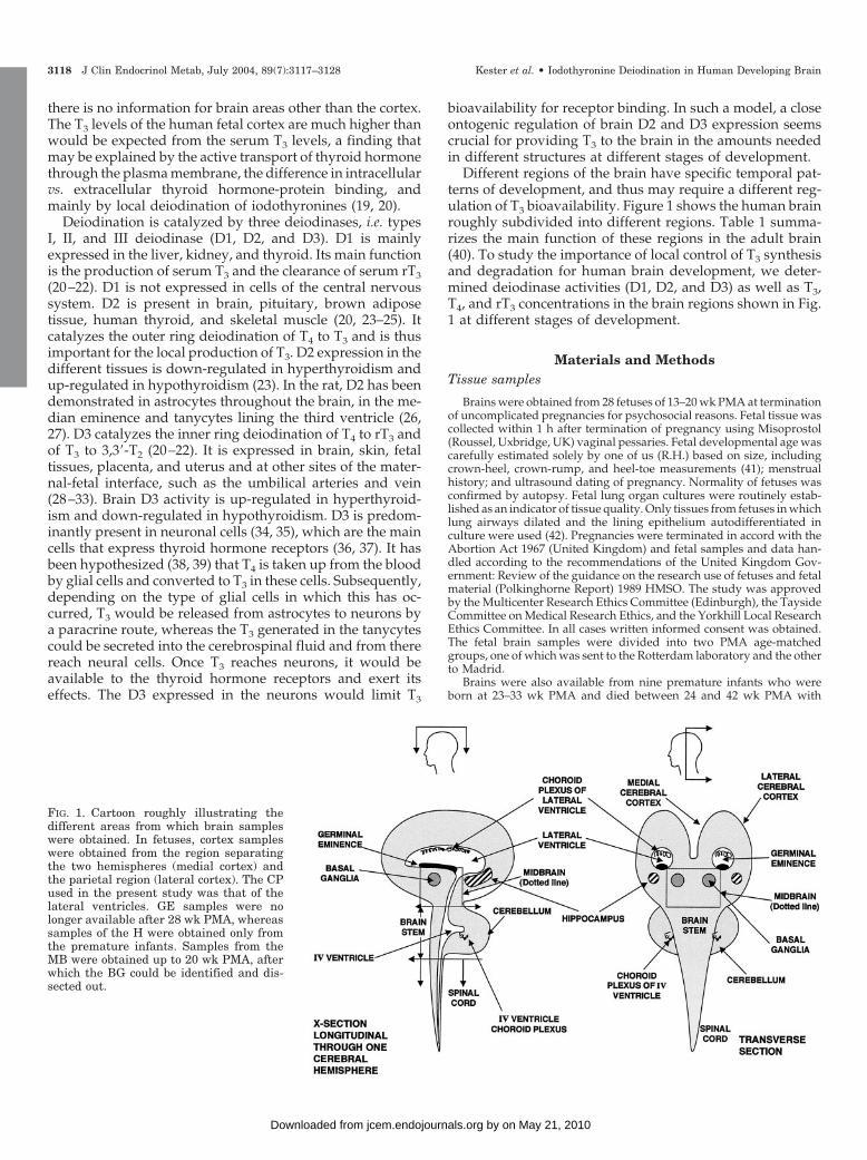
Different regions of the brain have specific temporal pat-terns of development, and thus may require a different reg-ulation of T3 bioavailability. Figure 1 shows the human brainroughly subdivided into different regions. Table 1 summa-rizes the main function of these regions in the adult brain(40). To study the importance of local control of T3 synthesisand degradation for human brain development, we deter-mined deiodinase activities (D1, D2, and D3) as well as T3,T4, and rT3 concentrations in the brain regions shown in Fig.1 at different stages of development.
Materials and MethodsTissue samples
Brains were obtained from 28 fetuses of 13–20 wk PMA at terminationof uncomplicated pregnancies for psychosocial reasons. Fetal tissue wascollected within 1 h after termination of pregnancy using Misoprostol(Roussel, Uxbridge, UK) vaginal pessaries. Fetal developmental age wascarefully estimated solely by one of us (R.H.) based on size, includingcrown-heel, crown-rump, and heel-toe measurements (41); menstrualhistory; and ultrasound dating of pregnancy. Normality of fetuses wasconfirmed by autopsy. Fetal lung organ cultures were routinely estab-lished as an indicator of tissue quality. Only tissues from fetuses in whichlung airways dilated and the lining epithelium autodifferentiated inculture were used (42). Pregnancies were terminated in accord with theAbortion Act 1967 (United Kingdom) and fetal samples and data han-dled according to the recommendations of the United Kingdom Gov-ernment: Review of the guidance on the research use of fetuses and fetalmaterial (Polkinghorne Report) 1989 HMSO. The study was approvedby the Multicenter Research Ethics Committee (Edinburgh), the TaysideCommittee on Medical Research Ethics, and the Yorkhill Local ResearchEthics Committee. In all cases written informed consent was obtained.The fetal brain samples were divided into two PMA age-matchedgroups, one of which was sent to the Rotterdam laboratory and the otherto Madrid.
Brains were also available from nine premature infants who wereborn at 23–33 wk PMA and died between 24 and 42 wk PMA with
FIG. 1. Cartoon roughly illustrating thedifferent areas from which brain sampleswere obtained. In fetuses, cortex sampleswere obtained from the region separatingthe two hemispheres (medial cortex) andthe parietal region (lateral cortex). The CPused in the present study was that of thelateral ventricles. GE samples were nolonger available after 28 wk PMA, whereassamples of the H were obtained only fromthe premature infants. Samples from theMB were obtained up to 20 wk PMA, afterwhich the BG could be identified and dis-sected out.
3118 J Clin Endocrinol Metab, July 2004, 89(7):3117–3128 Kester et al. • Iodothyronine Deiodination in Human Developing Brain
by on May 21, 2010 jcem.endojournals.orgDownloaded from
postnatal ages of 3 min to 15 wk (Table 2). Parental authorization forpostmortem examinations including full organ histology and ancillaryinvestigations was obtained, and examinations were performed by apediatric pathologist (A.H.). The major postmortem findings and diag-noses are given in Table 2. The samples from each hemisphere of thebrains of the premature infants were frozen separately for shipment tothe Madrid and Rotterdam laboratories. The tissues were divided andfrozen in liquid nitrogen and stored at �80 C until use.
Different areas from fetal and premature infants’ brains were dis-sected fresh and snap frozen immediately: choroid plexus (CP), medialand lateral cerebral cortex (CC), germinal eminence (GE), cerebellum(Cbl), brain stem (BS), spinal cord (SC), midbrain (MB), basal ganglia(BG), and hippocampus (H), as illustrated in Fig. 1. Exact dissection ofthe fetal brain areas was done as follows. The fetal brain was exposedby partial removal of the calvaria after incisions were made through thelambdoid, sagittal, and metopic sutures. In the now partially exposedfetal brain, a longitudinal incision was made through the superior aspectof the left CC, and this was extended into the lateral ventricle. The lateralventricle CP was lifted out, exposing the tissue ridge of the GE, whichwas removed by careful dissection. The tissue between the ventricularwall and the surface of the CC on the medial and lateral aspects wasremoved, thereafter called the medial and lateral CC. A similar dissec-tion procedure was carried out on the right cerebral hemisphere and thetissue retained. The remaining cerebral cortical tissue was then removed,
exposing the superior aspect of the MB region. A horizontal incision wasmade just superior to the upper border of the Cbl to define the superiorborder of the BS and the inferior aspect of the MB region, which was thendissected free. The fourth ventricle CP was carefully lifted free, and thecerebellar hemispheres dissected from the BS tissue. The inferior borderof the BS was defined as the level of the pyramidal decussation. Theentire SC was removed by dissection and retained. In all brain regionsthe pia-arachnoid membrane was removed.
The nine premature brains available for sampling ranged from 23 to33 wk. At these stages of development, readily identifiable landmarksand anatomical relationships allowed accurate sampling of brain areasof interest. The Cbl and BS were detached from the upper MB by atransverse incision. The brain was subjected to coronal sectioning, withthe first incision made at the level of the mamillary bodies, and if thesewere not readily identified, the incision was made immediately behindthe optic chiasma. Serial coronal sections, at intervals of 0.5 cm, werethen performed. The coronal sections of brain were then examined topermit accurate orientation for sampling. The medial cortical samplerepresented a block of cortex lining the interhemispheric sulcus betweenthe cerebral hemispheres. This cortical sample was made to a maximumdepth of 1 cm. The lateral cortical sample was of similar depth and wasfrom the parietal cortex. CP was taken from the lateral ventricles. TheBG mass was identifiable adjacent to and below the lateral ventricles.The GE was identifiable in the cases between 24 and 27 wk gestation as
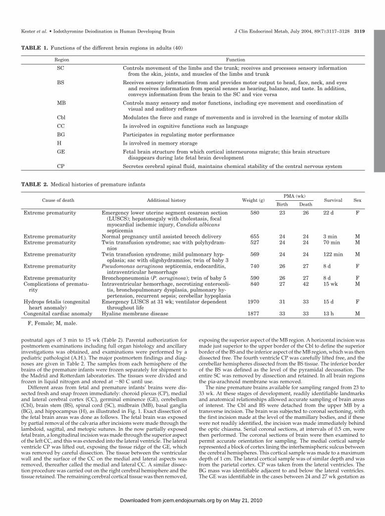
TABLE 2. Medical histories of premature infants
Cause of death Additional history Weight (g)PMA (wk)
Survival SexBirth Death
Extreme prematurity Emergency lower uterine segment cesarean section(LUSCS); hepatomegaly with cholestasis, focalmyocardial ischemic injury, Candida albicanssepticemia
580 23 26 22 d F
Extreme prematurity Normal pregnancy until assisted breech delivery 655 24 24 3 min MExtreme prematurity Twin transfusion syndrome; sac with polyhydram-
nios527 24 24 70 min M
Extreme prematurity Twin transfusion syndrome; mild pulmonary hyp-oplasia; sac with oligohydramnios; twin of baby 3
569 24 24 122 min M
Extreme prematurity Pseudomonas aeruginosa septicemia, endocarditis,intraventricular hemorrhage
740 26 27 8 d F
Extreme prematurity Bronchopneumonia (P. aeruginosa); twin of baby 5 590 26 27 8 d FComplications of prematu-
rityIntraventricular hemorrhage, necrotizing enterocoli-
tis, bronchopulmonary dysplasia, pulmonary hy-pertension, recurrent sepsis; cerebellar hypoplasia
840 27 42 15 wk M
Hydrops fetalis (congenitalheart anomaly)
Emergency LUSCS at 31 wk; ventilator dependentthroughout life
1970 31 33 15 d F
Congenital cardiac anomaly Hyaline membrane disease 1877 33 33 13 h M
F, Female; M, male.
TABLE 1. Functions of the different brain regions in adults (40)
Region Function
SC Controls movement of the limbs and the trunk; receives and processes sensory informationfrom the skin, joints, and muscles of the limbs and trunk
BS Receives sensory information from and provides motor output to head, face, neck, and eyesand receives information from special senses as hearing, balance, and taste. In addition,conveys information from the brain to the SC and vice versa
MB Controls many sensory and motor functions, including eye movement and coordination ofvisual and auditory reflexes
Cbl Modulates the force and range of movements and is involved in the learning of motor skills
CC Is involved in cognitive functions such as language
BG Participates in regulating motor performance
H Is involved in memory storage
GE Fetal brain structure from which cortical interneurons migrate; this brain structuredisappears during late fetal brain development
CP Secretes cerebral spinal fluid, maintains chemical stability of the central nervous system
Kester et al. • Iodothyronine Deiodination in Human Developing Brain J Clin Endocrinol Metab, July 2004, 89(7):3117–3128 3119
by on May 21, 2010 jcem.endojournals.orgDownloaded from
a subependymal protuberance over the head of the caudate nucleus onthe lateral wall of the lateral ventricle. The H was identified as a gyralstructure seen in the coronal section taken immediately posterior to theaqueduct identified in the cut surface of the MB. Cbl, BS, and SC sampleswere taken from these readily identifiable structures. The BS samplescomprise lower pons and medulla.
Materials
[3�-125I]T3 was obtained from Amersham (Amersham, UK) for thedetermination of D3; T4, T3, rT3, and 3,3�-T2 were purchased from Hen-ning Berlin GmbH (Berlin, Germany). High specific activity [3�,5�-131I]T4,[3�,5�-125I]T4, [3�-125I]T3, and [3�,5�-125I]rT3 (�3000 �Ci/�g) were pre-pared by radioiodination of T3, 3,5-T2, and 3,3�-T2, respectively, as pre-viously described (43), and used for the determinations of T4, T3, and rT3concentrations and D2 activities. [3,5-125I]T3 was obtained from FormulaGmbH (Berlin, Germany). Dithiothreitol (DTT) and 6-n-propyl-2-thio-uracil (PTU) were obtained from Sigma (St. Louis, MO); Sephadex LH-20from Pharmacia (Woerden, The Netherlands); and Dowex-50W-X2 andAG 1 � 2 resins from Bio-Rad Laboratories (Richmond, CA).
Determination of T4, T3, and rT3 concentrations in humanfetal brain
T4, T3, and rT3 were determined by highly sensitive and specific RIAsafter extensive extraction and purification of the iodothyronines fromtissues, as described elsewhere (43, 44). In brief, the sample was ho-mogenized directly in methanol, and [131I]T4 and [125I]T3 were added toeach sample as internal tracers for recovery calculations. These tracerswere added in amounts small enough to avoid interferences in the finalRIAs. Appropriate volumes of chloroform were added to extract withchloroform/methanol (2:1), twice. The iodothyronines were then back-extracted into an aqueous phase and purified by passing this aqueousphase through Bio-Rad AG 1 � 2 resin columns. After a pH gradient,the iodothyronines were eluted with 70% acetic acid, which was thenevaporated to dryness and the residue dissolved in RIA buffer. Eachextract was extensively counted to determine the recovery of the [131I]T4and [125I]T3 added to each sample during the initial homogenizationprocess. Average recovery was 50–60% for [131I]T4 and 60–70% for[125I]T3. T4 and T3 contents were determined by RIAs in triplicate at twodilutions. For the determination of rT3, we used the same procedure asfor T4 and T3 but using [125I]rT3 as recovery tracer. The limits of detectionare 3.3 fmol T4, 1.1 fmol T3, and 1.5 fmol rT3/tube. The molar cross-reactivities for the RIAs and the inter- and intraassay variations (�10%)have been previously described (10, 14, 45, 46). Concentrations were thencalculated using the amounts of T4 and T3 found in the respective RlAs,the individual recovery of the [131I]T4 and [125I]T3 added to each sampleduring the initial homogenization process, and the weight of the tissuesample submitted to extraction. The results are given throughout inpicomoles per gram wet weight.
No corrections for the amounts iodothyronines contributed by theblood trapped in the tissue aliquot could be carried out due to lack ofblood or serum from the fetuses or premature infants studied.
Determination of D1 and D3 activity
Tissues were homogenized on ice in 5 volumes 0.1 m phosphate (pH7.2), 2 mm EDTA, containing 1 mm DTT, using a Polytron (Kinematica,Lucerne, Switzerland). The tissue homogenates were stored at �80 Cuntil further analysis. Protein concentrations were determined using themethod of Bradford (47), using BSA as standard.
D1 activities were determined by incubation of 0.1 �m [125I]rT3(100,000 cpm) for 60 min at 37 C with 1 mg protein/ml tissue homog-enate in the presence or absence of 0.1 mm PTU in 0.1 ml 0.1 m phosphate(pH 7.2), 2 mm EDTA, 10 mm DTT. Reactions were stopped by theaddition of 0.1 ml 5% BSA. Protein-bound [125I]iodothyronines wereprecipitated by addition of 0.5 ml 10% trichloroacetic acid. After cen-trifugation, the supernatants were analyzed for 125I� production onSephadex LH-20 minicolumns (bed volume 0.25 ml), equilibrated, andeluted with 0.1 m HCl.
D3 activities were measured in the Rotterdam laboratory by incu-bation of 1 nm [125I]T3 (200,000 cpm) for 60 min at 37 C with 0.05 or 1mg protein/ml tissue homogenate in 0.1 ml 0.1 m phosphate buffer (pH
7.2), 2 mm EDTA, and 50 mm DTT. Reactions were stopped by theaddition of 0.1 ml ice-cold methanol. After centrifugation, 0.15 ml su-pernatant was mixed with 0.1 ml 0.02 m ammonium acetate (pH 4.0), and0.1 ml of the mixture was applied to a 4.6 � 250 mm Symmetry C18column connected to an Alliance HPLC system (Waters, Etten-Leur, TheNetherlands), and eluted with a gradient of acetonitrile in 0.02 m am-monium acetate (pH 4.0) at a flow of 1.2 ml/min. The proportion ofacetonitrile was increased linearly from 30 to 44% in 10 min. The ra-dioactivity in the eluate was determined using a Radiomatic A-500 flowscintillation detector (Packard, Meriden, CT). D3 activities are expressedin femtomoles per minute per milligram protein.
D3 activities in a smaller number of samples were also determined inthe Madrid laboratory by measuring the iodide released after incubationof tissue homogenates with 40,000 cpm of inner-ring labeled [3,5-125I]T3(80 �Ci/�g) at 37 C during 1 h. Assay final conditions were 25 nm T3,20 mm DTT, 1 mm PTU (pH 7.5), and 40–50 �g protein in a total volumeof 100 �l. [125I]iodide was separated from the rest of the reaction prod-ucts using Dowex 50W X2 columns as described (48). The amount ofiodide in the blanks was routinely less than 0.5% of the total radioac-tivity. Detection limits were 1.2–1.7 fmol/min�mg protein.
Determination of D2 activity
Brain samples were homogenized in buffer [0.32 m sucrose, 10 mmHEPES, and 10 mm DTT (pH 7.0)]. Before each assay [125I]T4 was purifiedby paper electrophoresis from contaminating iodide. D2 activity wasassayed as previously described (49), incubating 80–100 �g protein with80,000 cpm of [125I]T4, 2 nm T4, 1 �m T3, 20 mm DTT, and 1 mm PTU for1 h at 37 C. The total volume was 100 �l. The 125Iodide released wasseparated by ion-exchange chromatography on Dowex-5OW-X2 col-umns equilibrated in 10% acetic acid. The amount of iodide in the blankswas routinely less than 1% of the total radioactivity. Results were ex-pressed in femtomoles per hour per milligram protein. Detection limitswere 2–5 fmol/h�mg protein.
Statistical analysis
Unless data points are shown individually, results are given asmeans � se. These values, significance of differences between means(Student’s t test), and Pearson’s correlation coefficients, bivariate orpartial (correcting for PMA), were calculated using the SPSS statisticalpackage (SPSS Inc., Chicago, IL). P � 0.05 was considered significant.The regression coefficients r and P values shown in some panels of thefigures (see Figs. 2, 3, 4, and 6) were calculated with the SPSS statisticalpackage for curve estimation regression analysis, which evaluates thedegree of fitting of the different variables (iodothyronine concentrations,T3/T4 ratios, D2 activities, etc.) as different functions of PMA. Elevendifferent functions were tested (linear, logarithmic, inverse, quadratic,cubic, power, compound, logistic, growth, exponential, S mode). Onlywhen P � 0.05, the regression coefficients from the curve estimationanalysis are shown in the corresponding panels, the type of functionbeing indicated in the figure legend. Curves through data points shownin the same panels were obtained using the options provided by CA-Cricket Graph III for MacIntosh (Computer Associates International,Inc., Plaza Islandia, NY) for the type of function disclosed by the curveestimation regression analysis.
Results
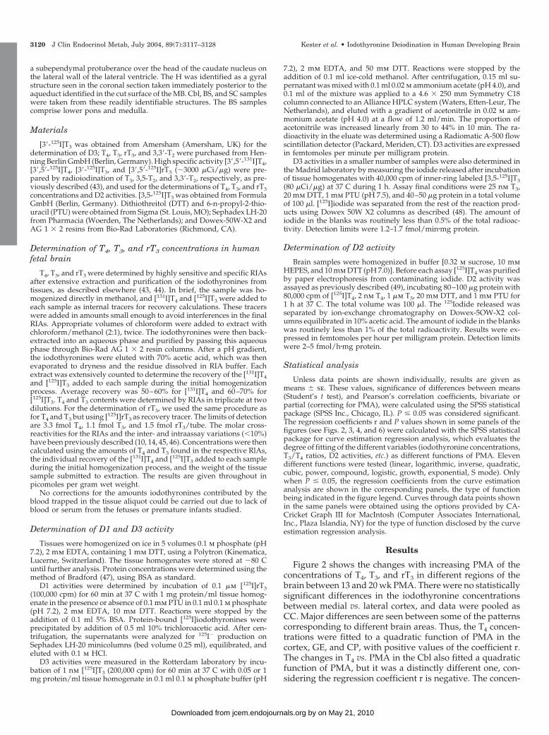
Figure 2 shows the changes with increasing PMA of theconcentrations of T4, T3, and rT3 in different regions of thebrain between 13 and 20 wk PMA. There were no statisticallysignificant differences in the iodothyronine concentrationsbetween medial vs. lateral cortex, and data were pooled asCC. Major differences are seen between some of the patternscorresponding to different brain areas. Thus, the T4 concen-trations were fitted to a quadratic function of PMA in thecortex, GE, and CP, with positive values of the coefficient r.The changes in T4 vs. PMA in the Cbl also fitted a quadraticfunction of PMA, but it was a distinctly different one, con-sidering the regression coefficient r is negative. The concen-
3120 J Clin Endocrinol Metab, July 2004, 89(7):3117–3128 Kester et al. • Iodothyronine Deiodination in Human Developing Brain
by on May 21, 2010 jcem.endojournals.orgDownloaded from
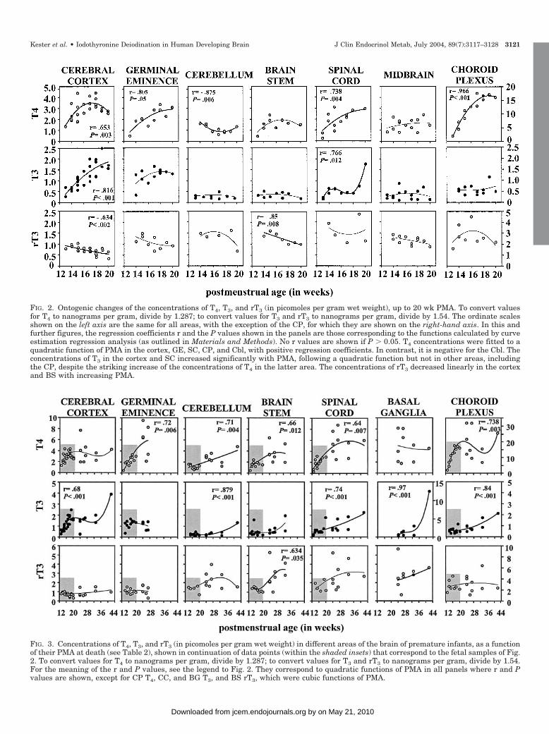
FIG. 3. Concentrations of T4, T3, and rT3 (in picomoles per gram wet weight) in different areas of the brain of premature infants, as a functionof their PMA at death (see Table 2), shown in continuation of data points (within the shaded insets) that correspond to the fetal samples of Fig.2. To convert values for T4 to nanograms per gram, divide by 1.287; to convert values for T3 and rT3 to nanograms per gram, divide by 1.54.For the meaning of the r and P values, see the legend to Fig. 2. They correspond to quadratic functions of PMA in all panels where r and Pvalues are shown, except for CP T4, CC, and BG T3, and BS rT3, which were cubic functions of PMA.
FIG. 2. Ontogenic changes of the concentrations of T4, T3, and rT3 (in picomoles per gram wet weight), up to 20 wk PMA. To convert valuesfor T4 to nanograms per gram, divide by 1.287; to convert values for T3 and rT3 to nanograms per gram, divide by 1.54. The ordinate scalesshown on the left axis are the same for all areas, with the exception of the CP, for which they are shown on the right-hand axis. In this andfurther figures, the regression coefficients r and the P values shown in the panels are those corresponding to the functions calculated by curveestimation regression analysis (as outlined in Materials and Methods). No r values are shown if P � 0.05. T4 concentrations were fitted to aquadratic function of PMA in the cortex, GE, SC, CP, and Cbl, with positive regression coefficients. In contrast, it is negative for the Cbl. Theconcentrations of T3 in the cortex and SC increased significantly with PMA, following a quadratic function but not in other areas, includingthe CP, despite the striking increase of the concentrations of T4 in the latter area. The concentrations of rT3 decreased linearly in the cortexand BS with increasing PMA.
Kester et al. • Iodothyronine Deiodination in Human Developing Brain J Clin Endocrinol Metab, July 2004, 89(7):3117–3128 3121
by on May 21, 2010 jcem.endojournals.orgDownloaded from

tration of T3 in the cortex and SC increased significantly withPMA, following quadratic functions, but not in the otherareas, including the CP, despite the striking increase of theconcentrations of T4 in this area. The concentrations of rT3decreased linearly in the cortex and BS and tended to de-crease in the GE and MB (not statistically significant).
The observed changes during this intrauterine period rep-resent ontogenic profiles because the fetuses and their moth-ers were presumably normal. This may not be so for the dataobtained from the brains of the premature infants because theillnesses suffered and the different causes of their deathmight affect the observed profiles. For this reason they areshown separately in Fig. 3 as a continuation of the data of thefetuses, and are referred to the PMA at death, to follow thesame criterion as used for Fig. 2.
It appears that T4 concentrations continue to increase inmost areas, following quadratic or cubic functions of PMA(Fig. 3). T3 concentrations also start increasing in areas inwhich they hardly changed before 20 wk PMA or in whichthey had actually been decreasing in fetuses (Cbl). The con-centrations of rT3 increase in the BS, in which they had beendecreasing before 20 wk PMA, with no well-defined patternsof change being found in the remaining areas. In the H (notshown in Fig. 3), obtained only from the premature infants,no correlations were found with PMA at death, mean valuesbeing 3.54 � 0.49 pmol T4/g (2.75 � 0.38 ng T4/g, n � 9),1.35 � 0.50 pmol T3/g (0.879 � 0.33 ng T3/g, n � 9), and1.98 � 0.57 pmol rT3/g (1.289 � 0.37 ng rT3/g, n � 9). Whendata from all brain areas obtained between 13 and 42 wkPMA are considered as a whole, positive correlations werefound vs. age for the three iodothyronines, with P � 0.003 forT4 and P � 0.001 for T3 and rT3. Considering the data of thefetuses alone, the positive correlation for rT3 was lost. Whenthe premature babies alone were considered, only the pos-itive correlation for T3 persisted (P � 0.001).
Tissue iodothyronine levels depend on not only local io-dothyronine deiodinase activities but also, among others, onthe supply of thyroid hormone, in particular T4, from thecirculation. As a means to correct for changes in T4 supply,the T3/T4, rT3/T4, and rT3/T3 ratios were calculated andplotted against PMA. Some correlations between the ratios
and PMA were found. Thus, for instance, the T3/T4 ratioincreased throughout the study period in the CC (r � 0.482;P � 0.005), whereas it tended to decrease in the GE (r ��0.473; P � 0.053). The rT3/T4 ratio decreased in the CC, butonly in fetuses (r � �0.721; P � 0.001), and increased in thesame area in premature infants (r � 0.669; P � 0.049). Itdecreased throughout the study period in the GE (r � �0.807;P � 0.001) and CP (r � �0.485; P � 0.048). The rT3/T3 ratiosshowed changes similar to those of the rT3/T4 ratios in theCC of the fetuses (r � �0.861; P � 0.001) and also decreasedin the CP (r � �0.804; P � 0.001) throughout the study periodand in the BG of the premature infants (r � �0.620; P �0.024). The changes with PMA of these ratios in the humandeveloping brain and of the concentrations of the iodothy-ronines shown in Figs. 2 and 3 clearly suggest that patternsare both area and age specific.
Therefore, they cannot be predicted from the circulatinglevels of the iodothyronines at different stages of develop-ment. This point is illustrated in Fig. 4, in which the T3/T4ratios in the CP, GE, and CC up to 20 wk PMA are comparedwith those obtained in sera from developing fetuses. Thelatter are taken from a previous study (14), in which the sameanalytical procedures had been used as for the present brainareas, thus permitting the determination of the very lowconcentrations of T3 in fetal serum. The T3/T4 ratios in serumtended to decrease with PMA, but the regression coefficientdid not reach statistical significance. In the GE and CP, theT3/T4 ratios decrease linearly with PMA. In contrast, theT3/T4 ratio in the CC increased with PMA.
In addition to the iodothyronine levels, deiodinase activ-ities were determined in the human developing brain sam-ples. No detectable D1 activity was found in any brain sam-ple (data not shown). D2 and D3 activities were determinedin the Cbl, BS, SC, CP, and CC from both fetal and prematureinfants’ samples (13–42 wk PMA), MB and GE from fetalsamples (13–20 wk PMA), and BG and H from prematureinfants (23–42 wk PMA at death). Average D2 an D3 activ-ities for the different brain regions are shown in Fig. 5 forfetuses and premature infants separately. D3 activity washighest in Cbl, but considerable D3 activities were also foundin MB, BG, BS, SC, and H, whereas D3 activity was low in the
FIG. 4. Comparison of the changes in T3 to T4 ratios in fetal serum, CP, GE, and cortex, as linear functions of PMA. The serum T3/T4 ratio tendedto decrease as a linear function of PMA but did not reach statistical significance (r � �0.403; P � 0.070). The ratios are plotted on a logarithmicscale to emphasize the differences among these three brain areas and between them and the serum. The functions fitting the CC data weredistinctly different from the others because there was no overlap between the 95% confidence intervals of its positive regression coefficient andthe negative ones of the other two areas. These coefficients did overlap when the CP and GE were compared.
3122 J Clin Endocrinol Metab, July 2004, 89(7):3117–3128 Kester et al. • Iodothyronine Deiodination in Human Developing Brain
by on May 21, 2010 jcem.endojournals.orgDownloaded from

GE, CP, and CC. In some fetal samples, D3 activities werehigher than in those of the same region obtained from thepremature infants. D2 activities were highest in CP and CC,in which D3 activities were the lowest. Most D2 activitiesranged between 8 and 20 fmol/h�mg protein. Although suchvalues are about 100 times lower than the D3 activities, theyare similar to, or higher than, those found in normal braintissue from adults (18).
Figure 6 shows the D2 activities in different brain areas,plotted against PMA. D2 activity was detected in all regionsand increased with PMA in the CC and Cbl (P � 0.05). Aftercontrolling for PMA and using all data from all regionsthroughout the fetal period as a whole, D2 activities werefound to correlate positively with the concentrations of T4(r � 0.65, P � 0.001) and rT3 (r � 0.42, P � 0.001) andnegatively with the T3/T4 ratios in the CC (r � �0.29, P �0.008). Except for the correlation between D2 activities and
T4 concentrations, the statistical significance disappearedwhen the data from the premature infants were included.
As shown in Fig. 7, D3 activities decreased with PMA inCbl, BS, and SC (P � 0.05). At all stages, highest D3 activitieswere found in the Cbl. D3 activities were low throughout thestudy period in GE, CP, and CC.
Table 3 shows the correlations between the average D3activities and the iodothyronine levels and ratios in the dif-ferent brain regions. Figure 8A shows the correlation be-tween the average D3 activities and the rT3/T3 and rT3/T4ratios. T4 levels tended to decrease and rT3 levels tended toincrease with D3 activity. A significant negative correlationwas found between D3 activity and the T3 level (r � �0.682).D3 activities were positively correlated with the rT3/T3 ratio(r � 0.812, P � 0.008) and the rT3/T4 ratio (r � 0.889, P �0.001). Figure 8B depicts the average D3 activities and rT3/T3and rT3/T4 ratios in the different brain regions. Except for theCP, the rT3/T3 and rT3/T4 ratios correlated positively withincreasing D3 activities.
The D3 activities shown in Figs. 5, 7, and 8 and in Table3 were measured in the Rotterdam laboratory. The fewerdeterminations performed in the Madrid laboratory with adifferent methodology fully supported these findings, in-cluding the different correlations that have been describedhere.
Discussion
Studies using the rat as an animal model have shown thatfetal and neonatal hypothyroidism lead to multiple struc-tural, functional, and biochemical alterations of brain devel-opment (see reviews in Refs. 38, 50, 51). These studies, to-gether with clinical evidence for the effects of maternalhypothyroxinemia on brain development (for review see Ref.1), indicate the importance of a tightly regulated thyroidhormone bioavailability during brain development. Iodo-thyronine deiodination contributes to this regulation. In thisstudy we determined local iodothyronine levels and deio-dinase activities in different brain regions at different stagesof development to evaluate the possible contribution of thedifferent deiodinases in controlling local T3 availability in thehuman developing brain.
As already pointed out previously, it is quite likely that thechanges in the concentrations of T4, T3, and rT3 observed withthe samples of the fetuses from normal mothers reflect thetrue ontogenic profile for different areas of the human brain.They are quite different for the different areas studied andcannot be predicted from the changes found in the fetalcirculation. Obviously we cannot exclude that there are alsodifferences within each area related to cellular heterogeneityand different timing of maturational events in each structure,but this point cannot yet be adequately resolved with thesensitivity of presently available techniques. A similar com-ment might also be pertinent for the D2 and D3 activities herereported.
The changes found in some areas before midgestationappear to merit closer attention. The highest T4 and rT3concentrations were observed in the choroid plexus. T4 in-creased significantly with PMA, despite little change in thelow T3 concentrations; indeed, the T3/T4 and rT3/T4 ratios
FIG. 5. Average D3 and D2 activities of the different brain regions.Results are the means � SE of samples from fetuses (13–20 wk PMA)and premature infants at death (Table 2). Asterisks identify statis-tically significant differences between the mean values for fetuses vs.premature infants. D3 activities decreased from Cbl to CP and CC(Cbl � MB � BS � SC � GE � CC � CP for fetuses; Cbl � BG � BS �H � SC � GE � CP � CC for premature infants). D2 activities werehighest in CP, followed by CC (CP � CC � Cbl � SC � MB � BS �GE for fetuses; CC � Cbl � H � SC � GE � BS � BG for prematureinfants). The � sign indicates a statistically significant differencebetween areas, whereas the � sign indicates that the differences werenot statistically significant.
Kester et al. • Iodothyronine Deiodination in Human Developing Brain J Clin Endocrinol Metab, July 2004, 89(7):3117–3128 3123
by on May 21, 2010 jcem.endojournals.orgDownloaded from

were decreasing with PMA. The lack of increase in the con-centration of T3 is also rather striking when we consider thatthe D2 activities were among the highest. This could not beattributed to high D3 activities because these were very low.However, it should be realized that the deiodination rates
measured under in vitro conditions may not represent deio-dination taking place in vivo. It is possible that only a smallproportion of the total T4 we have measured in the choroidplexus samples is actually available intracellularly for deio-dination by D2 and D3: most of it is likely to be bound by
FIG. 6. D2 activities in samples from fetuses (E) and premature infants (F) as a function of PMA from CP, CC, Cbl, H, BS, GE, SC, MB, andBG. For the meaning of r and P values, see the legend to Fig. 2. D2 activities changed with increasing PMA, the CC and Cbl following cubicfunctions of PMA, but no well-defined patterns were found in the remaining areas.
FIG. 7. D3 activities in fetuses (E) and premature infants (F) as a function of PMA from Cbl, MB, BG, BS, SC, H, GE, CP, and CC as a functionof PMA. The r values show Pearson’s correlation coefficient with PMA.
3124 J Clin Endocrinol Metab, July 2004, 89(7):3117–3128 Kester et al. • Iodothyronine Deiodination in Human Developing Brain
by on May 21, 2010 jcem.endojournals.orgDownloaded from

transthyretin, which is already synthesized in the human CPlong before 13 wk PMA (52). Despite the fact that transportof T4 from the plasma to the brain is normal in transthyretin-null mice (53), the CP is considered to be important for thetransport of T4 into the brain. The transthyretin that is syn-thesized in the CP epithelial cells would either transfer T4from the epithelial cells to the cerebrospinal fluid or facilitateits passage after the transthyretin is excreted into the cere-brospinal fluid (54). The amounts of substrate iodothyroni-nes actually reaching D2 and D3 deiodination sites may wellbe much lower than expected from the total concentrations.
It is therefore likely that in this unique and morphologicallyheterogeneous structure mechanisms other than deiodina-tion, such as thyroid hormone transport, are more importantfor the regulation of intracellular thyroid hormone levels.
The largest increase in T3 concentration up to midgestationwas observed in the CC, which appeared to continue evenwhen the T4 concentrations were no longer increasing. As aresult, the cortex T3/T4 ratio increased throughout this pe-riod, in contrast to the serum T3/T4 ratio, which tended todecrease. On the contrary, rT3 concentrations and rT3/T4
ratios were decreasing during the same developmental pe-riod. Thus, both the changes in T3 and rT3 concentrationswere consistent with the findings that D2 activities wereclearly detectable by 13 wk PMA, and D3 activities were thelowest found in the present study. We cannot exclude thatother regulatory mechanisms are also involved in determin-ing the concentration of T3 in the CC. The changes describedhere for the CC up to midgestation are consistent with pre-vious findings by others (8, 16, 17), showing that both T3 andthyroid hormone receptor concentrations are increasing inthe human brain between 8 and 18 wk PMA: at 13 wk ges-
FIG. 8. Average rT3/T3 and rT3/T4 ratios as a function of D3 activities (A) and average D3 activities and rT3/T3 and rT3/T4 ratios in the differentbrain regions (B). Results are the means per brain region � SE. The r value indicates the Pearson’s correlation between D3 activity and theiodothyronine ratio (B).
TABLE 3. Correlation of D3 activity with iodothyronine levelsand ratios in different brain regions from fetuses and prematureinfants
Iodothyronine levels Iodothyronine ratios
rT3 T4 T3 rT3/T4 rT3/T3 T3/T4
r 0.138 �0.454 �0.682 0.889 0.812 �0.236P 0.723 0.219 0.043 0.001 0.008 0.539
Pearson’s correlation coefficient r and P values were calculatedusing SPSS.
Kester et al. • Iodothyronine Deiodination in Human Developing Brain J Clin Endocrinol Metab, July 2004, 89(7):3117–3128 3125
by on May 21, 2010 jcem.endojournals.orgDownloaded from

tation T3 concentrations in the cortex had already reached60% of adult values. D2 and D3 activities have also beenpreviously reported in the human CC by 11–14 wk PMA (55).We point out that the present T3 concentrations in the humanCC are comparable with those reported in adults: 1.5–2.2pmol T3/g (1.0–1.4 ng T3/g) (18, 56). The fetal D2 activitiesare actually much higher than reported in the adult cortex(�8 fmol/h�mg protein) (18). The ontogenic changes of T4, T3,and D2 observed in the present study are in conceptualagreement with those reported for the rat brain (46, 49);between 18 and 22 d of gestation, there is a 4-fold increasein D2 activity, a 10-fold increase in T4 concentrations, and an18-fold increase in T3 concentrations in rat CC.
The present results also indirectly support the hypothesisthat T3 is relevant for the development of the human CC fromvery early in gestation, possibly soon after completion ofmorphogenesis of the pros-encephalon (57). Although thishypothesis is supported by epidemiological and clinical find-ings (1), no direct proof is available for man. It has, however,been directly confirmed (58) in the rat for a developmentalperiod corresponding to that occurring in man before mid-gestation. The tendency of T3 concentrations to increase inthe GE before midgestation suggests that this structure mightalready be thyroid hormone sensitive, but we are unaware ofany studies regarding abnormalities in this structure relatedto thyroid hormone insufficiency.
The ontogenic changes in the developing human Cbl con-trast with those described for the CC. The concentrations ofT4 and T3 remained low, and especially T3 was maintainedat levels that were appreciably lower than those found dur-ing the same period in other areas, such as the cortex, GE, SC,and even CP. D2 activities were similar to those found in thecortex, but D3 activities were the highest found in any of thebrain areas studied during this developmental period andare likely to be a very important factor in the maintenance ofthe low cerebellar T3 concentrations. So are the high D3activities found in MB, BS, and SC, all areas in which T3concentrations were low during most of the developmentalperiod up to midgestation.
Some caution should be applied to the interpretation ofdata obtained in the postnatal brain samples, insofar as it isnot excluded that they may to some extent be influenced bynonthyroidal illness, which is known to affect peripheralthyroid hormone metabolism (59, 60).
The present results confirm for the human developingbrain the same principles that appear to modulate T3 bio-availability in different developing structures, and in differ-ent species, in a temporally and spatially specific sequence ofevents, namely by the ontogenetically programed expressionof the iodothyronine deiodinase isoenzymes, mainly D2 andD3 (29, 61, 62). D1 activity was not detected in any brain area.This is in agreement with previous studies of Campos-Barroset al. (63), who found D2 and D3 activity, but no D1 activity,in adult human brain. We have already discussed the D2activities found in different areas, compared with those inadults. The activities of D3 during early development that wereport here for different brain areas show very high levels inspecific structures that, in general, tend to decrease withPMA. The highest D3 activities were found in Cbl and werehigher than in the adult brain (64). The spatial distribution
of D3, however, differs: in the adult brain, D3 activity is lowin Cbl, MB, and BS, whereas higher levels are found in theH and CC (Visser, T. J., E. Kaptein, and E. Fliers, unpublisheddata, and Ref. 64). Santini et al. (64) found that T3 levels arealso negatively correlated with D3 activity in the adult brain,as described here for the developing human brain.
The D3 activities found here for the brain are only 2-foldlower than those reported in human placenta (31). D3 ex-pression in the placenta is believed to protect the fetus fromexcessive maternal T3 (12, 15, 28). Thyroid hormone inducesneuronal differentiation such as dendritic and axonalgrowth, neuronal migration, and myelination (38). Strict reg-ulation of thyroid hormone bioavailability is critical becauseneuronal development is affected in the hypothyroid andhyperthyroid brain. The high D3 activities we found in thebrain, which tended to decrease with age, suggest that localD3 is important to limit T3 in the various brain regions duringcritical stages of development. It is unclear whether D3 hasan additional physiological role in the production of rT3 and3,3�-T2. Because rT3, but not T3, has profound and acuteeffects on the cytoskeleton in brain cells (65), it is not ex-cluded that rT3 also has a function in brain development.3,3�-T2 has been shown to increase the basal metabolic ratein adult rat. This effect may be mediated by direct mito-chondrial binding (66).
D2 and D3 are expressed in distinct cell types: D2 in as-trocytes and tanycytes and D3 in neurons. The hypothesishas been put forward (38) that astrocytes and tanycytes takeup T4 from the circulation and convert it to T3, which isdelivered to neurons (that contain most of the nuclear re-ceptors), in which D3 would limit T3 availability accordingto the local temporal needs for thyroid hormone action. Inaddition, although still poorly studied, metabolic pathwaysother than deiodination, such as sulfation, may play regu-latory roles in the developing brain.
A large number of cerebral genes are regulated by thyroidhormone (38). Although not much is known on the molecularbasis for the specific timing of action on gene expression, itis known that the different regions of the brain have specifictemporal patterns of development and thus require differentregulation of T3 bioavailability. In general, roughly, the ce-rebral cortex starts to develop in the second month of preg-nancy, whereas major events in cerebellar development donot occur until wk 34 (51). In agreement with this, we foundlow D3 activity in the CC, which would require T3 for dif-ferentiation early in development and high D3 activity in thelater developing Cbl.
In this study, we also compared the average D3 activitieswith the average thyroid hormone levels and ratios in thedifferent brain regions. Except for the CP, we observed thatD3 activity was high in the regions with low T3 and T4 andhigh rT3 levels and low in regions with high T3 and T4 andlow rT3 levels. We found a significant negative correlationbetween D3 activities and T3 levels and significant positivecorrelations between D3 activity and the ratio of rT3/T3 andthe ratio of rT3/T4. Because D3 catalyzes the degradation ofT3 and T4 and the production of rT3, our results suggest thatD3 is also important in humans for the regulation of theintracellular thyroid hormone levels in the different brainregions. Furthermore, no D1 activity was found in any brain
3126 J Clin Endocrinol Metab, July 2004, 89(7):3117–3128 Kester et al. • Iodothyronine Deiodination in Human Developing Brain
by on May 21, 2010 jcem.endojournals.orgDownloaded from

region. In addition to the presence of D3 activity, the absenceof D1 activity may contribute to the high tissue rT3 levels.
In conclusion, by determining and correlating the onto-genic patterns of deiodinase activities and thyroid hormonelevels in the human brain, we have shown that both D3- andD2-catalyzed deiodination are important pathways for theintracellular regulation of thyroid hormone in the differentregions of the developing human brain, this regulation beingregion and time specific. Although D3 is expressed to agreater extent than D2, the latter is clearly important inthyroid hormone activation at the cellular level. Further insitu hybridization and immunohistochemistry studies arerequired to confirm the hypothesis that a close regulation ofD2 and D3 activities is crucial for tailoring T3 bioavailabilityto changing needs of human developing brain structures.
Acknowledgments
We thank Asha S. P. D. Mangnoesing for her assistance with the D3activity determinations and Socorro Duran, Maria Jesus Presas, andRosalia Lavado-Autric for the determinations of the iodothyronine con-centrations. We are grateful to Professor Juan Jose de la Cruz (Facultyof Medicine, Madrid) for invaluable help with issues of statistics.
Received October 22, 2003. Accepted March 28, 2004.Address all correspondence and requests for reprints to: Theo J.
Visser, Department of Internal Medicine, Erasmus Medical Center,Room Ee 502, Dr Molewaterplein 50, 3015 GE Rotterdam, The Nether-lands. E-mail: [email protected].
This work was supported by European Community Grant QLG-2000-00930, Netherlands Organization for Scientific Research Grant 903-40-204, Fondo de Investigacion Sanitaria RCMN (C03/08) from Inst deSalud Carlos III, Chief Scientists Office Scottish Executive (K/MRS/50/C741), and Tenovus Scotland/Leng Trust.
References
1. Morreale de Escobar G, Obregon MJ, Escobar del Rey F 2000 Is neuropsy-chological development related to maternal hypothyroidism or to maternalhypothyroxinemia? J Clin Endocrinol Metab 85:3975–3987
2. Xue-Yi C, Xin-Min J, Zhi-Jong D, Rakeman MA, Ming-Li Z, O’Donnell K,Tai M, Amette K, DeLong N, DeLong GR 1994 Timing of vulnerability of thebrain to iodine deficiency in endemic cretinism. N Engl J Med 331:1739–1744
3. Bleichrodt N, Born M 1994 A metaanalysis of research on iodine and itsrelationship to cognitive development. In: Stanbury JB, ed. The damaged brainof iodine deficiency. Elmsford, NY: Cognizant Communication Co.; 195–200
4. Man EB, Serunian SA 1976 Thyroid function in human pregnancy. Devel-opment or retardation of 7-year-old progeny of hypothyroxinemic women.Am J Obst Gynecol 125:949–957
5. Pop VJ, Kuijpens JL, van Baar AL, Verkerk G, van Son MM, de Vijlder JJ,Vulsma T, Wiersinga WM, Drexhage HA, Vader HL 1999 Low maternal freethyroxine concentrations during early pregnancy are associated with impairedpsychomotor development in infancy. Clin Endocrinol (Oxf) 50:149–155
6. Haddow JE, Palomaki GE, Allan WC, Williams JR, Knight GJ, Gagnon J,O’Heir CE, Mitchell ML, Hermos RJ, Waisbren SE, Faix JD, Klein RZ 1999Maternal thyroid deficiency during pregnancy and subsequent neuropsycho-logical development of the child. N Engl J Med 341:549–555
7. Kopp P, van Sande J, Parma J, Duprez L, Gerber H, Joss E, Jameson JL,Dumont JE, Vassart G 1995 Brief report: congenital hyperthyroidism causedby a mutation in the thyrotropin-receptor gene. N Engl J Med 332:150–154
8. Bernal J, Pekonen F 1984 Ontogenesis of the nuclear 3,5,3�-triiodothyroxinereceptor in the human fetal brain. Endocrinology 114:677–679
9. Myant NB 1958 Passage of thyroxine and triiodothyronine from mother tofoetus in pregnant women. Clin Sci 17:75–79
10. Contempre B, Jauniaux E, Calvo R, Jurkovic D, Campbell S, Morreale deEscobar G 1993 Detection of thyroid hormones in human embryonic cavitiesduring the first trimester of pregnancy. J Clin Endocrinol Metab 77:1719–1722
11. Calvo R, Obregon MJ, Ruiz de Ona C, Escobar del Rey F, Morreale deEscobar G 1990 Congenital hypothyroidism, as studied in rats. Crucial role ofmaternal thyroxine but not of 3,5,3�-triiodothyronine in the protection of thefetal brain. J Clin Invest 86:889–899
12. Burrow GN, Fisher DA, Larsen PR 1994 Maternal and fetal thyroid function.N Engl J Med 331:1072–1078
13. Vulsma T, Gons MH, de Vijlder JJM 1989 Maternal-fetal transfer of thyroxine
in congenital hypothyroidism due to a total organification defect or thyroidagenesis. N Engl J Med 321:13–16
14. Calvo RM, Jauniaux E, Gulbis B, Asuncion M, Gervy C, Contempre B,Morreale de Escobar G 2002 Fetal tissues are exposed to biologically relevantfree thyroxine concentrations during early phases of development. Possibleconsequences of maternal hypothyroxinemia. J Clin Endocrinol Metab 87:1768–1777
15. Santini F, Chiovato L, Ghirri P, Lapi P, Mammoli C, Montanelli L, ScartabelliG, Ceccarini G, Coccoli L, Chopra IJ, Boldrini A, Pinchera A 1999 Serumiodothyronines in the human fetus and the newborn: evidence for an importantrole of placenta in fetal thyroid hormone homeostasis. J Clin Endocrinol Metab84:493–498
16. Ferreiro B, Bernal J, Goodyer CG, Branchard CL 1988 Estimation of nuclearthyroid hormone receptor saturation in human fetal brain and lung duringearly gestation. J Clin Endocrinol Metab 67:853–856
17. Bernal J, Perez-Castillo A, Pans T, Pekonen F 1984 Ontogenesis of thyroidhormone receptor. In: Labrie F, Proulx L, eds. Endocrinology. Amsterdam:Elsevier Science Publisher; 977–980
18. Calvo R, Roda JM, Obregon MJ, Morreale de Escobar G 1998 Thyroid hor-mones in human tumoral and normal nervous tissues. Brain Res 801:150–157
19. Hennemann G, Docter R, Friesema EC, de Jong M, Krenning EP, Visser TJ2001 Plasma membrane transport of thyroid hormones and its role in thyroidhormone metabolism and bioavailability. Endocr Rev 22:451–476
20. Bianco AC, Salvatore D, Gereben B, Berry MJ, Larsen PR 2002 Biochemistry,cellular and molecular biology, and physiological roles of the iodothyronineselenodeiodinases. Endocr Rev 23:38–89
21. St Germain DL, Galton VA 1997 The deiodinase family of selenoproteins.Thyroid 7:655–668
22. Kohrle J 1999 Local activation and inactivation of thyroid hormones: thedeiodinase family. Mol Cell Endocrinol 151:103–119
23. Croteau W, Davey JC, Galton VA, St. Germain DL 1996 Cloning of themammalian type II iodothyronine deiodinase. A selenoprotein differentiallyexpressed and regulated in human and rat brain and other tissues. J Clin Invest98:405–417
24. Salvatore D, Tu H, Harney JW, Larsen PR 1996 Type 2 iodothyronine deio-dinase is highly expressed in human thyroid. J Clin Invest 98:962–968
25. Bartha T, Kim SW, Salvatore D, Gereben B, Tu HM, Harney JW, Rudas P,Larsen PR 2000 Characterization of the 5�-flanking and 5�-untranslated regionsof the cyclic adenosine 3�,5�-monophosphate-responsive human type 2 iodo-thyronine deiodinase gene. Endocrinology 141:229–237
26. Guadano-Ferraz A, Obregon MJ, St. Germain DL, Bernal J 1997 The type 2iodothyronine deiodinase is expressed primarily in glial cells in the neonatalrat brain. Proc Natl Acad Sci USA 94:10391–10396
27. Tu HM, Kim SW, Salvatore D, Bartha T, Legradi G, Larsen PR, Lechan RM1997 Regional distribution of type 2 thyroxine deiodinase messenger ribonu-cleic acid in rat hypothalamus and pituitary and its regulation by thyroidhormone. Endocrinology 138:3359–3368
28. Galton VA, Martinez E, Hernandez A, St. Germain EA, Bates JM, St. Ger-main DL 1999 Pregnant rat uterus expresses high levels of the type 3 iodo-thyronine deiodinase. J Clin Invest 103:979–987
29. Bates JM, St. Germain DL, Galton VA 1999 Expression profiles of the threeiodothyronine deiodinases, D1, D2, and D3, in the developing rat. Endocri-nology 140:844–851
30. Kaplan MM, Shaw EA 1984 Type II iodothyronine 5�-deiodination by humanand rat placenta in vitro. J Clin Endocrinol Metab 59:253–257
31. Koopdonk-Kool JM, de Vijlder JJ, Veenboer GJ, Ris-Stalpers C, Kok JH,Vulsma T, Boer K, Visser TJ 1996 Type II and type III deiodinase activity inhuman placenta as a function of gestational age. J Clin Endocrinol Metab81:2154–2158
32. Richard K, Hume R, Kaptein E, Sanders JP, van Toor H, de Herder WW, denHollander JC, Krenning EP, Visser TJ 1998 Ontogeny of iodothyronine deio-dinases in human liver. J Clin Endocrinol Metab 83:2868–2874
33. Huang SA, Dorfman DM, Genest DR, Salvatore D, Larsen PR 2003 Type 3iodothyronine deiodinase is highly expressed in the human uteroplacental unitand in fetal epithelium. J Clin Endocrinol Metab 88:1384–1388
34. Tu HM, Legradi G, Bartha T, Salvatore D, Lechan RM, Larsen PR 1999Regional expression of the type 3 iodothyronine deiodinase messenger ribo-nucleic acid in the rat central nervous system and its regulation by thyroidhormone. Endocrinology 140:784–790
35. Escamez MJ, Guadano-Ferraz A, Cuadrado A, Bernal J 1999 Type 3 iodo-thyronine deiodinase is selectively expressed in areas related to sexual dif-ferentiation in the newborn rat brain. Endocrinology 140:5443–5446
36. Leonard JL, Farwell AP, Yen PM, Chin WW, Stula M 1994 Differential ex-pression of thyroid hormone receptor isoforms in neurons and astroglial cells.Endocrinology 135:548–555
37. Carlson DJ, Strait KA, Schwartz HL, Oppenheimer JH 1996 Thyroid hormonereceptor isoform content in cultured type 1 and type 2 astrocytes. Endocri-nology 137:911–917
38. Bernal J 2002 Action of thyroid hormone in brain. J Endocrinol Invest 25:268–288
39. Guadano-Ferraz A, Bernal J 2003 The role of deiodinases during brain de-
Kester et al. • Iodothyronine Deiodination in Human Developing Brain J Clin Endocrinol Metab, July 2004, 89(7):3117–3128 3127
by on May 21, 2010 jcem.endojournals.orgDownloaded from

velopment. In: Morreale de Escobar G, deVijlder JJM, Butz S, Hostalek U, eds.The thyroid and brain. Stuttgart, Germany: Schattauer GmbH; 161–173
40. Kandel ER 1991 Brain and behaviour. In: Kandel ER, Schwartz JH, Jessell TM,eds. Principles of neural science. 3rd ed. New York: Elsevier
41. Scammon RE, Calkins LA 1922 The development and growth of the externaldimensions of the human body in the fetal period. Minneapolis: University ofMinnesota Press
42. Hume R, Kelly R, Cossar D, Giles M, Hallas A, Gourlay M, Bell J 1991Self-differentiation of human lung organ culture: the role of prostaglandinsPGE2 and PGE2�. Exp Cell Res 194:111–117
43. Morreale de Escobar G, Pastor R, Obregon MJ, Escobar del Rey F 1985 Effectsof maternal hypothyroidism on the weight and thyroid hormone content of ratembryonic tissues, before and after onset of fetal thyroid function. Endocri-nology 117:1890–1900
44. Morreale de Escobar G, Calvo R, Escobar del Rey F, Obregon MJ 1994Thyroid hormones in tissues from fetal and adult rats. Endocrinology 134:2410–2415
45. Calvo R, Obregon MJ, Escobar del Rey F, Morreale de Escobar G 1992 Therat placenta and the transfer of thyroid hormones from the mother to the fetus.Effects of maternal thyroid status. Endocrinology 131:357–365
46. Ruiz de Ona C, Morreale de Escobar G, Calvo R, Escobar del Rey F, ObregonMJ 1991 Thyroid hormones and 5�-deiodinase in the rat fetus late in gestation:effects of maternal hypothyroidism. Endocrinology 128:422–432
47. Bradford MM 1976 A rapid and sensitive method for the quantitation ofmicrogram quantities of protein utilizing the principle of protein-dye binding.Anal Biochem 72:248–254
48. Hernandez A, Obregon MJ 1995 Presence of growth factors-induced type IIIiodothyronine 5-deiodinase in cultured rat brown adipocytes. Endocrinology136:4543–4550
49. Ruiz de Ona C, Obregon MJ, Escobar del Rey F, Morreale de Escobar G 1988Developmental changes in rat brain 5�-deiodinase and thyroid hormones dur-ing the fetal period: the effects of fetal hypothyroidism and maternal thyroidhormones. Pediatr Res 24:588–594
50. Morreale de Escobar G, Ruiz-Marcos A, Escobar del Rey F 1983 Thyroidhormones and the developing brain. In: Dussault JH, Walker P, eds. Congenitalhypothyroidism. New York: Marcel Dekker Inc.; 85–126
51. Porterfield SP, Hendrich CE 1993 The role of thyroid hormones in prenataland neonatal neurological development—current perspectives. Endocr Rev14:94–106
52. Jacobsson B 1989 Localization of transthyretin-mRNA and immunoreactivetransthyretin in the human fetus. Virchows Arch Pathol Anat 415:259–263
53. Palha JA, Hays MT, Morreale de Escobar G, Episkopou V, Gottesman M,
Saraiva MJM 1997 Transthyretin is not essential for thyroxine to reach thebrain and other tissues in a transthyretin-null mouse. Am J Physiol EndocrinolMetab 272:E485–E493
54. Southwell BR, Duan W, Alcorn D, Brack C, Richardson SJ, Kohrle J, Schre-iber G 1993 Thyroxine transport to the brain: role of protein synthesis by thechoroid plexus. Endocrinology 133:2116–2126
55. Karmarkar MG, Prabarkaran D, Godbole MM 1993 5�-Monodeiodinase ac-tivity in developing human cerebral cortex. Am J Clin Nutr 57(Suppl):291S–294S
56. Arem R, Wiener GJ, Kaplan SG, Kim H-S, Reichlin S, Kaplan MM 1993Reduced tissue thyroid hormone levels in fatal illness. Metabolism 42:1102–1108
57. DeLong GR 1993 The effects of nutrition on human brain development. Am JClin Nutrition 57:290S–295S
58. Lavado-Autric R, Auso E, Garcia-Velasco VJ, Arufe MC, Escobar del Rey F,Berbel P, Morreale de Escobar G 2003 Early maternal hypothyroxinemia altershistogenesis and cerebral cortex cytoarchitecture of the progeny. J Clin Invest111:1073–1082
59. DeGroot L 1999 Dangerous dogmas in medicine: the non-thyroidal illnesssyndrome. J Clin Endocrinol Metab 84:151–164
60. Peeters RP, Wouters PJ, Kaptein E, van Toor H, Visser TJ, Van den BergheG 2003 Reduced activation and increased inactivation of thyroid hormone intissues of critically ill patients. J Clin Endocrinol Metab 88:3202–3211
61. Becker KB, Stephens KC, Davey JC, Schneider MJ, Galton VA 1997 The type2 and type 3 iodothyronine deiodinases play important roles in coordinatingdevelopment in Rana catesbeiana tadpoles. Endocrinology 138:2989–2997
62. Marsh-Armstrong N, Huang H, Remo BF, Liu TT, Brown DD 1999 Asym-metric growth and development of the Xenopus laevis retina during metamor-phosis is controlled by type III deiodinase. Neuron 24:871–878
63. Campos-Barros A, Hoell T, Musa A, Sampaolo S, Stoltenburg G, Pinna G,Eravci M, Meinhold H, Baumgartner A 1996 Phenolic and tyrosyl ring io-dothyronine deiodination and thyroid hormone concentrations in the humancentral nervous system. J Clin Endocrinol Metab 81:2179–2185
64. Santini F, Pinchera A, Ceccarini G, Castagna M, Rosellini V, Mammoli C,Montanelli L, Zucchi V, Chopra IJ, Chiovato L 2001 Evidence for a role of thetype III-iodothyronine deiodinase in the regulation of 3,5,3�-triiodothyroninecontent in the human central nervous system. Eur J Endocrinol 144:577–583
65. Leonard JL, Farwell AP 1997 Thyroid hormone-regulated actin polymeriza-tion in brain. Thyroid 7:147–151
66. Moreno M, Lanni A, Lombardi A, Goglia F 1997 How the thyroid controlsmetabolism in the rat: different roles for triiodothyronine and diiodothyroni-nes. J Physiol 505:529–538
JCEM is published monthly by The Endocrine Society (http://www.endo-society.org), the foremost professional society serving theendocrine community.
3128 J Clin Endocrinol Metab, July 2004, 89(7):3117–3128 Kester et al. • Iodothyronine Deiodination in Human Developing Brain
by on May 21, 2010 jcem.endojournals.orgDownloaded from