Embed Size (px)
Citation preview

Introduction
Experimental
Results and Discussion
Resveratrol is a naturally occurringcompound in red wine and grape juice.Recent findings suggest that resveratrolmay also be anticarcinogenic (1) and apotent chemopreventive agent forbreast cancer (2). Previously wereported on a method for thedetermination of resveratrol in naturalsources and for its pharmacokineticstudy in rat (3,4). To continue ourresearch on this natural product, thispreliminary study is focused on tools tostudy the metabolism of this compoundand potential drug-drug interactionsfrom consuming wine or grape juice.
metabolism of resveratrol inrat liver microsomal incubations wasinvestigated. The main metabolite wasconfirmed to be piceatannol (a knownantileukemic agent) by LC and tandemmass spectrometry. This microsomalincubation result was consistent with arecent finding that resveratrolmetabolism by recombinant humancytochrome P450 isozyme CYP 1B1formed piceatannol (5). The metabolitepiceatannol has potential anticanceractivity by inhibiting an enzyme foundin human tumor cells (6). shows thes t ruc tu res of resvera t ro l andpiceatannol. Both metabolite andsubstrate were detected by electrosprayionization tandem mass spectrometry inthe negative ion mode using full scanMS and full scan MSMS experiments.This study is the first known report thatused LC/MSMS for the direct
identification of a resveratrol
Incubation of resveratrol with rat livermicrosomes was carried out at 37°C in abench-top Lab-Line shaker (Barnstead/Thermolyne, Dubuque, IA). Theincubation solution contained 50 mMpotassium phosphate buffer (pH 7.4), 1mg protein/mL microsomes, 10 mMMgCl , 2 mM NADPH and 1-50 µMresveratrol in a final volume of 200 µL.The enzyme reaction was initiated byadding NADPH after an initial 10-minpreincubation. The reaction wasterminated by adding 1 mL of ethylacetate. The solution was vortex-mixedand centrifuged (Allegra™ 6Rcentrifuge, Beckman Coulter, PaloAlto, CA) at 4°C for 10 min at 3750rpm. The supernatant was transferred toa test tube and dried under a stream ofnitrogen at room temperature. Theresidue was reconstituted in 200 µL ofmobile phase. The LC/MSMS systemwas equipped with a BASi PM-80 pumpcoupled to a Finnigan LCQ Deca iontrap mass spectrometer (ThermoQuest,San Jose, CA, USA). The massspectrometer was operated in ESInegative ion mode. A multi-channelamperometric detector (epsilon™,BASi, West Lafayette, IN, USA) wascoupled to a quad-glassy carbonworking electrode. Applied potentialswere +400, +500, +600 and +700 mV
vs. Ag/AgCl. The column was aSupecol Discovery C8, 5 µm, 150 x 2.1mm. The mobile phase consisted of0.5% acetic acid-acetonitrile (85:15,v/v) with a flow rate of 0.4 mL/min. Avolume of 20 µL of standard solution orincubation sample was injected intoLC/MS and LC/EC system.
In vitro
in vitro
F1
2 representative LC/MS chromatogramfor the incubation solution are shown in
. Besides the resveratrol peak at m/z227, the metabolite peak at m/z of 243corresponded to its oxidation productpiceatannol (M-H)-. For furtherconfirmation of the identity ofpiceatannol , fu l l scan MSMSexperiments were performed. and
show the chromatograms and fullscan MSMS mass spectra of a standardsmixture and of a microsomal incubationof resveratrol, respectively. A matchbetween the MSMS spectra of bothstandards and incubation solutionconfirms that the peak eluting at theretention time of 5 min is piceatannol.The results were consistent with thereport that resveratrol was metabolized
by recombinant humancytochrome P450 isozyme CYP 1B1 toform piceatannol, where the identitywas confirmed by GC/MS afterderivatisation (5).
F2
F3F4
in vitro
metabolite.
Mass spectra in negative ion mode and a
Yongxin Zhu* Hwa Chiang Jean Zhou Peter T. Kissinger, , and
Bioanalytical Systems, Inc., 2701 Kent Avenue, West Lafayette, Indiana 47906 USA*[email protected]
In Vitro Metabolism Study of Resveratrol and Identification andDetermination of Its Metabolite Piceatannol by LC/EC and LC/MSMS
This preliminary study focuses on tools to study the metabolism of resveratrol andpotential drug-to-drug interactions from consuming wine or grape juice, which bothcontain resveratrol.
93 Current Separations 20:3 (2003)

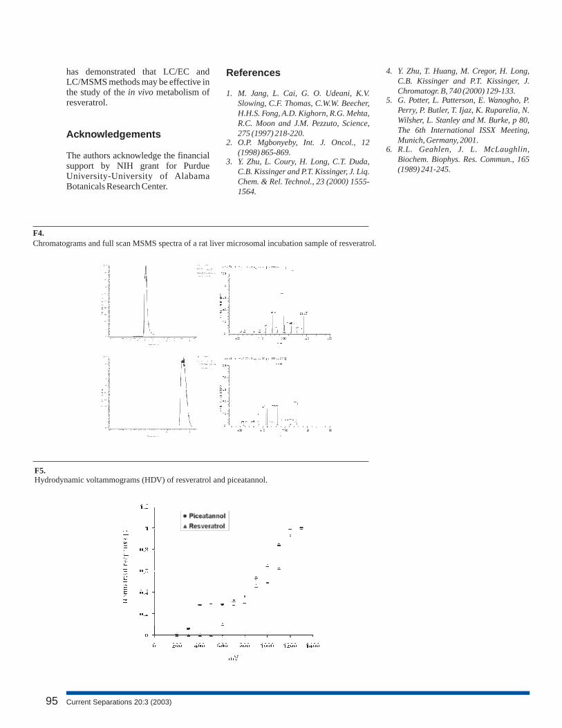
Both substrate and metabolite can beeasily detected by electrochemicalox ida t ion . The mul t i - channe lelectrochemical detector allows forsimultaneous application of differentoxidation potentials on four separateelectrodes. Therefore, the time neededto construct a hydrodynamic voltam-mogram (HDV) can be reducedsubstantially. The normalized responseof substrate and metabolite at theindividual electrodes was plottedagainst oxidation potential and ispresented in . Based on the HDV ofstandard piceatannol, the four channelswere set at oxidation potentials of +400,+500, +600 and +700 mV for allsubsequent determinations carried outin the study to obtain enzyme kineticparameters for resveratrol metabolism.Liquid chromatography with multi-channel electrochemical detection(LC/EC) provides a powerful tool forthe identification of metabolite.shows LC/EC chromatograms of apiceatannol standard and an incubationsample of resveratrol. Peak heightratios between different oxidationpotentials of a standard can be used forpeak identification of the retention-equivalent peak in the metabolismincubation sample. Such peak heightratios of standard and sample fromhave similar values. The closecorrelat ion confirms the peakassignment in the incubation sample.
F5
F6
F6
The Lineweaver-Burk plot of initialvelocity vs. substrate concentration(concentration of piceatannol producedvs. concentration of resveratrol inreaction mix) is presented in . TheK and V for resveratrol in them i c r o s o m a l i n c u b a t i o n w e r edetermined to be 15.64 ± 2.20 µM (n =3) and 28.13 ± 3.39 pmole/min/mgprotein (n = 3), respectively, under thereported conditions.
An LC/MSMS method has beendeveloped for identification anddetermination of resveratrol and its
m i c r o s o m a l m e t a b o l i t epiceatannol. Identity of the metabolitepiceatannol was confirmed by full scanMSMS experiments. The current study
F7m max
Conclusion
inv i t ro
www.currentseparations.com 94
F1.Structures of resveratrol and piceatannol.
F3.Chromatograms and full scan MSMS spectra of standard mixture of piceatannol and resveratrol.
F2.Mass spectra and representative LC/MS chromatogram of metabolite (A) and resveratrol (B).
OHHO
OH OH
HO OH
OH
[O]
Resveratrol Piceatannol
has demonstrated that LC/EC andLC/MSMS methods may be effective inthe study of the metabolism ofresveratrol.
The authors acknowledge the financialsupport by NIH grant for PurdueUniversity-University of AlabamaBotanicals Research Center.
in vivo
Acknowledgements
References
1. M. Jang, L. Cai, G. O. Udeani, K.V.Slowing, C.F. Thomas, C.W.W. Beecher,H.H.S. Fong, A.D. Kighorn, R.G. Mehta,R.C. Moon and J.M. Pezzuto, Science,275 (1997) 218-220.
2. O.P. Mgbonyeby, Int. J. Oncol., 12(1998) 865-869.
3. Y. Zhu, L. Coury, H. Long, C.T. Duda,C.B. Kissinger and P.T. Kissinger, J. Liq.Chem. & Rel. Technol., 23 (2000) 1555-1564.
4. Y. Zhu, T. Huang, M. Cregor, H. Long,C.B. Kissinger and P.T. Kissinger, J.Chromatogr. B, 740 (2000) 129-133.
5. G. Potter, L. Patterson, E. Wanogho, P.Perry, P. Butler, T. Ijaz, K. Ruparelia, N.Wilsher, L. Stanley and M. Burke, p 80,The 6th International ISSX Meeting,Munich, Germany, 2001.
6. R.L. Geahlen, J. L. McLaughlin,Biochem. Biophys. Res. Commun., 165(1989) 241-245.
95
F5.Hydrodynamic voltammograms (HDV) of resveratrol and piceatannol.
F4.Chromatograms and full scan MSMS spectra of a rat liver microsomal incubation sample of resveratrol.
Current Separations 20:3 (2003)

www.currentseparations.com 96
F7.Lineweaver-Burk plot for resveratrol. Resveratrol (1-50 µM) was incubated with rat liver microsomes(1.0 mg/mL) for 15 min at 37°C.
F6.Chromatograms of standard piceatannol (A) and incubation sample of resveratrol (B). Appliedpotentials were a) +400, b) +500, c) +600 and d) +700 mV vs Ag/AgCl.
Piceatannol
Resveratrol