Embed Size (px)
Citation preview
Resveratrol Mediated Modulation of Sirt-1/Runx2Promotes Osteogenic Differentiation of MesenchymalStem Cells: Potential Role of Runx2 DeacetylationMehdi Shakibaei1*, Parviz Shayan2, Franziska Busch1, Constance Aldinger1, Constanze Buhrmann1,
Cora Lueders3, Ali Mobasheri4
1 Institute of Anatomy, Ludwig-Maximilian-University Munich, Munich, Germany, 2 Investigating Institute Molecular Biological System Transfer, Tehran, Iran, 3 Department
of Thoracic and Cardiovascular Surgery, Laboratory for Tissue Engineering, German Heart Institute Berlin, Berlin, Germany, 4 Division of Veterinary Medicine, School of
Veterinary Medicine and Science, Faculty of Medicine and Health Sciences, University of Nottingham, Sutton Bonington Campus, Sutton Bonington, United Kingdom
Abstract
Objective: Osteogenic repair in response to bone injury is characterized by activation and differentiation of mesenchymalstem cells (MSCs) to osteoblasts. This study determined whether activation of Sirt-1 (a NAD+-dependent histonedeacetylase) by the phytoestrogen resveratrol affects osteogenic differentiation.
Methods: Monolayer and high-density cultures of MSCs and pre-osteoblastic cells were treated with an osteogenicinduction medium with/without the Sirt-1 inhibitor nicotinamide or/and resveratrol in a concentration dependent manner.
Results: MSCs and pre-osteoblastic cells differentiated to osteoblasts when exposed to osteogenic-induction medium. Theosteogenic response was blocked by nicotinamide, resulting in adipogenic differentiation and expression of the adiposetranscription regulator PPAR-c (peroxisome proliferator-activated receptor). However, in nicotinamide-treated cultures, pre-treatment with resveratrol significantly enhanced osteogenesis by increasing expression of Runx2 (bone specifictranscription factor) and decreasing expression of PPAR-c. Activation of Sirt-1 by resveratrol in MSCs increased its binding toPPAR-c and repressed PPAR-c activity by involving its cofactor NCoR (nuclear receptor co-repressor). The modulatory effectsof resveratrol on nicotinamide-induced expression of PPAR-c and its cofactor NCoR were found to be mediated, at least inpart, by Sirt-1/Runx2 association and deacetylation of Runx2. Finally, knockdown of Sirt-1 by using antisenseoligonucleotides downregulated the expression of Sirt-1 protein and abolished the inhibitory effects of resveratrol, namelynicotinamide-induced Sirt-1 suppression and Runx2 acetylation, suggesting that the acetylated content of Runx2 is relatedto downregulated Sirt-1 expression.
Conclusion: These data support a critical role for Runx2 acetylation/deacetylation during osteogenic differentiation in MSCsin vitro. (242 words in abstract)
Citation: Shakibaei M, Shayan P, Busch F, Aldinger C, Buhrmann C, et al. (2012) Resveratrol Mediated Modulation of Sirt-1/Runx2 Promotes OsteogenicDifferentiation of Mesenchymal Stem Cells: Potential Role of Runx2 Deacetylation. PLoS ONE 7(4): e35712. doi:10.1371/journal.pone.0035712
Editor: Jean-Marc Vanacker, Institut de Genomique Fonctionnelle de Lyon, France
Received January 10, 2012; Accepted March 20, 2012; Published April 23, 2012
Copyright: � 2012 Shakibaei et al. This is an open-access article distributed under the terms of the Creative Commons Attribution License, which permitsunrestricted use, distribution, and reproduction in any medium, provided the original author and source are credited.
Funding: The authors have no support or funding to report.
Competing Interests: The authors have declared that no competing interests exist.
* E-mail: [email protected]
Introduction
Mesenchymal stem cells (MSCs) are multipotent cells that can
differentiate into distinct connective tissue cell types (i.e.
osteoblasts, chondroblasts, adipocytes, myoblasts, etc.) [1,2].
MSCs may be used in tissue engineering to restore or replace
tissues and organs. Although bone marrow is a good source for
MSCs, the cells are available in limited quantities [2]. An
alternative source for MSCs is adipose tissue; adipose derived
MSCs can differentiate down the adipogenic, chondrogenic,
myogenic, neurogenic, and osteogenic cell lineage pathways [3].
However, more detailed information about differentiation of
MSCs to osteoblasts in vitro is essential for the understanding and
treatment of bone regeneration and osteoporosis. In age-related
osteoporosis, adipocytes are increased in bone marrow [4]. It is
known that osteoporosis is linked with estrogen deficiency after
menopause and this is one of the most common causes of age-
related bone loss [5]. Hormone replacement therapy (HRT)
inhibits endocrine-deficient postmenopausal osteoporosis and can
reduce the incidence of bone fractures [6], but adverse side effects
of these drugs have recently come to light. HRT increases the risk
of developing breast and endometrial cancer [7] and has other
undesirable side effects including fluid retention, headaches, mood
swings and depression, which can significantly reduce quality of
life in women. Therefore, safer, natural and more selective
pharmacotherapies and natural remedies for menopause-induced
osteoporosis are needed.
Resveratrol is a polyphenolic phytoestrogen (trans-3,5, 49-
trihydroxystilbene) found in the skin of red grapes, red vines,
various other fruits, peanuts and root extracts of Polygonum
PLoS ONE | www.plosone.org 1 April 2012 | Volume 7 | Issue 4 | e35712
cuspidatum [8]. Resveratrol acts as a mixed agonist/antagonist for
the estrogen receptors alpha and beta [9]. Through binding to the
estrogen receptor, resveratrol is thought to exert beneficial effects
on the cardiovascular system and may reverse osteoporosis by a
direct stimulatory effect on bone formation in osteoblastic cells
[10]. Many of the biological effects of resveratrol have already
been demonstrated in the literature; these include cardiovascular
protection [11], anticancer activity [12] and stimulation of
proliferation and osteoblastic differentiation in human and mouse
MSCs [13,14]. However, its effects on bone are less studied and
are particularly relevant to this investigation.
The sirtuins (silent information regulator 2- Sir2) are highly
conserved nicotinamide adenine dinucleotide (NAD)-dependent
enzymes that deacetylate residues of acetylated lysine. These
histone deacetylases (HDAC) are involved in deacetylation of
histones and non-histone proteins, including transcription factors,
proteins and enzymes playing an important role in chromatin
architectures, gene expression, control of cellular metabolism and
cancer in many species [15,16]. Mammals possess seven sirtuins
(SIRTs), whereas the histone deacetylase Sirt-1 is located in the
nucleus and shares identity with Sir2 [17]. The activity of the Sirt-
1 protein is known to be regulated by resveratrol and
nicotinamide, which activate and inhibit Sirt-1, respectively [18].
Activation of Sirt-1 decreases adipocyte formation during
osteoblastic differentiation of MSCs [14,19].
PPAR-c, a member of the nuclear receptors has been found to
be an important regulator of adipogenesis and plays a central role
in fat tissue development [20], inflammatory responses, cellular
proliferation and differentiation [21], as well as the balance
between osteogenesis and adipogenesis [22]. PPAR-c is activated
by a wide variety of substances including long chain fatty acids,
peroxisome proliferators and thiazolidinedione compounds [23].
In addition, it has been shown that adipocytes and osteoblasts
share a common progenitor, i.e., mesenchymal stem cells in which
expression of PPAR-c signaling can induce transdifferentiation of
osteoblasts to adipocytes in adipogenic medium [24]. Moreover,
several nuclear receptors have been found to interact with the
nuclear receptor co-repressor (NCoR) and silencing mediator for
retinoid and thyroid hormone receptor (SMRT) [25]. Further-
more, these co-repressors are required for the inhibition function
of nuclear receptors and transcription factors [26].
The transcription factor, Runt-related transcription factor 2
(Runx2) is one of the earliest and most specific markers during
osteogenesis. Runx2 induces osteoblast-specific gene expression in
vitro [14,27]. Different specific signals, like mechanical signals can
regulate Runx2 activation stimulating osteoblast differentiation
through the activation of the MAPKinase signal-transduction
pathway and Ras/Raf-dependent Erk1/2 activation [28].
In this study, we have established an in vitro model of
osteogenesis using adipose derived MSCs in monolayer and
high-density cultures and present new evidence to show that
resveratrol-activated Sirt-1 significantly favors osteogenic differen-
tiation over adipogenic differentiation. The question of whether an
interaction between resveratrol-activated Sirt-1 and Runx2 occurs
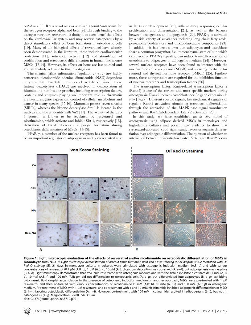
Figure 1. Light microscopic evaluation of the effects of resveratrol and/or nicotinamide on osteoblastic differentiation of MSCs inmonolayer culture. a–d: Light microscopic demonstration of osteoid-tissue formation with von Kossa staining (A) or adipose-tissue formation with OilRed O staining (B). 21 days in monolayer culture. In cultures were stimulated with osteogenic induction medium (A,B: a) and with variousconcentrations of resveratrol (0.1 mM (A,B: b), 1 mM (A,B: c), 10 mM (A,B: d)calcium deposition was observed (A: a–d), but adipogenesis was negative(B: a–d). Light microscopy demonstrated that MSC cultures treated with osteogenic medium and with the sirtuin inhibitor nicotinamide (1 mM (A, B:e), 10 mM (A,B: f) and 100 mM (A,B: g)), did not differentiate to osteoblastic cells (A, e–g), but differentiated into adipocytes (B, e–g), exhibitingcytoplasmic lipid droplet accumulation in the presence of osteogenic induction medium. In another approach, MSCs were pre-treated with 1 mMresveratrol and then co-treated with various concentrations of nicotinamide (1 mM (A,B: h), 10 mM (A,B: i) and 100 mM (A,B: j)) in osteogenicmedium. Pre-treatment of MSCs with 1 mM resveratrol and co-treatment with 1 and 10 mM nicotinamide inhibited adipogenic differentiation of MSCs(B: h–i), favoring osteoblastic differentiation (A: h–i). However, co-treatment with 100 mM nicotinamide resulted in adipogenesis (B: j), but not inosteogenesis (A: j). Magnification: 6200, bar 30 mm.doi:10.1371/journal.pone.0035712.g001
Resveratrol Promotes Osteogenesis of MSCs
PLoS ONE | www.plosone.org 2 April 2012 | Volume 7 | Issue 4 | e35712
and whether this causes deacetylation of Runx2 during osteogen-
esis is an important focus of this study.
Materials and Methods
AntibodiesPolyclonal anti-collagen type I antibody and alkaline phospha-
tase linked sheep anti-mouse and sheep anti-rabbit secondary
antibodies for immunoblotting were purchased from Millipore
(Schwalbach, Germany). Polyclonal anti- Runx2 was purchased
from Alpha Diagnostics Int. San Antonio, TX, USA. Monoclonal
anti-b-actin and nicotinamide were purchased from Sigma-
Aldrich (Munich, Germany). Polyclonal anti-Sirt-1 and anti-
NCoR were purchased from Abcam PLC (Cambridge, UK).
Polyclonal anti-PPAR-c antibodies were purchased from Acris
Antibodies GmbH, Germany. Acetylated-lysine (Ac-K-103) anti-
body was purchased from Cell Signaling Technology (Danvers,
MA, USA).
Resveratrol with purity greater than 98% was purchased from
Sigma-Aldrich (Munich, Germany). A 100-mM stock solution of
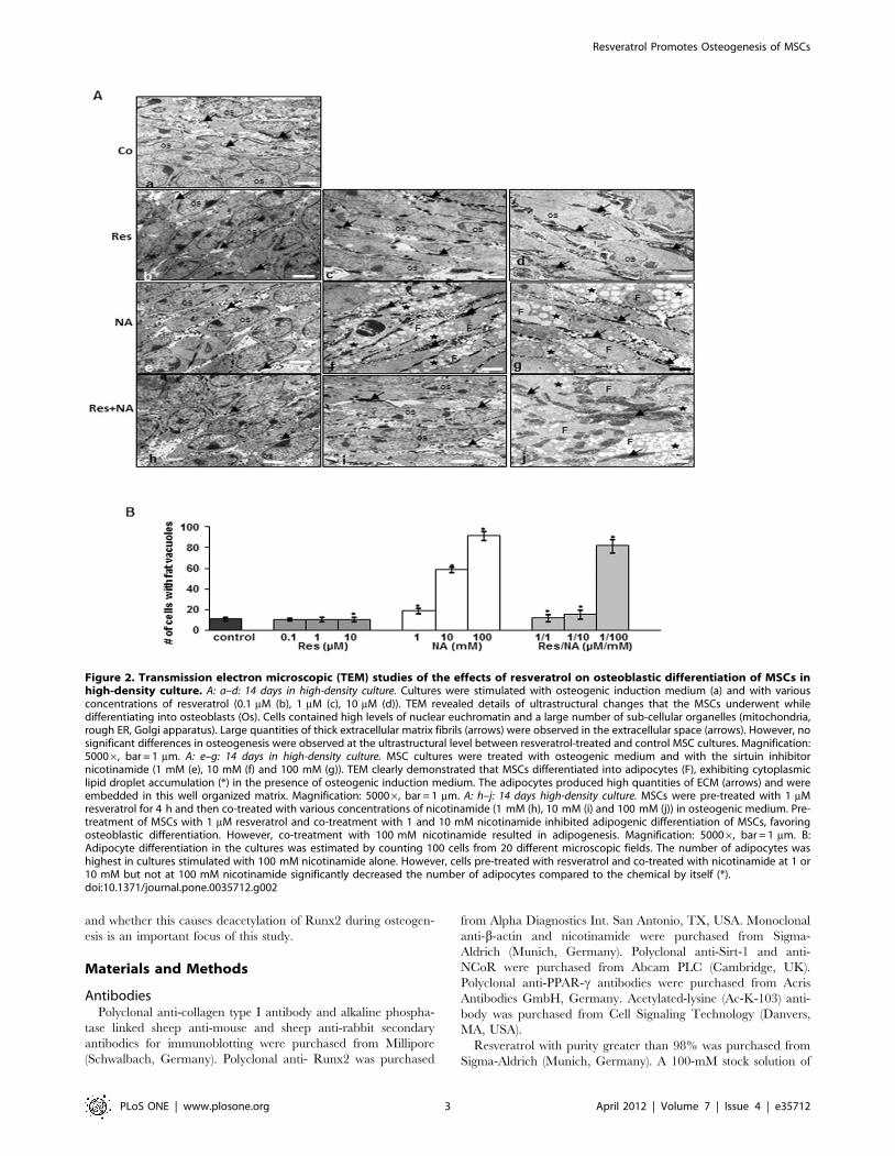
Figure 2. Transmission electron microscopic (TEM) studies of the effects of resveratrol on osteoblastic differentiation of MSCs inhigh-density culture. A: a–d: 14 days in high-density culture. Cultures were stimulated with osteogenic induction medium (a) and with variousconcentrations of resveratrol (0.1 mM (b), 1 mM (c), 10 mM (d)). TEM revealed details of ultrastructural changes that the MSCs underwent whiledifferentiating into osteoblasts (Os). Cells contained high levels of nuclear euchromatin and a large number of sub-cellular organelles (mitochondria,rough ER, Golgi apparatus). Large quantities of thick extracellular matrix fibrils (arrows) were observed in the extracellular space (arrows). However, nosignificant differences in osteogenesis were observed at the ultrastructural level between resveratrol-treated and control MSC cultures. Magnification:50006, bar = 1 mm. A: e–g: 14 days in high-density culture. MSC cultures were treated with osteogenic medium and with the sirtuin inhibitornicotinamide (1 mM (e), 10 mM (f) and 100 mM (g)). TEM clearly demonstrated that MSCs differentiated into adipocytes (F), exhibiting cytoplasmiclipid droplet accumulation (*) in the presence of osteogenic induction medium. The adipocytes produced high quantities of ECM (arrows) and wereembedded in this well organized matrix. Magnification: 50006, bar = 1 mm. A: h–j: 14 days high-density culture. MSCs were pre-treated with 1 mMresveratrol for 4 h and then co-treated with various concentrations of nicotinamide (1 mM (h), 10 mM (i) and 100 mM (j)) in osteogenic medium. Pre-treatment of MSCs with 1 mM resveratrol and co-treatment with 1 and 10 mM nicotinamide inhibited adipogenic differentiation of MSCs, favoringosteoblastic differentiation. However, co-treatment with 100 mM nicotinamide resulted in adipogenesis. Magnification: 50006, bar = 1 mm. B:Adipocyte differentiation in the cultures was estimated by counting 100 cells from 20 different microscopic fields. The number of adipocytes washighest in cultures stimulated with 100 mM nicotinamide alone. However, cells pre-treated with resveratrol and co-treated with nicotinamide at 1 or10 mM but not at 100 mM nicotinamide significantly decreased the number of adipocytes compared to the chemical by itself (*).doi:10.1371/journal.pone.0035712.g002
Resveratrol Promotes Osteogenesis of MSCs
PLoS ONE | www.plosone.org 3 April 2012 | Volume 7 | Issue 4 | e35712

resveratrol (molecular weight, 228.2) was prepared in ethanol and
further diluted in cell culture medium to prepare working
concentrations. The maximum final content of ethanol in cultures
was less than 0.1%. This concentration was also used as a control.
Isolation and culture of mesenchymal stem cellsMesenchymal stem cells were isolated from canine adipose
tissue biopsies obtained during orthopedic surgeries (from 3
animals between the age of ca. 5–7 years), as previously described
[29]. Fully informed owner consent was obtained and the project
was approved by the Ludwig-Maximilian University Ethical
Review committee. Briefly, adipose tissue was cut into small
pieces and digested with collagenase 0.2% in Ham’s-F12 in a
water bath at 37uC for 2 hours. Digested adipose tissue was
centrifuged at 1000 g/5 min and the pellet was resuspended in cell
culture medium consisting of DMEM/Ham’s-F12 1:1, 10% FCS,
1% partricin solution, 1% penicillin/streptomycin solution
(10 000 IU/10 000 IU), 75 mg/ml ascorbic acid, 1% essential
amino acids and 1% Glutamine, all obtained from Seromed
(Munich, FRG). The cells were seeded in a T75 cell culture flask
and incubated at 37uC/5%CO2, 95% humidity. After four days,
non-adherent cells were discarded by washing with Hank’s salt
solution. The medium was changed three times per week.
Adherent cells were split following formation of fibroblast-like cell
colonies and upon reaching 60–70% confluence, and were sub-
cultured until the third or fourth passage was achieved.
Pre-osteoblastic cell line culture. The mouse pre-
osteoblastic cell line MC3T3-E1 (DSMZ, Braunschweig,
Germany) was selected as an in vitro model of pre-osteoblastic
cells, as previously described [30]. The cells were cultured in
alpha-MEM containing 10% FCS, 100 U/mL penicillin and
100 mg/mL streptomycin. The cells were maintained in a
humidified, 95% air/5% CO2 atmosphere at 37uC. All
experiments were performed with third passage MC3T3-E1
cells. For induction of the osteoblast phenotype, cells were
cultured in differentiation medium (DMEM containing 10%
FCS, 10 mm b-glycerophosphate and 50 mg/mL ascorbate-2-
phosphate) [31].
Experimental designOsteogenic differentiation was performed in monolayer culture
or in high-density mass culture. Mesenchymal stem cell cultures
and pre-osteoblastic MC3T3-E1 cells were either left untreated, or
incubated with one of the following treatments: 0.1, 1 and 10 mM
resveratrol only; 1, 10 and 100 mM nicotinamide only; pre-
stimulated with resveratrol 1 mM for 4 h in suspension and then
brought into monolayer or high-density cultures and stimulated
with 1, 10 and 100 mM nicotinamide for the indicated time
Figure 3. Effect of resveratrol or/and nicotinamide on extracellular matrix, Runx2 and PPAR-c expression during osteogenesis ofMSC and pre-osteoblastic cells in high-density cultures. Whole cell lysates (500 ng of protein per lane) were probed with antibodies forcollagen type I (a), for the osteogenic specific transcription factor Runx2 (b) and for the adipogenic specific transcription factor PPAR-c (c) in MSC (A)and in pre-osteoblastic cells in high-density culture (B). Cultures were treated with 0.1, 1 and 10 mM resveratrol alone, or with 1, 10 and 100 mMnicotinamide alone or pre-treated with 1 mM resveratrol for 4 hours and then co-treated with 1, 10, 100 mM nicotinamide or left untreated for 2weeks with osteogenic induction medium in high-density cultures. Untreated cultures (without resveratrol or nicotinamide) produced collagen type I(a, A–B) and Runx2 (b, A–B) in both cultures. Incubation with nicotinamide reduced collagen type I and Runx2 production and increased theexpression of PPAR-c in a concentration dependent manner in MSC-cultures (c, A) and decreased the expression of PPAR-c in a concentrationdependent manner in pre-osteoblastic cultures (III, B). However, pre-treatment with resveratrol inhibited the adverse effects of nicotinamide and theosteoblasts produced large amounts of collagen type I and Runx2. Synthesis of the housekeeping protein b-actin was unaffected (d, A–B).doi:10.1371/journal.pone.0035712.g003
Resveratrol Promotes Osteogenesis of MSCs
PLoS ONE | www.plosone.org 4 April 2012 | Volume 7 | Issue 4 | e35712

periods. For monolayer culture 10,000 cells were seeded per well
in a four-well-plate and cultured until they reached confluency.
Cultures were treated as described below in osteogenic induction
medium and evaluated after 21 days. The high-density mass
culture was performed using procedures and specialized equip-
ment as previously described [32]. Briefly, an 8 ml drop of cells was
placed on a cellulose filter on top of a steel mesh bridge, containing
about 1 million cells. The osteogenic induction medium was
prepared as described by [2], consisting of DMEM base medium,
10% FCS, penicillin/streptomycin solution (10000 IU/10000 IU/
100 ml), 1027 M dexamethasone (Sigma-Aldrich, Cat. No. D-
8893), 10 mM b-glycerophosphate (Sigma-Aldrich, Cat. No. G-
9891) and 50 mM ascorbate-2-phosphate (Sigma-Aldrich, Cat.
No. A-8960). Medium changes were made every three days. For
the negative control, cells were cultured in cell culture medium
containing 10% FCS. To osteogenic induction medium, 0.1, 1 and
10 mM resveratrol and/or 1, 10 and 100 mM nicotinamide were
added for the indicated time periods. Cells were nurtured through
diffusion at the filter medium interface and evaluated after
indicated time periods.
Light microscopyMonolayer cultures were stained with von Kossa for mineral-
ized matrix deposition or stained with Oil Red O solution to
visualize the formation of fat vacuoles as previously described [33].
Antisense and lipofectin-mediated transfectionThe Sirt-1 antisense sequences used in these experiments were
designed using a computational neural network mode [34]. MSCs
were plated in 3 cm2 tissue culture dishes or in a four-well glass
plate at a concentration of 36105 cells/dish or 16104 cells/well
and were grown to confluence. All transfection experiments were
carried out on 50% confluent monolayer cultures. Antisense
oligonucleotide sequence (59-GTATTCCACATGAAACA-
GACA-39) was derived from the nucleotide at position 844 to
864 lying in upstream region of the nucleotide sequences coding
for the catalytic domain of Sirt-1 mRNA registered under
accession number NM012238 in GenBank. To overcome the
rapid degradation of antisense sequence by intracellular endo- and
exonucleases, the non-bridging oxygen on the phosphate linkage
was replaced with a sulfur atom (phosphothioate modification).
The phosphothioate modified sense oligonucleotide sequence (59-
TGTCTGTTTCATGTGGAATAC-39), complementary to the
antisense sequence, was used as control. The modified oligonu-
cleotides were purchased from MWG (Ebersberg, Germany). To
Figure 4. Effect of resveratrol on nicotinamide-induced inhibition of Sirt-1 expression. A: Sirt-1 protein expression during osteogenesis inmonolayer cultures. 21 days monolayer cultures of osteogenic induced fat tissue derived MSCs. Whole cell lysates (500 ng/lane) were probed for Sirt-1.MSCs express high levels of Sirt-1 before and after induction of osteogenic differentiation. Synthesis of the housekeeping protein b-actin wasunaffected. Sirt-1 control peptide was used as a control (co pep.). M = Marker for molecular weights. B–C: Effect of resveratrol on NA-induced inhibitionof Sirt-1 expression during osteogenesis in monolayer culture. 14 days osteogenic induction culture of control MSCs, cells treated with 0.1, 1, 10 mMresveratrol or with 1, 10, 100 mM nicotinamide or pre-treated with 1 mM resveratrol for 4 h followed by co-treatment with nicotinamide. Whole celllysates (500 ng/lane) were fractionated and subjected to western blotting with antibodies against Sirt-1. D: Densitometric evaluation was performedfor Sirt-1 expression from Fig. B–C. Each experiment was performed in triplicate and mean values and standard deviation are indicated. Values werecompared to the control and statistically significant values with p,0.05 were designated by an asterisk (*).doi:10.1371/journal.pone.0035712.g004
Resveratrol Promotes Osteogenesis of MSCs
PLoS ONE | www.plosone.org 5 April 2012 | Volume 7 | Issue 4 | e35712
provide enhanced transfection of oligonucleotides to the cytoplasm
of the target cells, lipofectin reagent (Life Technologies, Invitro-
gen, Darmstadt, Germany) was used according to the manufac-
turer’s instructions. Briefly, 10 ml lipofectin was mixed with 1, 0.5
and 0.2 mM of sense or antisense oligonucleotide (1000 mM)
respectively for 30 min at AT and subsequently the mixture was
added to 990 ml serum-free medium to obtain a working medium
with 1, 0.5 and 0.2 mM of the corresponding oligonucleotide. The
medium was then added to the already prepared cells (50–60%
confluent) and incubated for 24 h at 37uC. After 24 h of
incubation (colonies were pooled from each transfection condition
and used in the subsequent experiments), transfection media was
replaced by the regular culture or osteogenic induction media and
evaluated after 21 days.
Electron microscopyTransmission electron microscopy was performed as previously
described [35]. Briefly, high-density cultures were fixed for one
hour in Karnovsky’s fixative and then post-fixed in 1% OsO4
solution. After dehydration, pellets were embedded in Epon,
ultrathin cuts made on a Reichert-Ultracut E. and contrasted with
a mixture of 2% uranyl acetate/lead citrate. A transmission
electron microscope (Zeiss TEM900, Jena, Germany) was used to
examine the cultures.
To quantify adipocyte formation, the number of cells exhibiting
typical morphological features like multiple fat vacuoles was
determined by scoring 100 cells from 20 different microscopic
fields per culture and the number of adipocytes was expressed as
an indicator of adipogenic differentiation of MSCs.
Immunofluorescence analysis of Sirt-1The effect of specific Sirt-1 antisense or sense on the Sirt-1
expression was investigated by an immunofluorescence method as
previously described in detail [36]. Briefly, the MSCs were
cultured in 4-well glass plates and incubated for 24 h. Serum-
starved cells were treated with 1 mM end concentration of
antisense or sense for 24 hours in serum-starved medium. Glass
plates were rinsed three-times in Hanks solution before methanol
fixation for 10 min at ambient temperature (AT), and rinsing with
PBS. Cell membranes were permeabilized by treatment with 0.1%
Triton X-100 for 1 min on ice. Cells were overlaid with protease-
free bovine serum albumin (BSA) for 10 min at AT, rinsed with
PBS and incubated with primary antibodies (Sirt-1, 1:30 in PBS) in
a humid chamber overnight at 4uC. They were gently washed
several times with PBS before incubation with rhodamine-red
conjugated secondary antibody for 2 h at AT and finally washed
again three times with Aqua Dest laboratory water. Counterstain-
ing was performed with DAPI to visualize the cell nuclei. Samples
were evaluated under light microscope (Leica, Germany) and
photomicrographs were digitally captured and stored.
Immunoprecipitation and ImmunoblottingA detailed description of the technique used for the following
experiments has been previously published [37,38]. Briefly, high-
density cultures were rinsed in PBS and the proteins extracted with
lysis buffer (50 mM Tris/HCl (pH 7.2), 150 mM NaCl, l% (v/v)
Triton X-100, 1 mM sodium orthovanadate, 50 mM sodium
pyrophosphate, 100 mM sodium fluoride, 0.01% (v/v) aprotinin,
pepstatin A (4 mg/ml), leupeptin (10 mg/ml) and 1 mM phenyl-
methylsulfonyl fluoride (PMSF)) for 30 min on ice. After adjusting
the total protein concentration, samples were separated by SDS-
PAGE (5%–12% gels) under reducing conditions. For immuno-
precipitation, the extracts were pre-cleared by incubating them
first with 25 ml of either normal rabbit IgG-serum or normal
mouse IgG-serum and Staphylococcus (S.) aureus cells, then with
primary antibodies diluted in wash buffer (0.1% Tween 20,
150 mM NaCl, 50 mM Tris-HCl (pH 7.2), 1 mM CaCl2, 1 mM
MgCl2 and 1 mM PMSF) for 2 h at 4uC, and finally with S. aureus
cells for 1 h at 4uC. Control immunoprecipitations were
performed by incubating the samples with non-immune rabbit
anti-mouse IgG alone. S. aureus cells were washed five times with
wash buffer and once with 50 mM Tris-HCl (pH 7.2) and then
boiled in SDS-PAGE sample buffer. Separated proteins were
transferred to nitrocellulose membranes and incubated in blocking
buffer (5% (w/v) skimmed milk powder in PBS/0.1% Tween 20)
for 1 h at AT. Membranes were incubated overnight with the first
antibody diluted in blocking buffer at 4uC on a shaker, washed
three times with blocking buffer, and then incubated with the
secondary antibody conjugated with alkaline phosphatase for
90 min at AT. Membranes were rinsed with blocking buffer and
then washed three times in 0.1 M Tris (pH 9.5) containing 0.05 M
MgCl2 and 0.1 M NaCl. Specific antigen-antibody complexes
were rendered visible using nitro-blue tetrazolium and 5-bromo-4-
chloro-3-indoylphosphate (p-toluidine salt; Pierce, Rockford, IL,
USA) as the substrates for alkaline phosphatase. Total protein
concentration was determined according to the bicinchoninic acid
system (Pierce, Rockford, IL, USA) using bovine serum albumin as
a standard. Specific binding was quantified by densitometry using
‘‘quantity one’’ (Bio-Rad Laboratories Inc. CA, USA).
Runx2 acetylation assayRunx2 lysine acetylation was analyzed by immunoprecipitation
of Runx2 followed by western blotting using acetyl-lysine
antibodies. Cells were treated with 1 mM resveratrol for 4 hours
and then exposed to 1, 10 and 100 mM nicotinamide for indicated
times. Whole-cell extracts were prepared, immunoprecipitated
with an anti- Runx2 antibody, and subjected to western blot
analysis using an anti–acetyl-lysine antibody. To confirm these
observations, MSCs were treated with 1 mM resveratrol for 4 h
and then transfected with specific Sirt-1 antisense or sense
oligonucleotides at 1 mM end concentration for 24 h. After 24 h
of incubation transfection media were replaced with the regular
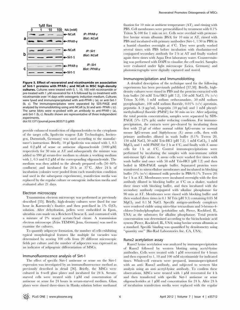
Figure 5. Effect of resveratrol and nicotinamide on associationof Sirt-1 proteins with PPAR-c and NCoR in MSC high-densitycultures. Cultures were treated with 0, 1, 10, 100 mM nicotinamide orpre-treated with 1 mM resveratrol for 4 h followed by co-treatment withnicotinamide over 14 days with osteogenic induction medium. Cultureswere lysed and immunoprecipitated with anti-PPAR-c (a), or anti-Sirt-1(b, c). The immunoprecipitates were separated by SDS-PAGE andanalyzed by immunoblotting using anti-NCoR (a, b) and anti- PPAR-c (c).The same blots were re-probed with an antibody to anti-PPAR-c (a),anti-Sirt-1 (b, c). Results shown are representative of three independentexperiments.doi:10.1371/journal.pone.0035712.g005
Resveratrol Promotes Osteogenesis of MSCs
PLoS ONE | www.plosone.org 6 April 2012 | Volume 7 | Issue 4 | e35712
culture medium or osteogenic induction medium with or without
nicotinamide (10 mM) and evaluated after 21 days. Whole cell
extracts were prepared and subjected to immunoprecipitation with
anti- Runx2 antibody and the precipitates were separated by SDS-
PAGE and immunoblotted using antibodies against acetyl-lysine
and Runx2.
Co-Immunoprecipitation of Runx2 and Sirt-1Endogenous protein interactions from high-density cultures
were evaluated by co-immunoprecipitation experiments using Sirt-
1 and Runx2 antibodies. Cells were treated with 1 mM resveratrol
for 4 hours and then exposed to 1, 10 and 100 mM nicotinamide
for the indicated times. Whole-cell extracts were prepared,
immunoprecipitated with an anti- Runx2 antibody, and precip-
itates were subjected to western blot analysis using an anti–Sirt-1
antibody. To confirm the protein-protein interactions in MSCs,
cells were treated with 1 mM resveratrol for 4 hours and then
transfected with 1 mM end concentration of specific Sirt-1
antisense or sense oligonucleotides for 24 h. After 24 h of
incubation transfection media were replaced by the regular
culture medium or osteogenic induction medium with or without
nicotinamide (10 mM) and evaluated after 21 days. Whole cell
extracts were prepared and subjected to immunoprecipitation with
anti-Runx2 antibody and the precipitates were separated by SDS-
PAGE and immunoblotted using antibodies against Sirt-1.
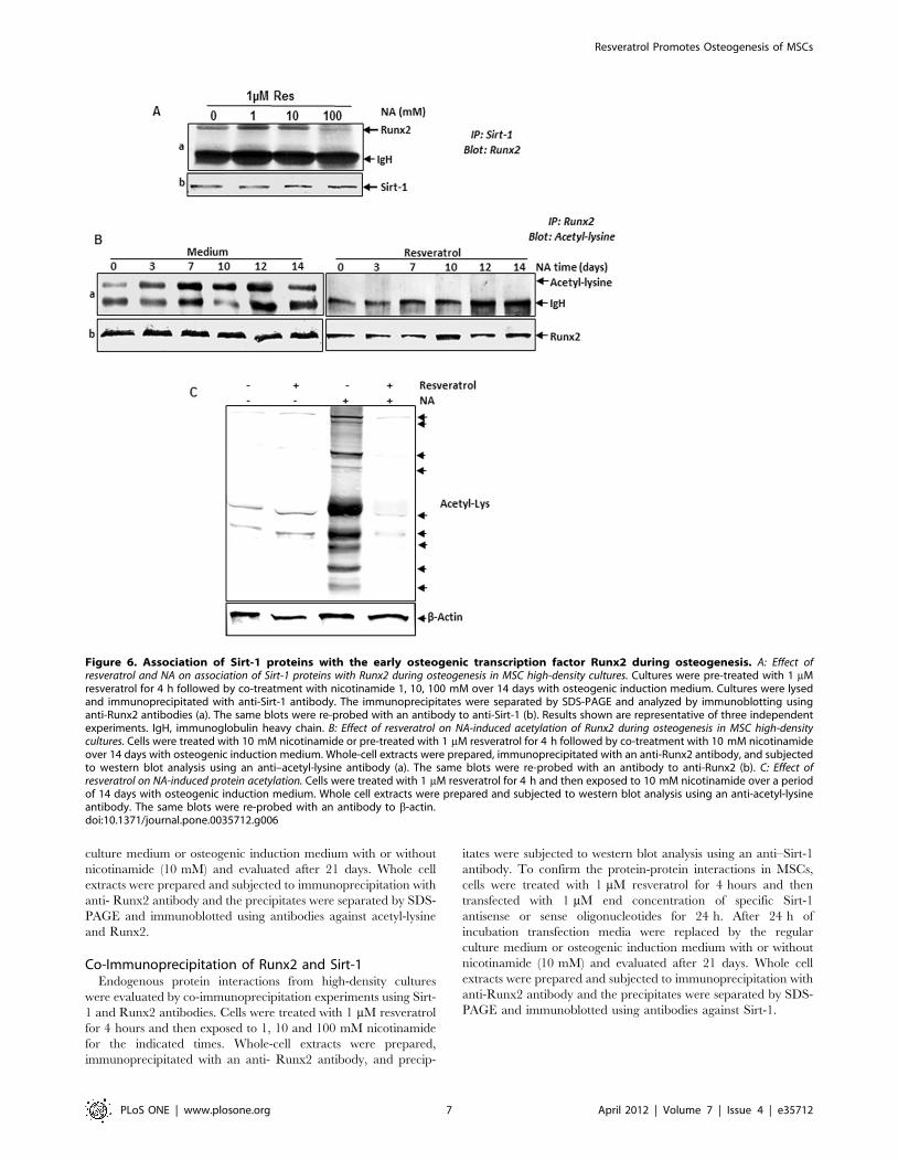
Figure 6. Association of Sirt-1 proteins with the early osteogenic transcription factor Runx2 during osteogenesis. A: Effect ofresveratrol and NA on association of Sirt-1 proteins with Runx2 during osteogenesis in MSC high-density cultures. Cultures were pre-treated with 1 mMresveratrol for 4 h followed by co-treatment with nicotinamide 1, 10, 100 mM over 14 days with osteogenic induction medium. Cultures were lysedand immunoprecipitated with anti-Sirt-1 antibody. The immunoprecipitates were separated by SDS-PAGE and analyzed by immunoblotting usinganti-Runx2 antibodies (a). The same blots were re-probed with an antibody to anti-Sirt-1 (b). Results shown are representative of three independentexperiments. IgH, immunoglobulin heavy chain. B: Effect of resveratrol on NA-induced acetylation of Runx2 during osteogenesis in MSC high-densitycultures. Cells were treated with 10 mM nicotinamide or pre-treated with 1 mM resveratrol for 4 h followed by co-treatment with 10 mM nicotinamideover 14 days with osteogenic induction medium. Whole-cell extracts were prepared, immunoprecipitated with an anti-Runx2 antibody, and subjectedto western blot analysis using an anti–acetyl-lysine antibody (a). The same blots were re-probed with an antibody to anti-Runx2 (b). C: Effect ofresveratrol on NA-induced protein acetylation. Cells were treated with 1 mM resveratrol for 4 h and then exposed to 10 mM nicotinamide over a periodof 14 days with osteogenic induction medium. Whole cell extracts were prepared and subjected to western blot analysis using an anti-acetyl-lysineantibody. The same blots were re-probed with an antibody to b-actin.doi:10.1371/journal.pone.0035712.g006
Resveratrol Promotes Osteogenesis of MSCs
PLoS ONE | www.plosone.org 7 April 2012 | Volume 7 | Issue 4 | e35712
Statistical analysisNumerical data are expressed as mean values (+/2SD) for a
representative experiment performed in triplicate. The means
were compared using student’s t-test assuming equal variances.
Differences were considered to be statistically significant if the P-
value was less than 0.05.
Results
Effects of resveratrol or/and nicotinamide on osteogenicdifferentiation of MSC in monolayer cultures
Incubation of MSCs in monolayer cultures with osteogenic
induction medium over 3 weeks resulted in osteogenesis; positive
von Kossa staining and high quantities of calcium deposition
(Fig. 1A, a) and negative oil Red O staining (Fig. 1B, a) was
observed in MSC cultures. In untreated pure MSC cultures, no
calcium deposition was observed (data not shown). Treatment of
MSC cultures with the osteogenic induction medium and
resveratrol (various concentrations, 0.1, 1 and 10 mM) induced
osteogenesis and produced positive von Kossa staining (Fig. 1A, b–
d) and negative oil Red O staining (Fig. 1B, b–d). In contrast, in
the presence of the sirtuin inhibitor nicotinamide, osteogenesis was
not observed (Fig. 1A, e–g). Cells differentiated to adipocytes and
contained more vacuoles compared to the resveratrol treated cells.
Oil Red O staining for fat deposition revealed the presence of fat
vacuoles containing neutral lipids (Fig. 1B, e–g). The number of
differentiated adipocytes in culture increased in the presence of 10
or 100 mM nicotinamide. To test whether activation of Sirt-1
inhibits adipogenesis during osteoblastic differentiation [19,39],
MSC cultures were treated with resveratrol and then co-treated
with various concentrations of nicotinamide in osteogenic
induction medium. Pre-treatment of MSCs with resveratrol and
co-treatment with nicotinamide promoted osteogenic differentia-
tion (Fig. 1A, h–i) and inhibited adipogenic differentiation (Fig. 1B,
h–i). However, the inhibition of adipogenesis by resveratrol was
concentration dependent. Pre-treatment of MSCs with 1 mM
resveratrol and co-treatment with 100 mM nicotinamide did not
result in osteogenesis (Fig. 1A, j), but stimulated adipogenesis
(Fig. 1B, j).
Effects of resveratrol or/and nicotinamide on osteogenicdifferentiation of MSC and pre-osteoblastic cells in high-density cultures
Incubation of MSCs with osteogenic induction medium resulted
in osteogenesis; cells exhibited high levels of nuclear euchromatin,
large numbers of morphologically normal cellular organelles
(mitochondria, rough ER, Golgi apparatus), numerous cell-cell
processes and large quantities of thick fibrils in a well-organized
extracellular matrix (Fig. 2A, a). Treatment of MSC cultures with
the osteogenic induction medium and resveratrol (various
concentrations, 0.1, 1 and 10 mM) induced osteogenesis (Fig. 2A,
b–d). However, no significant differences in osteogenesis were
observed at the ultrastructural level between with resveratrol-
treated and untreated MSC cultures.
In contrast, in the presence of the sirtuin inhibitor nicotinamide,
osteogenesis was not observed, and some MSCs underwent
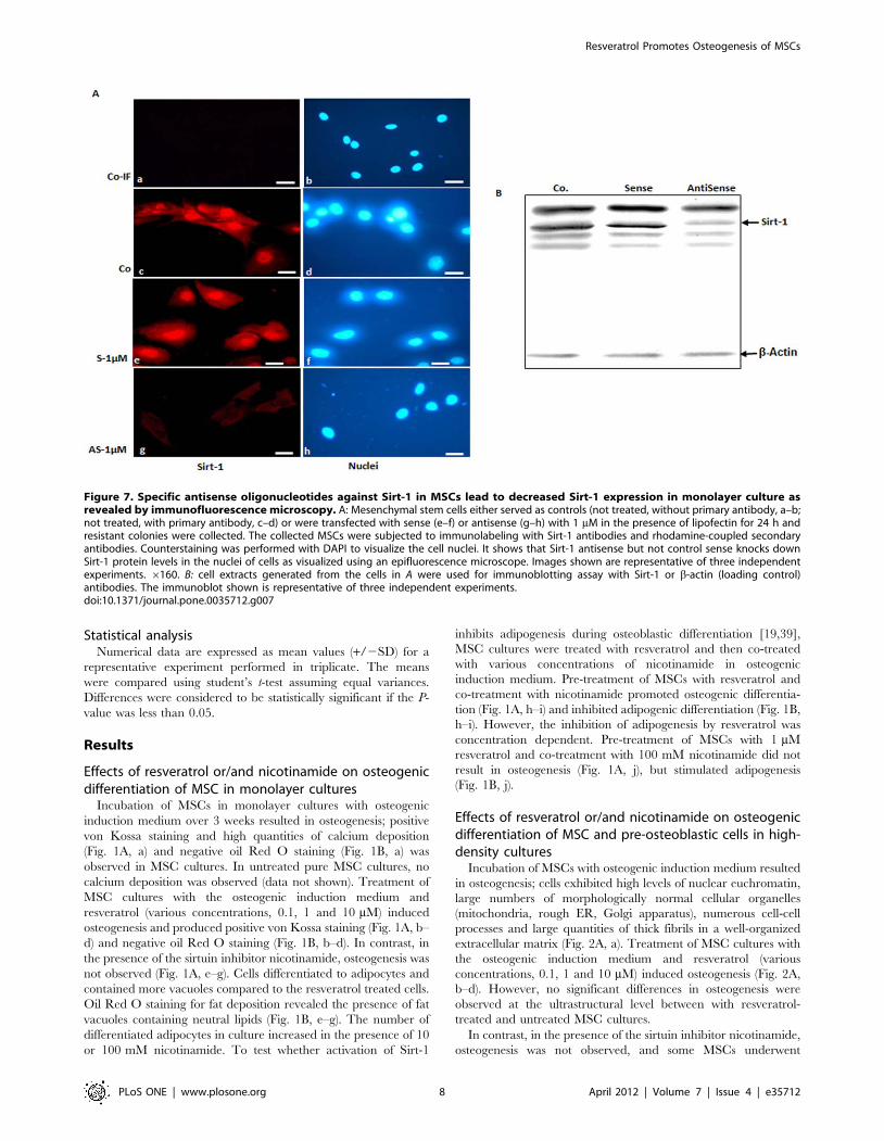
Figure 7. Specific antisense oligonucleotides against Sirt-1 in MSCs lead to decreased Sirt-1 expression in monolayer culture asrevealed by immunofluorescence microscopy. A: Mesenchymal stem cells either served as controls (not treated, without primary antibody, a–b;not treated, with primary antibody, c–d) or were transfected with sense (e–f) or antisense (g–h) with 1 mM in the presence of lipofectin for 24 h andresistant colonies were collected. The collected MSCs were subjected to immunolabeling with Sirt-1 antibodies and rhodamine-coupled secondaryantibodies. Counterstaining was performed with DAPI to visualize the cell nuclei. It shows that Sirt-1 antisense but not control sense knocks downSirt-1 protein levels in the nuclei of cells as visualized using an epifluorescence microscope. Images shown are representative of three independentexperiments. 6160. B: cell extracts generated from the cells in A were used for immunoblotting assay with Sirt-1 or b-actin (loading control)antibodies. The immunoblot shown is representative of three independent experiments.doi:10.1371/journal.pone.0035712.g007
Resveratrol Promotes Osteogenesis of MSCs
PLoS ONE | www.plosone.org 8 April 2012 | Volume 7 | Issue 4 | e35712
apoptosis, with degeneration of the cells, membrane blebbing,
nuclear damage and formation of apoptotic bodies. Remaining
cells differentiated to adipocytes as demonstrated by lipid
accumulation in fat vacuoles (Fig. 2A, e–g). The quantity of
differentiated adipocytes in culture increased in the presence of 10
or 100 mM nicotinamide. Transmission electron microscopy
clearly showed that the MSCs differentiated to adipocytes,
accumulating cytoplasmic lipid droplets and exhibiting well-
developed rough endoplasmic reticulum and mitochondria.
Pre-treatment of MSCs with resveratrol and co-treatment with
nicotinamide promoted osteogenic differentiation (Fig. 2A, h–j).
However, the inhibition of adipogenesis by resveratrol was
concentration dependent. Pre-treatment of MSCs with 1 mM
resveratrol and co-treatment with 100 mM nicotinamide resulted
in adipogenesis.
Incubation of pre-osteoblastic MC3T3-E1 cells with the
osteogenic induction medium or/and resveratrol resulted in
osteogenesis. However, in contrast to MSCs, treatment of pre-
osteoblastic MC3T3-E1 cells with nicotinamide, led to apoptosis
instead of to formation of adipocytes. Pre-treatment of pre-
osteoblastic MC3T3-E1 cells with resveratrol and co-treatment
with nicotinamide promoted osteogenic differentiation (data not
shown).
Statistical evaluation of the data clearly highlighted changes in
the number of cells with fat vacuole accumulation before and after
nicotinamide-treatment in MSC-osteogenesis high-density cul-
tures. Co-treatment with resveratrol decreased the number of
adipocytes with accumulated fat vacuoles (Fig. 2B).
Effect of resveratrol or/and nicotinamide on extracellularmatrix, Runx2 and PPAR-c expression during MSC-osteogenesis and in pre-osteoblastic cell-osteogenesis
To confirm the morphological results described above and to
demonstrate more precisely the identity of the osteogenesis or
adipogenesis by MSCs or pre-osteoblastic cell cultures, whole cell
extracts were probed for collagen type I, Runx2 and PPAR-c.
High collagen type I content was detected by immunoblotting in
the osteogenic-induced control cultures. Treatment of MSCs with
osteogenic induction medium and 0.1, 1 and 10 mM resveratrol in
high-density cultures resulted in a stimulation of collagen type I
production and expression of Runx2. MSC cultures treated with
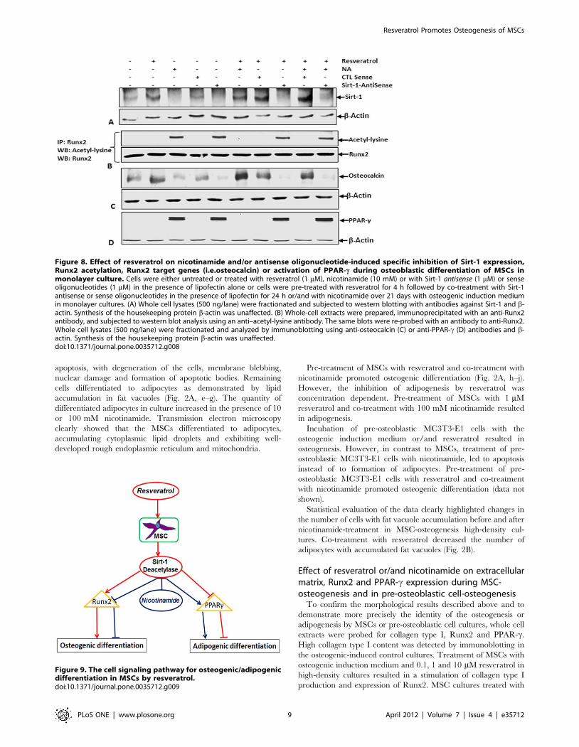
Figure 8. Effect of resveratrol on nicotinamide and/or antisense oligonucleotide-induced specific inhibition of Sirt-1 expression,Runx2 acetylation, Runx2 target genes (i.e.osteocalcin) or activation of PPAR-c during osteoblastic differentiation of MSCs inmonolayer culture. Cells were either untreated or treated with resveratrol (1 mM), nicotinamide (10 mM) or with Sirt-1 antisense (1 mM) or senseoligonucleotides (1 mM) in the presence of lipofectin alone or cells were pre-treated with resveratrol for 4 h followed by co-treatment with Sirt-1antisense or sense oligonucleotides in the presence of lipofectin for 24 h or/and with nicotinamide over 21 days with osteogenic induction mediumin monolayer cultures. (A) Whole cell lysates (500 ng/lane) were fractionated and subjected to western blotting with antibodies against Sirt-1 and b-actin. Synthesis of the housekeeping protein b-actin was unaffected. (B) Whole-cell extracts were prepared, immunoprecipitated with an anti-Runx2antibody, and subjected to western blot analysis using an anti–acetyl-lysine antibody. The same blots were re-probed with an antibody to anti-Runx2.Whole cell lysates (500 ng/lane) were fractionated and analyzed by immunoblotting using anti-osteocalcin (C) or anti-PPAR-c (D) antibodies and b-actin. Synthesis of the housekeeping protein b-actin was unaffected.doi:10.1371/journal.pone.0035712.g008
Figure 9. The cell signaling pathway for osteogenic/adipogenicdifferentiation in MSCs by resveratrol.doi:10.1371/journal.pone.0035712.g009
Resveratrol Promotes Osteogenesis of MSCs
PLoS ONE | www.plosone.org 9 April 2012 | Volume 7 | Issue 4 | e35712
nicotinamide alone at various concentrations showed a significant
downregulation of synthesis of collagen type I and Runx2, but up-
regulation of PPAR-c and this was in a concentration-dependent
manner (Fig. 3A, a, b, c left panel). In contrast to this, pre-
treatment of MSCs with resveratrol (1 mM, 4 h) followed by
stimulation with the sirtuin inhibitor, nicotinamide (1, 10 and
100 mM) resulted in an inhibition of nicotinamide-induced effects
on collagen type I production and Runx2 during MSC-
osteogenesis and downregulated PPAR-c in high-density cultures
(Fig. 3A, a, b, c right panel). However, 1 mM resveratrol could not
completely inhibit the blocking effect of 100 mM nicotinamide on
the synthesis of collagen type I and Runx2 during osteogenesis and
downregulated PPAR-c in high-density culture (Fig. 3A, a, b, c
right panel). Synthesis of the house-keeping protein b-actin
remained unaffected (Fig. 3A, d).
To see that the nicotinamide-induced inhibition of Runx2 and
stimulation of PPAR-c and adipogenesis during MSC-osteogenesis
occurs also transiently during osteogenesis with pre-osteoblastic
cells, we compared the effects of resveratrol or/and nicotinamide
on protein expression profiles of MSC and pre-osteoblastic
MC3T3-E1 cells during the osteogenesis in high-density culture
to further confirm their differentiation capacities. Pre-osteoblastic
MC3T3-E1 cells produced large quantities of collagen type I in
presence of 0.1, 1 and 10 mM resveratrol and Runx2 expression
was also stimulated. High collagen type I content was also detected
in the osteogenic-induced control cultures. Pre-osteoblastic cells
treated with nicotinamide alone at various concentrations showed
a significant downregulation of synthesis of collagen type I and
Runx2. Interestingly, in opposite to MSC-cultures, when nicotin-
amide was added to pre-osteoblastic MC3T3-E1 cells, no
significant effect was seen on formation of adipocytes and
PPAR-c expression compared with MSCs and this was in a
concentration-dependent manner (Fig. 3B, a, b, c left panel).
Moreover, pre-treatment of pre-osteoblastic MC3T3-E1 cells with
resveratrol (1 mM, 4 h) followed by stimulation with nicotinamide
(1, 10 and 100 mM) resulted in an inhibition of nicotinamide-
induced effects on collagen type I production and Runx2 and
downregulated PPAR-c in high-density cultures (Fig. 3B, a, b, c
right panel). However, 1 mM resveratrol could not completely
inhibit the blocking effect of 100 mM nicotinamide on the
synthesis of collagen type I and Runx2 in high-density culture
(Fig. 3B, a, b, c right panel). Taken together, these results indicate
that adipocytes and osteoblasts share a common progenitor, i.e.
MSCs expressing PPAR-c signaling can induce trans-differentia-
tion of osteoblasts to adipocytes by inhibiting of Runx2, whereas,
the pre-osteoblastic cells only have the capability to differentiate
into osteoblasts.
Expression of Sirt-1 in MSCs before and after osteoblasticdifferentiation in vitro
The NAD-dependent protein deacetylase Sirt-1 has been shown
to attenuate development of adipocytes from pre-adipocytes
through inhibition of PPAR-c activity [39]. Next, we wanted to
evaluate whether phytochemicals known to regulate the activity of
Sirt-1 could influence the formation of MSCs during osteoblast
differentiation in vitro. First, we could demonstrate the expression
of Sirt-1 in the MSCs derived from fat tissue. Whole cell lysate
from MSCs in monolayer cultures treated with osteogenic
induction medium for 0, 7, 14 and 21 days were analyzed by
western blot with anti-Sirt-1 antibody. Sirt-1 was expressed in the
MSCs before (day 0) and after induction of osteoblastic
differentiation (Fig. 4A).
Resveratrol inhibits nicotinamide-induced down-regulation of Sirt-1 during osteogenic differentiation ofMSCs in vitro
To investigate the possible mechanism for dedifferentiation of
MSCs to adipocytes during osteogenesis, we investigated the effect
of resveratrol on the expression of Sirt-1. As shown in Figures 4B–
C, when cells were treated with sirtuin inhibitor, nicotinamide (1,
10, 100 mM), the expression of endogenous Sirt-1 was decreased
in a concentration dependent manner. In contrast, resveratrol
treatment caused an increase of the Sirt-1 expression in a
concentration dependent manner (Fig. 4B). Interestingly, pre-
treatment of MSCs with resveratrol (1 mM, 4 h) followed by
stimulation with nicotinamide (1, 10 and 100 mM) caused a
concentration dependent up-regulation in Sirt-1 expression
(Fig. 4C). Densitometric analysis of a representative experiment
performed in triplicate from the effect of resveratrol on
nicotinamide-induced inhibition of Sirt-1 expression during
osteogenesis in monolayer culture showed that the increasing
concentration of nicotinamide decreased the amount of Sirt-1,
whereas pre-treatment with resveratrol markedly increased it
(Fig. 4D).
Sirt-1 blocks adipogenesis by repressing PPAR-c activityand NCoR involvement in this process
The nuclear receptor PPAR-c is known to regulate adipogenesis
[39]. It has also been shown that the nuclear receptor co-repressor,
NCoR, binds to known PPAR-c sites of promoters of adipogenic
genes in differentiated 3T3-L1 adipocytes [39]. To test whether
Sirt-1 is a PPAR-c co-repressor by means of NCoR, we pre-
treated the MSCs with resveratrol (1 mM, 4 h) followed by
stimulation with nicotinamide (1, 10 and 100 mM) in high-density
cultures, and co-immunoprecipitation assays. As shown in
Figure 5a, after immunoprecipitation with anti-PPAR-c antibod-
ies, the samples were probed by immunoblotting with anti-NCoR.
The results indicate that PPAR-c interacts with NCoR and this
interaction is dependent on the concentration of nicotinamide
(Fig. 5a). As shown in Figure 5b–c, after immunoprecipitation with
anti-Sirt-1 antibodies, the samples were probed by immunoblot-
ting with anti-NCoR and anti-PPAR-c. The results suggest that
Sirt-1 interacts with NCoR and PPAR-c. The expression of
endogenous NCoR was decreased and the expression of
endogenous PPAR-c was increased in a concentration dependent
manner with nicotinamide. Taken together, these results demon-
strate that NCoR may, at least in part, be involved in repression of
PPAR-c by Sirt-1.
Resveratrol blocks nicotinamide-induced inhibition ofthe association of Sirt-1 proteins with the earlyosteogenic transcription factor Runx2 in MSC high-density cultures
To determine possible downstream signaling proteins during
osteogenesis in high-density cultures, we examined whether Sirt-1
associates with the early osteogenic transcription factor Runx2
subsequently activating the pathway that stimulates osteogenesis.
The fact that the expression pattern of Sirt-1 and Runx2 protein
are similarly stimulated or inhibited by resveratrol or nicotinamide
in MSCs (Fig. 4A, B; Fig. 3A, B), suggests that these two proteins
could interact together. To examine this, we performed co-
immunoprecipitation of endogenous Sirt-1 protein and precipitat-
ed Runx2. As shown in Figure 6A, the cells were pre-treated with
resveratrol (1 mM, 4 h) and then co-treated with nicotinamide (1,
10 and 100 mM) for 14 days, then co-immunoprecipitation assays
were performed. After immunoprecipitation with anti-Sirt-1
Resveratrol Promotes Osteogenesis of MSCs
PLoS ONE | www.plosone.org 10 April 2012 | Volume 7 | Issue 4 | e35712
antibodies, the samples were probed by immunoblotting with anti-
Runx2. The results indicated that Runx2 was co-immunoprecip-
itated by anti-Sirt-1 antiserum but not by pre-immune serum in
high-density cultures (Fig. 6A). This interaction of Sirt-1 with
Runx2 was decreased with as little as 10 mM nicotinamide and
indicates that the expression and association of Runx2 with Sirt-1
is concentration-dependent. Taken together, these results indicate
that during osteogenesis resveratrol activates Sirt-1 and induces
Sirt-Runx2 complex formation, which activates the osteogenic
pathway.
Effect of resveratrol on nicotinamide-induced acetylationof Runx2 in MSC high-density cultures
Resveratrol has been shown to activate Sirt-1 deacetylase
activity [18,30]. The fact that the stimulation of Sirt-1 protein
correlated with the expression of Runx2 and in addition, that both
proteins interact together [30], suggests that Runx2 might be a
substrate for Sirt-1 deacetylation. As shown in Fig. 6B, nicotin-
amide treatment strongly induced Runx2 lysine acetylation in a
time dependent manner in high-density cultures. To examine the
functional impact of Sirt-1 regulation of nicotinamide-mediated
acetylation of Runx2, we pre-treated MSCs with resveratrol and
then co-treated them with nicotinamide during osteogenesis in
high-density cultures for the indicated time periods. Interestingly,
the nicotinamide-induced acetylation of Runx2 markedly de-
creased by pre-treatment with resveratrol, suggesting, at least in
part, a significant reduction in nicotinamide-induced Runx2
acetylation by Sirt-1 activity (Fig. 6B). To determine whether
resveratrol is able to block the nicotinamide-induced acetylation of
proteins, whole cell lysates from cells treated with nicotinamide,
resveratrol or combination of both of them were analyzed by
western blotting using anti-acetyl lysine antibody. As shown in
Fig. 6C, nicotinamide induced acetylation of several proteins,
whereas resveratrol suppressed the acetylation of these proteins.
These findings suggest that resveratrol-activated Sirt-1 plays an
important role in inhibiting nicotinamide-activated PPAR-c/
NCoR complex resulting in a decrease of Runx2 acetylation.
Specific antisense oligonucleotides downregulate Sirt-1in MSCs in vitro
To investigate whether specific antisense oligonucleotides
against Sirt-1 inhibit Sirt-1 expression, MSCs were transfected
with specific antisense or sense oligonucleotides derived from
nucleotide sequence coding upstream part of catalytic domain of
Sirt-1 protein. The immunofluorescence analysis (Fig. 7A) as well
as the immunoblot assays (Fig. 7B) showed that the specific
antisense oligonucleotides reduced the levels of Sirt-1 expression
and nuclear localization. In contrast, the control sense oligonu-
cleotide had no effect on Sirt-1 expression. The results indicated
that treatment with Sirt-1 antisense oligonucleotides inhibited Sirt-
1 expression specifically and concentration dependently (data not
shown) and the inhibition was not related to non-specific gene-
regulatory events.
Downregulation of Sirt-1 expression by antisenseoligonucleotides enhances Runx2 acetylation, PPAR-cactivation and inhibits expression of Runx2 target genes(osteocalcin) during osteogenic differentiation of MSCs inmonolayer cultures
Based on the results of co-immunoprecipitation assays
(Figure 6A), Sirt-1 interacts directly with Runx2 in vitro, which
raises the possibility that Runx2 may be a substrate for Sirt-1
deacetylase. Since Sirt-1 acts as a protein deacetylase, next we
examined whether the inhibitory effect of resveratrol on Runx2
acetylation is Sirt-1 dependent. The Sirt-1 specific oligonucleotide-
transfected cells efficiently knocked down Sirt-1 protein levels
during osteogenesis in vitro (Fig. 8A), and this abolished the ability
of resveratrol (activator of Sirt-1) to deacetylate Runx2 in
resveratrol and/or nicotinamide-stimulated cells (Fig. 8B) in
monolayer cultures. Interestingly, the acetylation content of
Runx2 was higher in cells treated with specific antisense
oligonucleotides than in cells treated with or without sense
oligonucleotides, suggesting the higher acetylated content of
Runx2 protein is related to downregulated Sirt-1 expression and
Runx2 could be a substrate for Sirt-1 deacetylase. To examine and
establish a correlation between Sirt-1 and the activity of Runx2,
western blot analysis with anti-osteocalcin (Runx2 target gene)
antibody was performed. As shown in Fig. 8C, osteocalcin protein
was downregulated in cells treated with nicotinamide and specific
antisense oligonucleotides, compared to the cells treated with or
without sense oligonucleotides. Taken together, these results
suggest that downregulation of Sirt-1 in MSCs can decrease
Runx2 activities and its downstream target genes.
To further investigate, if the acetylation of Runx2 by Sirt-1
downregulation, has an effect on the expression of PPAR-cprotein, western blot analysis with anti-PPAR-c antibody was
performed. As shown in Fig. 8C, PPAR-c protein was elevated in
cells treated with specific antisense oligonucleotides, compared to
the cells treated with or without sense oligonucleotides. These
results suggest that downregulation of Sirt-1 in MSCs can increase
adipogenic differentiation and expression of the adipose transcrip-
tion regulator PPAR-c and modulate the expression of down-
stream target genes.
Discussion
The aim of this study was to determine whether the naturally
occurring phytoestrogen resveratrol can influence the osteoblastic
differentiation of MSCs through its effects on Sirt-1-mediated
cellular responses in an in vitro model of osteogenesis. This study
leads to the following findings: (1) In the presence of nicotinamide
(sirtuin inhibitor) some MSCs differentiated into adipocytes. (2)
However, pre-treatment of MSCs with resveratrol protected them
from the effects of nicotinamide-induced sirtuin inhibition
resulting in osteogenesis (3) resveratrol blocked nicotinamide-
induced inhibition of Runx2, a well-known important transcrip-
tion factor involved in osteoblast recruitment and differentiation
[40]. Further, resveratrol simultaneously inhibited the nicotin-
amide-induced fat transcription regulator PPAR-c in the same
cultures. (4) In opposite to MSCs, pre-osteoblastic cells treated
with nicotinamide underwent apoptosis and did not differentiate to
adipocytes, suggesting that adipocytes and osteoblasts share a
common progenitor, while pre-osteoblastic cells only have the
capability to differentiate into osteoblasts. (5) Resveratrol-activated
Sirt-1 in MSCs increased its binding to PPAR-c and repressed
PPAR-c activity. (6) The modulatory effects of resveratrol-
activated Sirt-1 on nicotinamide-induced expression of PPAR-cwere found to be mediated, at least in part by the binding and
deacetylation of Runx2. (7) Finally, we describe, for the first time,
an antisense oligonucleotide approach to downregulate Sirt-1
expression in MSCs and demonstrate its ability to functionally
inhibit osteogenesis and induce adipogenesis.
The differentiation capacity of MSCs is a highly investigated
area of biology and medicine. However, little is known about the
behavior of MSCs and progenitor cells during osteogenic
differentiation. Therefore, we studied the differentiation of MSCs
and pre-osteoblastic cells (MC3T3-E1) to compare their differen-
Resveratrol Promotes Osteogenesis of MSCs
PLoS ONE | www.plosone.org 11 April 2012 | Volume 7 | Issue 4 | e35712
tiation capacities. In the presence of resveratrol or/and nicotin-
amide, MSCs differentiate into osteoblasts and adipocytes in high-
density cultures. In contrast to MSCs, pre-osteoblast cells were
programmed to differentiate into their committed target osteoblast
cells, as they were unable to differentiate into adipocytes [41]. For
this reason, this study demonstrates that the primary isolated
MSCs are stem cells, but pre-osteoblastic cells from the osteoblast
progenitor MC3T3-E1 are not. In our study, MSCs treated with
the sirtuin inhibitor (nicotinamide) downregulated bone-specific
matrix compounds. Furthermore, the pre-treatment of MSCs with
resveratrol lead to a recovery of osteoblastic differentiation and
production of collagen type I in co-nicotinamide-stimulated
MSCs. Thus, Sirt-1 appears to be a modulator of MSC
differentiation to osteogenic cells. Moreover, in contrast to MSCs,
pre-osteoblastic cells treated with nicotinamide downregulated
bone-specific matrix components and cells underwent apoptosis.
Activation of Sirt-1 in MSCs decreases adipocyte differentiation
and increases osteoblastic differentiation in high-density cultures.
This differentiation was accompanied by expression of the
osteoblastic transcription factor Runx2, which results in earlier
initiation of the osteoblast differentiation programme. Since Sirt-1
inhibits the adipogenic transcription factor PPAR-c, it also
stimulates mechanisms regulating osteoblast differentiation. The
most critical of these events is the activation of the master bone
gene Runx2 [40]. Runx2 is responsible for expression of
osteogenic marker genes, including osteopontin, osteocalcin and
ALP. It has been reported that differentiation of MSCs to
adipocytes can be inhibited by resveratrol and this process can be
inhibited by the sirtuin blocker nicotinamide [18]. The mecha-
nisms by which resveratrol and Sirt-1 mediate differentiation of
MSCs to osteoblasts and inhibit adipogenesis, appear to involve, at
least in part, the inhibition of PPAR-c and activation of Runx2.
Our co-immunoprecipitation data indicate that Sirt-1 interacts
with the nuclear receptor PPAR-c and this interaction was
downregulated by nicotinamide. Moreover, we demonstrated that
nuclear receptor PPAR-c interacts with the nuclear receptor co-
repressor NCoR. To test the possibility that Sirt-1 functionally
represses PPAR-c by the involvement of NCoR, we pre-treated
the cells with resveratrol and co-treated with nicotinamide in high-
density cultures. We found that PPAR-c, NCoR and Sirt-1 were in
a common complex, but in the presence of 1 mM resveratrol and 1
and 10 mM nicotinamide the amount of NCoR and Sirt-1
increased and the amount of PPAR-c decreased. In contrast, in
the presence of 1 mM resveratrol and 100 mM nicotinamide, the
amount of Sirt-1 and NCoR decreased and the amount PPAR-cincreased in these experiments (Fig. 5). It has also been reported
that Sirt-1 indirectly influences the transcriptional activity of the
nuclear receptor PPAR-c by docking the NCoR and SMRT to
PPAR-c [19]. The co-repressor protein, NCoR does not have an
enzymatic activity, but it can activate the catalytic activity of
histone deacetylases for deacetylation of histone proteins [42].
These data indicate that Sirt-1 interacts with the nuclear receptor
co-repressor NCoR suggesting that Sirt-1, at least in part represses
PPAR-c activity by involving the co-activators. However, it should
be considered that while resveratrol is known to activate Sirt-1, it
has also other additional target proteins in the cells, thus it cannot
be the only effect of Sirt-1.
Resveratrol’s enhancement of osteogenesis was, at least in part
regulated by Runx2 with additional contributions by Sirt-1.
Resveratrol increases alkaline phosphatase activity in osteoblastic
cells [10] an effect that is blocked by tamoxifen, an estrogen
antagonist, suggesting that some of resveratrol’s stimulatory
actions may be mediated through the estrogen receptor. Gehm
et al. have reported that resveratrol acts as a phytoestrogen (i.e.
activating the estrogen receptor) and decreases osteoporosis [43].
Moreover, resveratrol is one of the most potent Sirt-1 activators;
through binding to a special binding site it induces a conforma-
tional change in Sirt-1, lowering the Km for both the acetylated
substrate and NAD, thus resulting in increased enzymatic activity
[18]. Sirt-1 facilitates the differentiation of MSCs to osteoblasts by
directly regulating factors such as Runx2 and by modulation of
nuclear receptor co-repressor NCoR and PPAR-c.
It is known that the nuclear protein deacetylase Sirt-1 belongs to
class III of histone deacetylases, resulting in transcriptional
silencing. Thus, Sirt-1 participates in the regulation of genome
architecture and gene expression [15,16]. These results suggest
that Runx2 and Sirt-1 directly interact together and that Runx2
might be a substrate for Sirt-1 deacetylation. Furthermore, our
data demonstrate that nicotinamide treatment induced Runx2
acetylation and this was decreased and attenuated in the pre-
treatment cultures with resveratrol, suggesting that Sirt-1 activity is
increased in these cultures. This data suggest that resveratrol
suppresses nicotinamide-induced Runx2 acetylation through Sirt-
1 activation and at the same time through inhibition of NCoR/
PPAR-c complex.
Our study suggests that nicotinamide induces Runx2 acetylation
in MSCs during osteogenesis in vitro. Runx2 acetylation was
reversed by resveratrol, resulting in the suppression of nicotin-
amide-induced PPAR-c transcriptional activity including adipo-
genesis. Resveratrol activates the deacetylase Sirt-1, but it can also
inhibit a number of other signaling pathways [44,45,46,47].
Therefore, we used a specific gene knockdown approach to
investigate whether the ability of resveratrol to reverse Runx2
acetylation operates via Sirt-1. Knockdown of Sirt-1 protein levels
inhibited the effects of resveratrol, suggesting that it was not
operating via other signaling pathways. Furthermore, immuno-
precipitation and western blotting demonstrated functional and
physical interactions between Runx2 and Sirt-1, suggesting that
Sirt-1 directly deacetylates Runx2. This is the first description of
Runx2-Sirt-1 interactions; Sirt-1 mediated deacetylation of Runx2
suggests that this may play an important role in regulating
resveratrol-activated Sirt-1 during osteogenesis. Additionally, the
transcription factor Runx2 is modified by acetylation/deacetyla-
tion like other transcription factors such as p53, NF-kB, MyoD,
HMG I, E2F and FOXO [30,48,49,50].
In summary, this study identified Runx2 acetylation as an
important event in osteogenesis in vitro. Resveratrol-mediated
inhibition of adipogenesis in MSCs was attributed to Sirt-1
activation, which deacetylated Runx2 and suppressed the
nicotinamide-induced adipogenesis (Fig. 9). Thus, prevention or
reversal of Runx2 acetylation may represent a new therapeutic
strategy for suppression of osteoporosis.
Acknowledgments
The authors gratefully acknowledge the excellent technical assistance
provided by Ms. Christina Pfaff and Ms. Ursula Schwikowski.
Author Contributions
Conceived and designed the experiments: MS PS AM. Performed the
experiments: FB CA CL CB. Analyzed the data: PS AM MS. Contributed
reagents/materials/analysis tools: AM PS MS. Wrote the paper: MS AM
FB.
Resveratrol Promotes Osteogenesis of MSCs
PLoS ONE | www.plosone.org 12 April 2012 | Volume 7 | Issue 4 | e35712
References
1. Baksh D, Song L, Tuan RS (2004) Adult mesenchymal stem cells:
characterization, differentiation, and application in cell and gene therapy.J Cell Mol Med 8: 301–316.
2. Pittenger MF, Mackay AM, Beck SC, Jaiswal RK, Douglas R, et al. (1999)Multilineage potential of adult human mesenchymal stem cells. Science 284:
143–147.
3. Lee OK, Kuo TK, Chen WM, Lee KD, Hsieh SL, et al. (2004) Isolation ofmultipotent mesenchymal stem cells from umbilical cord blood. Blood 103:
1669–1675.4. Burkhardt R, Kettner G, Bohm W, Schmidmeier M, Schlag R, et al. (1987)
Changes in trabecular bone, hematopoiesis and bone marrow vessels in aplastic
anemia, primary osteoporosis, and old age: a comparative histomorphometricstudy. Bone 8: 157–164.
5. Kanis JA, Melton LJ, 3rd, Christiansen C, Johnston CC, Khaltaev N (1994) Thediagnosis of osteoporosis. J Bone Miner Res 9: 1137–1141.
6. Blank RD, Bockman RS (1999) A review of clinical trials of therapies forosteoporosis using fracture as an end point. J Clin Densitom 2: 435–452.
7. Vihtamaki T, Savilahti R, Tuimala R (1999) Why do postmenopausal women
discontinue hormone replacement therapy? Maturitas 33: 99–105.8. Baolin L, Inami Y, Tanaka H, Inagaki N, Iinuma M, et al. (2004) Resveratrol
inhibits the release of mediators from bone marrow-derived mouse mast cells invitro. Planta Med 70: 305–309.
9. Bowers JL, Tyulmenkov VV, Jernigan SC, Klinge CM (2000) Resveratrol acts as
a mixed agonist/antagonist for estrogen receptors alpha and beta. Endocrinol-ogy 141: 3657–3667.
10. Mizutani K, Ikeda K, Kawai Y, Yamori Y (1998) Resveratrol stimulates theproliferation and differentiation of osteoblastic MC3T3-E1 cells. Biochem
Biophys Res Commun 253: 859–863.11. Cao Z, Li Y (2004) Potent induction of cellular antioxidants and phase 2
enzymes by resveratrol in cardiomyocytes: protection against oxidative and
electrophilic injury. Eur J Pharmacol 489: 39–48.12. Savouret JF, Quesne M (2002) Resveratrol and cancer: a review. Biomed
Pharmacother 56: 84–87.13. Song LH, Pan W, Yu YH, Quarles LD, Zhou HH, et al. (2006) Resveratrol
prevents CsA inhibition of proliferation and osteoblastic differentiation of mouse
bone marrow-derived mesenchymal stem cells through an ER/NO/cGMPpathway. Toxicol In Vitro 20: 915–922.
14. Tseng PC, Hou SM, Chen RJ, Peng HW, Hsieh CF, et al. (2011) Resveratrolpromotes osteogenesis of human mesenchymal stem cells by upregulating
RUNX2 gene expression via the SIRT1/FOXO3A axis. J Bone Miner Res 26:2552–2563.
15. Haigis MC, Guarente LP (2006) Mammalian sirtuins–emerging roles in
physiology, aging, and calorie restriction. Genes Dev 20: 2913–2921.16. Imai S, Armstrong CM, Kaeberlein M, Guarente L (2000) Transcriptional
silencing and longevity protein Sir2 is an NAD-dependent histone deacetylase.Nature 403: 795–800.
17. Sherman JM, Stone EM, Freeman-Cook LL, Brachmann CB, Boeke JD, et al.
(1999) The conserved core of a human SIR2 homologue functions in yeastsilencing. Mol Biol Cell 10: 3045–3059.
18. Howitz KT, Bitterman KJ, Cohen HY, Lamming DW, Lavu S, et al. (2003)Small molecule activators of sirtuins extend Saccharomyces cerevisiae lifespan.
Nature 425: 191–196.19. Backesjo CM, Li Y, Lindgren U, Haldosen LA (2006) Activation of Sirt1
decreases adipocyte formation during osteoblast differentiation of mesenchymal
stem cells. J Bone Miner Res 21: 993–1002.20. Semple RK, Chatterjee VK, O’Rahilly S (2006) PPAR gamma and human
metabolic disease. J Clin Invest 116: 581–589.21. Michalik L, Wahli W (2006) Involvement of PPAR nuclear receptors in tissue
injury and wound repair. J Clin Invest 116: 598–606.
22. Nuttall ME, Gimble JM (2004) Controlling the balance between osteoblastogen-esis and adipogenesis and the consequent therapeutic implications. Curr Opin
Pharmacol 4: 290–294.23. Rosen ED, Spiegelman BM (2001) PPARgamma: a nuclear regulator of
metabolism, differentiation, and cell growth. J Biol Chem 276: 37731–37734.
24. Nuttall ME, Patton AJ, Olivera DL, Nadeau DP, Gowen M (1998) Humantrabecular bone cells are able to express both osteoblastic and adipocytic
phenotype: implications for osteopenic disorders. J Bone Miner Res 13:371–382.
25. Chen JD, Evans RM (1995) A transcriptional co-repressor that interacts withnuclear hormone receptors. Nature 377: 454–457.
26. Shibata H, Nawaz Z, Tsai SY, O’Malley BW, Tsai MJ (1997) Gene silencing by
chicken ovalbumin upstream promoter-transcription factor I (COUP-TFI) ismediated by transcriptional corepressors, nuclear receptor-corepressor (N-CoR)
and silencing mediator for retinoic acid receptor and thyroid hormone receptor(SMRT). Mol Endocrinol 11: 714–724.
27. Ducy P (2000) Cbfa1: a molecular switch in osteoblast biology. Dev Dyn 219:
461–471.
28. Kanno T, Takahashi T, Tsujisawa T, Ariyoshi W, Nishihara T (2007)
Mechanical stress-mediated Runx2 activation is dependent on Ras/ERK1/2MAPK signaling in osteoblasts. J Cell Biochem 101: 1266–1277.
29. Buhrmann C, Mobasheri A, Matis U, Shakibaei M (2010) Curcumin mediated
suppression of nuclear factor-kappaB promotes chondrogenic differentiation ofmesenchymal stem cells in a high-density co-culture microenvironment. Arthritis
Res Ther 12: R127.
30. Shakibaei M, Buhrmann C, Mobasheri A (2011) Resveratrol-mediated SIRT-1
interactions with p300 modulate receptor activator of NF-kappaB ligand(RANKL) activation of NF-kappaB signaling and inhibit osteoclastogenesis in
bone-derived cells. J Biol Chem 286: 11492–11505.
31. Fatokun AA, Stone TW, Smith RA (2006) Hydrogen peroxide-inducedoxidative stress in MC3T3-E1 cells: The effects of glutamate and protection
by purines. Bone 39: 542–551.
32. Shakibaei M (1998) Inhibition of chondrogenesis by integrin antibody in vitro.
Exp Cell Res 240: 95–106.
33. Csaki C, Matis U, Mobasheri A, Ye H, Shakibaei M (2007) Chondrogenesis,
osteogenesis and adipogenesis of canine mesenchymal stem cells: a biochemical,
morphological and ultrastructural study. Histochem Cell Biol 128: 507–520.
34. Chalk AM, Sonnhammer EL (2002) Computational antisense oligo prediction
with a neural network model. Bioinformatics 18: 1567–1575.
35. Shakibaei M, De Souza P, Merker HJ (1997) Integrin expression and collagen
type II implicated in maintenance of chondrocyte shape in monolayer culture:
an immunomorphological study. Cell Biol Int 21: 115–125.
36. Shakibaei M, John T, Schulze-Tanzil G, Lehmann I, Mobasheri A (2007)
Suppression of NF-kappaB activation by curcumin leads to inhibition ofexpression of cyclo-oxygenase-2 and matrix metalloproteinase-9 in human
articular chondrocytes: Implications for the treatment of osteoarthritis. BiochemPharmacol 73: 1434–1445.
37. Shakibaei M, John T, De Souza P, Rahmanzadeh R, Merker HJ (1999) Signal
transduction by beta1 integrin receptors in human chondrocytes in vitro:collaboration with the insulin-like growth factor-I receptor. Biochem J 342 Pt 3:
615–623.
38. Shakibaei M, Csaki C, Nebrich S, Mobasheri A (2008) Resveratrol suppresses
interleukin-1beta-induced inflammatory signaling and apoptosis in humanarticular chondrocytes: potential for use as a novel nutraceutical for the
treatment of osteoarthritis. Biochem Pharmacol 76: 1426–1439.
39. Picard F, Kurtev M, Chung N, Topark-Ngarm A, Senawong T, et al. (2004)Sirt1 promotes fat mobilization in white adipocytes by repressing PPAR-gamma.
Nature 429: 771–776.
40. Lian JB, Javed A, Zaidi SK, Lengner C, Montecino M, et al. (2004) Regulatory
controls for osteoblast growth and differentiation: role of Runx/Cbfa/AML
factors. Crit Rev Eukaryot Gene Expr 14: 1–41.
41. Quarles LD, Yohay DA, Lever LW, Caton R, Wenstrup RJ (1992) Distinct
proliferative and differentiated stages of murine MC3T3-E1 cells in culture: anin vitro model of osteoblast development. J Bone Miner Res 7: 683–692.
42. Guenther MG, Barak O, Lazar MA (2001) The SMRT and N-CoRcorepressors are activating cofactors for histone deacetylase 3. Mol Cell Biol
21: 6091–6101.
43. Gehm BD, McAndrews JM, Chien PY, Jameson JL (1997) Resveratrol, apolyphenolic compound found in grapes and wine, is an agonist for the estrogen
receptor. Proc Natl Acad Sci U S A 94: 14138–14143.
44. Bhardwaj A, Sethi G, Vadhan-Raj S, Bueso-Ramos C, Takada Y, et al. (2007)
Resveratrol inhibits proliferation, induces apoptosis, and overcomes chemore-sistance through down-regulation of STAT3 and nuclear factor-kappaB-
regulated antiapoptotic and cell survival gene products in human multiple
myeloma cells. Blood 109: 2293–2302.
45. Huang Z, Wang C, Wei L, Wang J, Fan Y, et al. (2008) Resveratrol inhibits
EMMPRIN expression via P38 and ERK1/2 pathways in PMA-induced THP-1cells. Biochem Biophys Res Commun 374: 517–521.
46. Kundu JK, Shin YK, Kim SH, Surh YJ (2006) Resveratrol inhibits phorbol
ester-induced expression of COX-2 and activation of NF-kappaB in mouse skinby blocking IkappaB kinase activity. Carcinogenesis 27: 1465–1474.
47. Venkatesan B, Ghosh-Choudhury N, Das F, Mahimainathan L, Kamat A, et al.(2008) Resveratrol inhibits PDGF receptor mitogenic signaling in mesangial
cells: role of PTP1B. FASEB J 22: 3469–3482.
48. Chen L, Fischle W, Verdin E, Greene WC (2001) Duration of nuclear NF-
kappaB action regulated by reversible acetylation. Science 293: 1653–1657.
49. Gu W, Roeder RG (1997) Activation of p53 sequence-specific DNA binding byacetylation of the p53 C-terminal domain. Cell 90: 595–606.
50. Martinez-Balbas MA, Bauer UM, Nielsen SJ, Brehm A, Kouzarides T (2000)Regulation of E2F1 activity by acetylation. EMBO J 19: 662–671.
Resveratrol Promotes Osteogenesis of MSCs
PLoS ONE | www.plosone.org 13 April 2012 | Volume 7 | Issue 4 | e35712