Embed Size (px)
Citation preview
Mediterranean Archaeology and Archaeometry, Vol. 15, No 3,(2015), pp. 187-199 Copyright © 2015 MAA
Printed in Greece. All rights reserved.
DOI: 10.5281/zenodo.27745
INVESTIGATION OF ENVIRONMENTALLY DRIVEN DETERIORATION OF DIORITE STATUES IN MUT TEMPLE,
EGYPT AND CONCEPTS FOR CONSERVATION
Nabil A. Bader1, Karim M. Moubark2, Abd El-Hakim El- Badry3
1Conservation Department, Faculty of Archaeology, South Valley University, Qena, Egypt.
2Geology Department, Faculty of Science, South Valley University, Qena, Egypt. 3Ministry of State for the Antiquities Affairs, Cairo, Egypt.
Received: 11/06/2015 Accepted: 04/07/2015
Corresponding author: Nabil Bader ([email protected])
ABSTRACT
Mut temple is located at the south of the great temple of Karnak. Most of parts at this temple be-longed to the king Amenhotep III, who furnished it with hundreds of statues of the goddess Mut in her leonine shape of Sekhmet. Amenhotep set up these statues in diorite which had been used in many important ancient Egyptian monuments during the heights of ancient Egyptian civilization. These diorite statues were subjected to different kinds of physical, chemical and biological altera-tion as a consequence of their exposure to the direct action of aggressive atmospheric agents (tem-perature, humidity, wind, chemical weathering and salts pressure) so, They suffer from different deterioration phenomena such as missing parts, erosion of stone, presence of cracks and micro cracks, disintegration of some parts, crystallization of salts and dirt. A correct evaluation of degra-dation processes is needed in order to find the appropriate conservation and restoration treat-ments. For this purpose, an integrated study of the diorite samples in the Mut temple and the evaluation of their conservation conditions were carried out. Little systematic investigation has been done as polarizing microscope (PM), scanning electron microscope (SEM) attached with en-ergy dispersive X-ray analysis (EDX), light optical microscope (LOM), X-ray diffraction (XRD), and chemical analysis of water to identify their components. Finally, discussed the important recom-mendations for the restoration, treatment and conservation of diorite statues were carried out.
KEYWORDS: Mut temple, Diorite, Alteration, groundwater, Salts, Analysis, Conservation
188 NABIL A. BADER et al
Mediterranean Archaeology & Archaeometry, 15, 3 (2015) 187-199
1. INTRODUCTION
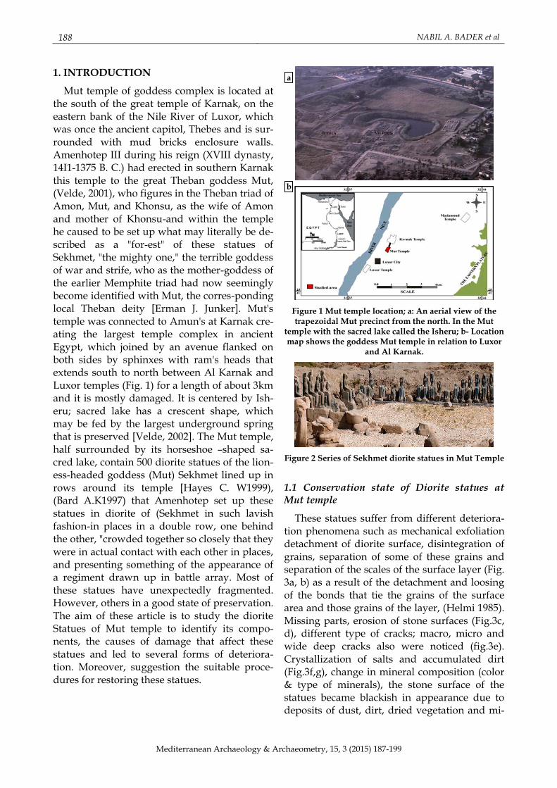
Mut temple of goddess complex is located at the south of the great temple of Karnak, on the eastern bank of the Nile River of Luxor, which was once the ancient capitol, Thebes and is sur-rounded with mud bricks enclosure walls. Amenhotep III during his reign (XVIII dynasty, 14I1-1375 B. C.) had erected in southern Karnak this temple to the great Theban goddess Mut, (Velde, 2001), who figures in the Theban triad of Amon, Mut, and Khonsu, as the wife of Amon and mother of Khonsu-and within the temple he caused to be set up what may literally be de-scribed as a "for-est" of these statues of Sekhmet, "the mighty one," the terrible goddess of war and strife, who as the mother-goddess of the earlier Memphite triad had now seemingly become identified with Mut, the corres-ponding local Theban deity [Erman J. Junker]. Mut's temple was connected to Amun's at Karnak cre-ating the largest temple complex in ancient Egypt, which joined by an avenue flanked on both sides by sphinxes with ram's heads that extends south to north between Al Karnak and Luxor temples (Fig. 1) for a length of about 3km and it is mostly damaged. It is centered by Ish-eru; sacred lake has a crescent shape, which may be fed by the largest underground spring that is preserved [Velde, 2002]. The Mut temple, half surrounded by its horseshoe –shaped sa-cred lake, contain 500 diorite statues of the lion-ess-headed goddess (Mut) Sekhmet lined up in rows around its temple [Hayes C. W1999), (Bard A.K1997) that Amenhotep set up these statues in diorite of (Sekhmet in such lavish fashion-in places in a double row, one behind the other, "crowded together so closely that they were in actual contact with each other in places, and presenting something of the appearance of a regiment drawn up in battle array. Most of these statues have unexpectedly fragmented. However, others in a good state of preservation. The aim of these article is to study the diorite Statues of Mut temple to identify its compo-nents, the causes of damage that affect these statues and led to several forms of deteriora-tion. Moreover, suggestion the suitable proce-dures for restoring these statues.
Figure 1 Mut temple location; a: An aerial view of the trapezoidal Mut precinct from the north. In the Mut
temple with the sacred lake called the Isheru; b- Location map shows the goddess Mut temple in relation to Luxor
and Al Karnak.
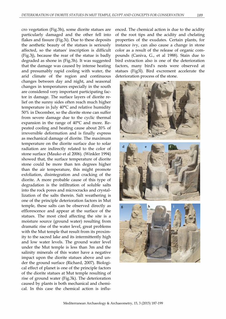
Figure 2 Series of Sekhmet diorite statues in Mut Temple
1.1 Conservation state of Diorite statues at Mut temple
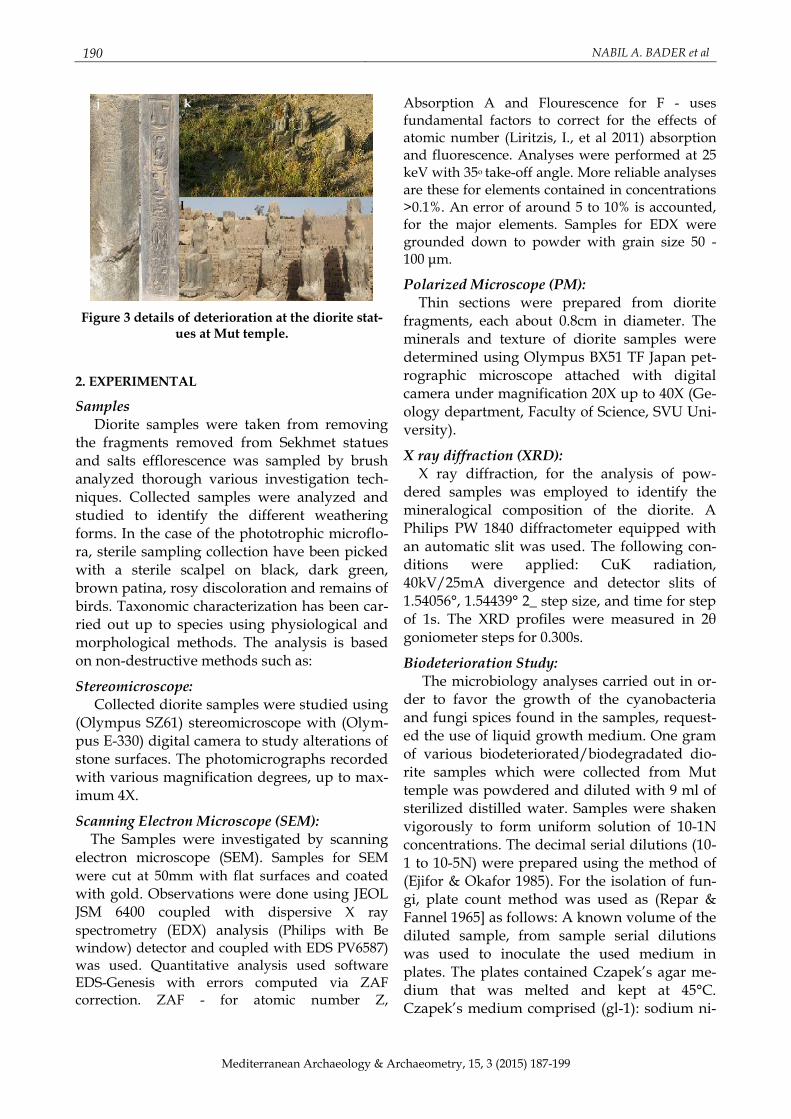
These statues suffer from different deteriora-tion phenomena such as mechanical exfoliation detachment of diorite surface, disintegration of grains, separation of some of these grains and separation of the scales of the surface layer (Fig. 3a, b) as a result of the detachment and loosing of the bonds that tie the grains of the surface area and those grains of the layer, (Helmi 1985). Missing parts, erosion of stone surfaces (Fig.3c, d), different type of cracks; macro, micro and wide deep cracks also were noticed (fig.3e). Crystallization of salts and accumulated dirt (Fig.3f,g), change in mineral composition (color & type of minerals), the stone surface of the statues became blackish in appearance due to deposits of dust, dirt, dried vegetation and mi-
a
b
DETERIORATION OF DIORITE STATUES IN MUT TEMPLE, EGYPT AND CONCEPTS FOR CONSERVATION 189
Mediterranean Archaeology & Archaeometry, 15, 3 (2015) 187-199
cro vegetation (Fig.3h), some diorite statues are particularly damaged and the other fell into flakes and fissure (Fig.3i). Due to these deposits the aesthetic beauty of the statues is seriously affected, so the statues' inscription is difficult (Fig.3j), because the rear of the statue is badly degraded as shone in (Fig.3h). It was suggested that the damage was caused by intense heating and presumably rapid cooling with water, the arid climate of the region and continuous changes between day and night, and seasonal changes in temperatures especially in the south are considered very important participating fac-tor in damage. The surface layers of diorite re-lief on the sunny sides often reach much higher temperature in July 40°C and relative humidity 50% in December, so the diorite stone can suffer from severe damage due to the cyclic thermal expansion in the range of 40°C and more. Re-peated cooling and heating cause about 20% of irreversible deformation and is finally express as mechanical damage of diorite. The maximum temperature on the diorite surface due to solar radiation are indirectly related to the color of stone surface (Mauko et al 2006). (Winkler 1994) showed that, the surface temperature of diorite stone could be more than ten degrees higher than the air temperature, this might promote exfoliation, disintegration and cracking of the diorite. A more probable cause of this type of degradation is the infiltration of soluble salts into the rock pores and microcracks and crystal-lization of the salts therein. Salt weathering is one of the principle deterioration factors in Mut temple, these salts can be observed directly as efflorescence and appear at the surface of the statues. The most cited affecting the site is a moisture source (ground water) resulting from dramatic rise of the water level, great problems with the Mut temple that result from its proxim-ity to the sacred lake and its intermittently high and low water levels. The ground water level under the Mut temple is less than 3m and the salinity minerals of this water have a negative impact upon the diorite statues above and un-der the ground surface (Richard, 2007). Biologi-cal effect of planet is one of the principle factors of the diorite statues at Mut temple resulting of rise of ground water (Fig.3k). The deterioration caused by plants is both mechanical and chemi-cal. In this case the chemical action is influ-
enced. The chemical action is due to the acidity of the root tips and the acidity and chelating properties of the exudates. Certain plants, for instance ivy, can also cause a change in stone color as a result of the release of organic com-pounds (Caniva, G., et al 1988). Stain due to bird extraction also is one of the deterioration factors, many bird's nests were observed at statues (Fig3l). Bird excrement accelerate the deterioration process of the stone.
a
f
g h
d
c b
i
e
190 NABIL A. BADER et al
Mediterranean Archaeology & Archaeometry, 15, 3 (2015) 187-199
Figure 3 details of deterioration at the diorite stat-ues at Mut temple.
2. EXPERIMENTAL
Samples Diorite samples were taken from removing
the fragments removed from Sekhmet statues and salts efflorescence was sampled by brush analyzed thorough various investigation tech-niques. Collected samples were analyzed and studied to identify the different weathering forms. In the case of the phototrophic microflo-ra, sterile sampling collection have been picked with a sterile scalpel on black, dark green, brown patina, rosy discoloration and remains of birds. Taxonomic characterization has been car-ried out up to species using physiological and morphological methods. The analysis is based on non-destructive methods such as:
Stereomicroscope: Collected diorite samples were studied using
(Olympus SZ61) stereomicroscope with (Olym-pus E-330) digital camera to study alterations of stone surfaces. The photomicrographs recorded with various magnification degrees, up to max-imum 4X.
Scanning Electron Microscope (SEM): The Samples were investigated by scanning
electron microscope (SEM). Samples for SEM
were cut at 50mm with flat surfaces and coated with gold. Observations were done using JEOL JSM 6400 coupled with dispersive X ray
spectrometry (EDX) analysis (Philips with Be window) detector and coupled with EDS PV6587) was used. Quantitative analysis used software EDS-Genesis with errors computed via ZAF correction. ZAF - for atomic number Z,
Absorption A and Flourescence for F - uses fundamental factors to correct for the effects of atomic number (Liritzis, I., et al 2011) absorption and fluorescence. Analyses were performed at 25 keV with 35o take-off angle. More reliable analyses are these for elements contained in concentrations >0.1%. An error of around 5 to 10% is accounted, for the major elements. Samples for EDX were grounded down to powder with grain size 50 - 100 μm.
Polarized Microscope (PM): Thin sections were prepared from diorite
fragments, each about 0.8cm in diameter. The minerals and texture of diorite samples were determined using Olympus BX51 TF Japan pet-rographic microscope attached with digital camera under magnification 20X up to 40X (Ge-ology department, Faculty of Science, SVU Uni-versity).
X ray diffraction (XRD): X ray diffraction, for the analysis of pow-
dered samples was employed to identify the mineralogical composition of the diorite. A Philips PW 1840 diffractometer equipped with an automatic slit was used. The following con-ditions were applied: CuK radiation, 40kV/25mA divergence and detector slits of 1.54056°, 1.54439° 2_ step size, and time for step of 1s. The XRD profiles were measured in 2θ goniometer steps for 0.300s.
Biodeterioration Study: The microbiology analyses carried out in or-
der to favor the growth of the cyanobacteria and fungi spices found in the samples, request-ed the use of liquid growth medium. One gram of various biodeteriorated/biodegradated dio-rite samples which were collected from Mut temple was powdered and diluted with 9 ml of sterilized distilled water. Samples were shaken vigorously to form uniform solution of 10-1N concentrations. The decimal serial dilutions (10-1 to 10-5N) were prepared using the method of (Ejifor & Okafor 1985). For the isolation of fun-gi, plate count method was used as (Repar & Fannel 1965] as follows: A known volume of the diluted sample, from sample serial dilutions was used to inoculate the used medium in plates. The plates contained Czapek’s agar me-dium that was melted and kept at 45°C. Czapek’s medium comprised (gl-1): sodium ni-
j k
l
DETERIORATION OF DIORITE STATUES IN MUT TEMPLE, EGYPT AND CONCEPTS FOR CONSERVATION 191
Mediterranean Archaeology & Archaeometry, 15, 3 (2015) 187-199
trate 3.0; potassium dihydrogen phosphate 1.0; magnesium sulfate 0.5; potassium chloride 0.5; ferrous sulfate 0.01; glucose 10; agar 15. Chlo-ramphenicol (0.05mg/ml) was used as bacterio-static agent. The plates were incubated at 28ºC for 5-7 days during which the developing fungi colonies were counted and identified. The mi-crobial population in the original sample was then calculated using the following equation: Organisms/g sample = number of colonies/ (amount plated x 1/dilution). The same method was used for the isolation of bacteria, by using nutrient agar medium (NA) instead of Czapek’s. The medium comprised (gl-1): beef extract 3.0; peotone 5.0; agar 15; pH = 7.0 (See-ley & Van Demark, 1981). The inoculated plates were incubated at 37°C from 24 to 48h. The evaluation of microorganism total concentration in each sample was determined by plate count-ing of serial dilutions according to the equation: Colony forming units: (CFU)/g = Number of colonies/dilution.
Chemical studies of groundwater: Water samples from the Mut temple was an-
alyzed in order to identify the soluble salts con-tent. Water sample was taken from Sacred Lake when its level was high in summer in sterile bottle in order to avoid any local contamination or evaporation. The samples were analyzed for the major cations (K+, Na+, Mg++ and Ca++) and the major anions (Cl-, NO3- SO42-, PO4-, CO32-, N-
NH4) using chemical methods, as well as water temperature. In added to, PH and Ec were measured.
3. RESULTS
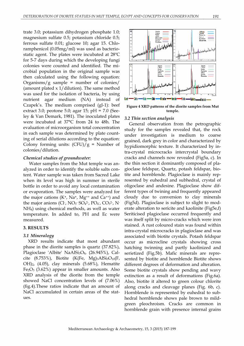
3.1 Mineralogy XRD results indicate that most abundant
phase in the diorite samples is quartz (37.82%), Plagioclase 'Albite' NaAlSi3O8, (26.945%), Cal-cite (8.753%), Biotite (K(Fe, Mg)3AlSi3O10(F, OH)2, (4.05), clay minerals (5.68%), Hematite Fe2O3 (3.62%) appear in smaller amounts. Also XRD analysis of the diorite from the temple showed NaCl concentration levels of (7.06%) (fig.4).These ratios indicate that an amount of NaCl accumulated in certain areas of the stat-ues.
Figure 4 XRD patterns of the diorite samples from Mut temple.
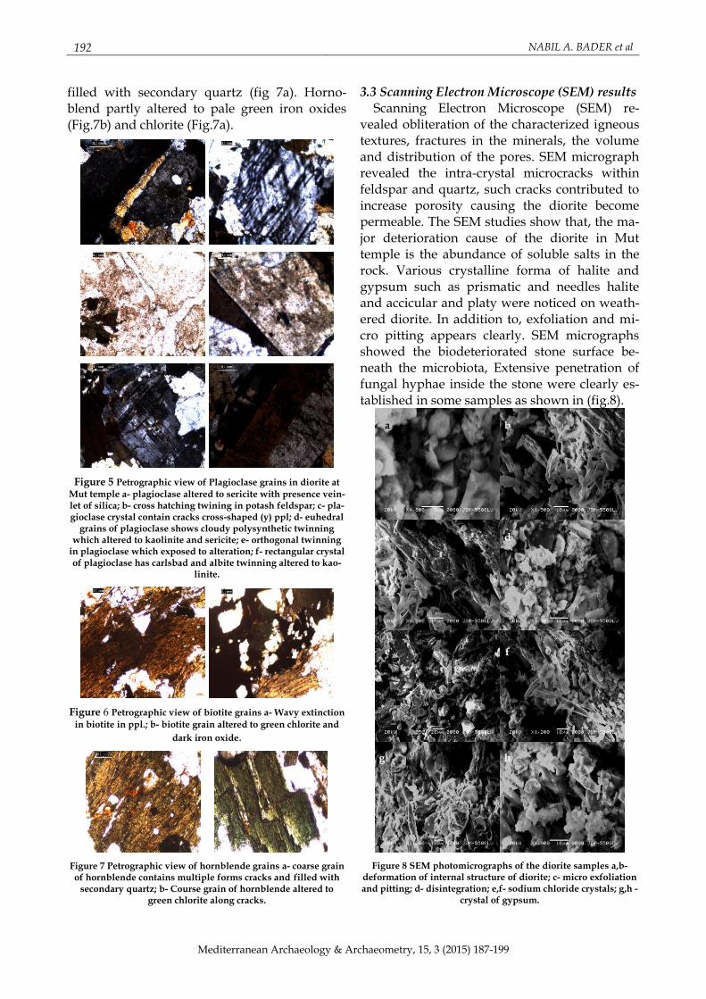
3.2 Thin section analysis General observation from the petrographic
study for the samples revealed that, the rock under investigation is medium to coarse grained, dark grey in color and characterized by hypidiomorphic texture. It characterized by in-tra-crystal microcracks intercrystal boundary cracks and channels now revealed (Fig5a, c). In the thin section it dominantly composed of pla-gioclase feldspar, Quartz, potash feldspar, bio-tite and hornblende. Plagioclase is mainly rep-resented by euhedral and subhedral, crystal of oligoclase and andesine. Plagioclase show dif-ferent types of twining and frequently appeared cloudy due to conversion to clay minerals (Fig5d). Plagioclase is subject to slight to mod-erate alteration to sericite and kaolinite (Fig5e,f) Seriticised plagioclase occurred frequently and was itself split by micro-cracks which were iron stained. A rust coloured stain was found within intra-crystal microcracks in plagioclase and was associated with biotite crystals. Potash feldspar occur as microcline crystals showing cross hatching twinning and partly kaolinized and serietized (Fig.5b). Mafic minerals are repre-sented by biotite and hornblende Biotite shows different degrees of deformation and alteration. Some biotite crystals show pending and wavy extinction as a result of deformations (Fig.6a). Also, biotite it altered to green colour chlorite along cracks and cleavage planes (Fig. 6b, c). Hornblende is represented by euhedral to sub-hedral hornblende shows pale brown to mild-green pleochroism. Cracks are common in hornblende grain with presence internal grains
192 NABIL A. BADER et al
Mediterranean Archaeology & Archaeometry, 15, 3 (2015) 187-199
filled with secondary quartz (fig 7a). Horno-blend partly altered to pale green iron oxides (Fig.7b) and chlorite (Fig.7a).
Figure 5 Petrographic view of Plagioclase grains in diorite at
Mut temple a- plagioclase altered to sericite with presence vein-let of silica; b- cross hatching twining in potash feldspar; c- pla-gioclase crystal contain cracks cross-shaped (y) ppl; d- euhedral
grains of plagioclase shows cloudy polysynthetic twinning which altered to kaolinite and sericite; e- orthogonal twinning
in plagioclase which exposed to alteration; f- rectangular crystal of plagioclase has carlsbad and albite twinning altered to kao-
linite.
Figure 6 Petrographic view of biotite grains a- Wavy extinction
in biotite in ppl.; b- biotite grain altered to green chlorite and
dark iron oxide.
Figure 7 Petrographic view of hornblende grains a- coarse grain of hornblende contains multiple forms cracks and filled with
secondary quartz; b- Course grain of hornblende altered to green chlorite along cracks.
3.3 Scanning Electron Microscope (SEM) results Scanning Electron Microscope (SEM) re-
vealed obliteration of the characterized igneous textures, fractures in the minerals, the volume and distribution of the pores. SEM micrograph revealed the intra-crystal microcracks within feldspar and quartz, such cracks contributed to increase porosity causing the diorite become permeable. The SEM studies show that, the ma-jor deterioration cause of the diorite in Mut temple is the abundance of soluble salts in the rock. Various crystalline forma of halite and gypsum such as prismatic and needles halite and accicular and platy were noticed on weath-ered diorite. In addition to, exfoliation and mi-cro pitting appears clearly. SEM micrographs showed the biodeteriorated stone surface be-neath the microbiota, Extensive penetration of fungal hyphae inside the stone were clearly es-tablished in some samples as shown in (fig.8).
Figure 8 SEM photomicrographs of the diorite samples a,b- deformation of internal structure of diorite; c- micro exfoliation and pitting; d- disintegration; e,f- sodium chloride crystals; g,h -
crystal of gypsum.
a
c
b
e
d
g h
f
DETERIORATION OF DIORITE STATUES IN MUT TEMPLE, EGYPT AND CONCEPTS FOR CONSERVATION 193
Mediterranean Archaeology & Archaeometry, 15, 3 (2015) 187-199
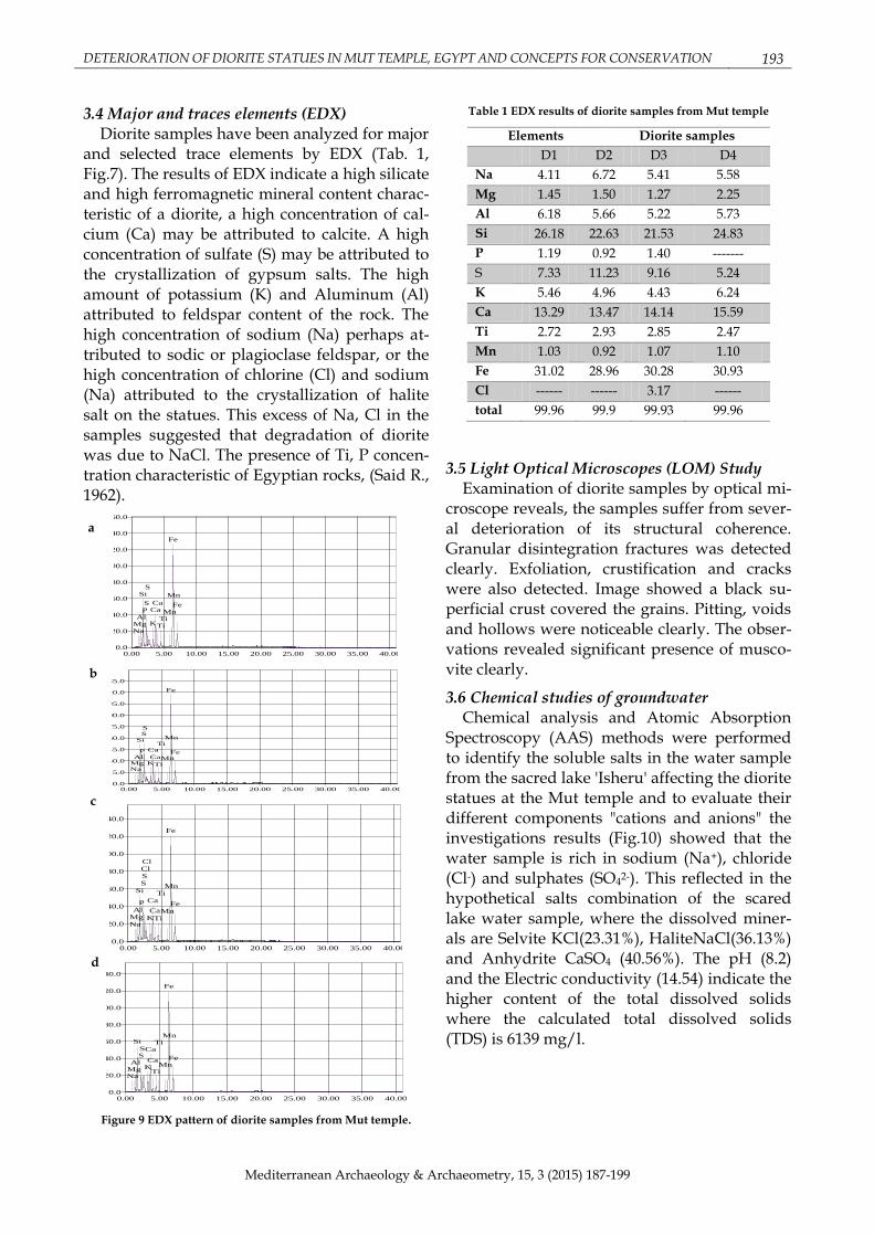
3.4 Major and traces elements (EDX) Diorite samples have been analyzed for major
and selected trace elements by EDX (Tab. 1, Fig.7). The results of EDX indicate a high silicate and high ferromagnetic mineral content charac-teristic of a diorite, a high concentration of cal-cium (Ca) may be attributed to calcite. A high concentration of sulfate (S) may be attributed to the crystallization of gypsum salts. The high amount of potassium (K) and Aluminum (Al) attributed to feldspar content of the rock. The high concentration of sodium (Na) perhaps at-tributed to sodic or plagioclase feldspar, or the high concentration of chlorine (Cl) and sodium (Na) attributed to the crystallization of halite salt on the statues. This excess of Na, Cl in the samples suggested that degradation of diorite was due to NaCl. The presence of Ti, P concen-tration characteristic of Egyptian rocks, (Said R., 1962).
keV
0.00 5.00 10.00 15.00 20.00 25.00 30.00 35.00 40.00
CPS
0.0
20.0
40.0
60.0
80.0
100.0
120.0
140.0
160.0
NaMgAl
Si
PS
S
K
CaCa
TiTiMn
Fe
Mn
Fe
keV
0.00 5.00 10.00 15.00 20.00 25.00 30.00 35.00 40.00
CPS
0.0
15.0
30.0
45.0
60.0
75.0
90.0
105.0
120.0
135.0
NaMgAl
Si
P
SS
K
Ca
Ca
Ti
Ti
Mn
Fe
Mn
Fe
keV
0.00 5.00 10.00 15.00 20.00 25.00 30.00 35.00 40.00
CPS
0.0
20.0
40.0
60.0
80.0
100.0
120.0
140.0
NaMgAl
Si
P
SSClCl
K
Ca
Ca
Ti
Ti
Mn
Fe
Mn
Fe
keV
0.00 5.00 10.00 15.00 20.00 25.00 30.00 35.00 40.00
CPS
0.0
20.0
40.0
60.0
80.0
100.0
120.0
140.0
NaMgAl
Si
SS
K
Ca
Ca
Ti
Ti
Mn
Fe
Mn
Fe
Figure 9 EDX pattern of diorite samples from Mut temple.
Table 1 EDX results of diorite samples from Mut temple
Elements Diorite samples
D1 D2 D3 D4
Na 4.11 6.72 5.41 5.58
Mg 1.45 1.50 1.27 2.25
Al 6.18 5.66 5.22 5.73
Si 26.18 22.63 21.53 24.83
P 1.19 0.92 1.40 -------
S 7.33 11.23 9.16 5.24
K 5.46 4.96 4.43 6.24
Ca 13.29 13.47 14.14 15.59
Ti 2.72 2.93 2.85 2.47
Mn 1.03 0.92 1.07 1.10
Fe 31.02 28.96 30.28 30.93
Cl ------ ------ 3.17 ------
total 99.96 99.9 99.93 99.96
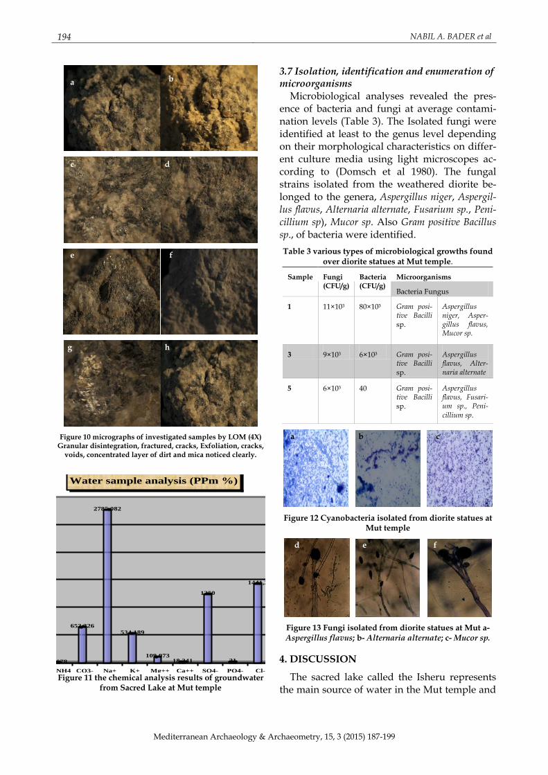
3.5 Light Optical Microscopes (LOM) Study Examination of diorite samples by optical mi-
croscope reveals, the samples suffer from sever-al deterioration of its structural coherence. Granular disintegration fractures was detected clearly. Exfoliation, crustification and cracks were also detected. Image showed a black su-perficial crust covered the grains. Pitting, voids and hollows were noticeable clearly. The obser-vations revealed significant presence of musco-vite clearly.
3.6 Chemical studies of groundwater Chemical analysis and Atomic Absorption
Spectroscopy (AAS) methods were performed to identify the soluble salts in the water sample from the sacred lake 'Isheru' affecting the diorite statues at the Mut temple and to evaluate their different components "cations and anions" the investigations results (Fig.10) showed that the water sample is rich in sodium (Na+), chloride (Cl-) and sulphates (SO42-). This reflected in the hypothetical salts combination of the scared lake water sample, where the dissolved miner-als are Selvite KCl(23.31%), HaliteNaCl(36.13%) and Anhydrite CaSO4 (40.56%). The pH (8.2) and the Electric conductivity (14.54) indicate the higher content of the total dissolved solids where the calculated total dissolved solids (TDS) is 6139 mg/l.
a
b
d
c
194 NABIL A. BADER et al
Mediterranean Archaeology & Archaeometry, 15, 3 (2015) 187-199
Figure 10 micrographs of investigated samples by LOM (4X) Granular disintegration, fractured, cracks, Exfoliation, cracks,
voids, concentrated layer of dirt and mica noticed clearly.
0,0078
652,326
2785,082
534,189
109,07318,241
1250
21
1441,2
1,20
500
1000
1500
2000
2500
3000
Per
cent
age
% (
PP
m)
N-NH4 CO3- Na+ K+ Mg++ Ca++ SO4- PO4- Cl- NO3-
Cations & Anions
Water sample analysis (PPm %)
Figure 11 the chemical analysis results of groundwater
from Sacred Lake at Mut temple
3.7 Isolation, identification and enumeration of microorganisms
Microbiological analyses revealed the pres-ence of bacteria and fungi at average contami-nation levels (Table 3). The Isolated fungi were identified at least to the genus level depending on their morphological characteristics on differ-ent culture media using light microscopes ac-cording to (Domsch et al 1980). The fungal strains isolated from the weathered diorite be-longed to the genera, Aspergillus niger, Aspergil-lus flavus, Alternaria alternate, Fusarium sp., Peni-cillium sp), Mucor sp. Also Gram positive Bacillus sp., of bacteria were identified.
Table 3 various types of microbiological growths found over diorite statues at Mut temple.
Sample Fungi (CFU/g)
Bacteria (CFU/g)
Microorganisms
Bacteria Fungus
1 11×103 80×10³ Gram posi-tive Bacilli sp.
Aspergillus niger, Asper-gillus flavus, Mucor sp.
3 9×10³ 6×10³ Gram posi-tive Bacilli sp.
Aspergillus flavus, Alter-naria alternate
5 6×10³ 40 Gram posi-tive Bacilli sp.
Aspergillus flavus, Fusari-um sp., Peni-cillium sp.
Figure 12 Cyanobacteria isolated from diorite statues at Mut temple
Figure 13 Fungi isolated from diorite statues at Mut a- Aspergillus flavus; b- Alternaria alternate; c- Mucor sp.
4. DISCUSSION
The sacred lake called the Isheru represents the main source of water in the Mut temple and
a
f e d
c b
a
e
d c
b
f
h g
DETERIORATION OF DIORITE STATUES IN MUT TEMPLE, EGYPT AND CONCEPTS FOR CONSERVATION 195
Mediterranean Archaeology & Archaeometry, 15, 3 (2015) 187-199
is one of the most arid saline lakes. Samples from the lake water were analyzed for compari-son with other samples from the Mut statues to see how much the lake water had affected them. The chemical analysis showed that the water sample is rich in sodium (Na+), chloride (Cl-), potassium (K+ ), magnesium (Mg2+ ), nitrate (NO3-) and calcium (Ca++) ions. The water also has a high content of sulphates (SO42-), Car-bonate (CO3--), phosphate (PO4--) and Ammonia (N-NH4). The XRF analysis of the diorite sam-ples showed similar results and the presence of the same salts in addition to a high percentage of Cl and Na, which are contained in halite. The samples also showed a saturation state with both calcium (Ca), magnesium (Mg) and phos-phate (P). This phenomenon is related to the effect of the lake groundwater. This excess of chloride ions, sulphates, nitrate and carbonates suggested that degradation of the diorite stat-ues. The presence of halite, sulphates and car-bonates in the diorite samples is derived from the lake or the soil of the temple. Generally, the soil of Egypt is known to be saline. This sug-gests that statues buried for thousands of years will eventually become salinized through salt-transporting ground water or through rare rains percolating through the earth. Since the con-struction of the first dam on the Nile and the High Dam near Aswan (1965). Irrigation has greatly increased the salt content of soil and monuments at Karnak. Upon excavation of the diorite, the absorbed water gradually evapo-rated and the salts present crystallized. Pres-sures on the micropores of the rock, due to the crystallization of the salts, would have likely caused its deterioration (McFarlanek J., et al 1983). Scanning electron microscope (SEM) re-sults confirm that a major deterioration is the abundance of soluble salts in the rock. SEM mi-crographs revealed the salt deposits on the stat-ues surface causes several alterations such cracks contributed to increased porosity causing the diorite to become permeable and losses of cohesion between grains. The microcracks and intergranular cracks had developed into chan-nels hence creating permeability for ingress of pollutants in gaseous and aqueous form. Vari-ous crystalline forms of halite and gypsum, such as needles crystal of halite and acicular and platy of gypsum were found in the weath-
ered diorite. The samples show a high incidence of rust colored iron staining. XRF, XRD and some of the rust colored staining were seen in the thin section. Mobilization of Fe from miner-als within the stone, such as biotite, may also be a source of the ferric Fe2+which during the weathering process goes to the more mobile fer-rous3+ (Jones, M.S et al 1996). The rate and ex-tent of degradation of Sekhmet diorite statues depends also on the weathering of individual minerals within its matrix. The study proved that, the feldspar, plagioclase and mica minerals in diorite were partially sericitised and com-pletely altered crystal. The petrographic study appeared plagioclase very cloudy due to re-placement by fine grained clay minerals such as sericite [K2Al4Si6O20 (OH4)] and kaolinite [Al4Si4O10 (OH8)]. The degradation and altera-tion of plagioclase in diorite may proceed by one or more mechanisms, including the leach-ing of cations from the silica lattice by hydrogen ion activity on its surface or to depths greater than one or two unit cells. The leaching of silica, which in turn renders the rock more porous is another suspected mechanism of igneous rock weathering, (McFarlanek J. et al 1983). Biotite and hornblende was found in all samples al-tered to green chlorite. Biotite undergoes vol-ume changes when it alters to chlorite, in addi-tion clay minerals are hydrophilic and react to varying humidities by expansion and contrac-tion. The activation of chlorite clay layers may destabilize the grains and cause decay. The weathering process altering biotite to chlorite involves loss of K, Na, Ti, OH and Si. The XRF data shows a loss of K and Ti.
LOM investigated showed a thick layer of dust covered the surface of the rock. Generally the accumulate of dirt coating the surfaces of monuments lead to degradation of the stone surface as result of the reaction between the dirt and the stone surface which affect the statues visibility and its aesthetic value (Eric, D., Price, C.A. 2010).
The biological investigation proved that dio-rite statues in the studied object are exposed to the attack of microorganisms, also SEM micro-graph revealed some microflora inside the pits at the diorite samples from the Mut temple. The biological investigation of the genera isolated from the diorite samples that were derived from
196 NABIL A. BADER et al
Mediterranean Archaeology & Archaeometry, 15, 3 (2015) 187-199
the forte of Sekhmet statues revealed that the fungal flora Aspergillus flavus represents the dominant fungi isolates from the collating sam-ples. Aspergillus niger was detected upon the other statues. The fungal flora Alternaria alter-nate has been detected also, while the fungal flora phoma sp., Fusarium sp., and Penicillium sp. were detected in the isolates taken from the dio-rite statues. In addition to, Gram positive Bacillus sp., of bacteria were identified. Recent research has demonstrated that microorganisms can ac-celerate elemental release from geologic materi-als, either directly through the acquisition of limiting nutrients required for biomass synthe-sis; e.g., P and Fe (Bennett P. 2001) or indirectly through the release of exoproducts that lower pH, complex cations, and/or change mineral saturation states (Liermann L. 2000). Other work has shown that microbial activities can inhibit elemental release by facilitating devel-opment of an amorphous leached layer (Ben-zerara et al., 2004, 2005), promoting adsorption of polysaccharides onto mineral surfaces (Welch et al., 1999) and releasing ferric iron that inter-acts with surface sites (Santelli et al., 2001).
Mechanical degradation of statues was sug-gested to occur upon their excavation In addi-tion, it is highly probably that microfractures caused by crystallization of soluble salts during burial; i.e. in dry periods between rains, accel-erates the destruction of the mineral structure.
5. CONSERVATION - SUGGESTION AND RECOMMENDATION
The previous study clarifies that the diorite statues at Mut temple had been exposed to ag-gressive deterioration factors. Therefore, the statues need to carry out different treatments and conservation processes, such as: - There are many problems need a lot of scien-
tific work before satisfying conservation plan will be established as preventive conserva-tion. Preventive conservation measures of more immediate effect are usually concerned with keeping water out of the stone and with controlling the relative humidity and tem-perature of the air around the stone (Price 1993). The main purpose of relative humidity control is to prevent salt damage because any efforts that taken to protect the statues when
water levels surge is worthwhile. The im-portant problem is the raised groundwater level due to increased irrigation. This can be solved in various ways, but probably the most sustainable solution is to change or im-prove the irrigation management in the area; restricting crop types.
- The present irrigation flood system should be changed to new systems such as sprin-kler and drop methods to reduce groundwa-ter recharge.
- Continuous monitoring of physical parame-ters e.g. pH, Electrical Conductivity, as a clue of changes in groundwater chemistry to trace the source of recharge water as well as pollutants.
- The present study shows the increased of concentration of ions (especially Na+, Cl- and SO4-2) in some low-lying areas in flood plains regions include Mut temple area due to the lack of agricultural drainage in newly reclaimed lands next to some other factors, such as lack of a sewage system in the ma-jority of rural areas and the intensive use of agricultural fertilizers.
- Construction of a sewage network in the temple area and the surroundings villages with a continuous care and detection of leakage in the drainage networks.
- Demolition all modern buildings (a few houses) near the site to preserving the his-torical environment, cultural and heritage values in addition to integrated preservation to the site.
- Completing excavation to discover all the buried statues. Careful observation of burial environments could save many dioritic stat-ues from rapid degradation. In the case of obviously damp soils, the statues should be immediately protected from evaporation.
- Reconstruction mud bricks wall enclosing the site that will be capable of excluding the animals which are damaging the site, with maintenance of the aesthetics and integrity of the site.
- Cleaning and removing of vegetation should be carried out to reduce the negative impacts by trees, plants, shrubs roots for all archeological remains at the site by mechan-ical removal for roots and rhizomes and chemical removal by using chemical pesti-
DETERIORATION OF DIORITE STATUES IN MUT TEMPLE, EGYPT AND CONCEPTS FOR CONSERVATION 197
Mediterranean Archaeology & Archaeometry, 15, 3 (2015) 187-199
cides as pesticide Glyphosate pesticide or Fuluazifop-p-butyl should be removed ac-cording to the law as they do not give any chance for the aesthetic appreciation of the temple in addition to give chance to com-plete the new excavation.in the context of other parameters-depth of penetration, sta-bility of surface appearance, and retention of porosity (Klaus J.H, et al 1988).
- After Preventive Conservation, conservation interventions have to be individually planned and Consolidation, other conserva-tion treatments must as always only be per-formed after thoroughly testing all proce-dures, recalling the often friable nature of these unique statues and avoid further damages.
- During field observation, highly areas have been detected. Here emergency measures should be carried out immediately in order to prevent the loss of original material. Emergency interventions are preliminary measures like gluing pieces or pre-consolidate.
- A partial pre‐consolidation should be car-ried out only on the crumbed and separated weak surface. According to experimental tests 5% Paraloid B.66 diluted at 5% in ace-tone by spray methods is the best consoli-dant material to apply in this environment.
- Friable and Unattractive dirt, Crusts, excrete of birds, microbial stains, unfavorable sur-face accumulations and different species of salt crusts should be removed by suitable scientific techniques. A wide range of tech-niques is available for cleaning stone, rang-ing from mechanical cleaning using manual and mechanical tools as scalpels, spatula, different types of brushes and sponge parti-cles containing mineral grains of varying
hardness at 100– 200 kPa can be used to minimize abrasion of substrate and reduce dust levels. Also, water cleaning can be used safely to remove dirt from the surfaces of the relives, supplemented with non-ionic detergent and steam or hot pressurized wa-ter cleaning (Mack et al 2003). Chemical cleaning can be done to remove what me-chanical cleaning failed to remove from stains and dirt by using organic solvents, the best results will obtained with mixture
of ethyl alcohol, acetone, toluene and tri-chloroethylene.
- Biocides must be used, not only to kill the growth in the relives, but also to be resistant to new strains.
- Reduction of salts should be done. Desalina-tion of statues and its inscription from solu-ble salts as sodium chloride is usually at-tempted through the use different types of poultices, which may consist of clay, paper pulp, or cellulose ethers. A mixture of clay and paper fiber produces an absorbent and plastic mixture that is often favored by con-servators of stone sculpture. (Aneta et al 2010).
- After removing the salts the walls become ready to consolidate, it should be consoli-dated not only for aesthetic reasons but also to ensure the correct conservation of the en-tire structure. Consolidation of weak parts, losing cohesion and adhesion of the diorite statues using Silane (The alkoxysilanes, or "silanes" for short, have undoubtedly been the most widely used stone consolidants over the past twenty years. Two com-pounds, in particular, have been dominant: methyltrimethoxysilane (MTMOS) and tet-raethoxysilane (TEOS). The silanes are hy-drolyzed by water to form silanols, which then polymerize in a condensation reaction to give a silicone polymer) (Price, C.A. 1996) which aims to eliminate or reduce capillary absorption of water in driving rain and en-hance durability of the stone. Laboratory re-search and experimental field work suggest that siloxanes, silanes and other alkoxysilanes consolidants are promising for treatment of diorite because of penetrating into the stone substrate slightly, increasing in compressive strength, modulus of rup-ture, and abrasion resistance. These im-provements seem remarkable, given the rel-atively small amount of consolidant depos-ited.
- On the other hand, filling the joints between the blocks of stones and completing the missing parts should be carried out. Our ex-periments have proved that the best materi-al for this target is mortar; diorite crushing under 5mm, dark sand, Wacker VB132+ Ac-
198 NABIL A. BADER et al
Mediterranean Archaeology & Archaeometry, 15, 3 (2015) 187-199
ryloid B.72 is the best one for weather dio-rite (Bader, N. 2011).
- Re-jointing and linking of the broken statues can be done by using stainless steel bars, Araldite 1092 and the same mortars and then complete the missing gaps and spaces with the same components.
- After conservation steps, all statues should be set on mastabas, further the possibility to waterproof the mastaba will be made all the more necessary by prevent intermittent ris-ing and lowering of the water table.
- The display area at the temple of Mut should be completed roofing to protect the statues.
- Preparing the site with restricting access, signs, low guidance fencing or barriers, walkways etc.
- Periodical monitoring with regular meas-urements (moisture, groundwater table, temperature, air pollution…etc.) will be more effective and help to observe any seri-ously impacting or damaging at the site.
REFERENCES Aneta S., Pikry, R., (2010) Determination of source areas of natural stones: A methodology ap-
proach applied to impure crystalline lime stones, In book: materials, technologies and practice in historic heritage structures, edited by, Maria Bostenaru Dan,Springer, New York.
Bader, N.A., (2010) Deterioration and conservation of Dioritic Relief in The Queen Hathepsut's chapel at Temple of Karnak, The 1st international conference for studies environments, South Valley University, Luxor, Egypt, 25-28 October, pp.62-77.
Bard A.K., (1999) Encyclopedia of the archaeology of ancient Egypt, Routledge, Canada, 1999, pp.360-398.,
Bennett P. C., Rogers J. R., Choi W. J., Hiebert F. K., (2001) Silicates, silicate weathering, and micro-bial ecology. Geomicrobiol. J. 18(1), pp.3–19.
Benzerara K., Barakat M., Menguy N., Guyot F., Luca G. D., Audrain C., Heulin T., (2004) Experi-mental colonization and alteration of orthopyroxene by the pleomorphic bacteria, Ramli-bacter tataouinensis. Geomicrobiol. J. 21, pp.341–349.
Caneva, G., Altieri A., (1988) Biochemical mechanisms of stone weathering induced by plant growth, Proceeding of the 6th International Congress on Deterioration and Conservation of Stone, Torun, Poland, pp.32-44.
Domsch,K.H., Gams, W., Anderson, T.H., (1980) Compendium of Soil Fungi. Vol. 1-2. London: Aca-demic Press.
Erman, Religion, p. 56 Eric, D., Price, C.A., (2010) Stone Conservation, An overview of current research, second edition,
Getty Conservation Institute pp. 1-25. Helmi, F., M., (1985) Deterioration of some granite in Egypt, V1 congress international sur
l`Alteration ET la conservation de la Pierre, Lausanne, Vol. 1, pp. 421-429 Jones, M.S., Obrien, P.F., Cooper, T.P., (1996) A study of decay occurring in the Leinster granite,
house No.9, Trinity College, Dublin, in, international cong. on det. And cons. of stone, V.1, Germany, pp.211-221.
Liritzis, I., Mavrikis, D., Zacharias, N., Sakalis, A., Tsirliganis, N. and Polymeris, G.S., (2011) Po-tassium determinations using SEM, FAAS and XRF: some experimental notes, Mediterra-nean Archaeology and Archaeometry, Vol. 11, No. 2, pp. 169-179
Velde, Hermane te, (2002). The Ancient Gods Speak: A Guide to Egyptian Religion. Oxford Univer-sity Press, NY, USA, pp.238.
Pinkowski, Jennifer, (2006). Egypt’s ageless goddess. Archaeology Magazine September/ October. Hayes C. W., (1997) Internal affairs from Tuthmois I to the death of Aminophis III, part 2, Cam-
bridge University, p.30-34 Liermann L. J., Kalinowski B. E., Brantley S. L., Ferry J. G. (2000) Role of bacterial siderophores in
dissolution of hornblende. Geochim. Cosmochim. Acta 64(4), pp.587–602.
DETERIORATION OF DIORITE STATUES IN MUT TEMPLE, EGYPT AND CONCEPTS FOR CONSERVATION 199
Mediterranean Archaeology & Archaeometry, 15, 3 (2015) 187-199
Mack, R.C. and Grimmer, F.A., (2003) Assessing cleaning and water-repellent treatments for his-toric masonry buildings, Washington DC.
Mauko, A., Mirtic B., Maldenovic A., Grlek B., (2006) Deterioration of the Granodiorite façade- case example, Maximarkit, Ljubljana, Martials and Geoenvironment, Vol.53,N.1, , pp.23-37.
McFarlanek J., Kristine M., Yang W., Joseph S., Fery C., Burns G., (1983) The mechanisms of the physicochemical reactions in diorite used in the construction of ancient royal Egyptian statues, Can.J.CHEM. Vol.61, pp.718-723.
Okafor, N., Ejiofor, M.A.N., (1985) The linamarase of Leuconostoc mesenteroides, production, iso-lation and some properties, Journal of the Science of Food and Agriculture, 36, 1985, pp 667-678.
Price C.A, (1993) Preventive conservation of salt-contaminated masonry in the Wakefield Tower, HM Tower of London, Institute of Archaeology, Bulletin 30, pp. 121-33.
Price C.A, (1996) Stone Conservation, an overview of current research, The Getty Conservation Insti-tute, p. 20
Richard, A. Fazzini, (2007) internal report from the SCA (Mut temple excavations in Luxor city), Brooklyn Museum, New York.
Raper, K.B., Fennell, D.I., (1965) the Genus Aspergillus, Williams & Wilkins Company, Baltimore, pp 686.
Said R., The geology of Egypt. Elsevier Publishing Co., New York. 1962. p. 259. Santelli C. M., Welch S. A., Westrich H. R., Banfield J. F. (2001) The effect of Fe-oxidizing bacteria
on Fe–silicate mineral dissolution. Chem. Geol. 180(1–4), pp.99–115.
Seeley, H.W., Vandemark, P.J., John J.L., (1990) Selected Exercises for Microbes in Action, (4th Edi-tion), Published by W. H. Freeman ISBN-13: 978-0-7167-2111-6.
Welch S. A., Ullman W. J. (1999) The effect of microbial glucose metabolism on bytownite feldspar dissolution rates between 5ºC and 35ºC, Geochim. Cosmochim. Acta No. 63(19-20), 1999, pp.3247–3259.
Winkler, E., M., (1994) Stone properties durability in mans`s environment, Springer – Verlag, pp. 193.