Embed Size (px)
Citation preview
INVESTIGATION OF DIABETIC SENSORIMOTOR
POLYNEUROPATHY THROUGH NEUROPHYSIOLOGY AND NERVE
ULTRASONOGRAPHY IN TYPE 2 DIABETES MELLITUS
THAARANI D/O ARUMUGAM
FACULTY OF MEDICINE
UNIVERSITY OF MALAYA
KUALA LUMPUR
2016
INVESTIGATION OF DIABETIC SENSORIMOTOR
POLYNEUROPATHY THROUGH NEUROPHYSIOLOGY AND NERVE
ULTRASONOGRAPHY IN TYPE 2 DIABETES MELLITUS
THAARANI D/O ARUMUGAM
DISSERTATION SUBMITTED IN FULFILMENT OF THE
REQUIREMENTS FOR THE DEGREE OF MEDICAL SCIENCE
FACULTY OF MEDICINE
UNIVERSITY OF MALAYA
KUALA LUMPUR
2016
iii
UNIVERSITY OF MALAYA
ORIGINAL LITERARY WORK DECLARATION
Name of Candidate: Thaarani d/o Arumugam I.C No: 891127145912
Registration/Matric No: MGN 130076
Name of Degree: Masters of Medical Science (MMedSc)
Title of Dissertation: Investigation of diabetic sensorimotor polyneuropathy through
neurophysiology and nerve ultrasonography in Type 2 diabetes mellitus
Field of Study: Neurology
I do solemnly and sincerely declare that:
(1) I am the sole author/writer of this Work;
(2) This Work is original;
(3) Any use of any work in which copyright exists was done by way of fair
dealing and for permitted purposes and any excerpt or extract from, or
reference to or reproduction of any copyright work has been disclosed
expressly and sufficiently and the title of the Work and its authorship have
been acknowledged in this Work;
(4) I do not have any actual knowledge nor do I ought reasonably to know that
the making of this work constitutes an infringement of any copyright work;
(5) I hereby assign all and every rights in the copyright to this Work to the
University of Malaya (“UM”), who henceforth shall be owner of the
copyright in this Work and that any reproduction or use in any form or by any
means whatsoever is prohibited without the written consent of UM having
been first had and obtained;
(6) I am fully aware that if in the course of making this Work I have infringed
any copyright whether intentionally or otherwise, I may be subject to legal
action or any other action as may be determined by UM.
Candidate’s Signature Date:
Subscribed and solemnly declared before,
Witness’s Signature Date:
Name:
Designation:
iv
ABSTRACT
The present study explored the role of ultrasonography (US) and neurophysiology in
characterizing type 2 diabetes patients according to their neuropathy severity as
determined by the Toronto Clinical Scoring System (TCSS). This study also aimed to
comprehensively determine the relationship between nerve US and nerve conduction
study (NCS) parameters in diabetic patients. The second research question was to
investigate if there are any imaging markers that could possibly differentiate diabetic
patients with neurophysiological evidence of demyelination and true chronic
inflammatory demyelinating polyneuropathy (CIDP) patients. The study subjects were
100 symptomatic distal symmetrical polyneuropathy (DSP) patients and 40 age-matched
healthy controls. A subset of nine DSP patients with neurophysiological features of
demyelination (D-DSP) and six true CIDP patients were also recruited. DSP severity
was ascertained through TCSS where patients are grouped into mild (score 6-8),
moderate (9-11) and severe (12-19). Nerve electrophysiology and ultrasound were
performed on both lower limbs and the non-dominant upper limb in DSP subjects, and
in both upper and lower limbs in true CIDP and D-DSP subjects. Nerves cross sectional
area (CSA) recordings were taken at standard anatomical sites. A diagnosis of DSP and
CIDP was made based on existing criteria. Statistical analyses were performed using
SPSS version 22. Our findings revealed that sural nerve was inexcitable in 19.1% of
mild, 40.0% of moderate and 69.0% of severe DSP groups. In contrast, CSAs were
measureable in all nerves of DSP patients and were significantly larger compared to
controls. Patients with severe DSP had significantly larger nerves in the ulnar, peroneal,
tibial and sural, compared to mild DSP patients. By receiver operating characteristic
analysis, the cut-off value for sural at 2 mm2 was a good discriminator with area under
curve (AUC) of 0.88 between the presence and absence of DSP (sensitivity 0.90
;specificity 0.74) but performed less well in discriminating between severity of DSP
v
(cut-off 2.75mm2; AUC 0.62; sensitivity 0.59; specificity 0.73). Significant correlations
were demonstrated between TCSS, most neurophysiology parameters and nerve CSA of
ulnar, peroneal, tibial and sural. Significant enlargement of nerves was also found in
true CIDP patients compared to D-DSP patients at non-entrapment sites in the proximal
regions of the upper extremities. This research found that nerve US in DSP revealed
enlarged CSA and these changes worsen with increasing disease severity thus serving as
a useful, reliable and practical diagnostic tool especially when neurophysiology is
unrevealing. The present study also found nerve US aids in differentiation of true CIDP
from D-DSP patients by differences in the nerve enlargement and electrophysiological
profile between these two groups. This is important when managing these groups of
patients as CIDP is treatable.
vi
ABSTRAK
Kajian ini bertujuan untuk menerokai peranan ultrasonografi (US) dan neurofisiologi
dalam mengenalpasti pesakit diabetes mellitus (DM) jenis 2 berdasarkan severiti
neuropati seperti yang dikenalpasti melalui sistem skor klinikal Toronto (TCSS). Kami
juga bermatlamat untuk mengkaji hubungan di antara US saraf dan parameter kajian
konduksi saraf (NCS) secara komprehensif untuk menentukan sama ada US boleh
digunakan untuk membezakan neuropati dengan pelbagai gred severiti.Persoalan kajian
kedua adalah untuk menyiasat sama ada terdapat ciri-ciri pengimejan yang boleh
membezakan pesakit ‘distal symmetrical polyneuropathy’(DSP) dengan neurofisiologi
menunjukkan ciri-ciri penyahmielinan (D-DSP) dengan pesakit ‘Chronic Inflammatory
Demyelinating Polyneuropathy’(CIDP). Subjek kajian merupakan 100 pesakit DM
dengan simptom DSP dan 40 subjek sihat yang dipadankan umur sebagai kawalan.
Subset 9 pesakit D-DSP dan 6 pesakit CIDP juga direkrut. Severiti neuropati
berdasarkan TCSS; ringan (skor 6-8), sederhana (9-11) dan teruk (12-19). US dilakukan
pada kedua dua kaki dan satu tangan tidak dominan dalam pesakit DSP,tetapi pada
kedua dua belah kaki dan tangan dalam pesakit D-DSP dan CIDP. Bacaan CSA diambil
pada tempat anatomi secara seragam. Diagnosis DSP dan CIDP dibuat berdasarkan
kriteria yang sedia ada. Analisis statistik dibuat melalui SPSS versi 22. Kajian kami
mendapati perbezaan signifikasi antara ketiadaan aksi potensi deria sural di antara
pesakit neuropati ringan (19.1%), sederhana (40.0%) dan teruk (69.0%).
Walaubagaimanapun, kawasan kerataan rentas (CSA) dapat dinilai dalam semua saraf
pesakit DSP dan secara signifikasinya, lebih besar berbanding kumpulan orang sihat
yang dijadikan kawalan. CSA saraf dalam kumpulan pesakit gred severiti teruk adalah
lebih besar secara signifikasi dalam saraf ulnar , peroneal , tibial dan sural berbanding
kumpulan pesakit gred severiti ringan . Analisis kawasan bawah lengkung ( AUC )
dijalankan untuk membezakan pesakit dengan DSP dan pesakit yang tiada DSP.Kami
vii
mendapati bahawa saraf sural adalah yang diskriminator terbaik dengan nilai CSA 2
mm2 yang mempunyai AUC 0.88, sensitiviti 0.90 and spesifisiti 0.74 tetapi tidak
menunjukkan perbezaan teliti dalam membezakan CSA saraf pesakit DSP teruk dari
DSP tidak teruk dengan nilai 2.75 mm2; AUC 0.62; sensitiviti 0.59; spesifisiti
0.73.Hubungan secara signifikasi didapati antara TCSS,hampir kesemua parameter
neurofisiologi dan CSA saraf ulnar, peroneal, tibial dan sural. Pembengkakan saraf
secara signifikasi didapati dalam saraf pada bahagian proximal pada tangan dalam
pesakit CIDP. Penyelidikan ini telah mendapati bahawa US saraf dalam DSP
menunjukkan pembengkakan saraf dan menjadi lebih teruk dengan bertambahnya
severiti neuropati. Dengan ini,US menjadi alat diagnostik yang berguna,praktikal dan
boleh dipercayai khususnya apabila aksi potensi melalui neurofisiologi tidak dapat
dirangsang. Kajian ini juga membantu dalam membezakan pesakit CIDP dengan D-DSP
melalui definisi keratan rentas saraf dan profil neurofisiologi yang berbeza di antara dua
kumpulan pesakit ini yang membantu dalam pengurusan rawatan. Kajian ini
menunjukkan corak pembengkakan saraf pada bahagian proximal pada tangan dalam
pesakit CIDP berbanding pesakit D-DSP melalui kajian US yang membantu
mengenalpasti blok konduksi saraf yang selalunya sukar dikenalpasti melalui NCS,
khususnya dalam bahagian proximal saraf yang boleh membantu pengurusan rawatan
bagi pesakit yang terjejas.

viii
ACKNOWLEDGEMENTS
I would never have been able to finish my dissertation without the guidance of my
supervisor, support from fellow coursemates, friends, and my family.
I would like to express my deepest gratitude to my supervisor, Associate Professor
Dr Nortina Shahrizaila, for her excellent guidance, patience, and persistent motivation in
completing this research. I am deeply grateful to her for the long discussions that
helped me sort out the technical details of my work and undoubtedly, her expertise,
considerably to my graduate experience. Her insightful comments and constructive
criticisms at different stages of my research were thought provoking and they helped me
focus my ideas. I am very much thankful to her for holding me to a high research
standard and enforcing strict validations for each research result, and thus teaching me
how to do research.
I would like to also express my heartiest thanks to my fellow coursemates for the
stimulating discussions, for the sleepless nights we were working together before
deadlines, and for all the fun we have had in the last two years.
Many friends have always been there for me and their support and care helped me
overcome setbacks and stay focused on my graduate study. I greatly value their
friendship and I deeply appreciate their belief in me.
Last but not least, I would also like to thank my family for their support emotionally
and financially that they provided me throughout my graduate studies and without their
love and encouragement, this dream would have never come true.

ix
TABLE OF CONTENTS
Abstract ............................................................................................................................................. iv
Abstrak ........................................................................................................................................... ..vi
Acknowledgements ........................................................................................................................ viii
Table of Contents ............................................................................................................................. ix
List of Figures .................................................................................................................................. xii
List of Tables .................................................................................................................................. xiii
List of Symbols and Abbreviations ................................................................................................ xiv
List of Appendices .......................................................................................................................... xvi
CHAPTER 1: INTRODUCTION .................................................................................................. 1
1.0 Introduction .................................................................................................................1
1.1 Objective/s...................................................................................................................4
CHAPTER 2: LITERATURE REVIEW ...................................................................................... 5
2.1 Burden of diabetic DN in the global context………….. .......................................... 5
2.2 Pathophysiology of DN……….. .............................................................................. 6
2.2.1 Formation of advanced glycation end products ......................................................... 8
2.2.2 Oxidative stress…….. .............................................................................................. 8
2.2.3 Accumulation of polyol…… ..................................................................................... 9
2.2.4 Deficiency of NO……….......................................................................................... 9
2.3 Classification and characteristics of DN .............................................................. 10
2.3.1 DSP………………… .............................................................................................. 11
2.3.2 Autonomic neuropathy (AN) .................................................................................. 13
2.3.3 Focal and multifocal neuropathies ........................................................................... 13
2.4 Diagnosis of DSP……… ....................................................................................... 14
2.4.1 Clinical examination. .. .... …………………………………………………………15
x
2.4.4.1 Clinical Scoring System ......................................................................................... 15
2.5 Neurophysiological examination ........................................................................ 16
2.6 US….…………………………. . …………………………………………………18
2.7 CIDP……………………………………… .......................................................... 21
2.7.1 Clinical presentation and diagnosis .......................................................................... 22
2.8 Disease treatment and management ................................................................... 23
2.8.1 DSP treatment and management.............................................................................. 23
2.8.1.1 Pharmacological treatment of DSP ..................................................................... 23
2.8.1.2 Non pharmacological treatment of DSP .............................................................. 24
2.8.2 CIDP disease treatment and management ............................................................... 24
CHAPTER 3: METHODOLOGY ............................................................................................... 26
3.1 Subjects and study design ...................................................................................... 26
3.2 DSP Clinical Screening .......................................................................................... 27
3.3 Diagnosis of CIDP and D-DSP patients ................................................................ 28
3.4 Neurophysiological examination ........................................................................... 34
3.5 Sonographic examination ....................................................................................... 35
3.6 Statistical analyses ................................................................................................. 37
CHAPTER 4: RESULTS……… .................................................................................................. 38
4.1 Clinical Characteristics .......................................................................................... 38
4.2 Ultrasound studies .............................................................................................. 40
4.2.1 Comparison of nerve CSAs between DM patients and healthy
group……………………… ................................................................................................ 40
4.2.2 Comparison of nerve CSAs between DM patients according to neuropathy
severity ...... ……………………………………………………………………………….43
4.2.3 ROC analysis of the nerve CSAs ........................................................................... 47
4.2.4 Comparison of CSAs between CIDP patients and D-DSP patients ....................... 50
4.2.5 Sonogram images of DM patients with different neuropathy severities vs healthy
controls…………………. ................................................................................................... 53
xi
4.2.6 Sonogram images of CIDP patient and D-DSP patients ........................................ 56
4.3 Neurophysiological studies .................................................................................. 57
4.3.1 Electrophysiological findings according to severity classification (based on TCSS) of
DM patients………… ......................................................................................................... 57
4.3.2 Comparison of electrophysiological findings between CIDP patients and D-DSP
patients………………… ..................................................................................................... 61
4.4 Correlation studies ............................................................................................... 65
4.4.1 Correlation studies of nerve CSAs and electrophysiology in DM patients ........... 65
4.4.2 Correlation studies of nerve CSAs at different sites versus different disease
markers……… .................................................................................................................... 66
4.4.3 Correlation studies of NCS parameters versus different disease markers ............. 68
4.4.4 Association between HbA1C values with neuropathy severity and DM
duration ...............................................................................................................................70
CHAPTER 5: DISCUSSION ........................................................................................................ 71
5.1 Evaluation of DSP .................................................................................................. 71
5.2 US…………….. .................................................................................................... 71
5.3 Neurophysiological studies (NCS)......................................................................... 75
5.4 Correlation studies ................................................................................................. 77
5.5 Study limitations .................................................................................................... 79
CHAPTER 6: CONCLUSION ..................................................................................................... 80
References ...........................................................................................................................................
List of Publications and Papers Presented ...........................................................................................
Appendix .............................................................................................................................................
xii
LIST OF FIGURES
Figure 2.1: Pathopysiology of DN ................................................................................... 7
Figure 3.1: Anatomical sites of US ................................................................................ 36
Figure 4.1 : CSAs of tibial and sural nerves at the ankle in type 2 DM patients vs.
healthy controls ..............................................................................................................42
Figure 4.2: Distribution of mean nerve CSAs at the tibial and sural nerves at ankle of
DM patients with different neuropathy severities..........................................................46
Figure 4.3: Distribution of mean CSAs of D-DSP patients vs. true CIDP patients at non
entrapment sites at the median nerve of the upper extremities.......................................52
Figure 4.4: Distribution of mean CSAs of D-DSP patients vs. true CIDP patients at non
entrapment sites at the ulnar nerve of the upper extremities...........................................52
Figure 4.5: Sonogram of the tibial nerve at medial malleolus (transverse view) of
healthy controls and DM patients with different neuropathy severity showing a
hypoechoic structure with hyperechoic dots within ....................................................... 54
Figure 4.6: Sonogram of the sural nerve at 10 cm above the lateral malleolus
(transverse view) of healthy controls and DM patients with different neuropathy
severity showing an hypoechoic structure with hyperechoic dots
within.............................................................................................................................. 55
Figure 4.7: Sonogram images of the median mid arm of true CIDP patient versus
D-DSP patients ................................................................................................................ 56
xiii
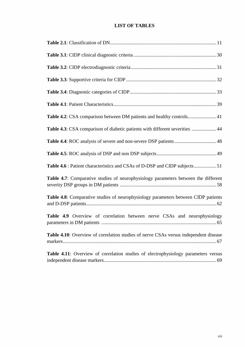
LIST OF TABLES
Table 2.1: Classification of DN ...................................................................................... 11
Table 3.1: CIDP clinical diagnostic criteria ................................................................... 30
Table 3.2: CIDP electrodiagnostic criteria ..................................................................... 31
Table 3.3: Supportive criteria for CIDP ......................................................................... 32
Table 3.4: Diagnostic categories of CIDP ...................................................................... 33
Table 4.1: Patient Characteristics ................................................................................... 39
Table 4.2: CSA comparison between DM patients and healthy controls ....................... 41
Table 4.3: CSA comparison of diabetic patients with different severities .................... 44
Table 4.4: ROC analysis of severe and non-severe DSP patients .................................. 48
Table 4.5: ROC analysis of DSP and non DSP subjects ................................................ 49
Table 4.6: Patient characteristics and CSAs of D-DSP and CIDP subjects .................. 51
Table 4.7: Comparative studies of neurophysiology parameters between the different
severity DSP groups in DM patients .............................................................................. 58
Table 4.8: Comparative studies of neurophysiology parameters between CIDP patients
and D-DSP patients ......................................................................................................... 62
Table 4.9 Overview of correlation between nerve CSAs and neurophysiology
parameters in DM patients ............................................................................................. 65
Table 4.10: Overview of correlation studies of nerve CSAs versus independent disease
markers ............................................................................................................................ 67
Table 4.11: Overview of correlation studies of electrophysiology parameters versus
independent disease markers ........................................................................................... 69
xiv
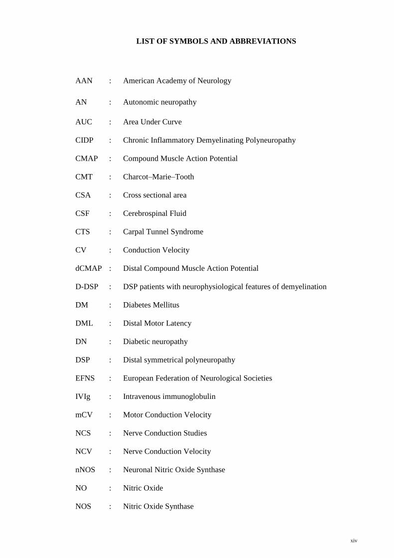
LIST OF SYMBOLS AND ABBREVIATIONS
AAN : American Academy of Neurology
AN : Autonomic neuropathy
AUC : Area Under Curve
CIDP : Chronic Inflammatory Demyelinating Polyneuropathy
CMAP : Compound Muscle Action Potential
CMT : Charcot–Marie–Tooth
CSA : Cross sectional area
CSF : Cerebrospinal Fluid
CTS : Carpal Tunnel Syndrome
CV : Conduction Velocity
dCMAP : Distal Compound Muscle Action Potential
D-DSP : DSP patients with neurophysiological features of demyelination
DM : Diabetes Mellitus
DML : Distal Motor Latency
DN : Diabetic neuropathy
DSP : Distal symmetrical polyneuropathy
EFNS : European Federation of Neurological Societies
IVIg : Intravenous immunoglobulin
mCV : Motor Conduction Velocity
NCS : Nerve Conduction Studies
NCV : Nerve Conduction Velocity
nNOS : Neuronal Nitric Oxide Synthase
NO : Nitric Oxide
NOS : Nitric Oxide Synthase
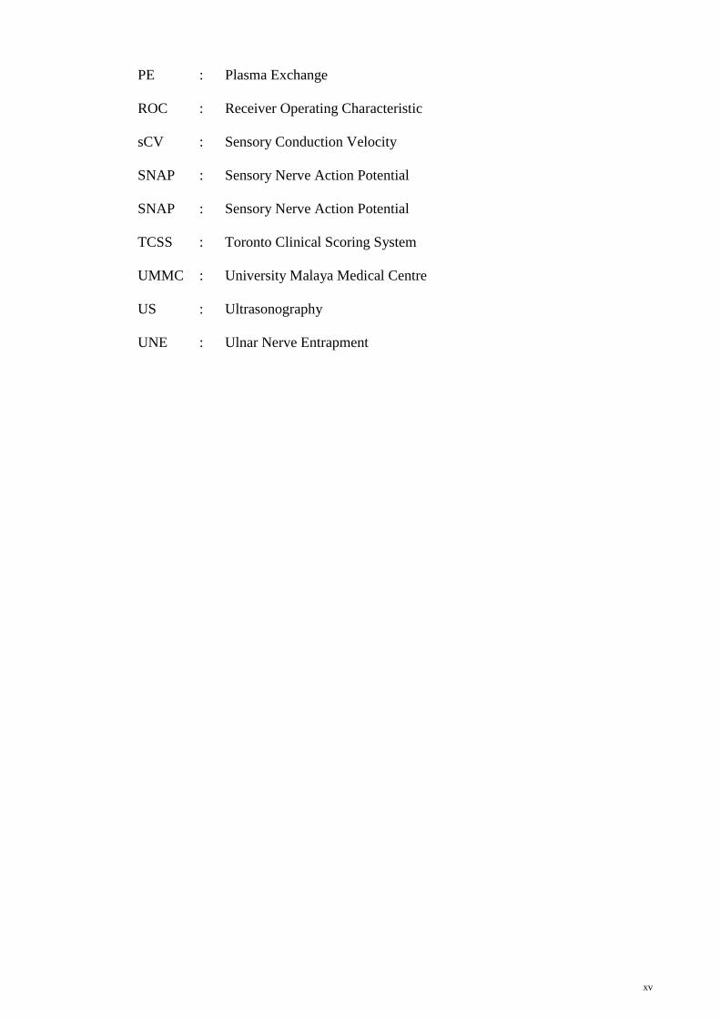
xv
PE : Plasma Exchange
ROC : Receiver Operating Characteristic
sCV : Sensory Conduction Velocity
SNAP : Sensory Nerve Action Potential
SNAP : Sensory Nerve Action Potential
TCSS : Toronto Clinical Scoring System
UMMC : University Malaya Medical Centre
US : Ultrasonography
UNE : Ulnar Nerve Entrapment
xvi
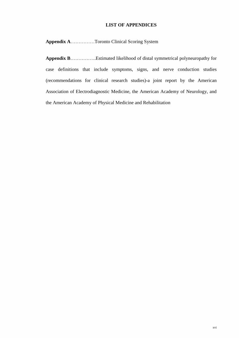
LIST OF APPENDICES
Appendix A……………Toronto Clinical Scoring System
Appendix B…………….Estimated likelihood of distal symmetrical polyneuropathy for
case definitions that include symptoms, signs, and nerve conduction studies
(recommendations for clinical research studies)-a joint report by the American
Association of Electrodiagnostic Medicine, the American Academy of Neurology, and
the American Academy of Physical Medicine and Rehabilitation
1
CHAPTER 1: INTRODUCTION
1.0 Introduction
Diabetes mellitus (DM) is increasingly prevalent in both developed and developing
countries. A decreased quality of life, mortality, and morbidity are major consequences
from diabetes and its complications. One of the long-term complications
that affect the nerves is neuropathy. There are a broad diversity of neuropathies in
diabetes, affecting single (mononeuropathy), several (mononeuropathy multiplex), or
many nerves (polyneuropathy). The most common presentation of diabetic neuropathy
(DN) that presents with symmetrical sensorimotor symptoms is distal symmetrical
polyneuropathy (DSP), with a reported prevalence of about 50% (P. J. Dyck et al,
2010). In DSP, there are indications that small fiber sensory modalities are involved and
minor distal motor weakness may follow. The fundamental pathology in DSP has been
shown to be of distal axonal degeneration of dying back type (distal axonopathies)
(Said, Slama, & Selva, 1983) which are typically seen as ‘length-dependent’ or ‘glove-
and-stocking’ neuropathies with relative preservation of dorsal root ganglion cells
(Dolman, 1963; Watkins et al, 1995).
The clinical diagnosis usually relies on the patients’ description of pain, where
symptoms are distal, symmetrical, often associated with nocturnal exacerbations, and
commonly described as prickling, sharp and burning with hyperalgesia and frequent
allodynia upon examination. Validated scales and questionnaires such as Toronto
Clinical Scoring System (TCSS) and McGill’s Pain Questionnaire can be used to
estimate the severity of the neuropathic pain (Tesfaye et al, 2010).
2
Conventionally, nerve conduction studies (NCS) have been widely used to diagnose
DSP (England et al., 2009). For electrodiagnostic confirmation of DSP, the minimum
criterion is abnormality of any attribute of nerve conduction in two separate nerves, one
of which must be the sural nerve (Callaghan, Cheng, Stables, Smith, & Feldman, 2012).
Since DSP presents in a length-dependent manner, NCS in the lower limbs would be
more suitable to assess DSP severity. However, NCS in the lower limbs is time
consuming and in patients with severe DSP, action potential in the lower limbs often
cannot be stimulated (Tsuneo Watanabe et al., 2010).
In recent years, peripheral nerve ultrasonography (US) have emerged as an additional
tool in the assessment of peripheral nerve disorders demonstrating morphological
changes in patients with different forms of neuropathy.
In the diagnosis of entrapment neuropathies, there is a substantial body of literature
on US, however, the US changes of polyneuropathy, particularly in DM has not been
fully explored. Another scope of interest is in making the distinction between DSP
patients with neurophysiological evidence of demyelination (D-DSP) and true chronic
inflammatory symmetrical polyneuropathy (CIDP) patients. CIDP characteristically
affects the most proximal regions of the peripheral nervous system, nerve roots and
major plexuses. In clinical practice, simultaneous occurrence of CIDP and DM (diabetic
CIDP or CIDP-DM) is frequently seen; however, it is still unclear whether the two
disorders are pathogenetically correlated (Lozeron et al, 2002; Sharma et al,2002;
Stewart, McKelvey, Durcan, Carpenter, & Karpati, 1996).
3
In the current study, we prospectively recruited DM patients with clinical
symptoms suggestive of neuropathy and assessed them for objective evidence of
DSP through NCS in patients with different severities of DSP as determined by the
TCSS. As measured by sural nerve morphology and NCS, the TCSS is a valid
instrument to reflect the presence and severity of DSP (Bril & Perkins, 2002). To
our knowledge, no studies have investigated the severity of DSP. We also aimed to
determine if US could reliably discriminate between the different grades of severity
of DSP. This is crucial as currently, there is no objective evaluation of assessing
DSP severity, and our approach might enable appropriate specialist referral for
treatment.
We also assessed the validity of ultrasound as an additional adjunct diagnostic
modality in DSP. This is important as the current ‘gold standard’ for diagnosis and
staging of DSP severity, which is NCS, is unable to assess small fiber involvement
in DSP and nerves are frequently inexcitable in patients with severe disease. We
hypothesized that US can perform as an equitable evaluation to determine severity
of DSP. One of our objectives is also to examine the validity of the TCSS in our
cohort by correlating this tool with NCS parameters. We examined a subset of D-
DSP patients and compared this group to patients with true CIDP to investigate if
there are any sonographic features that can differentiate these two groups of patients.
It is vital to distinguish true CIDP patients from D-DSP patients due to the
implications of prognosis and therapy because CIDP is treatable whereas DSP is not
(Sharma et al, 2002) . To date, no specific nerve parameters have shown to
specifically distinguish between CIDP and DSP.
4
1.1 Objective/s
1. To investigate the patterns of peripheral neuropathy in diabetic patients
using clinical symptom scores, NCS and US
2. To assess the validity of ultrasound as a diagnostic modality in diabetic
peripheral neuropathy
3. To correlate the findings of nerve conduction studies and ultrasound with
clinical symptom scores in patients with DM
4. To investigate the utility of ultrasound as a tool to distinguish
demyelinating diabetic neuropathies from true CIDP patients
5
CHAPTER 2: LITERATURE REVIEW
2.1 Burden of DN in the global context
With more industrialization, globalization and reformation in lifestyles worldwide,
we are experiencing a shift in disease paradigm, with chronic disease such as DM
becoming more widespread. DM is increasingly prevalent in both developed and
developing countries. In 2010, the world prevalence of DM among adults was 6.4%
affecting 285 million adults and by 2030, this number is likely to increase to 7.7 % i.e.
439 million (Shaw, Sicree, & Zimmet, 2010). The International Diabetes Federation
(IDF) estimated that 371 million people worldwide were living with DM in 2012, of
which about half live in South Asia, the Western Pacific, and Eastern Mediterranean
regions (Abdullah, Attia, Oldmeadow, Scott, & Holliday, 2014). Asia is now the focal
point of a growing diabetes epidemic, largely due to population growth and ageing in
India and China. By 2030, projections indicate that more than 60% of worldwide
diabetes cases will come from Asia (Shaw et al, 2010; Wild, Roglic G Fau - Green,
Green A Fau - Sicree, Sicree R Fau - King, & King, 2004) with the vast majority of
these being type 2 DM. A cross-sectional multicentre study performed in the United
Kingdom hospital clinic population revealed the prevalence of type 2 (non-insulin-
dependent) DM patients was 32.1 % (30.6–33.6 %) and diabetic peripheral neuropathy
increased with age, from 5% (3.1– 6.9 %) in the 20–29 year age group to 44.2 % (41.1–
47.3 %) in the 70–79 year age group (Young, Boulton, MacLeod, Williams, & Sonksen,
1993). A report from Pittsburgh epidemiology of diabetes complications study
demonstrated 34% (18%, 18-29 yr old, 58% ≥30 yr old) prevalence of DN (Maser et al.,
1989). In a study done in the Malaysian cohort, the prevalence of DN was found to be
54.7% (Abougalambou & Abougalambou, 2012).
6
The most common presentation of DN is DSP, affecting more than 90% of the
patients (Tesfaye, Boulton, & Dickenson, 2013). Small and/or large nerve fibers may be
affected. DSP is a major and independent risk factor for mortality (Forsblom et al, 1998)
and morbidity because of foot ulceration and amputation (Abbott et al., 2002).
2.2 Pathophysiology of DN
The pathophysiological mechanisms of DN are not yet fully established, although
pain is one of the main symptoms. It is generally accepted that the toxic effects of
hyperglycemia plays a significant role in the development of this complication, but
several other hypotheses have been proposed (Dobretsov, Hastings, Romanovsky,
Stimers, & Zhang, 2003; Oyibo, Prasad, Jackson, Jude, & Boulton, 2002). Both
metabolic and vascular factors are involved in the pathophysiology of DN.
Hyperlipidemia, hypertension, cigarette smoking, consumption of alcohol, and obesity
are other comorbid factors associated with DN. Hyperglycemia plays a prominent role
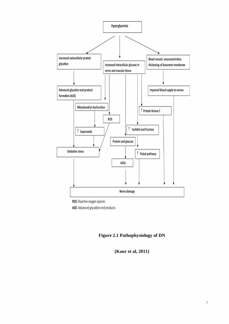
in the pathogenesis which results in the following (Kaur, Pandhi, & Dutta, 2011) (Fig
2.1 ):
7
Figure 2.1 Pathophysiology of DN
(Kaur et al, 2011)
8
2.2.1 Formation of advanced glycation end products
DM results in an increase in oxidation products. Increased flux through one or more
glucose metabolism pathways leads to excess intracellular glucose production which
leads to incorporation of glucose into proteins nonenzymatically by an unregulated
glycation reaction (Head, 2006). Haemoglobin, plasma albumin, lipoproteins, fibrin and
collagen are among the different proteins that gets glycated. All these glycated end
products are responsible for causing the tissue damage.
2.2.2 Oxidative stress
Production of reactive oxygen species or defective scavenging of free radicals
provides major evidence that points to increased oxidative stress in DN. Excess glucose
undergoes auto-oxidation and this leads to formation of reactive oxygen species. By
obstructing the nitric oxide (NO) production by the endothelium and thereby leading to
ischemia of nerves, these oxygen free radicals cause damage of the nerves. A study
analyzed markers of oxidative stress in 189 people with diabetes and 85 controls
(Ziegler, Sohr, & Nourooz-Zadeh, 2004). Subjects with DN exhibited significant rise of
all oxidative stress markers, as well as significant decreases in the protective antioxidant
vitamins C and E. In another study to examine effects of pro-oxidants, rats that were
exposed to two pro-oxidant interventions show diminished nerve conduction velocity
(NCV), nerve growth factor in the sciatic nerve, and neuropeptides compared to diabetic
rats that were not exposed to additional oxidative stress (Hounsom, Corder, Patel, &
Tomlinson, 2001).
9
2.2.3 Accumulation of polyols
Glucose is capable of diffusing passively without insulin into certain type of cells,
including nerve cells. Once inside the cell, glucose undergoes conversion to sorbitols
and other polyols by the enzyme aldose reductase. Due to polyols inability to passively
diffuse out of cells, polyols concentrate within cells such as neurons, thus creating a
concentration gradient that allows excess sodium and water to flow (Raccah et al, 1998).
With polyol accumulation, free carnitine and myo-inositol content in the caudal nerves
of diabetic rats were remarkably decreased. (Nakamura et al, 1998).
2.2.4 Deficiency of NO
The pathogenesis of DN has also been associated with vascular factors. NO plays a
vital role in controlling (Na+ /K+)-ATPase activity (Gupta et al, 2002), a reduction of
which has been implicated in the pathogenesis of DN (Stevens et al, 1994).
Experimental analysis showed hyperglycemia results in an excess of endothelial
superoxide radicals that result in decreased stimulation of NO on (Na+ /K+ )-ATPase
activity; this effect is inhibited by L-arginine (Gupta et al, 2002). Nerve blood flow is
reduced in experimental DN, and many studies have shown it may be mediated by
variation in NO metabolism. One such study investigated nerve blood flow and nitric
oxide synthase (NOS) activity in the microvasculature serving peripheral nerves in
diabetic rats (Kihara & Low, 1995). A significant decrease of nerve blood flow was
observed compared to controls due to hyperglycemia. An in vivo study also
demonstrated disruptions in neuronal nitric oxide synthase (nNOS) in experimental
diabetes. Reduced nNOS expression was associated with a higher degree of neuropathic
pain (Sasaki, Yasuda, Maeda, & Kikkawa, 1998).
10
Experimental analysis has further demonstrated that hyperglycemia results in an
excess of endothelial superoxide radicals that result in diminished stimulation of NO on
(Na+ /K+ )-ATPase activity (Gupta et al, 2002). However, no relationship between
altered NO activity and the development of sensory peripheral neuropathy was found in
another study (Thomsen, Rubin, & Lauritzen, 2002).
The interrelationships between the various pathogenetic features of DN are not
clearly understood. An animal study aimed to clarify a possible connection between
aldose reductase activity (enhanced polyol pathway activity) and decreased NO activity.
The study demonstrated NO to be an important mediator of nerve (Na+ /K+)-ATPase
and aldose reductase activity on NCV. The study concluded that hyperglycemia
increases the activity of aldose reductase, subsequently reducing NO synthase activity
via cofactor competition (Stevens et al., 1994).
2.3 Classifications and characteristics of DN
Multifarious neurological complications in DM are seen, affecting different parts of
the nervous system, and may manifest in various clinical presentations. There are many
classification of DN (Boulton, Malik, Arezzo, & Sosenko, 2004). One such
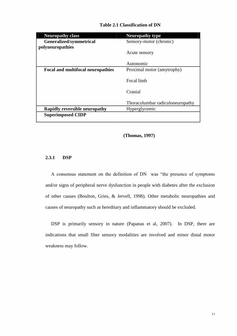
classification is shown in Table 2.1 (Thomas, 1997). The most common form of DN is
DSP, representing 70% of DN (Zochodne, 2007). DSP is the focus of this thesis and
will be discussed in further detail. Other forms of DN are also described to a lesser
extent.
11
Table 2.1 Classification of DN
Neuropathy class Neuropathy type
Generalized/symmetrical
polyneuropathies
Sensory-motor (chronic)
Acute sensory
Autonomic
Focal and multifocal neuropathies Proximal motor (amytrophy)
Focal limb
Cranial
Thoracolumbar radiculoneuropathy
Rapidly reversible neuropathy Hyperglycemic
Superimposed CIDP
(Thomas, 1997)
2.3.1 DSP
A consensus statement on the definition of DN was “the presence of symptoms
and/or signs of peripheral nerve dysfunction in people with diabetes after the exclusion
of other causes (Boulton, Gries, & Jervell, 1998). Other metabolic neuropathies and
causes of neuropathy such as hereditary and inflammatory should be excluded.
DSP is primarily sensory in nature (Papanas et al, 2007). In DSP, there are
indications that small fiber sensory modalities are involved and minor distal motor
weakness may follow.
12
Accumulation of sorbitol due to increased flux in the polyol pathway secondary
to hyperglycemia is the major metabolic abnormality of peripheral nerves in DSP
(Chalk, Benstead, & Moore, 2007). In human DM patients, axonal proteins have been
shown to be abnormally glycated (Brownlee, Cerami, & Vlassara, 1988).
Since DSP is a distal axonopathy of the dying back type, there is a possible
interference with the operation of growth factors by the diabetic state resulting in nerve
cells being unable to maintain their distal axons. Failure of axonal regeneration is an
important aspect of DSP (Thomas, 1994).
DSP is characterized by burning or aching pain, numbness, paraesthesia, and
hyperalgesia in both feet and lower limbs (symmetrical). These symptoms begin in the
feet and spread proximally in a length-dependent fashion, eventually involving distal
hands, in what is referred to as ‘stocking-and-glove’ distribution. Sensory symptoms
appear to be more prominent than motor involvement. The array of symptoms
associated with DSP has many downstream effects that can affect patients’ quality of
life, both physically and mentally. DSP-associated numbness frequently results in
balance difficulties, which can lead to falls (Callaghan et al, 2012). Additionally,
patients with severe DSP are at risk of ulcerations and lower-extremity amputations,
with 15% developing an ulcer during the course of their disease. Despite the availability
of many successful therapies, however, less than half of patients are treated for pain.
Currently, the only treatments available to patients with DSP are improved glucose
control and pain management (Bril, Hirose, Tomioka, & Buchanan, 2009).
13
2.3.2 Autonomic neuropathy (AN)
Diabetic AN can involve the complete autonomic nervous system. It is manifested by
impairment of one or more organ systems (e.g., cardiovascular, gastrointestinal [GI]
genitourinary, sudomotor, or ocular) (Vinik, Maser, Mitchell, & Freeman, 2003).
General symptoms include dizziness (orthostatic hypertension), resting tachycardia,
oedema, bladder dysfunction, and erectile dysfunction. An increased mortality risk is
seen among DM patients with AN compared to DM patients without AN, chiefly due to
renal failure, sudden death and cardiovascular events (Vinik et al, 2003).
Autoimmunity is thought to play an important role in the pathogenesis of AN
(Granberg, Ejskjaer, Peakman, & Sundkvist, 2005; Sundkvist, Lind,Bergstrom, Lilja, &
Rabinowe, 1991; Vinik et al., 2003). Autoantibodies against adrenal medulla,
sympathetic ganglia, and the vagal nerve have been identified and associated to future
development of cardiac and peripheral AN (Granberg et al., 2005). Treatment is mainly
symptomatic, but glycaemic control seems to improve AN (Vinik et al, 2003).
2.3.3 Focal and multifocal neuropathies
Focal/asymmetrical diabetic neuropathies may involve a single nerve
(mononeuropathy), or few different nerves (mononeuropathy multiplex). Nerve
entrapments, usually involving the ulnar, median, and peroneal nerves are one of the
common causes of some focal neuropathies (Boulton et al, 2004; Boulton et al, 2005).
DM patients have an increased susceptibility to nerve compression (Dahlin, Stenberg,
Luthman, & Thomsen, 2008) and about one third of them have nerve entrapments
(Boulton et al, 2005).
14
Carpal tunnel syndrome (CTS) is the most common nerve entrapment i.e.
compression of the median nerve at the wrist (Boulton et al, 2004). Focal neuropathies
such as mononeuritis multiplex has an acute onset, associated with pain and heal
spontaneously, between 6-8 weeks. They are caused by vascular obstruction typically in
the cranial nerves III, VI, and VII, ulnar, median, and peroneal nerves. Other
focal/multifocal neuropathies in DM may have an ischemic basis and often exist with
sudden onset of severe pain. Microvascular nerve infarct results in cranial neuropathies,
typically involving the third, fourth, sixth, and seventh cranial nerves (Boulton et al,
2004; Boulton et al, 2005).
Cranial neuropathies are rare and usually present in older individuals with a long
duration of DM (Boulton et al., 2004). Mostly, cranial neuropathies resolve
spontaneously over several months but can recur in 25% of patients (Boulton et al,
2004). In the proximal lower limb motor neuropathy (amyotrophy), nerve infarcts have
also been indicated but there is evidence that focal inflammatory lesions (including
vasculitic) may be related (Boulton et al, 2004; Thomas, 1997).
2.4 Diagnosis of DSP
Although DSP can be diagnosed by experienced clinicians with a clinical
examination, there are still inconsistencies in the diagnostic criteria that exists in the
literature. The American Academy of Neurology (AAN) in conjunction with the
American Association of Electrodiagnostic Medicine and the American Academy of
Physical Medicine and Rehabilitation reported a case definition of DSP to systematize
and facilitate clinical research and epidemiologic studies (England et al, 2005). The
combination of neuropathic symptoms, signs, and electrodiagnostic findings
accordingly gives the most accurate diagnosis of DSP.
15
2.4.1 Clinical examination
The clinical examination consists of a detailed inspection of peripheral sensation,
tendon reflexes, and muscle strength. Neuropathic symptoms with distal sensory loss,
absent tendon reflexes, and abnormal nerve conduction studies are distinctly suggestive
of DSP (England et al, 2005).
2.4.4.1 Clinical Scoring System
The clinical diagnosis usually relies on the patients’ decription of pain, where
symptoms are distal, symmetrical, often associated with nocturnal exacerbations, and
commonly described as prickling, sharp and burning with hyperalgesia and frequent
allodynia upon examination. Validated scales and questionnaires such as TCSS and
McGill’s Pain Questionnaire are used to estimate the severity of the neuropathic pain
(Tesfaye et al, 2010). In recent years, different clinical scoring systems were developed
to document the presence and severity of DSP quantitatively (Dyck, 1988; Perkins,
Olaleye, Zinman, & Bril, 2001). One study has indicated that the Michigan
Neuropathy Screening Instrument (MNSI) is a good screening tool for diabetic
neuropathy and that the Michigan Diabetic Neuropathy Score (MDNS) coupled with
NCS gives a simple means to confirm this diagnosis (Feldman et al, 1994). However,
none of these methods was validated against morphological criteria for DSP. In another
study, the TCSS was implemented for a simple screening for DSP to classify patients
into severity categories and correlated well with NCS findings and complications in
subjects with DSP in that study (Perkins et al, 2001) . Consequently, TCSS was
considered as a simple method for evaluation of DSP. However, further justification in
patients with documented DSP was required.
16
A study has assessed the correlation of TCSS with the presence and severity of
DSP as ascertained by electrophysiological criteria and the additional morphological
gold standard of myelinated fiber density on sural nerve biopsy in an independent group
of DSP patients. The study demonstrated that the clinical neuropathy as characterized by
the TCSS is associated with the morphological severity of DSP and hence suggests that
the TCSS may prove to be useful in documenting and monitoring DSP in the clinic and
in clinical research trials (Bril & Perkins, 2002).
2.5 Neurophysiological examination
NCS are the most widely accepted objective evaluation for the diagnosis of DSP
(Bae & Kim, 2007; Kim, Kwon, Lee, & Sunwoo, 2000) and CIDP (Dyck et al, 1975).
NCS are non-invasive, standardized technique that provides an objective and sensitive
measure of the functional status of sensory and motor nerves. An electrical impulse, an
action potential, is evoked by stimulating the nerve and conducted along a motor or
sensory axon. The distribution of abnormality (focal, multifocal, or diffuse), and
whether the pathophysiology is predominantly a segmental demyelination or axonal
degeneration can be determined using NCS. The axonal degeneration and progressive
loss of nerve fibers are the most important features in DSP. These axonal changes are
identified by reduced motor and sensory action potential amplitudes, with normal or
slightly reduced conduction velocities secondary to loss of the largest and fastest-
conducting axons (Callaghan et al, 2012). In CIDP, the diagnosis is based on NCS
evidence of conduction block and temporal dispersion (Stewart et al, 1996).
17
A study has suggested that the residual latency (RL), terminal latency index (TLI)
and modified F ratio (MFR) which indicates distal conduction slowing, may be a useful
guide to identify subclinical DN. The study also demonstrated that electrophysiological
changes that are unclear in routine NCS are present before the clinical manifestation
(Bae & Kim, 2007). Another study has evaluated the reproducibility of NCS. The study
demonstrated that the median and tibial F-wave latencies produce the most reproducible
measures for NCS, serving as one of the best measures in multicentre drug trials for DN
(Kohara et al, 2000). Another study aimed at evaluating the relationship of abnormal
parameters in commonly tested peripheral nerves and clinical findings in DN through
NCS and found the amplitude of sensory nerve action potential to be a vital parameter in
detection of early DN (K. W. Lee, Hwang, & Kim, 1999).
Motor nerve conduction was studied along the entire course of nerves from the spinal
cord to the muscle in diabetic and normal controls. The study found a diffuse pattern of
motor conduction abnormalities in DN over the total length of the nerve, being extreme
in the distal than proximal segment. Additionally, both proximal and distal segments
were more often affected in the lower than in the upper extremities (Kimura, Yamada,
& Stevland, 1979). NCS in the lower limbs would be more suitable to assess DSP
severity as it presents in a length-dependent fashion. However, in patients with severe
disease, action potential in the lower limbs cannot be stimulated. A study reported that
the sensory nerve conduction velocity was not measurable in many patients, and instead
they looked at distal motor latency (DML) and motor conduction velocities (mCV) to
evaluate DSP (Mizumoto, Hashizume, Senda, Nagoshi, & Inoue, 2003). At times, these
too cannot be recorded due to the small foot muscle wasting.
18
The sensory nerve action potential amplitude (SNAP) and compound muscle
action potential (CMAP) amplitude is reduced due to axonal loss. Reduction of the
nerve conduction velocity and increased dispersion are caused by demyelination. Motor
and sensory neuropathies can be diagnosed since both afferent and efferent nerves can
be tested. When polyneuropathy is suspected, the ulnar and median nerves in the arm
and the peroneal, posterior tibial, and sural nerves in the leg are commonly tested. For
electrodiagnostic confirmation of DSP, the minimum criterion is an abnormality of any
attribute of nerve conduction in two separate nerves, one of which must be the sural
nerve (England et al, 2005). To test whether physicians can validly and reproducibly
diagnose DSP, one study demonstrated that there was significant agreement between
75% group diagnosis and confirmed nerve conduction abnormality. When compared to
nerve conduction score, individual physicians' clinical diagnoses were greatly variable
and mostly inaccurate, often overestimating DSP (Peter J Dyck et al, 2010).
2.6 US
While NCS remain fundamental to confirm the diagnosis of DSP and CIDP, the test
is time consuming and uncomfortable for patients. Sonographic examinations can
potentially be an alternative to assess peripheral nerves with less discomfort and there
have been studies that have proven its clinical use in the evaluation of disorders of the
peripheral nervous systems. (Goedee et al, 2013). In recent years, the role of US of
peripheral nerves has been investigated. In the diagnosis of entrapment neuropathies,
there is a substantial body of literature on nerve ultrasound, however, the US changes of
polyneuropathy, especially in DM has not been thoroughly explored.
19
Nerve US when performed along with NCS aids in visualizing nerve
morphology, not only in mononeuropathies but also in peripheral neuropathies. Most
US studies have concentrated on entrapment neuropathies. In DN, ultrasound studies
have been less comprehensive, typically only looking at distal lower limb nerves.
Currently, US has proven its usefulness for the diagnosis of compressive
neuropathy in CTS, and there is evidence that substantiates its usefulness in ulnar
neuropathy at the elbow (Suk, Walker, & Cartwright, 2013). In the diagnosis of
peripheral nerve damage in entrapment syndromes, nerve tumors, and focal nerve
lesions, the use of peripheral nerve US has been demonstrated clearly and has gained
vast interest in recent times (Grimm, Heiling, Schumacher, Witte, & Axer, 2014). In one
study, larger nerve cross sectional area (CSA) was observed in patients with common
fibular neuropathy than controls (Visser et al, 2013). An increased CSA was also
observed on magnetic resonance imaging and on US in immune-mediated and
demyelinating hereditary peripheral neuropathies with more prominent changes in
hereditary neuropathy (Grimm et al, 2014).
Peripheral nerve polyneuropathy US data, especially axonal forms, so far have
been based chiefly on small patient numbers or single case studies (Goedee et al, 2013).
An enlargement of distal parts of the tibial nerves in patients with DM were
observed in one study (D. Lee & Dauphinee, 2005). There have been reports on
enlargement of affected nerves attributed to the process of attempted remyelination in
patients with Charcot-Marie-Tooth disease (CMT), multifocal motor neuropathy
(MMN) and CIDP (Beekman et al, 2005; Cartwright et al, 2009; Heinemeyer &
Reimers, 1999; Martinoli et al, 2002; Taniguchi et al, 2000) .
20
A sonographic distinction was seen through a larger nerve area and fascicular
diameter in the CMT 1A disease compared to patients with the other types of
disease (including CMT2 and CMTX) and the control subjects (Martinoli et al, 2002).
Another study studied the peripheral nerves of individuals with CMT type 1B and
found that patients with CMT 1B have significantly larger median and vagus nerves
than healthy controls, but no difference was observed in cranial nerve size between
those with versus those without cranial neuropathies (Cartwright et al, 2009). An
increased nerve CSA has also been demonstrated in vasculitic neuropathy, amyloidosis,
neurofibromatosis and POEMS syndrome (AD, Skare, Sakuma, & Barros, 2015; Bohm,
2009; Ito, Kijima, Watanabe, Sakuta, & Nishiyama, 2007; Lucchetta, Pazzaglia,
Granata, Briani, & Padua, 2011). One study has evaluated the usefulness of US to detect
abnormalities in tibial vasculitic neuropathy at the medial ankle and they found the
affected nerve area was significantly larger than in controls (Ito et al, 2007).
High-frequency sonography was found helpful in one study in the diagnosis of
vasculitic neuropathy in their two cases of mononeuritis multiplex and two cases of
DSP and was able to detect focal morphologic lesions which could not be identified
electrophysiologically due to the axonopathy (Bohm, 2009). Another study aimed to
establish the value of US in the diagnosis of CTS and found the measurement of
median nerve area by US performs well and could be used as first choice for the
investigation of patients with CTS (AD et al, 2015).
In DN, US studies have been less comprehensive, typically only looking at distal
lower limb nerves. One study aimed to determine the sonographic characteristics of
lower extremity nerves in DN and correlate them with electrodiagnostic findings. The
results showed measurements of lower extremity nerves in DN do not differ from
controls or correlate with electrodiagnostic findings.

21
They concluded that further innovative US techniques might be necessary to
detect differences (Hobson-Webb, Massey, & Juel, 2013). Another study determined the
morphological changes of sural nerves in patients with type 2 DM using US and found
that 22-MHz US may be a worthy tool for assessing diabetic cutaneous nerve
neuropathy (Liu, Zhu, Wei, Bao, & Hu, 2012).
Riazi et.al demonstrated a larger nerve CSA in posterior tibial nerve in DM
patients compared to control subjects and this large study of DM patients concluded US
is a promising point-of-care screening tool for DM patients with DSP (Riazi et al,
2012).
2.7 CIDP
CIDP is an immune-mediated disorder. It characteristically affects the most proximal
regions of the peripheral nervous system, nerve roots, and major plexuses. Simultaneous
occurrence of CIDP and DM (diabetic CIDP or CIDP-DM) is frequently seen in clinical
practice; however, it is still unclear whether the two disorders are pathogenetically
correlated (Chio et al, 2009;Lozeron et al, 2002; Sharma et al, 2002). Making the
distinction between chronic symmetric sensorimotor DPN and CIDP can be
challenging. CIDP may be diagnosed in a DM patient when motor symptoms are
predominant, however, it is more difficult to diagnose in DM patients with
neurophysiological features of demyelination without clinical motor weakness (Ayyar &
Sharma, 2004).
22
Stewart et al described seven DM patients with distal greater than proximal,
symmetric neuropathy (polyneuropathy) that had more features in keeping with CIDP
rather than a length-dependent pattern of neuropathy typically described in DN (Stewart
et al, 1996). Krendel described six insulin dependent diabetics with a demyelinating
neuropathy indistinguishable from CIDP, all of whom improved with varying types of
immunotherapy (Krendel, Costigan, & Hopkins, 1995). To date no specific nerve
parameters have been shown to specifically distinguish between CIDP and DSP.
Proposals for diagnostic tools that can help clinicians to determine the probability of a
patient with diabetes having CIDP exist (Lotan, Hellman, & Steiner, 2015). By listing
several clinical, electrophysiological, and laboratory parameters that, when combined,
were able to powerfully discriminate an immune-mediated neuropathy in patients with
diabetes mellitus. Four levels of probability for a patient with diabetes to have CIDP
were defined by summing the points assigned to each of these parameters.
The results demonstrated that this diagnostic tool enables the identification of
diabetic patients with overlapping CIDP (Lotan et al, 2015). This is of great importance
as unlike DSP, CIDP is a treatable condition.
2.7.1 Clinical presentation and diagnosis
CIDP typically arises between the ages of 30 and 60 years and is characterized by the
occurrence of progressive (more than two months), symmetric proximal and distal
muscle weakness. The condition also demonstrates impaired sensation, absent or
reduced tendon reflexes, an elevated cerebrospinal fluid protein level, demyelinating
NCS, and indications of demyelination in nerve-biopsy experiments (Barohn, Kissel,
Warmolts, & Mendell, 1989).
23
2.8 Disease treatment and management
2.8.1 DSP treatment and management
Poor glycaemic control has been implicated as a pathogenetic mechanism in the
etiology of DSP. An increase in blood glucose flux has been reported to cause pain in
DSP (Oyibo et al., 2002). Currently, strategies that has been implemented for
management of DSP are based on (i) improving glucose control ; (ii) symptomatic
control of DSP and (iii) treatment centered on pathogenetic mechanisms (Tesfaye,
2011).
2.8.1.1 Pharmacological treatment of DSP
Some pharmacological therapies have proven to be effective in management of DSP.
Tricyclic compounds have been used as first-line therapy for many years and its efficacy
has been supported by several randomized clinical trials (Finnerup, Otto, McQuay,
Jensen, & Sindrup, 2005;Max et al,1992; Tesfaye, 2007).
However, the uses of tricyclic drugs are limited due to its side effects including
anticholinergic effects such as dry mouth and dizziness. Usually, titration of the dose of
tricyclic drugs is recommended to avoid side effects (Tesfaye, 2007). Selective
serotonin-reuptake inhibitors (SNRI), such as duloxetine and venlafaxine relieves pain
by increasing synaptic accessibility of 5-hydroxytryptamine and noradrenalin in the
descending pathways that inhibit pain impulses (Tesfaye, 2011). Anticonvulsants such
as gabapentin and pregabalin have been used in the management of neuropathic pain for
many years. There have been few clinical trials involving pregabalin in DSP, and these
demonstrated clear efficacy in management of DSP (Freeman, Durso-Decruz, & Emir,
2008). Topical treatment, such as topical lidocaine in the form of a 5% patch is
potentially effective in management of pain associated with DSP (Bril et al., 2011).
24
The most often used antioxidant is α-lipoic acid, which is the pathogenetically
oriented treatment for DSP. It has been shown that only α-lipoic acid administered
intravenously over 3 weeks (600 mg i.v. per day) is effective in improving several
neuropathic symptoms and nerve function in patients with DSP (Ziegler, Nowak,
Kempler, Vargha, & Low, 2004).
2.8.1.2 Non pharmacological treatment of DSP
Alternative therapies, such as acupuncture (Abuaisha, Costanzi, & Boulton, 1998),
low intensity laser therapy (Zinman et al., 2004) and transcutaneous electrical
stimulation (Somers & Somers, 1999) have been used due to lack of response and
unwanted side effects of conventional pharmacological treatments. These might be
useful as add-on therapy at any stage of DSP.
2.8.2 CIDP disease treatment and management
The first line therapy in CIDP are steroids since the first report of their use (Austin,
1958). In one study of an unblinded randomized controlled trial with 28 subjects,
prednisone was superior to no treatment (Dyck et al., 1982). It has been clearly
demonstrated that that intravenously administered immune globulin (IVIg) plays a role
in immunomodulation and has anti-inflammatory effects (Gelfand, 2012). In a meta-
analysis of four double blind randomized control trials, IVIg showed a significant
improvement was seen in disability lasting 2-6 weeks in 235 subjects (Eftimov, Winer,
Vermeulen, de Haan, & van Schaik, 2013). The treatment of IVIg needs to be repeated
at intervals and doses needs to be determined on individual basis (Kuitwaard & van
Doorn, 2009).
25
Plasma exchange (PE) aims to remove circulating autoantibodies, cytokines, immune
complexes, and immune cells (Lehmann, Hartung, Hetzel, Stüve, & Kieseier, 2006) to
achieve fast immunosuppression. Conventionally, PE is used in acute forms of
dysimmune peripheral neuropathies such as Guillain-Barré syndrome (GBS), but also
patients with chronic disease such as CIDP may respond to PE in the short term, usually
for 2–4 weeks (Lehmann & Hartung, 2011). Immunosuppressive drugs such as
azathioprine, methotrexate , cyclosporin A and rituximab may be considered when the
response to steroids, IVIg or PE is inadequate. Treatment option will rely on several
variables such as initial disease severity, age, general health status, and potential
contraindications.
26
CHAPTER 3: METHODOLOGY
3.1 Subjects and study design
The study design is a prospective cohort study. DM patients were recruited
prospectively by direct approach at the outpatient clinic at University Malaya Medical
Centre (UMMC), Kuala Lumpur, Malaysia. Patients with Type 2 DM irrespective of
treatment type were included in the study. Exclusion criteria included all patients with
previous history or concurrent history of significant exposure to potential neurotoxins
(chronic alcohol consumption, environmental toxin, heavy metals such as lead, mercury
and arsenic, chemotherapy drugs) and previous or concurrent neurological disease,
compression or trauma to the peripheral nerves involving the lower limbs were
excluded. This was based on medical history. Existing patients with CIDP from the
neurology clinic at UMMC were also recruited.
The diagnosis of DSP was determined by applying the TCSS questionnaire (see
next section). Age and gender matched control subjects were recruited from relatives
and colleagues. Diabetic control was ascertained by HbA1C values. The values of
HbA1C were determined by the National Glycohemoglobin Standardization Program
(NGSP) method. Demographic data such as height and weight were collected at the time
of the study. Other relevant information acquired includes HbA1C values and DM
disease duration. Written consent was obtained from all the patients participating in this
research. Patients with longstanding chronic renal failure were also excluded. Ethical
approval was obtained from the UMMC Medical Research Ethical Committee.
27
3.2 DSP Clinical Screening
Patients underwent a series of examination through questionnaire and clinical
examination by the candidate, who had received prior training. Clinical assessment of
DSP was determined by the TCSS and AAN’s estimated likelihood of DSP for case
definitions that include symptoms, signs, and nerve conduction studies for assessment
of diabetic neuropathy (Bril & Perkins, 2002; England et al., 2005). TCSS symptom
scores include lower extremity pain, numbness, tingling, weakness, walking imbalance
and upper extremity symptoms. Normal was drafted as 0 point, abnormal as 1 point, and
a total of 6 points can be obtained in this section. Reflex scores, including the bilateral
knee reflex and ankle reflex, were 0 point for normal, reduced 1 point and absent 2
points, providing a total of 8 points in this section. Sensory score, including light touch
in the right great toe, joint position sense, vibration sense, pinprick, temperature
sensation, were normal- 0 point, abnormal- 1 point, giving a total of 5 points in this
section.
The total possible score in TCSS taking into account the symptom score, reflex
score, and sensory score was 19 points. A score of six or greater was considered
abnormal, suggesting the presence of DSP. The diabetic patients were grouped into
three groups of DSP according to severity: mild, moderate and severe according to
TCSS scores. The AAN estimates of the likelihood of DSP were also assessed and these
include symptoms, signs and NCS (England et al, 2005).
28
3.3 Diagnosis of CIDP and D-DSP patients
We identified eight patients that presented with a progressive symmetrical or
asymmetrical polyradiculoneuropathy where the clinical course is relapsing or remitting
and progressing for more than two months. Two patients with CIDP were excluded due
to a concurrent history of DM . The diagnosis of CIDP was made as specified by the
European Federation of Neurological Societies/Peripheral Nerve Society’s (EFNS/PNS)
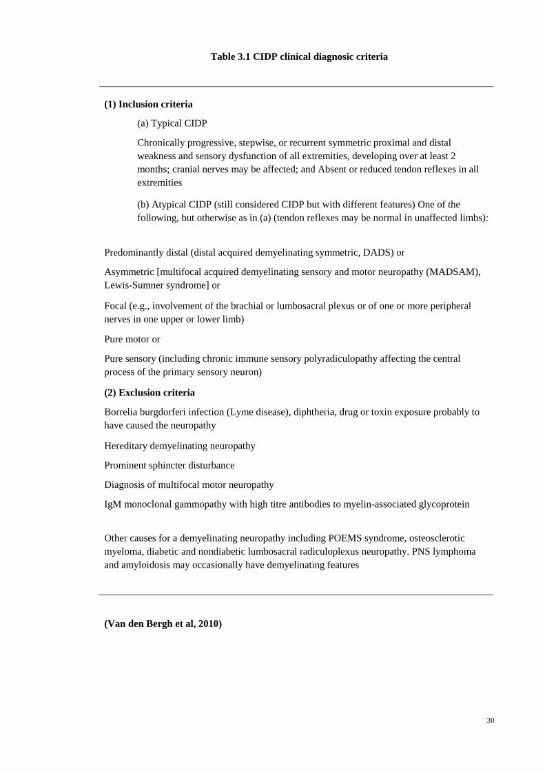
diagnostic criteria (Van den Bergh et al, 2010). The patients fulfilled the mandatory
diagnostic criteria, with evidence of sensory and motor impairment, disease
duration/progression of at least 8 weeks, hyporeflexia/areflexia upon clinical
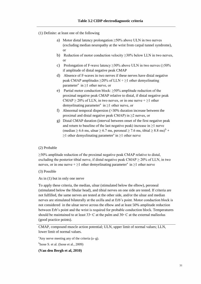
examination and NCS shows evidence of demyelination [Table 3.1 and 3.2] (Van den
Bergh et al, 2010). Cerebrospinal fluid (CSF) analysis also showed albuminocytologic
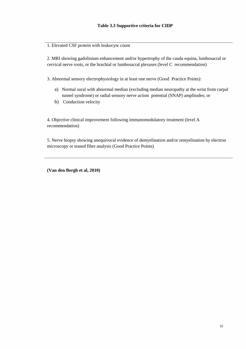
dissociation in all patients. Magnetic resonance imaging (MRI) of spinal roots, brachial
plexus,and lumbosacral plexus along with nerve biopsies are additional investigations to
diagnose CIDP, but not mandatory as shown in Table 3.3 (Van den Bergh et al., 2010).
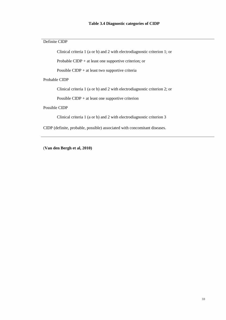
The diagnostic criteria of CIDP are shown in Table 3.4. We employed the criteria for
definite CIDP which comprises of clinical criteria 1 (a or b) and 2 with electrodiagnostic
criterion 1.
DM patients with DSP were defined as having demyelination (D-DSP) out of
proportion to axonal loss if amplitudes were preserved and at least two NCS parameters
showed conduction slowing as suggested by the EFNS criteria for CIDP (Van den
Bergh et al., 2010).The diagnosis of D-DSP patients were made based on
electrophysiological criteria and the patients demonstrated no clinical weakness.
Cerebrospinal fluid (CSF) analysis was not performed, as patients did not consent to this
procedure.
29
Clinical examination with quantification of muscle strength was done in all patients
according to the Medical Research Council (MRC) score, ranging from 0 (absence of
contraction) to 5 (full strength), in both proximal and distal muscles of four limbs.
30
Table 3.1 CIDP clinical diagnosic criteria
(1) Inclusion criteria
(a) Typical CIDP
Chronically progressive, stepwise, or recurrent symmetric proximal and distal
weakness and sensory dysfunction of all extremities, developing over at least 2
months; cranial nerves may be affected; and Absent or reduced tendon reflexes in all
extremities
(b) Atypical CIDP (still considered CIDP but with different features) One of the
following, but otherwise as in (a) (tendon reflexes may be normal in unaffected limbs):
Predominantly distal (distal acquired demyelinating symmetric, DADS) or
Asymmetric [multifocal acquired demyelinating sensory and motor neuropathy (MADSAM),
Lewis-Sumner syndrome] or
Focal (e.g., involvement of the brachial or lumbosacral plexus or of one or more peripheral
nerves in one upper or lower limb)
Pure motor or
Pure sensory (including chronic immune sensory polyradiculopathy affecting the central
process of the primary sensory neuron)
(2) Exclusion criteria
Borrelia burgdorferi infection (Lyme disease), diphtheria, drug or toxin exposure probably to
have caused the neuropathy
Hereditary demyelinating neuropathy
Prominent sphincter disturbance
Diagnosis of multifocal motor neuropathy
IgM monoclonal gammopathy with high titre antibodies to myelin-associated glycoprotein
Other causes for a demyelinating neuropathy including POEMS syndrome, osteosclerotic
myeloma, diabetic and nondiabetic lumbosacral radiculoplexus neuropathy. PNS lymphoma
and amyloidosis may occasionally have demyelinating features
(Van den Bergh et al, 2010)
31
Table 3.2 CIDP electrodiagnostic criteria
(1) Definite: at least one of the following
a) Motor distal latency prolongation ≥50% above ULN in two nerves
(excluding median neuropathy at the wrist from carpal tunnel syndrome),
or
b) Reduction of motor conduction velocity ≥30% below LLN in two nerves,
or
c) Prolongation of F-wave latency ≥30% above ULN in two nerves (≥50%
if amplitude of distal negative peak CMAP
d) Absence of F-waves in two nerves if these nerves have distal negative
peak CMAP amplitudes ≥20% of LLN + ≥1 other demyelinating
parametera in ≥1 other nerve, or
e) Partial motor conduction block: ≥50% amplitude reduction of the
proximal negative peak CMAP relative to distal, if distal negative peak
CMAP ≥ 20% of LLN, in two nerves, or in one nerve + ≥1 other
demyelinating parametera in ≥1 other nerve, or
f) Abnormal temporal dispersion (>30% duration increase between the
proximal and distal negative peak CMAP) in ≥2 nerves, or
g) Distal CMAP duration (interval between onset of the first negative peak
and return to baseline of the last negative peak) increase in ≥1 nerve
(median ≥ 6.6 ms, ulnar ≥ 6.7 ms, peroneal ≥ 7.6 ms, tibial ≥ 8.8 ms)b +
≥1 other demyelinating parametera in ≥1 other nerve
(2) Probable
≥30% amplitude reduction of the proximal negative peak CMAP relative to distal,
excluding the posterior tibial nerve, if distal negative peak CMAP ≥ 20% of LLN, in two
nerves, or in one nerve + ≥1 other demyelinating parametera in ≥1 other nerve
(3) Possible
As in (1) but in only one nerve
To apply these criteria, the median, ulnar (stimulated below the elbow), peroneal
(stimulated below the fibular head), and tibial nerves on one side are tested. If criteria are
not fulfilled, the same nerves are tested at the other side, and/or the ulnar and median
nerves are stimulated bilaterally at the axilla and at Erb’s point. Motor conduction block is
not considered in the ulnar nerve across the elbow and at least 50% amplitude reduction
between Erb’s point and the wrist is required for probable conduction block. Temperatures
should be maintained to at least 33◦ C at the palm and 30◦ C at the external malleolus
(good practice points).
CMAP, compound muscle action potential; ULN, upper limit of normal values; LLN,
lower limit of normal values.
aAny nerve meeting any of the criteria (a–g).
bIsose S. et al. (Isose et al., 2009)
(Van den Bergh et al, 2010)
32
Table 3.3 Supportive criteria for CIDP
1. Elevated CSF protein with leukocyte count
2. MRI showing gadolinium enhancement and/or hypertrophy of the cauda equina, lumbosacral or
cervical nerve roots, or the brachial or lumbosacral plexuses (level C recommendation)
3. Abnormal sensory electrophysiology in at least one nerve (Good Practice Points):
a) Normal sural with abnormal median (excluding median neuropathy at the wrist from carpal
tunnel syndrome) or radial sensory nerve action potential (SNAP) amplitudes; or
b) Conduction velocity
4. Objective clinical improvement following immunomodulatory treatment (level A
recommendation)
5. Nerve biopsy showing unequivocal evidence of demyelination and/or remyelination by electron
microscopy or teased fibre analysis (Good Practice Points)
(Van den Bergh et al, 2010)
33
Table 3.4 Diagnostic categories of CIDP
Definite CIDP
Clinical criteria 1 (a or b) and 2 with electrodiagnostic criterion 1; or
Probable CIDP + at least one supportive criterion; or
Possible CIDP + at least two supportive criteria
Probable CIDP
Clinical criteria 1 (a or b) and 2 with electrodiagnostic criterion 2; or
Possible CIDP + at least one supportive criterion
Possible CIDP
Clinical criteria 1 (a or b) and 2 with electrodiagnostic criterion 3
CIDP (definite, probable, possible) associated with concomitant diseases.
(Van den Bergh et al, 2010)
34
3.4 Neurophysiological examination
A single assessor (neurologist) who was blinded to the patient’s DSP severity at the
time of study performed NCS using a standard electro-neurophysiologic device
(CareFusion Nicolet EDX Systems with Synergy Software; Synergy EDX). The
neurophysiological examination was performed at the Neurology Lab, UMMC.
Standard techniques of supramaximal percutaneous stimulation and surface electrode
recording were applied. Nerves were considered inexcitable when unrecordable after at
least three attempts made with supramaximal stimulation. Recordings were performed
with temperature control (32ᵒC). All diabetic and CIDP patients had bilateral nerve
conduction testing of the peroneal and tibial motor nerves and sural sensory nerves in
the lower limbs using standardized protocols. Diabetic patients had nerve conduction
testing of the median and ulnar motor nerves and radial sensory nerve in the non-
dominant upper limb, while CIDP patients had the testing in both upper limbs.
Electrodiagnostic data examined includes SNAP, CMAP amplitude, conduction
block/temporal dispersion, conduction velocity, distal latency, and minimal F-wave
latency in motor nerves. In the D-DSP and CIDP patients, distal CMAP duration were
also examined. Reference values were derived from previously established normal
ranges at our laboratory. A diagnosis of DSP was made based on existing criteria
(England et al, 2009). A diagnosis of co-existing median nerve entrapment across the
wrist and ulnar neuropathy at the elbow was made according to previously described
criteria (Bahou & Elhadidy, 2005;England et al; 2005; Moon, Kwon, Kim, Lee, & Lee,
2014).
35
3.5 Sonographic examination
US was performed using a 12-MHz linear array transducer (E Logic book®, GE,
USA) by a single assessor who was blinded to the severity of DSP and disease category
in all recruited patients. Patients were in the supine position for imaging of the median,
ulnar and radial nerve in the upper limbs and in prone position for imaging of the
common peroneal, tibial and sural nerves in the lower limbs. The ultrasound transducer
was placed in the transverse position. The CSA recordings of each nerve were measured
at standardised anatomical sites and values from individual nerves were obtained.US
was performed in both lower limbs and one non-dominant upper limb in DM patients.
This was to limit the time of image acquisition. As DSP is a length dependent
neuropathy, it was important to include both lower limbs. Both upper limbs and lower
limbs were examined in D-DSP and true CIDP patients. CSA of the median and ulnar
nerves were assessed at the standard anatomical sites at distal wrist crease, mid-forearm,
elbow, and mid-arm. Superficial radial nerve was assessed at mid-forearm after the split
from the main trunk of the radial nerve prior to its entrance to the supinator. In the lower
limbs, the peroneal nerve was assessed at the fibular head and popliteal fossa, the tibial
nerve at the medial malleolus, and the sural nerve at 10 cm above the lateral malleolus
(Figure 3.1). These anatomical sites were chosen based on previous studies of nerve
ultrasound (Hobson-Webb et al, 2013; (Tsuneo Watanabe et al., 2010). The proximal
nerves such as the cervical roots were excluded, as there were limited views on
ultrasound.
The frequency was set at 12 MHz. The depth and gain were kept constant. The CSA
at the relevant point of each nerve was measured by tracing inside the hyperechoic rim
of the nerve using an electronic tracer. The sonographer was also blinded to the NCS
results.
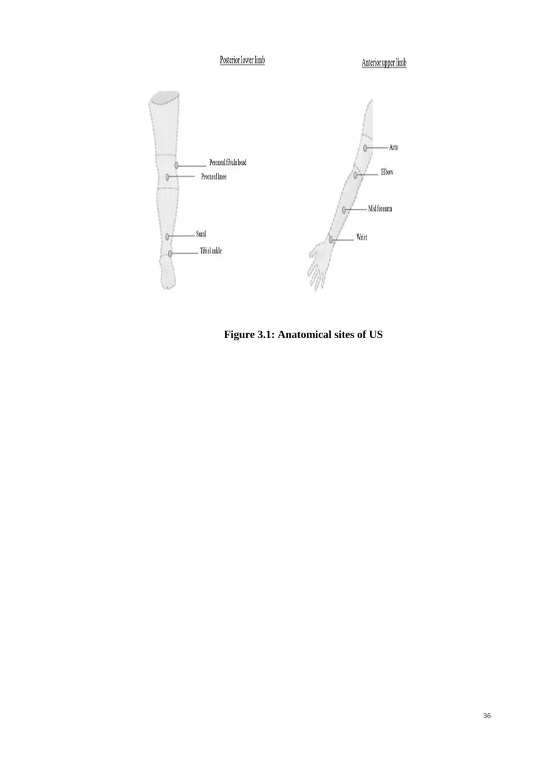
36
Figure 3.1: Anatomical sites of US
37
3.6 Statistical analyses
Statistical analyses were performed using SPSS version 22. Normality of data
was tested using Kolmogrov-Smirnov normality test. Demographic data and CSAs were
compared between diabetic patients and control groups using independent t-test for
parametric and Mann Whitney test for non-parametric variables respectively and
similarly for comparison between true CIDP patients and D-DSP patients. Comparative
studies between groups were done with ANOVA for parametric variables or Kruskal-
Wallis for non-parametric variables. Receiver operating characteristic (ROC) analysis
was done to determine the nerve CSA cut-off values that best predict the presence of
DSP as well as differentiate between severe and non-severe DSP, based on TCSS
values. Correlation studies were done with Spearman’s rank correlation for non-
parametric variables and Pearson’s for parametric variables. Statistical significance was
established at p<0.05.
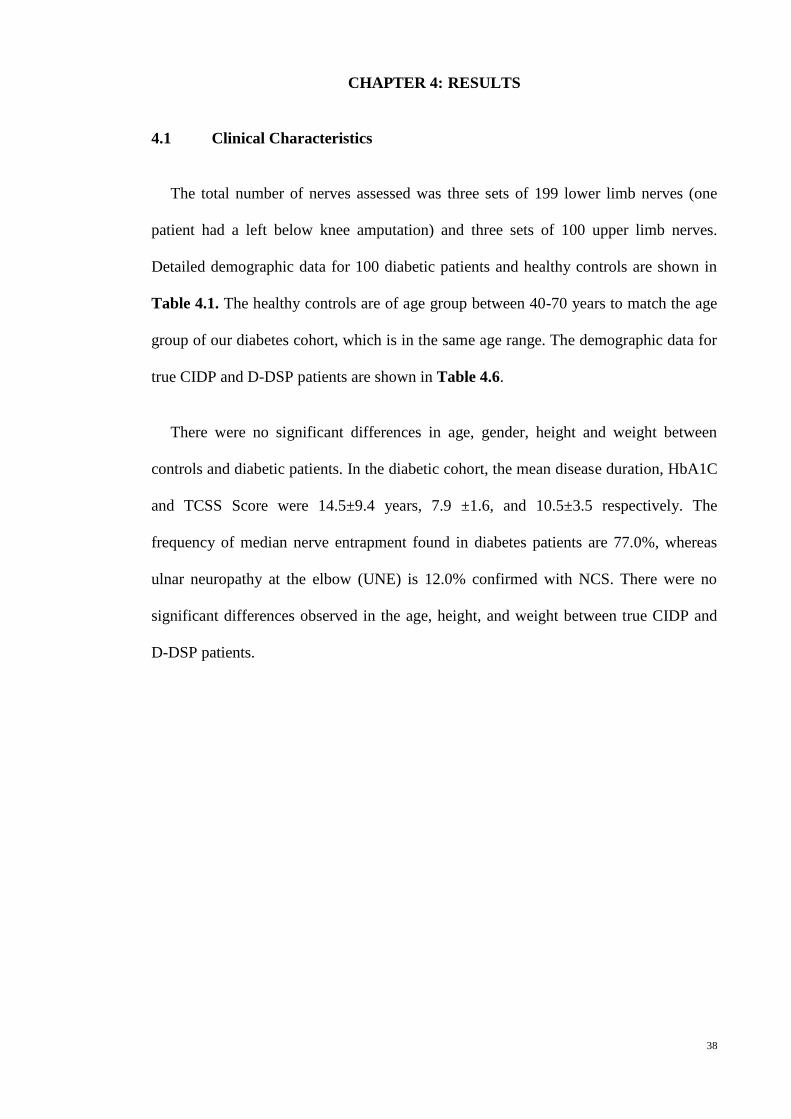
38
CHAPTER 4: RESULTS
4.1 Clinical Characteristics
The total number of nerves assessed was three sets of 199 lower limb nerves (one
patient had a left below knee amputation) and three sets of 100 upper limb nerves.
Detailed demographic data for 100 diabetic patients and healthy controls are shown in
Table 4.1. The healthy controls are of age group between 40-70 years to match the age
group of our diabetes cohort, which is in the same age range. The demographic data for
true CIDP and D-DSP patients are shown in Table 4.6.
There were no significant differences in age, gender, height and weight between
controls and diabetic patients. In the diabetic cohort, the mean disease duration, HbA1C
and TCSS Score were 14.5±9.4 years, 7.9 ±1.6, and 10.5±3.5 respectively. The
frequency of median nerve entrapment found in diabetes patients are 77.0%, whereas
ulnar neuropathy at the elbow (UNE) is 12.0% confirmed with NCS. There were no
significant differences observed in the age, height, and weight between true CIDP and
D-DSP patients.
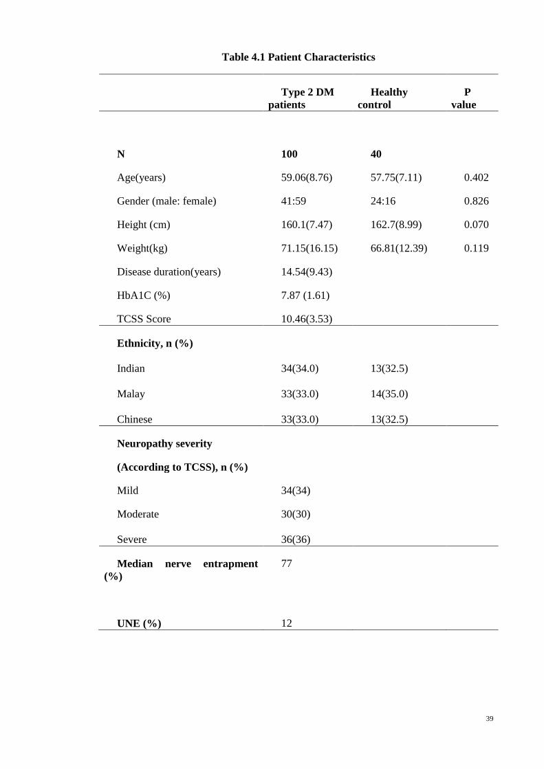
39
Table 4.1 Patient Characteristics
Type 2 DM
patients
Healthy
control
P
value
N
100
40
Age(years) 59.06(8.76) 57.75(7.11) 0.402
Gender (male: female) 41:59 24:16 0.826
Height (cm) 160.1(7.47) 162.7(8.99) 0.070
Weight(kg) 71.15(16.15) 66.81(12.39) 0.119
Disease duration(years) 14.54(9.43)
HbA1C (%) 7.87 (1.61)
TCSS Score 10.46(3.53)
Ethnicity, n (%)
Indian
Malay
Chinese
34(34.0)
33(33.0)
33(33.0)
13(32.5)
14(35.0)
13(32.5)
Neuropathy severity
(According to TCSS), n (%)
Mild 34(34)
Moderate
Severe
30(30)
36(36)
Median nerve entrapment
(%)
77
UNE (%) 12
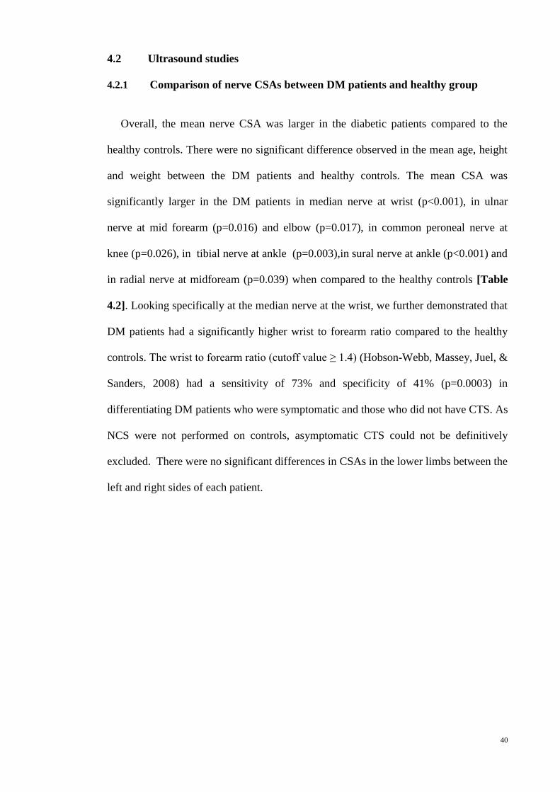
40
4.2 Ultrasound studies
4.2.1 Comparison of nerve CSAs between DM patients and healthy group
Overall, the mean nerve CSA was larger in the diabetic patients compared to the
healthy controls. There were no significant difference observed in the mean age, height
and weight between the DM patients and healthy controls. The mean CSA was
significantly larger in the DM patients in median nerve at wrist (p<0.001), in ulnar
nerve at mid forearm (p=0.016) and elbow (p=0.017), in common peroneal nerve at
knee (p=0.026), in tibial nerve at ankle (p=0.003),in sural nerve at ankle (p<0.001) and
in radial nerve at midfoream (p=0.039) when compared to the healthy controls [Table
4.2]. Looking specifically at the median nerve at the wrist, we further demonstrated that
DM patients had a significantly higher wrist to forearm ratio compared to the healthy
controls. The wrist to forearm ratio (cutoff value ≥ 1.4) (Hobson-Webb, Massey, Juel, &
Sanders, 2008) had a sensitivity of 73% and specificity of 41% (p=0.0003) in
differentiating DM patients who were symptomatic and those who did not have CTS. As
NCS were not performed on controls, asymptomatic CTS could not be definitively
excluded. There were no significant differences in CSAs in the lower limbs between the
left and right sides of each patient.
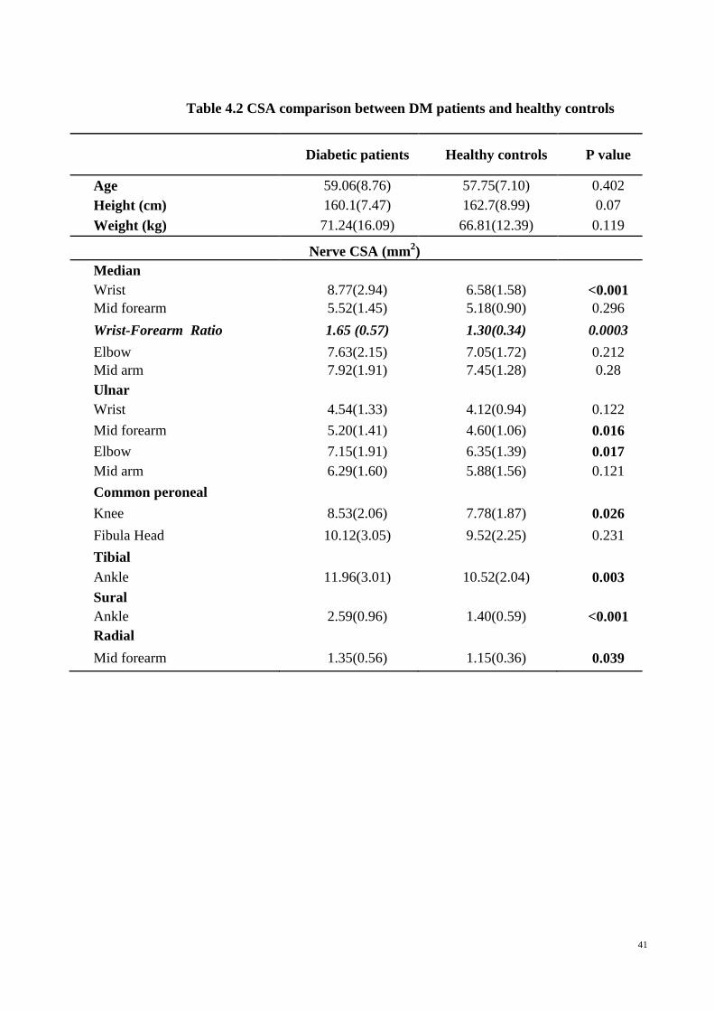
41
Table 4.2 CSA comparison between DM patients and healthy controls
Diabetic patients Healthy controls P value
Age 59.06(8.76) 57.75(7.10) 0.402
Height (cm) 160.1(7.47) 162.7(8.99) 0.07
Weight (kg) 71.24(16.09) 66.81(12.39) 0.119
Nerve CSA (mm2)
Median
Wrist 8.77(2.94) 6.58(1.58) <0.001
Mid forearm 5.52(1.45) 5.18(0.90) 0.296
Wrist-Forearm Ratio 1.65 (0.57) 1.30(0.34) 0.0003
Elbow 7.63(2.15) 7.05(1.72) 0.212
Mid arm 7.92(1.91) 7.45(1.28) 0.28
Ulnar
Wrist 4.54(1.33) 4.12(0.94) 0.122
Mid forearm 5.20(1.41) 4.60(1.06) 0.016
Elbow 7.15(1.91) 6.35(1.39) 0.017
Mid arm 6.29(1.60) 5.88(1.56) 0.121
Common peroneal
Knee 8.53(2.06) 7.78(1.87) 0.026
Fibula Head 10.12(3.05) 9.52(2.25) 0.231
Tibial
Ankle 11.96(3.01) 10.52(2.04) 0.003
Sural
Ankle 2.59(0.96) 1.40(0.59) <0.001
Radial
Mid forearm 1.35(0.56) 1.15(0.36) 0.039
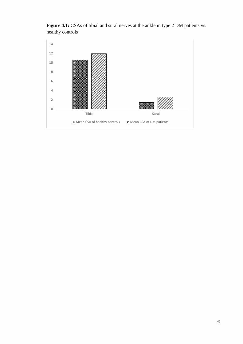
42
Figure 4.1: CSAs of tibial and sural nerves at the ankle in type 2 DM patients vs.
healthy controls
0
2
4
6
8
10
12
14
Tibial Sural
Mean CSA of healthy controls Mean CSA of DM patients
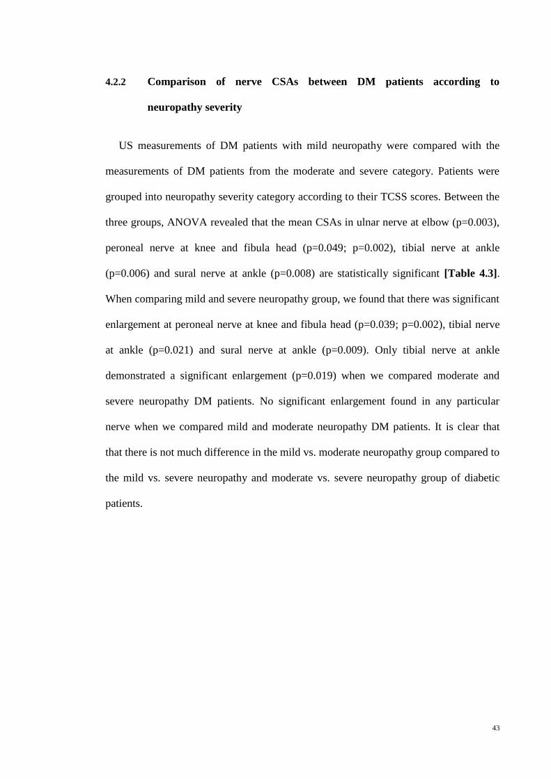
43
4.2.2 Comparison of nerve CSAs between DM patients according to
neuropathy severity
US measurements of DM patients with mild neuropathy were compared with the
measurements of DM patients from the moderate and severe category. Patients were
grouped into neuropathy severity category according to their TCSS scores. Between the
three groups, ANOVA revealed that the mean CSAs in ulnar nerve at elbow (p=0.003),
peroneal nerve at knee and fibula head (p=0.049; p=0.002), tibial nerve at ankle
(p=0.006) and sural nerve at ankle (p=0.008) are statistically significant [Table 4.3].
When comparing mild and severe neuropathy group, we found that there was significant
enlargement at peroneal nerve at knee and fibula head (p=0.039; p=0.002), tibial nerve
at ankle (p=0.021) and sural nerve at ankle (p=0.009). Only tibial nerve at ankle
demonstrated a significant enlargement (p=0.019) when we compared moderate and
severe neuropathy DM patients. No significant enlargement found in any particular
nerve when we compared mild and moderate neuropathy DM patients. It is clear that
that there is not much difference in the mild vs. moderate neuropathy group compared to
the mild vs. severe neuropathy and moderate vs. severe neuropathy group of diabetic
patients.
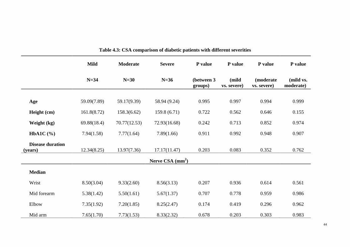
44
Table 4.3: CSA comparison of diabetic patients with different severities
Mild Moderate Severe P value P value P value P value
N=34 N=30 N=36 (between 3
groups)
(mild
vs. severe)
(moderate
vs. severe)
(mild vs.
moderate)
Age 59.09(7.89) 59.17(9.39) 58.94 (9.24) 0.995 0.997 0.994 0.999
Height (cm) 161.8(8.72) 158.3(6.62) 159.8 (6.71) 0.722 0.562 0.646 0.155
Weight (kg) 69.88(18.4) 70.77(12.53) 72.93(16.68) 0.242 0.713 0.852 0.974
HbA1C (%) 7.94(1.58) 7.77(1.64) 7.89(1.66) 0.911 0.992 0.948 0.907
Disease duration
(years) 12.34(8.25) 13.97(7.36) 17.17(11.47) 0.203 0.083 0.352 0.762
Nerve CSA (mm2)
Median
Wrist 8.50(3.04) 9.33(2.60) 8.56(3.13) 0.207 0.936 0.614 0.561
Mid forearm 5.38(1.42) 5.50(1.61) 5.67(1.37) 0.707 0.778 0.959 0.986
Elbow 7.35(1.92) 7.20(1.85) 8.25(2.47) 0.174 0.419 0.296 0.962
Mid arm 7.65(1.70) 7.73(1.53) 8.33(2.32) 0.678 0.203 0.303 0.983
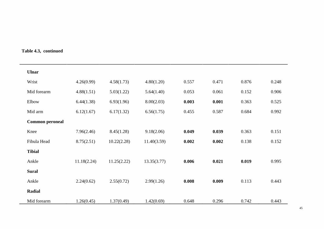
45
Table 4.3, continued
Ulnar
Wrist 4.26(0.99) 4.58(1.73) 4.80(1.20) 0.557 0.471 0.876 0.248
Mid forearm 4.88(1.51) 5.03(1.22) 5.64(1.40) 0.053 0.061 0.152 0.906
Elbow 6.44(1.38) 6.93(1.96) 8.00(2.03) 0.003 0.001 0.363 0.525
Mid arm 6.12(1.67) 6.17(1.32) 6.56(1.75) 0.455 0.587 0.684 0.992
Common peroneal
Knee 7.96(2.46) 8.45(1.28) 9.18(2.06) 0.049 0.039 0.363 0.151
Fibula Head 8.75(2.51) 10.22(2.28) 11.40(3.59) 0.002 0.002 0.138 0.152
Tibial
Ankle 11.18(2.24) 11.25(2.22) 13.35(3.77) 0.006 0.021 0.019 0.995
Sural
Ankle 2.24(0.62) 2.55(0.72) 2.99(1.26) 0.008 0.009 0.113 0.443
Radial
Mid forearm 1.26(0.45) 1.37(0.49) 1.42(0.69) 0.648 0.296 0.742 0.443
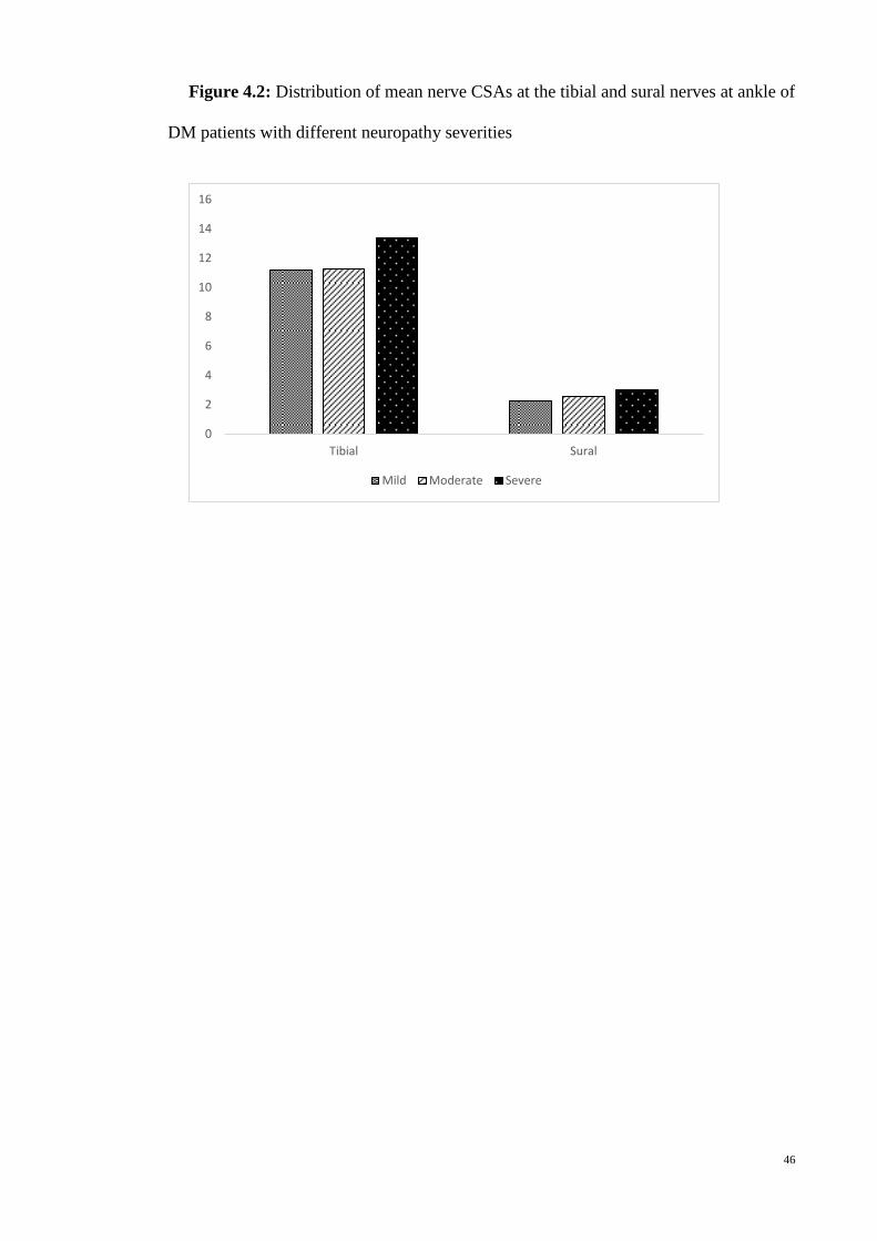
46
Figure 4.2: Distribution of mean nerve CSAs at the tibial and sural nerves at ankle of
DM patients with different neuropathy severities
0
2
4
6
8
10
12
14
16
Tibial Sural
Mild Moderate Severe
47
4.2.3 ROC analysis of the nerve CSAs
We analyzed the area under the curve (AUC) that best discriminate nerve CSAs of
DM patients with severe DSP (TCSS ≥ 12) from non-severe DSP (TCSS<12) which is
shown in Table 4.4. The majority of cut-off CSA values were extremely poor
discriminators (AUC < 60%). The tibial CSA at 11.75 mm2 and sural nerve CSA at 2.75
mm2 nerves had an AUC value of 69% and 62% respectively. Based on these values, the
tibial nerve could discriminate between DSP severity at 65.1% sensitivity and 65.0
specificity whereas sural nerve at 59.0% sensitivity and 73.0% specificity. The AUC in
the ROC analysis [Table 4.5] was determined to differentiate between patients with
DSP (TCSS≥6) and without DSP (TCSS≤5). We found that the sural nerves performed
the best and a cut-off value of 2 mm2 had an AUC of 88%, sensitivity of 90% and
specificity of 74%.
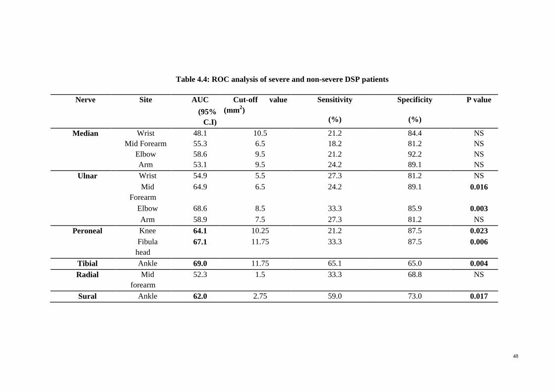
48
Table 4.4: ROC analysis of severe and non-severe DSP patients
Nerve Site AUC Cut-off value
(mm2)
Sensitivity
(%)
Specificity
(%)
P value
(95%
C.I)
Median Wrist 48.1 10.5 21.2 84.4 NS
Mid Forearm 55.3 6.5 18.2 81.2 NS
Elbow 58.6 9.5 21.2 92.2 NS
Arm 53.1 9.5 24.2 89.1 NS
Ulnar Wrist 54.9 5.5 27.3 81.2 NS
Mid
Forearm
64.9 6.5 24.2 89.1 0.016
Elbow 68.6 8.5 33.3 85.9 0.003
Arm 58.9 7.5 27.3 81.2 NS
Peroneal Knee 64.1 10.25 21.2 87.5 0.023
Fibula
head
67.1 11.75 33.3 87.5 0.006
Tibial Ankle 69.0 11.75 65.1 65.0 0.004
Radial Mid
forearm
52.3 1.5 33.3 68.8 NS
Sural Ankle 62.0 2.75 59.0 73.0 0.017
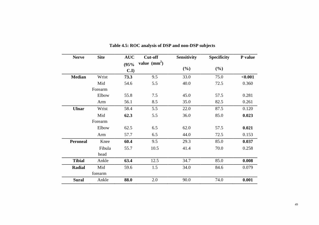
49
Table 4.5: ROC analysis of DSP and non-DSP subjects
Nerve Site AUC Cut-off
value (mm2)
Sensitivity
(%)
Specificity
(%)
P value
(95%
C.I)
Median Wrist 73.3 9.5 33.0 75.0 <0.001
Mid
Forearm
54.6 5.5 40.0 72.5 0.360
Elbow 55.8 7.5 45.0 57.5 0.281
Arm 56.1 8.5 35.0 82.5 0.261
Ulnar Wrist 58.4 5.5 22.0 87.5 0.120
Mid
Forearm
62.3 5.5 36.0 85.0 0.023
Elbow 62.5 6.5 62.0 57.5 0.021
Arm 57.7 6.5 44.0 72.5 0.153
Peroneal Knee 60.4 9.5 29.3 85.0 0.037
Fibula
head
55.7 10.5 41.4 70.0 0.258
Tibial Ankle 63.4 12.5 34.7 85.0 0.008
Radial Mid
forearm
59.6 1.5 34.0 84.6 0.079
Sural Ankle 88.0 2.0 90.0 74.0 0.001
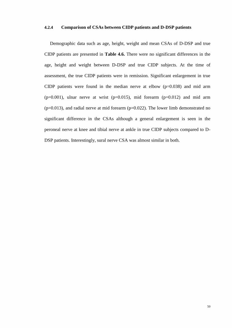
50
4.2.4 Comparison of CSAs between CIDP patients and D-DSP patients
Demographic data such as age, height, weight and mean CSAs of D-DSP and true
CIDP patients are presented in Table 4.6. There were no significant differences in the
age, height and weight between D-DSP and true CIDP subjects. At the time of
assessment, the true CIDP patients were in remission. Significant enlargement in true
CIDP patients were found in the median nerve at elbow (p=0.038) and mid arm
(p=0.001), ulnar nerve at wrist (p=0.015), mid forearm (p=0.012) and mid arm
(p=0.013), and radial nerve at mid forearm (p=0.022). The lower limb demonstrated no
significant difference in the CSAs although a general enlargement is seen in the
peroneal nerve at knee and tibial nerve at ankle in true CIDP subjects compared to D-
DSP patients. Interestingly, sural nerve CSA was almost similar in both.
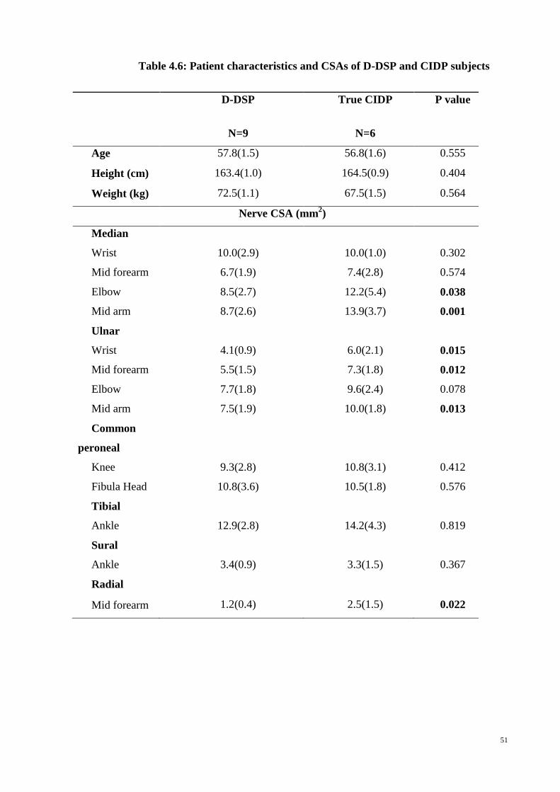
51
Table 4.6: Patient characteristics and CSAs of D-DSP and CIDP subjects
D-DSP True CIDP P value
N=9 N=6
Age 57.8(1.5) 56.8(1.6) 0.555
Height (cm) 163.4(1.0) 164.5(0.9) 0.404
Weight (kg) 72.5(1.1) 67.5(1.5) 0.564
Nerve CSA (mm2)
Median
Wrist 10.0(2.9) 10.0(1.0) 0.302
Mid forearm 6.7(1.9) 7.4(2.8) 0.574
Elbow 8.5(2.7) 12.2(5.4) 0.038
Mid arm 8.7(2.6) 13.9(3.7) 0.001
Ulnar
Wrist 4.1(0.9) 6.0(2.1) 0.015
Mid forearm 5.5(1.5) 7.3(1.8) 0.012
Elbow 7.7(1.8) 9.6(2.4) 0.078
Mid arm 7.5(1.9) 10.0(1.8) 0.013
Common
peroneal
Knee 9.3(2.8) 10.8(3.1) 0.412
Fibula Head 10.8(3.6) 10.5(1.8) 0.576
Tibial
Ankle 12.9(2.8) 14.2(4.3) 0.819
Sural
Ankle 3.4(0.9) 3.3(1.5) 0.367
Radial
Mid forearm 1.2(0.4) 2.5(1.5) 0.022
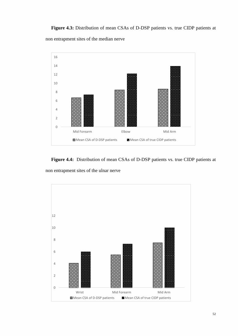
52
Figure 4.3: Distribution of mean CSAs of D-DSP patients vs. true CIDP patients at
non entrapment sites of the median nerve
Figure 4.4: Distribution of mean CSAs of D-DSP patients vs. true CIDP patients at
non entrapment sites of the ulnar nerve
0
2
4
6
8
10
12
14
16
Mid Forearm Elbow Mid Arm
Mean CSA of D-DSP patients Mean CSA of true CIDP patients
0
2
4
6
8
10
12
Wrist Mid Forearm Mid Arm
Mean CSA of D-DSP patients Mean CSA of true CIDP patients
53
4.2.5 Sonogram images of DM patients with different neuropathy severities vs.
healthy controls
Figure 4.5 shows transverse sonography of the tibial and Figure 4.6 shows
transverse sonography of sural nerves of healthy controls and DM patients of different
neuropathy severities. Transverse sonography of the tibial and sural nerves showed a
hypoechoic structure with hyperechoic dots within it. We observed a general nerve
enlargement in the DM patients when compared with healthy controls. CSA is also seen
to increase with progressing severity of neuropathy in DM patients.
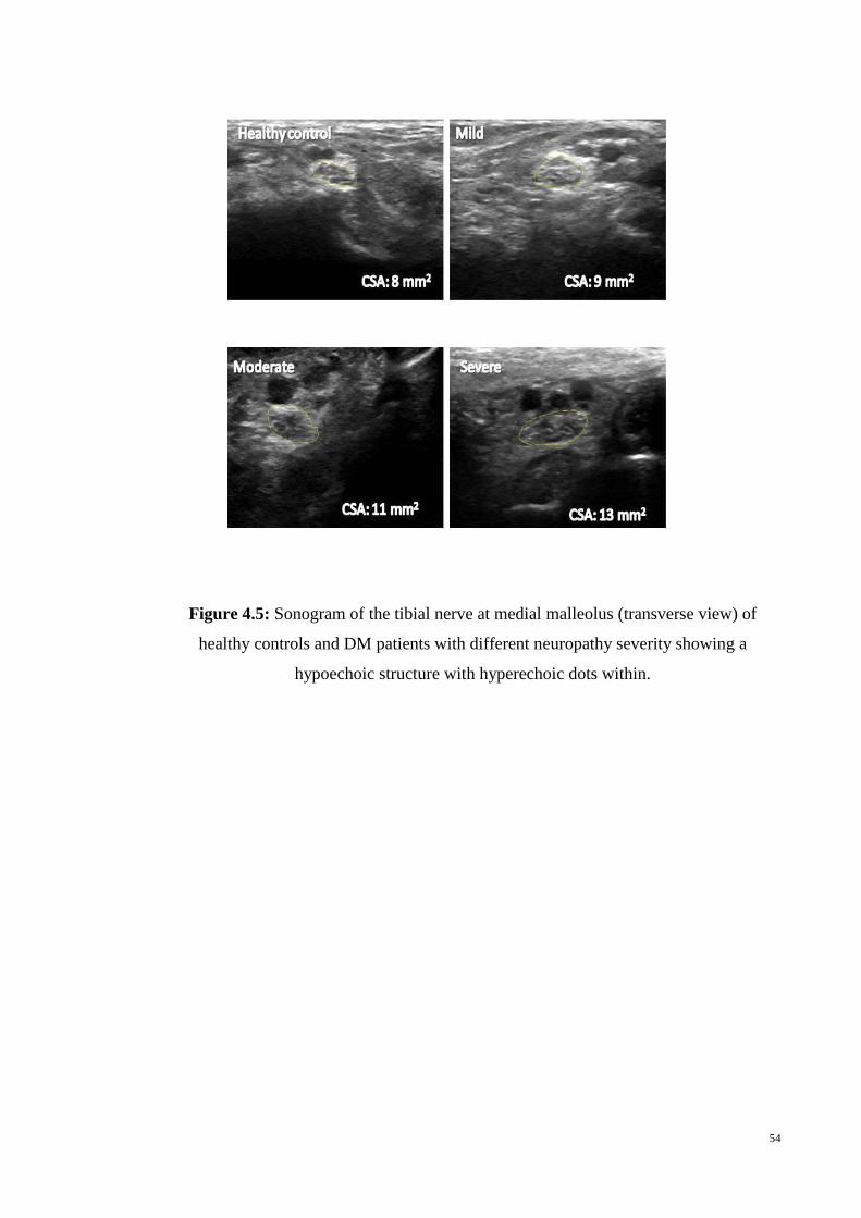
54
Figure 4.5: Sonogram of the tibial nerve at medial malleolus (transverse view) of
healthy controls and DM patients with different neuropathy severity showing a
hypoechoic structure with hyperechoic dots within.
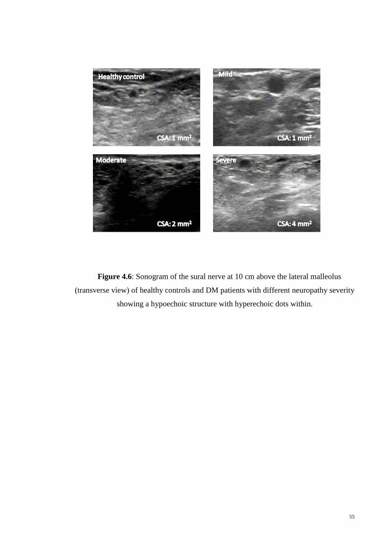
55
Figure 4.6: Sonogram of the sural nerve at 10 cm above the lateral malleolus
(transverse view) of healthy controls and DM patients with different neuropathy severity
showing a hypoechoic structure with hyperechoic dots within.
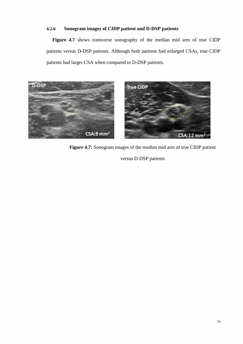
56
4.2.6 Sonogram images of CIDP patient and D-DSP patients
Figure 4.7 shows transverse sonography of the median mid arm of true CIDP
patients versus D-DSP patients. Although both patients had enlarged CSAs, true CIDP
patients had larger CSA when compared to D-DSP patients.
Figure 4.7: Sonogram images of the median mid arm of true CIDP patient
versus D-DSP patients
57
4.3 Neurophysiological studies
4.3.1 Electrophysiological findings according to severity classification (based on
TCSS) of DM patients
Looking specifically at the lower limb nerve conduction parameters, these were
unrecordable in 43.2% sural, 13.1% peroneal and 8.0% tibial nerve potentials. There
were significant differences (p<0.001) between absence of sural sensory potentials in
the mild (19.1%), moderate (40.0%) and severe (69.0%) groups and peroneal nerve
motor potentials in the mild (2.9%), moderate (10.0%) and severe (25.4%), in keeping
with worsening DSP. The mean values and comparison between the DML, distal CMAP
(dCMAP), mCV, SNAP and sensory conduction velocity (sCV) for the three groups of
diabetic patients according to their DSP severity are presented in Table 4.7. The DSP
patients in the severe neuropathy category (according to TCSS scores) significantly
demonstrated prolonged distal latency in the ulnar nerve at wrist (p<0.001) and in tibial
nerve at ankle (p<0.001).
The dCMAP in the peroneal nerve at knee (p=0.005) and in tibial nerve at knee
(p=0.001) is significantly reduced when the DM patient neuropathy severity increases.
The mCV is significantly slowed across the median nerve at elbow (p<0.001), ulnar
nerve at above and below elbow (p=0.001,p<0.001), peroneal nerve at fibula head
(p<0.001) and tibial nerve at knee (p<0.001) with worsening DSP severity. The SNAP
amplitude was markedly reduced when the neuropathy severity increases in the ulnar
nerve at wrist (p=0.007), radial nerve at forearm (p=0.005) and sural nerve at calf
(p=0.005).We observed a significant slowing of sCV at the radial forearm (p=0.004) in
keeping with worsening of DSP. Patients in the most severe category demonstrated
significant changes in their neurophysiological parameters.
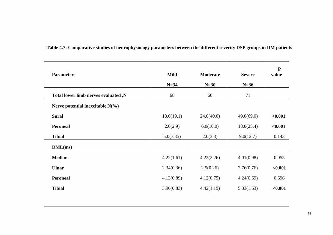
58
Table 4.7: Comparative studies of neurophysiology parameters between the different severity DSP groups in DM patients
Parameters Mild Moderate Severe
P
value
N=34 N=30 N=36
Total lower limb nerves evaluated ,N 68 60 71
Nerve potential inexcitable,N(%)
Sural 13.0(19.1) 24.0(40.0) 49.0(69.0) <0.001
Peroneal 2.0(2.9) 6.0(10.0) 18.0(25.4) <0.001
Tibial 5.0(7.35) 2.0(3.3) 9.0(12.7) 0.143
DML(ms)
Median 4.22(1.61) 4.22(2.26) 4.01(0.98) 0.055
Ulnar 2.34(0.36) 2.5(0.26) 2.76(0.76) <0.001
Peroneal 4.13(0.89) 4.12(0.75) 4.24(0.69) 0.696
Tibial 3.96(0.83) 4.42(1.19) 5.33(1.63) <0.001
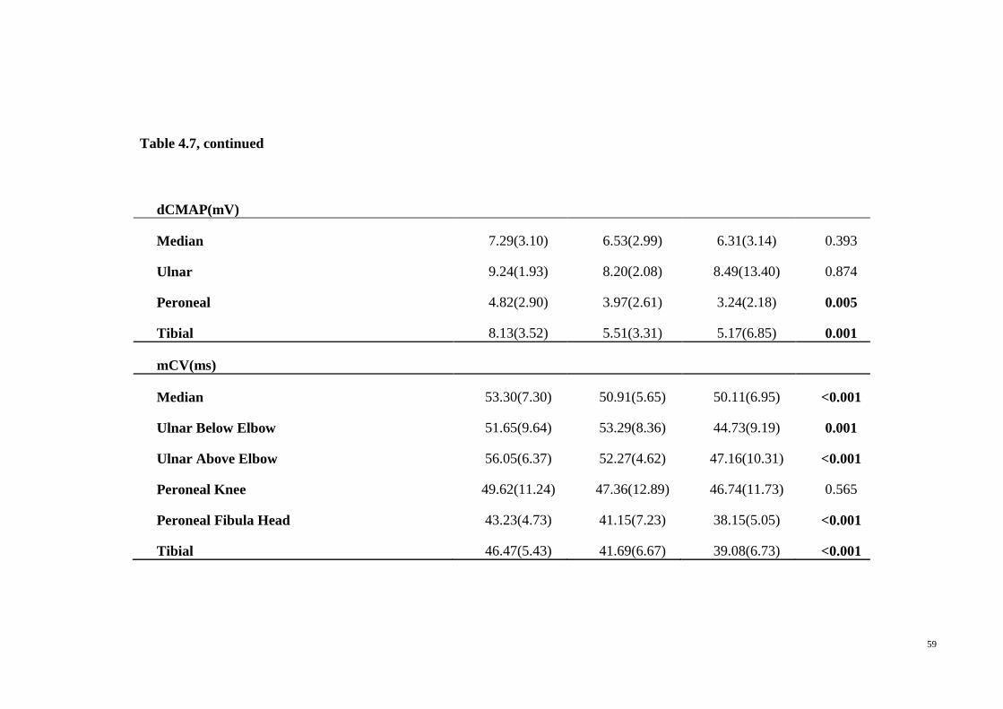
59
Table 4.7, continued
dCMAP(mV)
Median 7.29(3.10) 6.53(2.99) 6.31(3.14) 0.393
Ulnar 9.24(1.93) 8.20(2.08) 8.49(13.40) 0.874
Peroneal 4.82(2.90) 3.97(2.61) 3.24(2.18) 0.005
Tibial 8.13(3.52) 5.51(3.31) 5.17(6.85) 0.001
mCV(ms)
Median 53.30(7.30) 50.91(5.65) 50.11(6.95) <0.001
Ulnar Below Elbow 51.65(9.64) 53.29(8.36) 44.73(9.19) 0.001
Ulnar Above Elbow 56.05(6.37) 52.27(4.62) 47.16(10.31) <0.001
Peroneal Knee 49.62(11.24) 47.36(12.89) 46.74(11.73) 0.565
Peroneal Fibula Head 43.23(4.73) 41.15(7.23) 38.15(5.05) <0.001
Tibial 46.47(5.43) 41.69(6.67) 39.08(6.73) <0.001
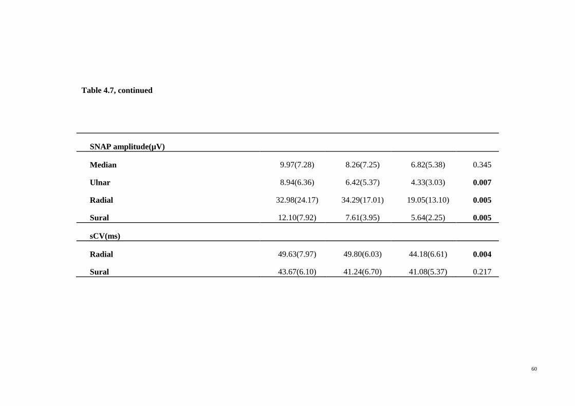
60
Table 4.7, continued
SNAP amplitude(µV)
Median 9.97(7.28) 8.26(7.25) 6.82(5.38) 0.345
Ulnar 8.94(6.36) 6.42(5.37) 4.33(3.03) 0.007
Radial 32.98(24.17) 34.29(17.01) 19.05(13.10) 0.005
Sural 12.10(7.92) 7.61(3.95) 5.64(2.25) 0.005
sCV(ms)
Radial 49.63(7.97) 49.80(6.03) 44.18(6.61) 0.004
Sural 43.67(6.10) 41.24(6.70) 41.08(5.37) 0.217
61
4.3.2 Comparison of electrophysiological findings between CIDP patients and
D-DSP patients
NCS revealed significantly prolonged latency in the peroneal nerve at ankle in true
CIDP patients (p=0.009). A significantly reduced CMAP amplitude was observed in
true CIDP patients (p=0.011) in the ulnar nerve above elbow [Table 4.8]. The CIDP
cohort also exhibited bigger distal to proximal ratios in CMAP amplitudes in both upper
and lower extremities. Looking at the duration, we observed a prolonged duration in the
median nerve at wrist (p=0.011) and elbow (p=0.001) and in the ulnar nerve below
elbow (p=0.035) and in tibial nerve at knee (p=0.004) in true CIDP patients. Significant
reduction in the sCV were observed in the ulnar (p=0.004) and radial nerve (p=0.020).
Generally, the distal latencies and duration were prolonged; CMAP amplitude, mCV,
SNAP amplitude, and sCV were reduced in true CIDP patients compared to D-DSP
patients. No statistically significant differences observed in the distal to proximal
CMAP ratio between true CIDP and D-DSP subjects.
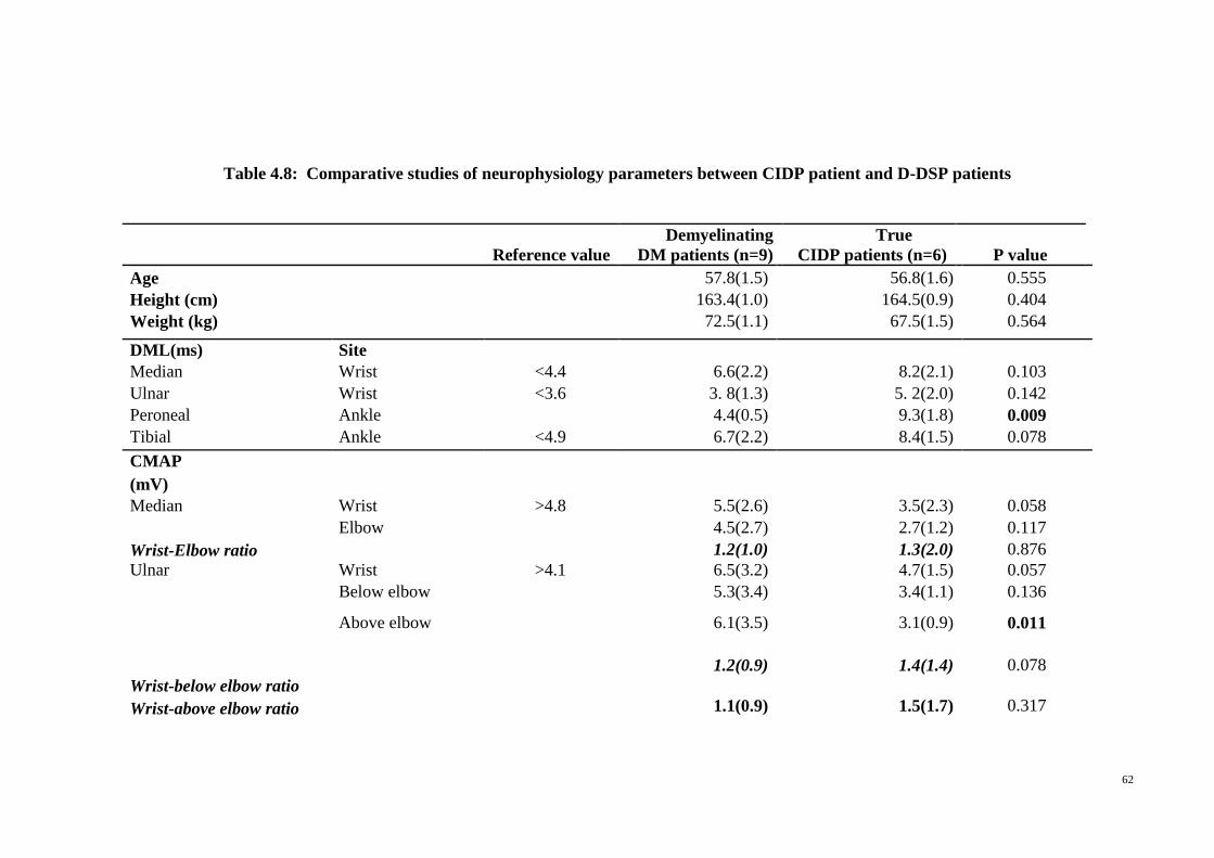
62
Table 4.8: Comparative studies of neurophysiology parameters between CIDP patient and D-DSP patients
Reference value
Demyelinating
DM patients (n=9)
True
CIDP patients (n=6) P value
Age 57.8(1.5) 56.8(1.6) 0.555
Height (cm) 163.4(1.0) 164.5(0.9) 0.404
Weight (kg) 72.5(1.1) 67.5(1.5) 0.564
DML(ms) Site
Median Wrist <4.4 6.6(2.2) 8.2(2.1) 0.103
Ulnar Wrist <3.6 3. 8(1.3) 5. 2(2.0) 0.142
Peroneal Ankle 4.4(0.5) 9.3(1.8) 0.009
Tibial Ankle <4.9 6.7(2.2) 8.4(1.5) 0.078
CMAP
(mV)
Median Wrist >4.8 5.5(2.6) 3.5(2.3) 0.058
Elbow 4.5(2.7) 2.7(1.2) 0.117
Wrist-Elbow ratio
1.2(1.0) 1.3(2.0) 0.876
Ulnar Wrist >4.1 6.5(3.2) 4.7(1.5) 0.057
Below elbow 5.3(3.4) 3.4(1.1) 0.136
Above elbow 6.1(3.5) 3.1(0.9) 0.011
Wrist-below elbow ratio
1.2(0.9) 1.4(1.4) 0.078
Wrist-above elbow ratio 1.1(0.9) 1.5(1.7) 0.317
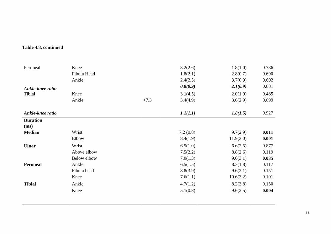
63
Table 4.8, continued
Peroneal Knee 3.2(2.6) 1.8(1.0) 0.786
Fibula Head 1.8(2.1) 2.8(0.7) 0.690
Ankle 2.4(2.5) 3.7(0.9) 0.602
Ankle-knee ratio 0.8(0.9) 2.1(0.9) 0.881
Tibial Knee 3.1(4.5) 2.0(1.9) 0.485
Ankle >7.3 3.4(4.9) 3.6(2.9) 0.699
Ankle-knee ratio 1.1(1.1) 1.8(1.5) 0.927
Duration
(ms)
Median Wrist 7.2 (0.8) 9.7(2.9) 0.011
Elbow 8.4(1.9) 11.9(2.0) 0.001
Ulnar Wrist 6.5(1.0) 6.6(2.5) 0.877
Above elbow 7.5(2.2) 8.8(2.6) 0.119
Below elbow 7.0(1.3) 9.6(3.1) 0.035
Peroneal Ankle 6.5(1.5) 8.3(1.8) 0.117
Fibula head 8.8(3.9) 9.6(2.1) 0.151
Knee 7.6(1.1) 10.6(3.2) 0.101
Tibial Ankle 4.7(1.2) 8.2(3.8) 0.150
Knee 5.1(0.8) 9.6(2.5) 0.004
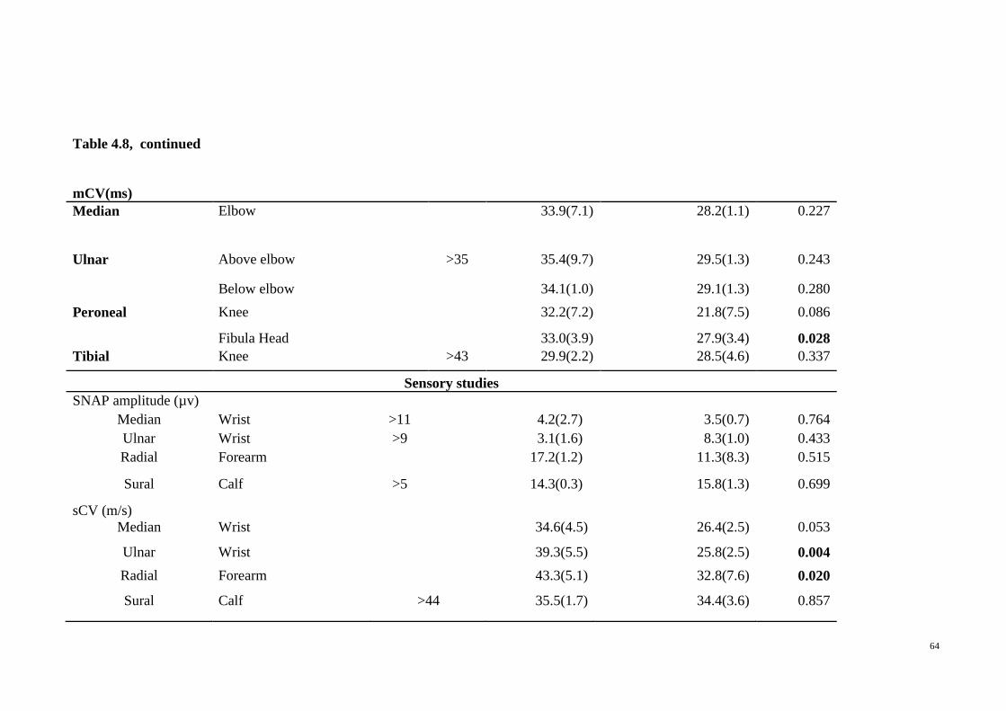
64
Table 4.8, continued
mCV(ms)
Median Elbow 33.9(7.1) 28.2(1.1) 0.227
Ulnar Above elbow >35 35.4(9.7) 29.5(1.3) 0.243
Below elbow 34.1(1.0) 29.1(1.3) 0.280
Peroneal Knee 32.2(7.2) 21.8(7.5) 0.086
Fibula Head 33.0(3.9) 27.9(3.4) 0.028
Tibial Knee >43 29.9(2.2) 28.5(4.6) 0.337
Sensory studies
SNAP amplitude (µv)
Median Wrist >11 4.2(2.7) 3.5(0.7) 0.764
Ulnar Wrist >9 3.1(1.6) 8.3(1.0) 0.433
Radial Forearm 17.2(1.2) 11.3(8.3) 0.515
Sural Calf >5 14.3(0.3) 15.8(1.3) 0.699
sCV (m/s)
Median Wrist 34.6(4.5) 26.4(2.5) 0.053
Ulnar Wrist 39.3(5.5) 25.8(2.5) 0.004
Radial Forearm 43.3(5.1) 32.8(7.6) 0.020
Sural Calf >44 35.5(1.7) 34.4(3.6) 0.857
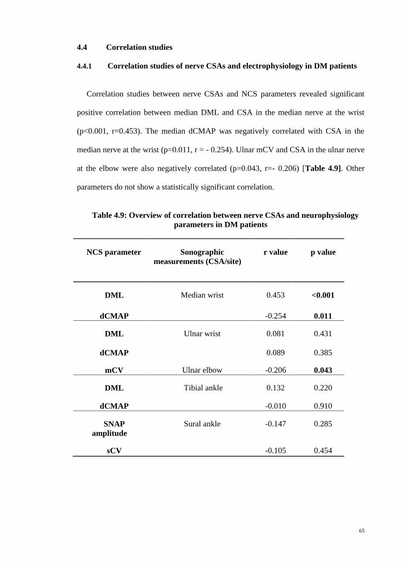
65
4.4 Correlation studies
4.4.1 Correlation studies of nerve CSAs and electrophysiology in DM patients
Correlation studies between nerve CSAs and NCS parameters revealed significant
positive correlation between median DML and CSA in the median nerve at the wrist
(p<0.001, r=0.453). The median dCMAP was negatively correlated with CSA in the
median nerve at the wrist (p=0.011, r = - 0.254). Ulnar mCV and CSA in the ulnar nerve
at the elbow were also negatively correlated (p=0.043, r=- 0.206) [Table 4.9]. Other
parameters do not show a statistically significant correlation.
Table 4.9: Overview of correlation between nerve CSAs and neurophysiology
parameters in DM patients
NCS parameter Sonographic
measurements (CSA/site)
r value p value
DML Median wrist 0.453 <0.001
dCMAP -0.254 0.011
DML Ulnar wrist 0.081 0.431
dCMAP 0.089 0.385
mCV Ulnar elbow -0.206 0.043
DML Tibial ankle 0.132 0.220
dCMAP -0.010 0.910
SNAP
amplitude
Sural ankle -0.147 0.285
sCV -0.105 0.454

66
4.4.2 Correlation studies of nerve CSAs at different sites versus different
disease markers
Table 4.10 shows correlation studies of nerve CSAs with different disease markers.
TCSS scores show a significant, linear correlation with nerve CSA in the ulnar nerve at
mid forearm (p=0.010, r=0.310) and at elbow (p=0.006, r=0.300), in the common
peroneal nerve at fibula head (p=0.002, r=0.301), in tibial nerve at ankle (p=0.005,
r=0.200) and in sural nerve at ankle (p=0.0008, r=0.335). HbA1C shows a significant,
linear relationship with nerve CSA in the median nerve at wrist (p=0.001, r=0.313)
only. DM disease duration correlated well with a direct, significant association between
nerve CSAs in the median nerve at wrist (p=0.041, r=0.026) and in the ulnar nerve at
elbow (p=0.001, r=0.321) and arm (p=0.021, r=0.231).
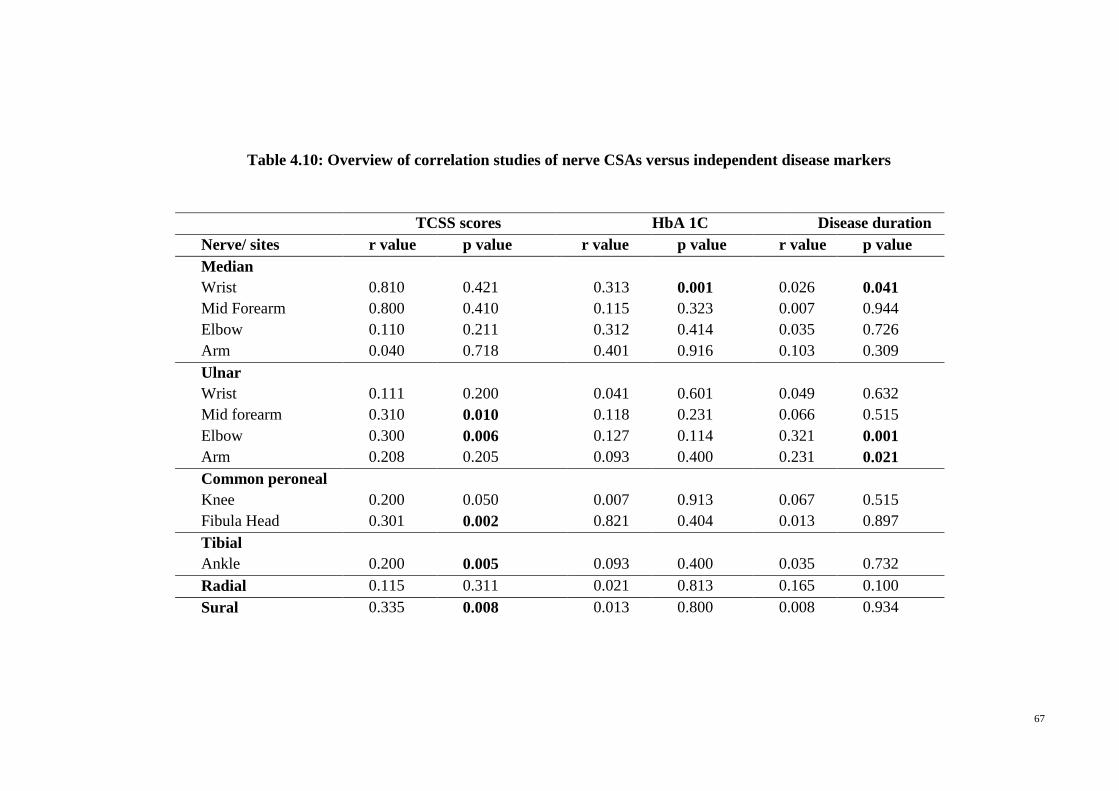
67
Table 4.10: Overview of correlation studies of nerve CSAs versus independent disease markers
TCSS scores HbA 1C Disease duration
Nerve/ sites r value p value r value p value r value p value
Median
Wrist 0.810 0.421 0.313 0.001 0.026 0.041
Mid Forearm 0.800 0.410 0.115 0.323 0.007 0.944
Elbow 0.110 0.211 0.312 0.414 0.035 0.726
Arm 0.040 0.718 0.401 0.916 0.103 0.309
Ulnar
Wrist 0.111 0.200 0.041 0.601 0.049 0.632
Mid forearm 0.310 0.010 0.118 0.231 0.066 0.515
Elbow 0.300 0.006 0.127 0.114 0.321 0.001
Arm 0.208 0.205 0.093 0.400 0.231 0.021
Common peroneal
Knee 0.200 0.050 0.007 0.913 0.067 0.515
Fibula Head 0.301 0.002 0.821 0.404 0.013 0.897
Tibial
Ankle 0.200 0.005 0.093 0.400 0.035 0.732
Radial 0.115 0.311 0.021 0.813 0.165 0.100
Sural 0.335 0.008 0.013 0.800 0.008 0.934
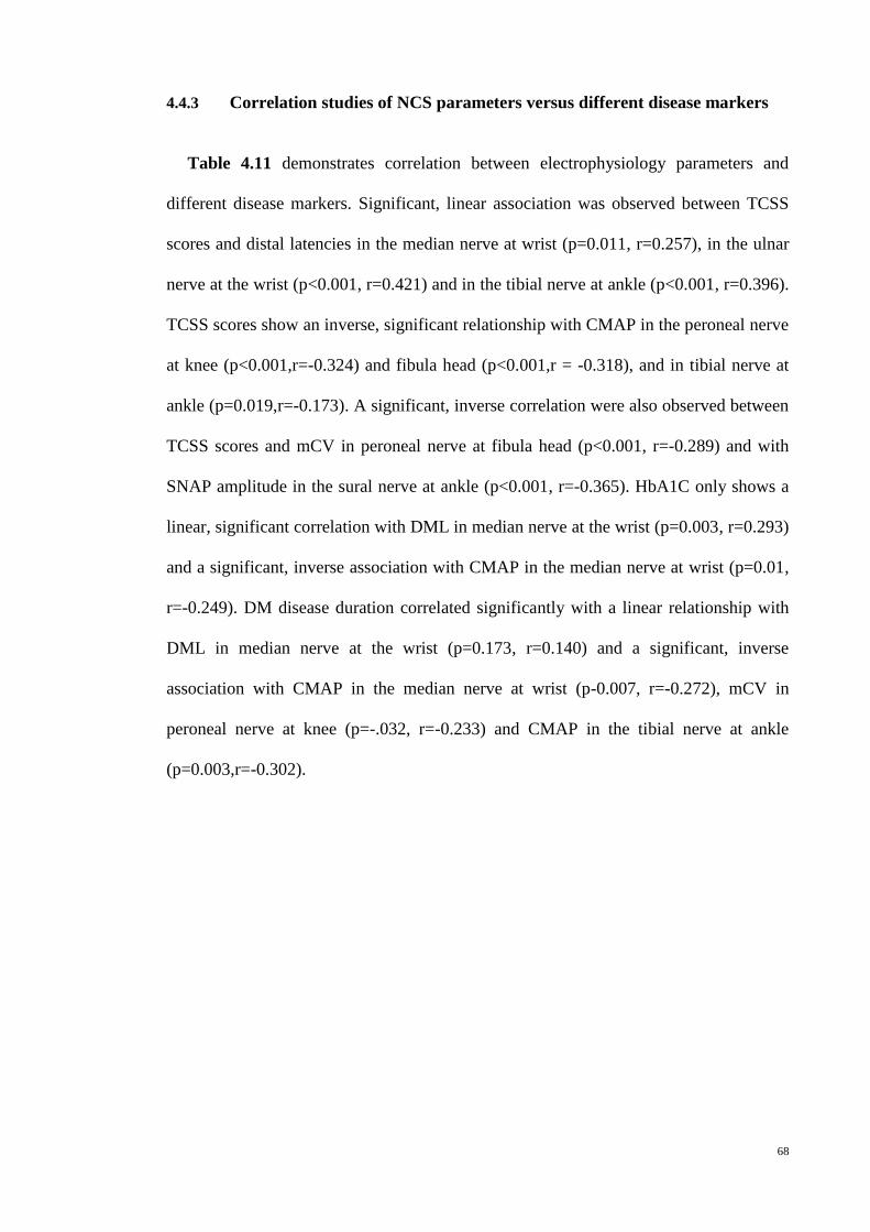
68
4.4.3 Correlation studies of NCS parameters versus different disease markers
Table 4.11 demonstrates correlation between electrophysiology parameters and
different disease markers. Significant, linear association was observed between TCSS
scores and distal latencies in the median nerve at wrist (p=0.011, r=0.257), in the ulnar
nerve at the wrist (p<0.001, r=0.421) and in the tibial nerve at ankle (p<0.001, r=0.396).
TCSS scores show an inverse, significant relationship with CMAP in the peroneal nerve
at knee (p<0.001,r=-0.324) and fibula head (p<0.001,r = -0.318), and in tibial nerve at
ankle (p=0.019,r=-0.173). A significant, inverse correlation were also observed between
TCSS scores and mCV in peroneal nerve at fibula head (p<0.001, r=-0.289) and with
SNAP amplitude in the sural nerve at ankle (p<0.001, r=-0.365). HbA1C only shows a
linear, significant correlation with DML in median nerve at the wrist (p=0.003, r=0.293)
and a significant, inverse association with CMAP in the median nerve at wrist (p=0.01,
r=-0.249). DM disease duration correlated significantly with a linear relationship with
DML in median nerve at the wrist (p=0.173, r=0.140) and a significant, inverse
association with CMAP in the median nerve at wrist (p-0.007, r=-0.272), mCV in
peroneal nerve at knee (p=-.032, r=-0.233) and CMAP in the tibial nerve at ankle
(p=0.003,r=-0.302).
69
TCSS Scores HbA1C Disease duration
Nerve Site NCS
parameter r
value p value r value p value r value p value
Median Wrist DML 0.257 0.011 0.293 0.003 0.140 0.173
CMAP -0.129 0.206 -0.249 0.013 -0.272 0.007
Ulnar Wrist DML 0.421 <0.001 0.072 0.481 0.147 0.153
CMAP -0.004 0.972 -0.084 0.437 0.136 0.887
Peroneal Knee CMAP -0.324 <0.001 -0.088 0.286 -0.067 0.569
mCV -0.046 0.551 -0.086 0.265 -0.233 0.032
Fibula
Head CMAP -0.318 <0.001 -0.132 0.080 -0.082 0.449
mCV -0.289 <0.001 -0.134 0.097 -0.176 0.128
Tibial Ankle DML 0.396 <0.001 0.071 0.311 0.132 0.207
CMAP -0.173 0.019 -0.140 0.060 -0.302 0.003
Sural Ankle SNAP
amplitude -0.365 <0.001 0.144 0.122 -0.067 0.623
CV -0.121 0.201 -0.061 0.528 -0.007 0.957
Table 4.11: Overview of correlation studies of electrophysiology parameters versus independent
disease markers
70
4.4.4 Association between HbA1C values with neuropathy severity and DM
duration
The correlation between HbA1C values with the neuropathy severity (as per TCSS)
of diabetic patients failed to reach significance. However, there was a significant, linear
relationship between HbA1C values and the DM duration (p=0.013, r=0.248). A
significant correlation were also found between DM duration and neuropathy severity
(p=0.045, r=0.201).
71
CHAPTER 5: DISCUSSION
5.1 Evaluation of DSP
DM is the most common cause of neuropathy. The frequency of neuropathy in
patients with DM is about 30%, and up to 50% of patients will eventually develop
neuropathy during the course of their disease (Callaghan et al, 2012). The diagnosis of
DSP is primarily based on its characteristic symptoms. TCSS is proven to be a valid
instrument to reflect the presence and severity of DSP (Bril & Perkins, 2002) .
Conventionally, NCS has been widely used to diagnose DSP (Dyck, Karnes, Daube,
O'Brien, & Service, 1985). However, NCS is time consuming and uncomfortable for
patients. Sonographic examination can potentially be an alternative to assess peripheral
nerves with less discomfort and there have been studies that have proven its clinical use
in the evaluation of disorders of the peripheral nervous system (Goedee et al, 2013).
5.2 US
In the current study, US revealed that nerve CSAs varied along the length of nerves.
In one study, the CSAs of multiple nerves in the upper and lower extremities of normal,
healthy controls were examined (Cartwright et al, 2008). The mean area of tibial nerve
at ankle observed in their study was 13.7 mm2, which is slightly greater than the value
10.5 mm2 that we obtained [Table 4.2]. The participants in their study varied from other
populations with regard to body composition and racial distribution and this may
account for the discrepancy seen in comparison to our findings. Another study reported
a mean area of the tibial nerve at the ankle, which was 7.2 mm2 in 35 healthy
individuals, which is lower than our finding. On average, nerves were smaller in the
median and ulnar nerve at forearm than in the arm of the diabetic patients as well as in
the healthy controls.
72
Nerves also appear to be larger in entrapment sites compared to their more proximal
nerve segment. In asymptomatic, older subjects, where it is common for nerve to be
relatively enlarged, slight nerve enlargement at the elbow or wrist relative to the more
proximal site does not necessarily suggest important focal pathology (Hassan, Leep
Hunderfund, Watson, Boon, & Sorenson, 2013). In DSP, nerve enlargement in the
median and tibial nerves has been reported (D. Lee & Dauphinee, 2005; Watanabe et
al., 2009). In our study, we found that the CSA in the median nerve at wrist and in the
ulnar nerve at elbow is significantly larger than in healthy controls. This may be because
the wrist is an entrapment site and most of our diabetic patients are susceptible to
developing CTS i.e. median nerve entrapment [Table 4.1] and UNE. CTS has been
documented as the frequent form of median nerve entrapment (Lo, Raskin, Lester, &
Lester, 2002; Padua, Lo Monaco, Padua, Gregori, & Tonali, 1997; Pfeffer, Gelberman,
Boyes, & Rydevik, 1988) and accounts for almost 90% of all entrapment neuropathies
(Aroori & Spence, 2008). Entrapment of the median nerve at the level of the carpal
tunnel, by the carpal bones and by the transverse carpal ligament results in CTS
(Alfonso, Jann, Massa, & Torreggiani, 2010). CTS reflect a decreased function of the
median nerve at that level due to increased pressure within the carpal tunnel. In DM,
CTS can be considered to be caused by both chronic compression and nerve dysfunction
(Comi et al, 1985). Aside from this, we found that nerves are also significantly larger in
the ulnar and radial nerve at mid forearm in diabetic patients than in healthy controls.
The lower extremity nerves in DSP subjects exhibit significant enlargement compared
to healthy controls. This is particularly true in the peroneal nerve at knee, tibial and
sural nerve at ankle [Table 4.2]. However, the mean CSA of diabetic patients in the
tibial nerve at ankle in our study (11.96 mm2) is lower than previously reported where
the threshold value was found to be 19.01 mm2 (Riazi et al., 2012).
73
This might be due to the variation in the patient population where their patients are
older and had longer duration of diabetes. Our findings are also in keeping with
previous studies that have found sural nerves in diabetes patients to be enlarged (Liu et
al., 2012; T. Watanabe et al., 2010). However, the mean CSA of sural nerve in our study
of 2.59 mm2
is higher than previously reported (1.88 mm2
) (Liu et al., 2012). The
possible explanation could be their subjects had shorter duration of DM (7.5±2.6 years)
compared to our subjects (14.54± 9.43).
Looking specifically at the median nerve at the wrist, we further demonstrated that
the DM patients had a significantly higher wrist to forearm ratio compared to the
healthy controls. The wrist to forearm ratio (cutoff value ≥ 1.4) (Hobson-Webb et al,
2008) had a sensitivity of 73% and specificity of 41% in differentiating DM patients
who were symptomatic and those who were not for carpal tunnel syndrome. We found
this ratio to be sensitive but not specific for clinically symptomatic median nerve
entrapment in DM patients with DSP.
We also found that the CSA of all lower limb nerves examined demonstrated a
significant difference between the mild and severe neuropathy group, where the severe
neuropathy patient group demonstrated larger CSA than the mild neuropathy patient
group [Table 4.3]. Generally, peroneal, tibial and sural nerves showed progressively
larger nerve CSAs with worsening severity. This is similar to a study that has reported a
close association between morphological parameters such as nerve hyperechoic area and
nerve CSA with the severity of diabetic neuropathy (Ishibashi et al, 2015).
AUC in ROC analysis revealed fair discriminatory cut-off CSA values for tibial and
sural nerves at 11.75 mm2
and 2.75 mm2
respectively to differentiate between severe and
less severe DSP as determined by TCSS [Table 4.4]. Previous studies have suggested
optimum threshold value that identifies DSP without specifically focusing on severity.
74
In one study, a cut-off value for tibial nerve of 19.1 mm2 was suggested to identify
DSP (Riazi et al, 2012). In the same study, NCS was utilized to detect the presence of
neuropathy. We find that whilst NCS is useful as an objective marker of DSP, it may
not be as useful in discriminating between DSP severities.
For this, TCSS has been validated as an instrument that can indicate the presence and
severity of DSP (Bril & Perkins, 2002). TCSS is also a universally accessible tool that is
easily implemented in DM patients. Using ROC analysis, we also found the sural nerve
CSA to be a good discriminator between the presence and absence of DSP in DM
patients using a cut-off value of 2.0 mm2
[Table 4.5] . This is similar to another study,
that reported a cut-off value of 1.685 mm2 of the sural nerve to differentiate diabetic
patients with neuropathy and diabetic patients without neuropathy (Liu et al, 2012).
To our knowledge, there has been no study that has compared the nerve sizes of true
CIDP patients and DM patients with demyelinating neurophysiological characteristic.
We explored the difference in CSAs of the nerves between DM patients with
demyelinating neurophysiological characteristics and true CIDP patients. We found that
the nerves in true CIDP patients were significantly enlarged in the upper extremities,
especially at the proximal and non-entrapment sites [Table 4.6]. This is similar to a
study that exhibits bigger mean nerve CSAs especially at proximal and non-entrapment
sites (Jang, Cho, Yang, Seok, & Kim, 2014). CIDP is mainly characterized
pathologically by segmental demyelination. There have been previous pathological
studies of CIDP that have demonstrated consistently a widespread segmental
demyelination and ‘onion bulb formation’, which describes magnified fascicles with
elevated endoneural connective tissues in which many myelinated fibers are enclosed by
concentrically arranged Schwann cells (Dyck et al., 1975; Matsuda et al, 1996).
75
The increase in nerve size in sonographic images is likely to be due to multiple
enlarged fascicles resulting from repeated demyelination and remyelination (Oguz,
Oguz, Cila, & Tan, 2003). There have also been studies that have reported the diffuse
nerve enlargement at multiple nerves in CIDP patients (Jang et al., 2014; Taniguchi et
al., 2000). Our study further demonstrates that CSA enlargement was more prominent at
sites where entrapment or compression is not common, and at proximal regions.
This may be due to the fact that DSP presents in a length dependent fashion, whereas
CIDP is rather patchy or segmental. Proximal and distal weakness strongly indicates
CIDP (Koski et al, 2009). Regional nerve enlargement have been reported in acquired
demyelinating neuropathies (CIDP) (Zaidman, Harms, & Pestronk, 2013) which is in
contrast to D-DSP, which is more likely a form of axonal neuropathy, exhibiting
“demyelinating” neurophysiology. US studies in patients with CIDP have shown diffuse
nerve enlargement of the median and ulnar nerves and are more common than in axonal
neuropathies, and these distinction may reflect the pathologic findings associated with
repeated demyelination and remyelination (Zaidman, Al-Lozi, & Pestronk, 2009).
5.3 Neurophysiological studies (NCS)
NCS was found to be useful in staging of neuropathy severity in DSP patients with
significant differences found in DML in the ulnar and tibial nerves, where the severe
patient group demonstrated a prolonged DML. The CMAP is also significantly reduced
in the peroneal and tibial nerves examined in the severe group. The motor conduction
velocities are decreased in the median nerve, ulnar nerve, peroneal nerve at fibula head
and tibial nerve in the severe neuropathy patient group. As for the sensory nerves, ulnar,
radial and sural nerves demonstrated a significant reduction in the sensory nerve action
potential among the severe neuropathy patient group.
76
Only the radial nerve showed a reduced sensory conduction velocity in the severe
neuropathy patient group [Table 4.7]. It is important to note that the lower limb nerve
potentials were inexcitable in 43%, 13% and 8% of total sural, peroneal and tibial
nerves respectively. The unrecordable potentials were likely due to significant axonal
degeneration resulting in too few remaining axons that are able to conduct an
electrically quantifiable signal. Previous studies have suggested the latter to reflect more
severe neuropathy with an increased risk of developing foot ulcers (Behse, Buchthal, &
Carlsen, 1977; Severinsen & Andersen, 2007; Veves et al, 1991; Vinik, Bril, Litchy,
Price, & Bastyr, 2005). Other possibilities for inexcitable nerves include peripheral
nerve edema but this was not a significant feature in our cohort, suggesting that the
nerves were truly diseased.
Comparative studies of neurophysiology parameters between true CIDP patients and
D-DSP patients [Table 4.8] revealed that DML in the peroneal nerve and duration in the
median nerve, ulnar nerve and tibial nerve were prolonged in true CIDP patients. True
CIDP patients demonstrated reduced CMAP in the ulnar nerve and a slowed velocity in
the peroneal nerve at fibula head. Ulnar and radial nerve in true CIDP patients exhibited
decreased sensory conduction velocities. This is identical to a study that has reported
similar results where they compared the NCS parameters between CIDP patients and
DM patients with neuropathy (Wilson, Chawla, & Fisher, 2005). Sensory NCS
abnormalities studies in CIDP have reported a high proportion of nerves with absent
responses and a susceptibility for sensory nerve to exhibit somewhat lesser degree of
CV slowing than is evident in motor nerves of the same patient (Krarup & Trojaborg,
1996). In other studies, evidence of demyelination on electrodiagnostic tests was found
(e.g. slowed conduction velocity or prolonged DML) in sensory nerves (Oh, Joy, &
Kuruoglu, 1992; Sinnreich et al., 2004).
77
Another study has also shown that sCV slowing is a highly specific marker for
differentiating CIDP from axonal polyneuropathy (Bragg & Benatar, 2008). This is
particularly true in our study where we found a significant reduction in conduction
velocity in the sensory ulnar and radial nerve.
5.4 Correlation studies
Correlation analysis in the current study revealed significant associations between
NCS parameters and nerve size in DSP patients [Table 4.9]. DMLs of median nerve
were positively correlated with nerve CSAs whereas CMAP amplitudes were negatively
correlated with nerve CSAs in the median nerve at the wrist. Ulnar mCV was also
negatively correlated with nerve CSA across the elbow. We failed to detect significant
correlations in the lower limb NCS parameters and nerve CSAs. This is similar to a
study that has reported the CSAs were negatively correlated with both a reduced mCV
and delayed latency (Tsuneo Watanabe et al, 2010). Another study has found an
increased CSA of the median nerve at wrist, which inversely correlated with conduction
velocity (Watanabe et al, 2009). A study also reported that the NCV might be decreased
not only due to loss of the fastest conducting axons, but also because of demyelination
and acute metabolic dysregulation (Severinsen & Andersen, 2007). A secondary
sodium accumulation and an increase in sorbitol may be major contributors to an
increase in intracellular hydration using a 1H-nuclear magnetic resonance study (Suzuki
et al, 1994). The peripheral nerve in DM patients may be swollen due to increased
water content related to increased aldose reductase conversion of glucose to sorbitol
further causing enlarged peripheral nerves i.e. larger CSAs (D. Lee & Dauphinee,
2005).
78
A significant positive correlation was also seen between disease duration; nerve CSA
of the median at the wrist and ulnar at the elbow, and arm [Table 4.10]. No correlation
was found between DM duration and CSA in the lower extremities. This is similar to
one study that has studied lower extremity nerve CSAs and found no correlation
between DM duration and nerve CSAs (Hobson-Webb et al, 2013).
We also investigated the relationship between disease duration and glycaemic
control (HbA1C) with DSP severity, NCS parameters and nerve CSA. Having excluded
entrapment sites, we failed to demonstrate a significant relationship between glycaemic
control and DSP severity or NCS and CSA parameters [Table 4.10 and 4.11]. This is
similar to a previous study where no significant association was found between HbA1C
and DSP severity (Sachedina & Toth, 2013). In one study, the authors investigated
glycaemic control based on glycated haemoglobin and nerve morphology based on sural
nerve biopsy (Perkins et al, 2001). The latter is an invasive method and unlikely to be a
feasible measure of nerve morphology in a clinical setting. However, we did
demonstrate a significant positive correlation between duration of DM with HbA1C and
disease severity. This is similar to other studies that have also found disease duration to
be a better predictor of DSP severity (Dyck et al., 1999; Mimi, Teng, & Chia, 2003).
Our study also demonstrated significant positive correlations between TCSS and most
neurophysiology parameters and with nerve CSAs [Table 4.10 and 4.11]. This is
supported by a study that had found strong correlations between TCSS and sural nerve
fiber density with electrophysiology, by both summed amplitude and summed
conduction velocity values. It was further concluded that TCSS is a valid instrument to
reflect the presence and severity of DSP as measured by sural nerve morphology and
electrophysiology (Bril & Perkins, 2002).
79
5.5 Study limitations
Some of the limitations of our study include the small number of patients in the
individual DSP severity groups. However, we were able to demonstrate that nerve US
has a role in discriminating between DSP severities. A separate cohort of patients with
asymptomatic DSP was not identified. Future studies involving a larger cohort and
identification of a separate group of asymptomatic DSP patients may yield further
useful information such as CSA cut-off values with better performance when
discriminating between DSP severities and the presence of DSP. The use of more
advanced US techniques incorporating color Doppler imaging might also provide
further insight.
This study aimed to distinguish D-DSP patients from true CIDP patients, which was
a pilot study with only preliminary results. However, our results show some promise as
to the utility of nerve US to differentiate the two cohorts. This diagnostic value will
require further validation in prospective studies involving a larger sample of patients at
different clinical stages of CIDP. We also did not attempt histological evaluation,
although these are not as easily available but may provide additional information and
lead to evidence that is more robust.
80
CHAPTER 6: CONCLUSION
In the current study, we investigated the nerve morphology of DM patients through
nerve US by assessing nerve CSAs in DM patients of different DSP severities as
assessed by the clinical tool, TCSS. We demonstrated that US correlated well with NCS
parameters in our cohort of patients, which suggests US can potentially be used to
objectively evaluate for severity of DSP and can be utilized in future studies.
We found that the nerve CSA of DM patients were larger as the severity of DSP
progresses. To our knowledge, there have been no previous studies that have described
such a relationship utilizing nerve CSA values. Traditionally, NCS has been the gold
standard for the diagnosis of DSP. However, patients with advanced DSP are likely to
have inexcitable nerves thus making an objective assessment of injured nerves
challenging. In these situations, we found US to be useful in assessing DSP in patients
and could serve as a valid bedside evaluation method to identify and determine the
severity of DSP, as currently, there is no objective practical method to determine the
presence and/or severity of DSP at the bedside.
In the current study, we found this ratio to be sensitive but not specific for clinically
symptomatic median nerve entrapment in DM patients with DSP. To further improve
the sensitivity of US, measurement of the median nerve at different levels and using a
proximal median nerve to median nerve at carpal tunnel ratio has been recommended
(Pastare, Therimadasamy, Lee, & Wilder-Smith, 2009). This would be helpful in
demonstrating the focality of median nerve swelling at the carpal tunnel similar to that
used in NCS. Besides this, when assessment of nerve conduction abnormalities across
the carpal tunnel becomes difficult in patients with DSP as median neuropathy and DSP
might affect median nerve conduction, US may serve as an alternative tool to detect
median neuropathy.
81
Both tibial and sural nerves showed progressively larger nerve CSAs with
worsening severity. This suggests that both tibial and sural nerve CSAs can potentially
be used as markers of disease severity in DSP. Further studies based on large multi-
center cohorts of patients with broader spectrum of neuropathy including asymptomatic
DSP patients may yield additional information, which may provide a more specific and
sensitive cut-off values to differentiate severe and non-severe DSP patients as well as
DSP and non DSP patients.
In the current study, we were also able to demonstrate significant associations
between NCS parameters and nerve size in DSP patients. US can be an alternative
diagnostic modality when NCS results are not confirmatory in patients suspected of
DSP. NCS and US used in combination may be able to estimate DSP more accurately.
Future serial studies with long-term follow up are needed to investigate correlations
between changes in US findings and other parameters such as NCS and clinical
characteristics. Significant positive correlations between TCSS and most
neurophysiology parameters and with nerve CSAs were presented in this study. Further
investigation to examine relationships between morphological changes and other
clinical scoring systems in DSP patients would yield extra information on the validity of
TCSS as well.
We did not detect any significant correlation between HbA1C and DSP severity or
NCS and CSA parameters, but detected significant correlation between duration of DM
with HbA1C and disease severity. Significant association between duration of diabetes
and DM severity from our study implies that although not a modifiable risk factor, the
duration of diabetes is of great significance for early identification and management of
diabetic polyneuropathy. Our findings strongly suggest that nerve CSA values have a
82
role in determining severity of DSP, particularly when other existing objective
parameters such as nerve action potentials are inexcitable.
Making the distinction between chronic DSP and CIDP can be challenging. The
current study exhibits a pattern of significant nerve enlargement in non-entrapment sites
in the upper extremities of true CIDP patients compared to D-DSP patients through US
studies. This is of significant clinical importance, as earlier initiation of treatment is
required to improve the overall prognosis and clinical outcome. Future prospective and
serial studies in a larger cohort of patients are required to confirm the current findings.
In conclusion, work presented here has demonstrated the promising use of nerve US
in the diagnosis of DSP including the ability to differentiate disease severity and
distinguishing between DSP and CIDP.
REFERENCES
Abbott, C. A, Carrington, A. L, Ashe, H., Bath, S, Every, L. C, Griffiths, J Boulton, A.
J. (2002). The North-West Diabetes Foot Care Study: incidence of, and risk
factors for, new diabetic foot ulceration in a community-based patient cohort.
Diabet Med, 19(5), 377-384.
Abdullah, N. A.-O., Attia, J., Oldmeadow, C., Scott, R. J., & Holliday, E. G. (2014).
The architecture of risk for type 2 diabetes: understanding Asia in the context of
global findings. Int J Endocrinol., 2014 (1687-8337 (Print)).
Abougalambou, S. S., & Abougalambou, A. S. (2012). Explorative study on diabetes
neuropathy among type II diabetic patients in Universiti Sains Malaysia
Hospital. Diabetes Metab Syndr, 6(3), 167-172. doi: 10.1016/j.dsx.2012.09.002
Abuaisha, B. B., Costanzi, J. B., & Boulton, A. J. (1998). Acupuncture for the treatment
of chronic painful peripheral diabetic neuropathy: a long-term study. Diabetes
Res Clin Pract, 39(2), 115-121.
AD, E. C, Skare, T. L., Sakuma, A. K., & Barros, W. H. (2015). [Ultrasonography as a
tool in diagnosis of carpal tunnel syndrome]. Rev Bras Reumatol. doi:
10.1016/j.rbr.2014.12.002
Alfonso, C., Jann, S., Massa, R., & Torreggiani, A. (2010). Diagnosis, treatment and
follow-up of the carpal tunnel syndrome: a review. Neurol Sci, 31(3), 243-252.
doi: 10.1007/s10072-009-0213-9
Aroori, S, & Spence, R. A. (2008). Carpal tunnel syndrome. Ulster Med J, 77(1), 6-17.
Austin, J. H. (1958). Recurrent polyneuropathies and their corticosteroid treatment; with
five-year observations of a placebo-controlled case treated with corticotrophin,
cortisone, and prednisone. Brain, 81(2), 157-192.
Ayyar, D. R., & Sharma, K. (2004). Chronic inflammatory demyelinating
polyradiculoneuropathy in diabetes mellitus. Current Diabetes Reports, 4(6),
409-412. doi: 10.1007/s11892-004-0048-y
Bae, J. S, & Kim, B. J. (2007). Subclinical diabetic neuropathy with normal
conventional electrophysiological study. J Neurol, 254(1), 53-59. doi:
10.1007/s00415-006-0261-5
Bahou, Y. G., & Elhadidy, S. (2005). Ulnar neuropathy at Jordan University Hospital.
Neurosciences (Riyadh), 10(2), 144-148.
Barohn, R. J., Kissel, J. T., Warmolts, J. R., & Mendell, J. R. (1989). Chronic
inflammatory demyelinating polyradiculoneuropathy. Clinical characteristics,
course, and recommendations for diagnostic criteria. Arch Neurol, 46(8), 878-
884.
Beekman, R., van den Berg, L. H., Franssen, H., Visser, L. H., van Asseldonk, J. T., &
Wokke, J. H. (2005). Ultrasonography shows extensive nerve enlargements in
multifocal motor neuropathy. Neurology, 65(2), 305-307. doi:
10.1212/01.wnl.0000169179.67764.30
Behse, F, Buchthal, F, & Carlsen, F. (1977). Nerve biopsy and conduction studies in
diabetic neuropathy. J Neurol Neurosurg Psychiatry, 40(11), 1072-1082.
Bohm, J. (2009). [Assessment of peripheral nerves in vascular neuropathy with high-
resolution ultrasonography ]. Ideggyogy Sz, 62(7-8), 277-281.
Boulton, A. J, Gries, F. A., & Jervell, J. A. (1998). Guidelines for the diagnosis and
outpatient management of diabetic peripheral neuropathy. Diabet Med, 15(6),
508-514. doi: 10.1002/(sici)1096-9136(199806)15:6<508::aid-dia613>3.0.co;2-l
Boulton, A. J., Malik, R. A., Arezzo, J. C., & Sosenko, J. M. (2004). Diabetic somatic
neuropathies. Diabetes Care, 27(6), 1458-1486.
Boulton, A. J., Vinik, A. I., Arezzo, J. C., Bril, V., Feldman, E. L., Freeman, R, Ziegler,
D. (2005). Diabetic neuropathies: a statement by the American Diabetes
Association. Diabetes Care, 28(4), 956-962.
Bragg, J. A, & Benatar, M. G. (2008). Sensory nerve conduction slowing is a specific
marker for CIDP. Muscle Nerve, 38(6), 1599-1603. doi: 10.1002/mus.21186
Bril, V, England, J., Franklin, G. M., Backonja, M., Cohen, J., Del Toro, D., . . .
Zochodne, D. (2011). Evidence-based guideline: Treatment of painful diabetic
neuropathy: report of the American Academy of Neurology, the American
Association of Neuromuscular and Electrodiagnostic Medicine, and the
American Academy of Physical Medicine and Rehabilitation. Pm r, 3(4), 345-
352, 352.e341-321. doi: 10.1016/j.pmrj.2011.03.008
Bril, V, Hirose, T., Tomioka, S., & Buchanan, R. (2009). Ranirestat for the management
of diabetic sensorimotor polyneuropathy. Diabetes Care, 32(7), 1256-1260. doi:
10.2337/dc08-2110
Bril, V, & Perkins, B. A. (2002). Validation of the Toronto Clinical Scoring System for
diabetic polyneuropathy. Diabetes Care, 25(11), 2048-2052.
Brownlee, M., Cerami, A, & Vlassara, H. (1988). Advanced glycosylation end products
in tissue and the biochemical basis of diabetic complications. N Engl J Med,
318(20), 1315-1321. doi: 10.1056/nejm198805193182007
Callaghan, B. C., Cheng, H. T., Stables, C. L., Smith, A. L., & Feldman, E. L. (2012).
Diabetic neuropathy: clinical manifestations and current treatments. The Lancet
Neurology, 11(6), 521-534. doi: http://dx.doi.org/10.1016/S1474-
4422(12)70065-0
Cartwright, M. S, Brown, M. E., Eulitt, P, Walker, F. O, Lawson, V. H., & Caress, J. B.
(2009). Diagnostic nerve ultrasound in Charcot-Marie-Tooth disease type 1B.
Muscle Nerve, 40(1), 98-102. doi: 10.1002/mus.21292
Cartwright, M. S., Passmore, L. V., Yoon, J. S., Brown, M. E., Caress, J. B., & Walker,
F. O. (2008). Cross-sectional area reference values for nerve ultrasonography.
Muscle Nerve, 37(5), 566-571. doi: 10.1002/mus.21009
Chalk, C, Benstead, T. J, & Moore, F. (2007). Aldose reductase inhibitors for the
treatment of diabetic polyneuropathy. Cochrane Database Syst Rev(4),
Cd004572. doi: 10.1002/14651858.CD004572.pub2
Chio, A., Plano, F., Calvo, A., Leone, M., Mutani, R., & Cocito, D. (2009).
Comorbidity between CIDP and diabetes mellitus: only a matter of chance? Eur
J Neurol, 16(6), 752-754.
Comi, G., Lozza, L., Galardi, G., Ghilardi, M. F., Medaglini, S., & Canal, N. (1985).
Presence of carpal tunnel syndrome in diabetics: effect of age, sex, diabetes
duration and polyneuropathy. Acta Diabetol Lat, 22(3), 259-262.
Dahlin, L. B., Stenberg, L., Luthman, H., & Thomsen, N. O. (2008). Nerve compression
induces activating transcription factor 3 in neurons and Schwann cells in diabetic
rats. Neuroreport, 19(9), 987-990. doi: 10.1097/WNR.0b013e328302f4ec
Dalakas, M. C., & Engel, W. K. (1981). Chronic relapsing (dysimmune)
polyneuropathy: pathogenesis and treatment. Ann Neurol, 9 Suppl, 134-145.
Dobretsov, M., Hastings, S. L., Romanovsky, D., Stimers, J. R., & Zhang, J. M. (2003).
Mechanical hyperalgesia in rat models of systemic and local hyperglycemia.
Brain Res, 960(1-2), 174-183.
Dolman, C. L. (1963). The morbid anatomy of diabetic neuropathy. Neurology, 13, 135-
142.
Dyck, P. J. (1988). Detection, characterization, and staging of polyneuropathy: assessed
in diabetics. Muscle Nerve, 11(1), 21-32. doi: 10.1002/mus.880110106
Dyck, P. J. (1991). Evaluative procedures to detect, characterize, and assess the severity
of diabetic neuropathy. Diabet Med, 8 Spec No, S48-51.
Dyck, P. J., Davies, J. L., Wilson, D. M., Service, F. J., Melton, L. J., 3rd, & O'Brien, P.
C. (1999). Risk factors for severity of diabetic polyneuropathy: intensive
longitudinal assessment of the Rochester Diabetic Neuropathy Study cohort.
Diabetes Care, 22(9), 1479-1486.
Dyck, P. J., Karnes, J. L., Daube, J., O'Brien, P., & Service, F. J. (1985). Clinical and
neuropathological criteria for the diagnosis and staging of diabetic
polyneuropathy. Brain, 108 ( Pt 4), 861-880.
Dyck, P. J., Lais, A. C., Ohta, M., Bastron, J. A., Okazaki, H., & Groover, R. V. (1975).
Chronic inflammatory polyradiculoneuropathy. Mayo Clin Proc, 50(11), 621-
637.
Dyck, P. J., O'Brien, P. C., Oviatt, K. F., Dinapoli, R. P., Daube, J. R., Bartleson, J. D,
Windebank, A. J. (1982). Prednisone improves chronic inflammatory
demyelinating polyradiculoneuropathy more than no treatment. Ann Neurol,
11(2), 136-141. doi: 10.1002/ana.410110205
Dyck, P. J, Overland, C. J., Low, P. A., Litchy, W. J, Davies, J. L, Dyck, P. J., . . .
Vella, A. (2010). Signs and symptoms versus nerve conduction studies to
diagnose diabetic sensorimotor polyneuropathy: Cl vs. NPhys trial. Muscle
Nerve, 42(2), 157-164. doi: 10.1002/mus.21661
Dyck, P. J., Overland, C. J., Low, P. A., Litchy, W. J., Davies, J. L., Dyck, P. J. B., &
O'Brien, P. C. (2010). Signs and symptoms versus nerve conduction studies to
diagnose diabetic sensorimotor polyneuropathy: Cl vs. NPhys trial. Muscle
Nerve, 42(2), 157-164.
Eftimov, F, Winer, J. B, Vermeulen, M, de Haan, R., & van Schaik, I. N. (2013).
Intravenous immunoglobulin for chronic inflammatory demyelinating
polyradiculoneuropathy. Cochrane Database Syst Rev, 12, Cd001797. doi:
10.1002/14651858.CD001797.pub3
England, J. D, Gronseth, G. S, Franklin, G., Carter, G. T, Kinsella, L. J, Cohen, J. A.,
Sumner, A. J. (2009). Practice parameter: the evaluation of distal symmetric
polyneuropathy: the role of autonomic testing, nerve biopsy, and skin biopsy (an
evidence-based review). Report of the American Academy of Neurology, the
American Association of Neuromuscular and Electrodiagnostic Medicine, and
the American Academy of Physical Medicine and Rehabilitation. Pm r, 1(1), 14-
22. doi: 10.1016/j.pmrj.2008.11.011
England, J. D., Gronseth, G. S., Franklin, G., Miller, R. G., Asbury, A. K., Carter, G. T.,
Sumner, A. J. (2005). Distal symmetric polyneuropathy: a definition for clinical
research: report of the American Academy of Neurology, the American
Association of Electrodiagnostic Medicine, and the American Academy of
Physical Medicine and Rehabilitation. Neurology, 64(2), 199-207. doi:
10.1212/01.wnl.0000149522.32823.ea
Feldman, E. L, Stevens, M. J., Thomas, P. K, Brown, M. B., Canal, N, & Greene, D. A.
(1994). A practical two-step quantitative clinical and electrophysiological
assessment for the diagnosis and staging of diabetic neuropathy. Diabetes Care,
17(11), 1281-1289.
Finnerup, N. B., Otto, M., McQuay, H. J., Jensen, T. S., & Sindrup, S. H. (2005).
Algorithm for neuropathic pain treatment: an evidence based proposal. Pain,
118(3), 289-305. doi: 10.1016/j.pain.2005.08.013
Forsblom, C. M., Sane, T., Groop, P. H, Totterman, K. J, Kallio, M, Saloranta, C., . . .
Groop, L. (1998). Risk factors for mortality in Type II (non-insulin-dependent)
diabetes: evidence of a role for neuropathy and a protective effect of HLA-DR4.
Diabetologia, 41(11), 1253-1262. doi: 10.1007/s001250051062
Freeman, R., Durso-Decruz, E., & Emir, B. (2008). Efficacy, safety, and tolerability of
pregabalin treatment for painful diabetic peripheral neuropathy: findings from
seven randomized, controlled trials across a range of doses. Diabetes Care,
31(7), 1448-1454. doi: 10.2337/dc07-2105
Gelfand, E. W. (2012). Intravenous immune globulin in autoimmune and inflammatory
diseases. N Engl J Med, 367(21), 2015-2025. doi: 10.1056/NEJMra1009433
Goedee, H. S., Brekelmans, G. J., van Asseldonk, J. T., Beekman, R., Mess, W. H., &
Visser, L. H. (2013). High resolution sonography in the evaluation of the
peripheral nervous system in polyneuropathy--a review of the literature. Eur J
Neurol, 20(10), 1342-1351. doi: 10.1111/ene.12182
Granberg, V., Ejskjaer, N., Peakman, M., & Sundkvist, G. (2005). Autoantibodies to
autonomic nerves associated with cardiac and peripheral autonomic neuropathy.
Diabetes Care, 28(8), 1959-1964.
Grimm, A, Heiling, B., Schumacher, U., Witte, O. W, & Axer, H. (2014). Ultrasound
differentiation of axonal and demyelinating neuropathies. Muscle Nerve, 50(6),
976-983. doi: 10.1002/mus.24238
Gupta, S, Chough, E, Daley, J, Oates, P, Tornheim, K, Ruderman, N. B, & Keaney, J.
F., Jr. (2002). Hyperglycemia increases endothelial superoxide that impairs
smooth muscle cell Na+-K+-ATPase activity. Am J Physiol Cell Physiol, 282(3),
C560-566. doi: 10.1152/ajpcell.00343.2001
Hassan, A, Leep Hunderfund, A. N, Watson, J, Boon, A. J, & Sorenson, E. J. (2013).
Median nerve ultrasound in diabetic peripheral neuropathy with and without
carpal tunnel syndrome. Muscle Nerve, 47(3), 437-439. doi: 10.1002/mus.23677
Head, K. A. (2006). Peripheral neuropathy: pathogenic mechanisms and alternative
therapies. Altern Med Rev, 11(4), 294-329.
Heinemeyer, O, & Reimers, C. D. (1999). Ultrasound of radial, ulnar, median, and
sciatic nerves in healthy subjects and patients with hereditary motor and sensory
neuropathies. Ultrasound Med Biol, 25(3), 481-485.
Hobson-Webb, L. D., Massey, J. M., & Juel, V. C. (2013). Nerve ultrasound in diabetic
polyneuropathy: correlation with clinical characteristics and electrodiagnostic
testing. Muscle Nerve, 47(3), 379-384. doi: 10.1002/mus.23625
Hobson-Webb, L. D., Massey, J. M., Juel, V. C., & Sanders, D. B. (2008). The
ultrasonographic wrist-to-forearm median nerve area ratio in carpal tunnel
syndrome. Clin Neurophysiol, 119(6), 1353-1357. doi:
10.1016/j.clinph.2008.01.101
Hounsom, L, Corder, R., Patel, J., & Tomlinson, D. R. (2001). Oxidative stress
participates in the breakdown of neuronal phenotype in experimental diabetic
neuropathy. Diabetologia, 44(4), 424-428. doi: 10.1007/s001250051638
Ishibashi, F, Taniguchi, M., Kojima, R., Kawasaki, A., Kosaka, A., & Uetake, H.
(2015). Morphological changes of the peripheral nerves evaluated by high-
resolution ultrasonography are associated with the severity of diabetic
neuropathy, but not corneal nerve fiber pathology in patients with type 2
diabetes. J Diabetes Investig, 6(3), 334-342. doi: 10.1111/jdi.12299
Ito, T, Kijima, M, Watanabe, T, Sakuta, M., & Nishiyama, K. (2007). Ultrasonography
of the tibial nerve in vasculitic neuropathy. Muscle Nerve, 35(3), 379-382. doi:
10.1002/mus.20673
Jang, J. H, Cho, C. S, Yang, K. S, Seok, H. Y. & Kim, B. J. (2014). Pattern analysis of
nerve enlargement using ultrasonography in chronic inflammatory
demyelinating polyneuropathy. Clin Neurophysiol, 125(9), 1893-1899. doi:
10.1016/j.clinph.2013.12.115
Kaur, S, Pandhi, P, & Dutta, P. (2011). Painful diabetic neuropathy: an update. Ann
Neurosci, 18(4), 168-175. doi: 10.5214/ans.0972-7531.1118409
Kihara, M., & Low, P. A. (1995). Impaired vasoreactivity to nitric oxide in
experimental diabetic neuropathy. Exp Neurol, 132(2), 180-185.
Kim, W. K, Kwon, S. H., Lee, S. H, & Sunwoo, I. N. (2000). Asymptomatic
electrophysiologic carpal tunnel syndrome in diabetics: entrapment or
polyneuropathy. Yonsei Med J, 41(1), 123-127.
Kimura, J, Yamada, T, & Stevland, N. P. (1979). Distal slowing of motor nerve
conduction velocity in diabetic polyneuropathy. Journal of the Neurological
Sciences, 42(2), 291-302. doi: http://dx.doi.org/10.1016/0022-510X(79)90061-3
Kohara, N, Kimura, J., Kaji, R., Goto, Y., Ishii, J, Takiguchi, M, & Nakai, M. (2000). F-
wave latency serves as the most reproducible measure in nerve conduction
studies of diabetic polyneuropathy: multicentre analysis in healthy subjects and
patients with diabetic polyneuropathy. Diabetologia, 43(7), 915-921.
Koski, C. L, Baumgarten, M., Magder, L. S., Barohn, R. J., Goldstein, J., Graves, M.,
Cornblath, D. R. (2009). Derivation and validation of diagnostic criteria for
chronic inflammatory demyelinating polyneuropathy. J Neurol Sci, 277(1-2), 1-
8. doi: 10.1016/j.jns.2008.11.015
Krarup, C, & Trojaborg, W. (1996). Sensory pathophysiology in chronic acquired
demyelinating neuropathy. Brain, 119 ( Pt 1), 257-270.
Krendel, D. A., Costigan, D. A., & Hopkins, L. C. (1995). Successful treatment of
neuropathies in patients with diabetes mellitus. Arch Neurol, 52(11), 1053-1061.
Kuitwaard, K, & van Doorn, P. A. (2009). Newer therapeutic options for chronic
inflammatory demyelinating polyradiculoneuropathy. Drugs, 69(8), 987-1001.
doi: 10.2165/00003495-200969080-00004
Lee, D, & Dauphinee, D. M. (2005). Morphological and functional changes in the
diabetic peripheral nerve: using diagnostic ultrasound and neurosensory testing
to select candidates for nerve decompression. J Am Podiatr Med Assoc, 95(5),
433-437.
Lee, K. W, Hwang, J. H, & Kim, J. W. (1999). Nerve Conduction Study in Diabetic
Neuropathy. Journal of Korean Academy of Rehabilitation Medicine, 23(6),
1183-1190.
Lehmann, H. C, & Hartung, H.-P. (2011). Plasma exchange and intravenous
immunoglobulins: mechanism of action in immune-mediated neuropathies.
Journal of neuroimmunology, 231(1), 61-69.
Lehmann, H. C, Hartung, H.-P, Hetzel, G. R., Stüve, O, & Kieseier, B. C. (2006).
Plasma exchange in neuroimmunological disorders: part 2. Treatment of
neuromuscular disorders. Arch Neurol, 63(8), 1066-1071.
Liu, F, Zhu, J, Wei, M., Bao, Y, & Hu, B. (2012). Preliminary evaluation of the sural
nerve using 22-MHz ultrasound: a new approach for evaluation of diabetic
cutaneous neuropathy. PLoS One, 7(4), e32730. doi:
10.1371/journal.pone.0032730
Lo, S. L, Raskin, K, Lester, H, & Lester, B. (2002). Carpal tunnel syndrome: a historical
perspective. Hand Clin, 18(2), 211-217, v.
Lotan, I., Hellman, M. A., & Steiner, I. (2015). Diagnostic criteria of chronic
inflammatory demyelinating polyneuropathy in diabetes mellitus. Acta Neurol
Scand. doi: 10.1111/ane.12394
Lozeron, P, Nahum, L, Lacroix, C, Ropert, A, Guglielmi, J. M., & Said, G. (2002).
Symptomatic diabetic and non-diabetic neuropathies in a series of 100 diabetic
patients. J Neurol, 249(5), 569-575. doi: 10.1007/s004150200066
Lucchetta, M., Pazzaglia, C., Granata, G., Briani, C., & Padua, L. (2011). Ultrasound
evaluation of peripheral neuropathy in POEMS syndrome. Muscle Nerve, 44(6),
868-872. doi: 10.1002/mus.22258
Martinoli, C., Schenone, A., Bianchi, S., Mandich, P., Caponetto, C., Abbruzzese, M.,
& Derchi, L. E. (2002). Sonography of the median nerve in Charcot-Marie-
Tooth disease. AJR Am J Roentgenol, 178(6), 1553-1556. doi:
10.2214/ajr.178.6.1781553
Maser, R. E, Steenkiste, A. R., Dorman, J. S., Nielsen, V. K., Bass, E. B., Manjoo, Q. et
al. (1989). Epidemiological correlates of diabetic neuropathy. Report from
Pittsburgh Epidemiology of Diabetes Complications Study. Diabetes, 38(11),
1456-1461.
Matsuda, M., Ikeda, S., Sakurai, S., Nezu, A., Yanagisawa, N., & Inuzuka, T. (1996).
Hypertrophic neuritis due to chronic inflammatory demyelinating
polyradiculoneuropathy (CIDP): a postmortem pathological study. Muscle
Nerve, 19(2), 163-169. doi: 10.1002/(SICI)1097-
4598(199602)19:2<163::AID-MUS6>3.0.CO;2-C
Max, M. B., Lynch, S. A., Muir, J., Shoaf, S. E., Smoller, B., & Dubner, R. (1992).
Effects of desipramine, amitriptyline, and fluoxetine on pain in diabetic
neuropathy. N Engl J Med, 326(19), 1250-1256. doi:
10.1056/nejm199205073261904
Mimi, O, Teng, C. L., & Chia, Y. C. (2003). The prevalence of diabetic peripheral
neuropathy in an outpatient setting. Med J Malaysia, 58(4), 533-538.
Mizumoto, D., Hashizume, H., Senda, M., Nagoshi, M., & Inoue, H. (2003).
Electrophysiological assessment of the carpal tunnel syndrome in hemodialysis
patients: formula for predicting surgical results. J Orthop Sci, 8(1), 8-15. doi:
10.1007/s007760300001
Moon, H. I., Kwon, H. K., Kim, L., Lee, H. J., & Lee, H. J. (2014). Ultrasonography of
palm to elbow segment of median nerve in different degrees of diabetic
polyneuropathy. Clin Neurophysiol, 125(4), 844-848. doi:
10.1016/j.clinph.2013.10.041
Nakamura, J, Koh, N., Sakakibara, F, Hamada, Y, Hara, T, Sasaki, H, . . . Hotta, N.
(1998). Polyol pathway hyperactivity is closely related to carnitine deficiency in
the pathogenesis of diabetic neuropathy of streptozotocin-diabetic rats. J
Pharmacol Exp Ther, 287(3), 897-902.
Oguz, B, Oguz, K. K, Cila, A, & Tan, E. (2003). Diffuse spinal and intercostal nerve
involvement in chronic inflammatory demyelinating polyradiculoneuropathy:
MRI findings. Eur Radiol, 13 Suppl 6, L230-234. doi: 10.1007/s00330-003-
1996-3
Oh, S. J, Joy, J. L, & Kuruoglu, R. (1992). "Chronic sensory demyelinating
neuropathy": chronic inflammatory demyelinating polyneuropathy presenting as
a pure sensory neuropathy. J Neurol Neurosurg Psychiatry, 55(8), 677-680.
Oyibo, S. O, Prasad, Y. D, Jackson, N. J, Jude, E. B, & Boulton, A. J. (2002). The
relationship between blood glucose excursions and painful diabetic peripheral
neuropathy: a pilot study. Diabet Med, 19(10), 870-873.
Padua, L, Lo Monaco, M., Padua, R., Gregori, B., & Tonali, P. (1997).
Neurophysiological classification of carpal tunnel syndrome: assessment of 600
symptomatic hands. Ital J Neurol Sci, 18(3), 145-150.
Papanas, N, Giassakis, G., Papatheodorou, K, Papazoglou, D, Monastiriotis, C,
Christakidis, D Maltezos, E. (2007). Sensitivity and specificity of a new
indicator test (Neuropad) for the diagnosis of peripheral neuropathy in type 2
diabetes patients: a comparison with clinical examination and nerve conduction
study. J Diabetes Complications, 21(6), 353-358. doi:
10.1016/j.jdiacomp.2006.08.003
Pastare, D, Therimadasamy, A. K., Lee, E, & Wilder-Smith, E. P. (2009). Sonography
versus nerve conduction studies in patients referred with a clinical diagnosis of
carpal tunnel syndrome. J Clin Ultrasound, 37(7), 389-393. doi:
10.1002/jcu.20601
Perkins, B. A, Olaleye, D., Zinman, B, & Bril, V. (2001). Simple screening tests for
peripheral neuropathy in the diabetes clinic. Diabetes Care, 24(2), 250-256.
Pfeffer, G. B, Gelberman, R. H., Boyes, J. H., & Rydevik, B. (1988). The history of
carpal tunnel syndrome. J Hand Surg Br, 13(1), 28-34.
Raccah, D, Coste, T, Cameron, N. E., Dufayet, D, Vague, P, & Hohman, T. C. (1998).
Effect of the aldose reductase inhibitor tolrestat on nerve conduction velocity,
Na/K ATPase activity, and polyols in red blood cells, sciatic nerve, kidney
cortex, and kidney medulla of diabetic rats. J Diabetes Complications, 12(3),
154-162.
Research criteria for diagnosis of chronic inflammatory demyelinating polyneuropathy
(CIDP). Report from an Ad Hoc Subcommittee of the American Academy of
Neurology AIDS Task Force. (1991). Neurology, 41(5), 617-618.
Riazi, S., Bril, V., Perkins, B. A., Abbas, S., Chan, V. W., Ngo, M., . . . Brull, R.
(2012). Can ultrasound of the tibial nerve detect diabetic peripheral neuropathy?
A cross-sectional study. Diabetes Care, 35(12), 2575-2579. doi: 10.2337/dc12-
0739
Sachedina, S, & Toth, C. (2013). Association of comorbidities with increasing severity
of peripheral neuropathy in diabetes mellitus. World Journal of Diabetes, 4(4),
135-144. doi: 10.4239/wjd.v4.i4.135
Said, G, Slama, G, & Selva, J. (1983). Progressive centripetal degeneration of axons in
small fibre diabetic polyneuropathy. Brain, 106 ( Pt 4), 791-807.
Sasaki, T, Yasuda, H, Maeda, K, & Kikkawa, R. (1998). Hyperalgesia and decreased
neuronal nitric oxide synthase in diabetic rats. Neuroreport, 9(2), 243-247.
Severinsen, K, & Andersen, H. (2007). Evaluation of atrophy of foot muscles in diabetic
neuropathy -- a comparative study of nerve conduction studies and
ultrasonography. Clin Neurophysiol, 118(10), 2172-2175. doi:
10.1016/j.clinph.2007.06.019
Sharma, K. R., Cross, J., Farronay, O., Ayyar, D. R., Shebert, R. T., & Bradley, W. G.
(2002). Demyelinating neuropathy in diabetes mellitus. Arch Neurol, 59(5), 758-
765.
Shaw, J. E., Sicree, R. A., & Zimmet, P. Z. (2010). Global estimates of the prevalence
of diabetes for 2010 and 2030. Diabetes Res Clin Pract, 87(1), 4-14. doi:
10.1016/j.diabres.2009.10.007
Sinnreich, M., Klein, C. J., Daube, J. R., Engelstad, J., Spinner, R. J., & Dyck, P. J.
(2004). Chronic immune sensory polyradiculopathy: a possibly treatable sensory
ataxia. Neurology, 63(9), 1662-1669.
Somers, D. L., & Somers, M. F. (1999). Treatment of neuropathic pain in a patient with
diabetic neuropathy using transcutaneous electrical nerve stimulation applied to
the skin of the lumbar region. Phys Ther, 79(8), 767-775.
Stevens, M. J., Dananberg, J., Feldman, E. L., Lattimer, S. A., Kamijo, M., Thomas, T.
P Greene, D. A. (1994). The linked roles of nitric oxide, aldose reductase and,
(Na+,K+)-ATPase in the slowing of nerve conduction in the streptozotocin
diabetic rat. J Clin Invest, 94(2), 853-859. doi: 10.1172/jci117406
Stewart, J. D., McKelvey, R., Durcan, L., Carpenter, S., & Karpati, G. (1996). Chronic
inflammatory demyelinating polyneuropathy (CIDP) in diabetics. J Neurol Sci,
142(1-2), 59-64.
Suk, J. I., Walker, F. O., & Cartwright, M. S. (2013). Ultrasound of Peripheral Nerves.
Current neurology and neuroscience reports, 13(2), 328-328. doi:
10.1007/s11910-012-0328-x
Sundkvist, G., Lind, P., Bergstrom, B., Lilja, B., & Rabinowe, S. L. (1991). Autonomic
nerve antibodies and autonomic nerve function in type 1 and type 2 diabetic
patients. J Intern Med, 229(6), 505-510.
Suzuki, E., Yasuda, K., Yasuda, K., Miyazaki, S., Takeda, N., Inouye, H., . . . Miura, K.
(1994). 1H-NMR analysis of nerve edema in the streptozotocin-induced diabetic
rat. J Lab Clin Med, 124(5), 627-637.
Taniguchi, N, Itoh, K, Wang, Y, Omoto, K, Shigeta, K, Fujii, Y, Nakano, I. (2000).
Sonographic detection of diffuse peripheral nerve hypertrophy in chronic
inflammatory demyelinating polyradiculoneuropathy. J Clin Ultrasound, 28(9),
488-491.
Tesfaye, S. (2007). Advances in the management of painful diabetic neuropathy. Clin
Med, 7(2), 113-114.
Tesfaye, S. (2011). Recent advances in the management of diabetic distal symmetrical
polyneuropathy. J Diabetes Investig, 2(1), 33-42. doi: 10.1111/j.2040-
1124.2010.00083.x
Tesfaye, S, Boulton, A. J, & Dickenson, A. H. (2013). Mechanisms and management of
diabetic painful distal symmetrical polyneuropathy. Diabetes Care, 36(9), 2456-
2465. doi: 10.2337/dc12-1964
Tesfaye, S., Boulton, A. J., Dyck, P. J., Freeman, R., Horowitz, M., Kempler, P., . . .
Valensi, P. (2010). Diabetic neuropathies: update on definitions, diagnostic
criteria, estimation of severity, and treatments. Diabetes Care, 33(10), 2285-
2293. doi: 10.2337/dc10-1303
Thomas, P. K. (1994). Growth factors and diabetic neuropathy. Diabet Med, 11(8), 732-
739.
Thomas, P. K. (1997). Classification, differential diagnosis, and staging of diabetic
peripheral neuropathy. Diabetes, 46 Suppl 2, S54-57.
Thomsen, K, Rubin, I, & Lauritzen, M. (2002). NO- and non-NO-, non-prostanoid-
dependent vasodilatation in rat sciatic nerve during maturation and developing
experimental diabetic neuropathy. J Physiol, 543(Pt 3), 977-993.
Van Dam, P. S. (2002). Oxidative stress and diabetic neuropathy: pathophysiological
mechanisms and treatment perspectives. Diabetes Metab Res Rev, 18(3), 176-
184. doi: 10.1002/dmrr.287
Van den Bergh, P. Y., Hadden, R. D., Bouche, P., Cornblath, D. R., Hahn, A., Illa, Ivan
Schaik, I. N. (2010). European Federation of Neurological Societies/Peripheral
Nerve Society guideline on management of chronic inflammatory demyelinating
polyradiculoneuropathy: report of a joint task force of the European Federation
of Neurological Societies and the Peripheral Nerve Society - first revision. Eur J
Neurol, 17(3), 356-363. doi: 10.1111/j.1468-1331.2009.02930.x
Veves, A, Malik, R. A, Lye, R. H., Masson, E. A, Sharma, A. K, Schady, ., & Boulton,
A. J. (1991). The relationship between sural nerve morphometric findings and
measures of peripheral nerve function in mild diabetic neuropathy. Diabet Med,
8(10), 917-921.
Vinik, A. I, Bril, V, Litchy, W. J, Price, K. L, & Bastyr, E. J., 3rd. (2005). Sural sensory
action potential identifies diabetic peripheral neuropathy responders to therapy.
Muscle Nerve, 32(5), 619-625. doi: 10.1002/mus.20423
Vinik, A. I., Maser, R. E., Mitchell, B. D., & Freeman, R. (2003). Diabetic autonomic
neuropathy. Diabetes Care, 26(5), 1553-1579.
Visser, L. H., Hens, V., Soethout, M., De Deugd-Maria, V., Pijnenburg, J., &
Brekelmans, G. J. (2013). Diagnostic value of high-resolution sonography in
common fibular neuropathy at the fibular head. Muscle Nerve, 48(2), 171-178.
doi: 10.1002/mus.23729
Watanabe, T., Ito, H., Morita, A., Uno, Y., Nishimura, T., Kawase, Seishima, M.
(2009). Sonographic evaluation of the median nerve in diabetic patients:
comparison with nerve conduction studies. J Ultrasound Med, 28(6), 727-734.
Watanabe, T., Ito, H., Sekine, A., Katano, Y., Nishimura, T., Kato, Y Matsuoka, T.
(2010). Sonographic Evaluation of the Peripheral Nerve in Diabetic Patients The
Relationship Between Nerve Conduction Studies, Echo Intensity, and Cross-
sectional Area. Journal of Ultrasound in Medicine, 29(5), 697-708.
Watanabe, T, Ito, H, Sekine, A, Katano, Y, Nishimura, T, Kato, Matsuoka, T. (2010).
Sonographic evaluation of the peripheral nerve in diabetic patients: the
relationship between nerve conduction studies, echo intensity, and cross-
sectional area. J Ultrasound Med, 29(5), 697-708.
Watkins, P. J, Gayle, C, Alsanjari, N., Scaravilli, F, Zanone, M, & Thomas, P. K.
(1995). Severe sensory-autonomic neuropathy and endocrinopathy in insulin-
dependent diabetes. Qjm, 88(11), 795-804.
Wild, S., Roglic G Fau - Green, A, Green A Fau - Sicree, R., Sicree R Fau - King, H., &
King, H. (2004). Global prevalence of diabetes: estimates for the year 2000 and
projections for 2030. Diabetes Care (0149-5992 (Print)), 1047-1053.
Wilson, J., Chawla, J., & Fisher, M. (2005). Sensitivity and specificity of
electrodiagnostic criteria for CIDP using ROC curves: comparison to patients
with diabetic and MGUS associated neuropathies. J Neurol Sci, 231(1-2), 19-28.
doi: 10.1016/j.jns.2004.12.004
Young, M. J, Boulton, A. J, MacLeod, A. F, Williams, D. R., & Sonksen, P. H. (1993).
A multicentre study of the prevalence of diabetic peripheral neuropathy in the
United Kingdom hospital clinic population. Diabetologia, 36(2), 150-154.
Zaidman, C. M., Al-Lozi, M., & Pestronk, A. (2009). Peripheral nerve size in normals
and patients with polyneuropathy: an ultrasound study. Muscle Nerve, 40(6),
960-966. doi: 10.1002/mus.21431
Zaidman, C. M., Harms, M. B., & Pestronk, A. (2013). Ultrasound of inherited vs.
acquired demyelinating polyneuropathies. J Neurol, 260(12), 3115-3121. doi:
10.1007/s00415-013-7123-8
Ziegler, D., Nowak, H., Kempler, P., Vargha, P., & Low, P. A. (2004). Treatment of
symptomatic diabetic polyneuropathy with the antioxidant alpha-lipoic acid: a
meta-analysis. Diabet Med, 21(2), 114-121.
Ziegler, D., Sohr, C. G., & Nourooz-Zadeh, J. (2004). Oxidative stress and antioxidant
defense in relation to the severity of diabetic polyneuropathy and cardiovascular
autonomic neuropathy. Diabetes Care, 27(9), 2178-2183.
Zinman, L. H., Ngo, M., Ng, E. T., Nwe, K. T., Gogov, S., & Bril, V. (2004). Low-
intensity laser therapy for painful symptoms of diabetic sensorimotor
polyneuropathy: a controlled trial. Diabetes Care, 27(4), 921-924.
Zochodne, D. W. (2007). Diabetes mellitus and the peripheral nervous system:
manifestations and mechanisms. Muscle Nerve, 36(2), 144-166. doi:
10.1002/mus.20785

LIST OF PUBLICATIONS AND PAPERS PRESENTED
1. Serial peripheral nerve ultrasound in Guillain–Barré syndrome, Clinical
Neurophysiology (2015)
2. Relationship between ultrasonographic nerve morphology and severity
of diabetic sensorimotor polyneuropathy-Peripheral Nerve Society
Meeting,Canada
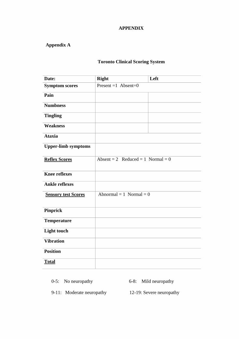
APPENDIX
Appendix A
Toronto Clinical Scoring System
Date: Right Left
Symptom scores Present =1 Absent=0
Pain
Numbness
Tingling
Weakness
Ataxia
Upper-limb symptoms
Reflex Scores Absent = 2 Reduced = 1 Normal = 0
Knee reflexes
Ankle reflexes
Sensory test Scores Abnormal = 1 Normal = 0
Pinprick
Temperature
Light touch
Vibration
Position
Total
0-5: No neuropathy 6-8: Mild neuropathy
9-11: Moderate neuropathy 12-19: Severe neuropathy
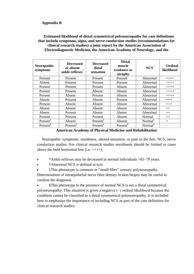
Appendix B
Estimated likelihood of distal symmetrical polyneuropathy for case definitions
that include symptoms, signs, and nerve conduction studies (recommendations for
clinical research studies)-a joint report by the American Association of
Electrodiagnostic Medicine, the American Academy of Neurology, and the
American Academy of Physical Medicine and Rehabilitation
Neuropathic symptoms: numbness, altered sensation, or pain in the feet. NCS, nerve
conduction studies. For clinical research studies enrollment should be limited to cases
above the bold horizontal line (i.e. ++++).
*Ankle reflexes may be decreased in normal individuals >65–70 years.
†Abnormal NCS is defined in text.
‡This phenotype is common in “small-fiber” sensory polyneuropathy.
Determination of intraepithelial nerve fiber density in skin biopsy may be useful to
confirm the diagnosis.
§This phenotype in the presence of normal NCS is not a distal symmetrical
polyneuropathy. This situation is given a negative (−) ordinal likelihood because the
condition cannot be classified as a distal symmetrical polyneuropathy. It is included
here to emphasize the importance of including NCS as part of the case definition for
clinical research studies.
Neuropathic
symptoms
Decreased
or absent
ankle reflexes*
Decreased
distal
sensation
Distal
muscle
weakness or
atrophy
NCS†
Ordinal
likelihood
Present Present Present Present Abnormal ++++ Absent Present Present Present Abnormal ++++ Present Present Present Absent Abnormal ++++ Present Present Absent Absent Abnormal ++++ Present Absent Present Absent Abnormal ++++ Absent Present Absent Present Abnormal +++ Present Absent Absent Absent Abnormal +++ Absent Absent Absent Absent Abnormal ++ Absent Present Absent Absent Abnormal ++ Present Present Present Absent Normal ++ Present
‡ Absent Present‡ Absent Normal
‡ +
Present§ Present
§ Present§ Present
§ Normal§ −


![METHODOLOGY REVIEW Assessing Decreased Sensation and ...€¦ · rimotor polyneuropathies (typical and atypical diabetic sensorimotor polyneuropathy [DSPN]); compression and entrapment](https://img.pdfslide.us/doc/110x75/60d3087a24053204b72786ba/methodology-review-assessing-decreased-sensation-and-rimotor-polyneuropathies.jpg)