Embed Size (px)
Citation preview
Guillain Barre Syndrome
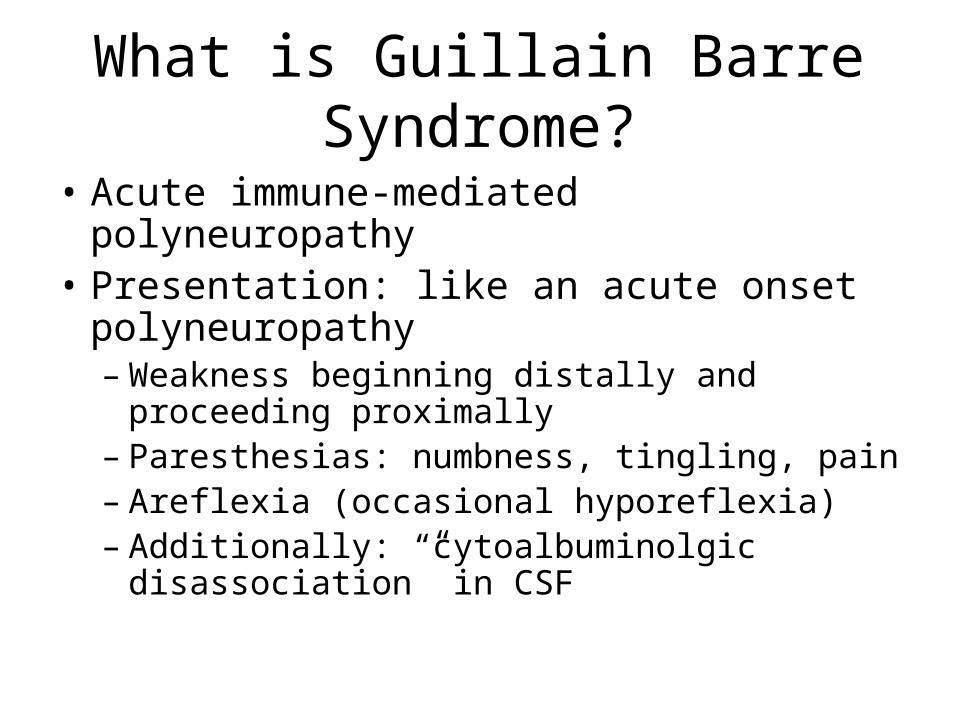
What is Guillain Barre Syndrome?
• Acute immune-mediated polyneuropathy• Presentation: like an acute onset
polyneuropathy– Weakness beginning distally and proceeding
proximally– Paresthesias: numbness, tingling, pain– Areflexia (occasional hyporeflexia)– Additionally: “cytoalbuminolgic disassociation” in
CSF
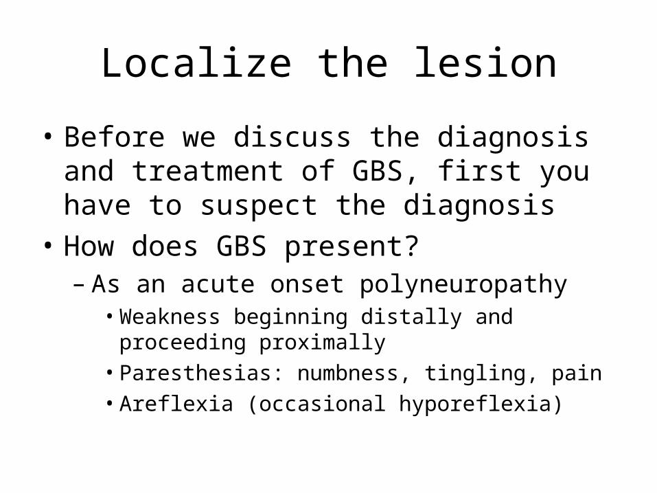
Localize the lesion
• Before we discuss the diagnosis and treatment of GBS, first you have to suspect the diagnosis
• How does GBS present?– As an acute onset polyneuropathy• Weakness beginning distally and proceeding proximally• Paresthesias: numbness, tingling, pain• Areflexia (occasional hyporeflexia)
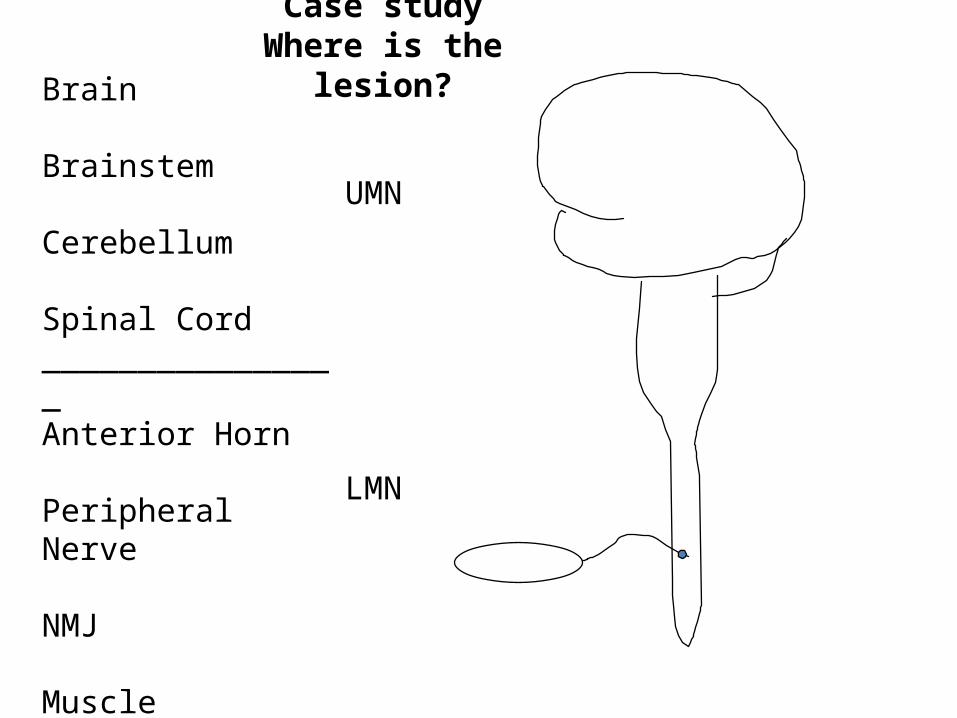
Brain
Brainstem
Cerebellum
Spinal Cord________________Anterior Horn
Peripheral Nerve
NMJ
Muscle
UMN
LMN
Case studyWhere is the lesion?
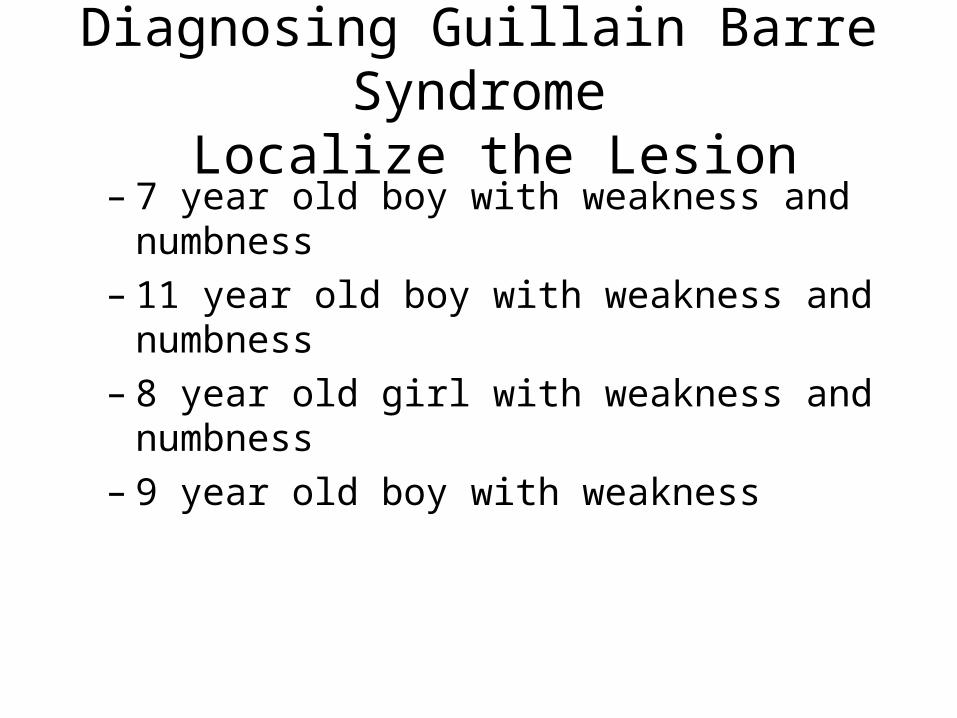
Diagnosing Guillain Barre Syndrome Localize the Lesion
– 7 year old boy with weakness and numbness– 11 year old boy with weakness and numbness– 8 year old girl with weakness and numbness– 9 year old boy with weakness
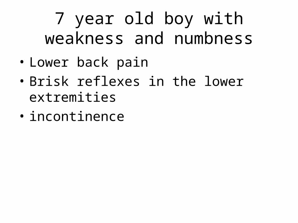
7 year old boy with weakness and numbness
• Lower back pain• Brisk reflexes in the lower extremities• incontinence
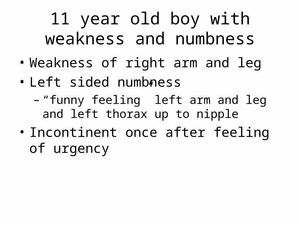
11 year old boy with weakness and numbness
• Weakness of right arm and leg• Left sided numbness– “funny feeling” left arm and leg and left thorax up
to nipple
• Incontinent once after feeling of urgency
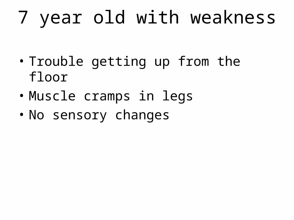
7 year old with weakness
• Trouble getting up from the floor• Muscle cramps in legs• No sensory changes
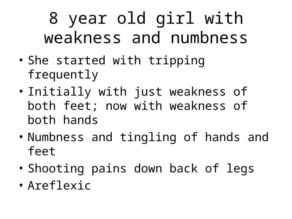
8 year old girl with weakness and numbness
• She started with tripping frequently• Initially with just weakness of both feet; now
with weakness of both hands• Numbness and tingling of hands and feet• Shooting pains down back of legs• Areflexic
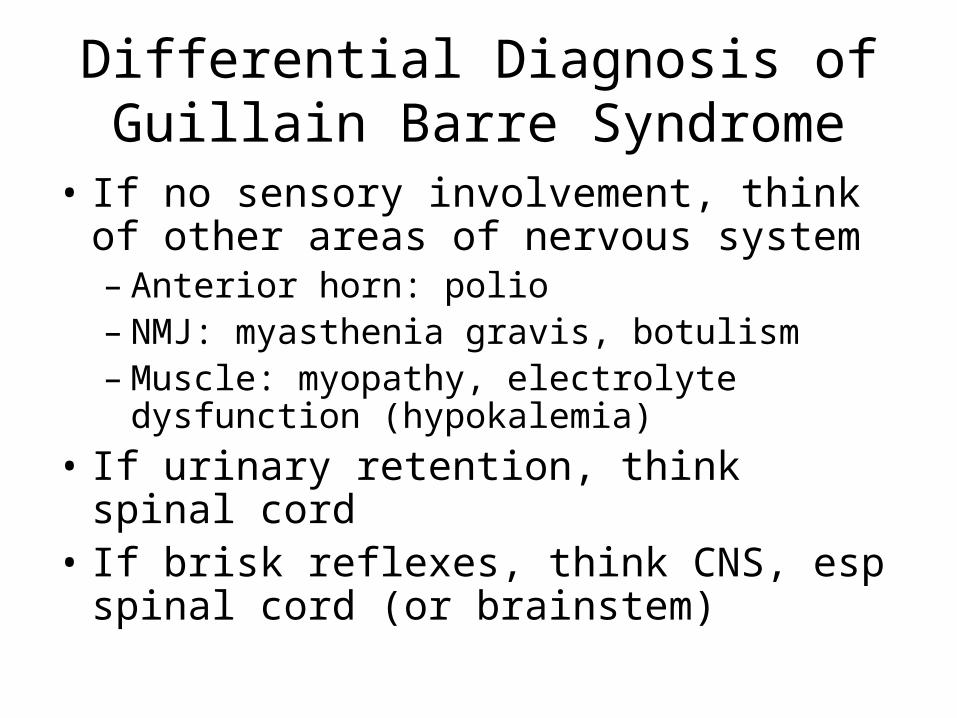
Differential Diagnosis of Guillain Barre Syndrome
• If no sensory involvement, think of other areas of nervous system– Anterior horn: polio– NMJ: myasthenia gravis, botulism– Muscle: myopathy, electrolyte dysfunction
(hypokalemia)• If urinary retention, think spinal cord• If brisk reflexes, think CNS, esp spinal cord (or
brainstem)
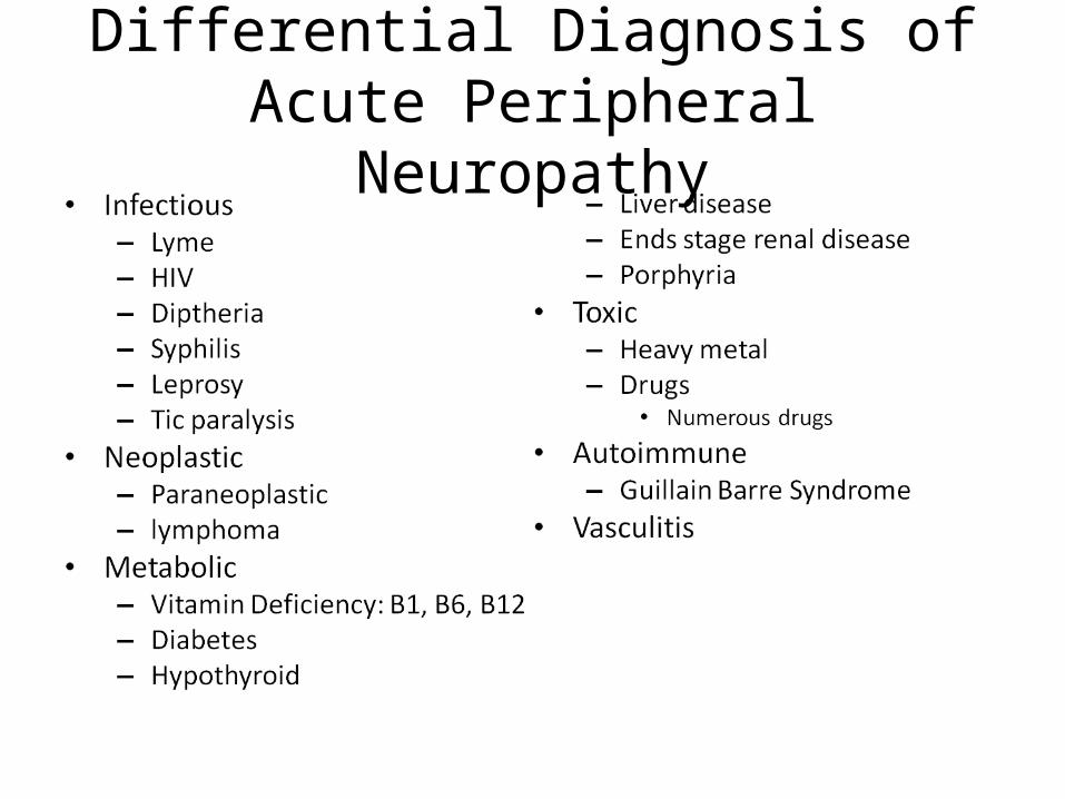
Differential Diagnosis of Acute Peripheral Neuropathy
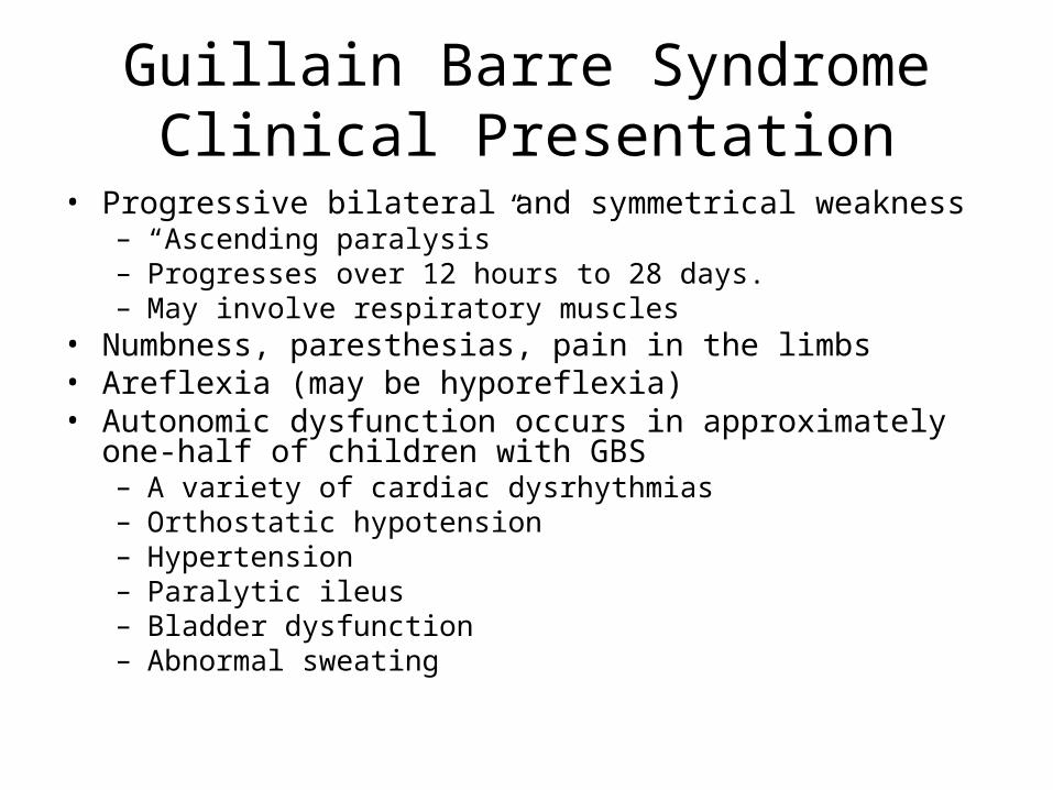
Guillain Barre SyndromeClinical Presentation
• Progressive bilateral and symmetrical weakness– “Ascending paralysis”– Progresses over 12 hours to 28 days. – May involve respiratory muscles
• Numbness, paresthesias, pain in the limbs• Areflexia (may be hyporeflexia)• Autonomic dysfunction occurs in approximately one-half of children
with GBS– A variety of cardiac dysrhythmias– Orthostatic hypotension– Hypertension– Paralytic ileus– Bladder dysfunction– Abnormal sweating
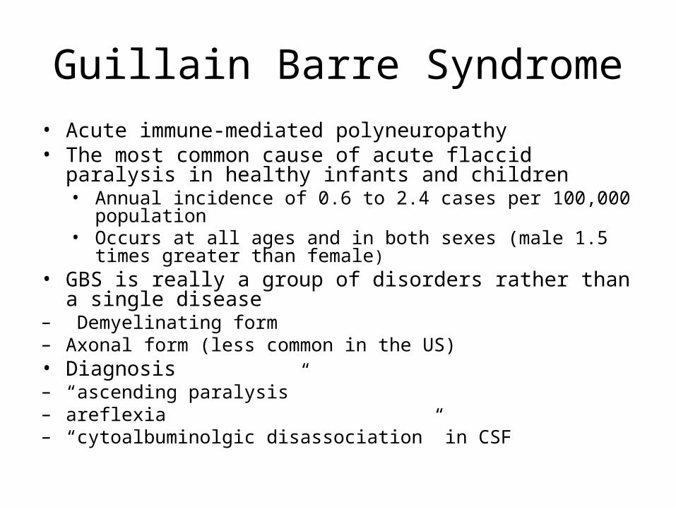
Guillain Barre Syndrome• Acute immune-mediated polyneuropathy• The most common cause of acute flaccid paralysis in healthy infants
and children• Annual incidence of 0.6 to 2.4 cases per 100,000 population• Occurs at all ages and in both sexes (male 1.5 times greater than
female)• GBS is really a group of disorders rather than a single disease– Demyelinating form– Axonal form (less common in the US)• Diagnosis– “ascending paralysis”– areflexia– “cytoalbuminolgic disassociation” in CSF
Etiology of Guillain Barre Syndrome
• Two thirds of cases have preceding URI or diarrhea– Campylobacter jejuni identified in 30%– CMV identified in 10%– Also identified: Epstein-Barr virus, Haemophilus influenzae
mycoplasma pneumoniae, the enteroviruses, hepatitis A and B, herpes simplex, and Chlamydophila (formerly Chlamydia) pneumoniae
• Post vaccine: other than the 1976 swine flu vaccine, no clear linkage seen in other vaccines
Etiology of Guillain Barre Syndrome
• Acute immune-mediated polyneuropathy– Antiganglioside antibodies develop• Some evidence supports the presence of molecular
mimicry between gangliosides and antecedent infectious agents• Gangliosides are a component of peripheral nerves• Different autoantibodies are associated with different
forms of GBS– IgG autoantibodies to GM1 and GD1a associated with acute
motor axonal neuropathy– IgG autoantibodies to GQ1b associated with Miller Fisher
sindrome
Diagnosis of GBS• Clinical triad of ascending paralysis, areflexia and cytoalbuminologic
association• CSF
– elevated protein concentration (> 45 mg/dL)• Maximum protein values may not be seen for four to five weeks
– A normal opening pressure– fewer than 10 cells (typically mononuclear)
• An initial pleocytosis of less than 100 lymphocytes may occur• If this is noted, consider other diseases associated with GBS such as HIV
infection, Lyme disease, and malignancy– These findings may be delayed and a repeat lumbar puncture may be required.
• Electrophysiologic studies– the most specific and sensitive tests for diagnosis of the disease. – Evidence of conduction block usually is the earliest abnormality– Electromyogram abnormalities typically are delayed for two to three weeks
VARIANTS OF GUILLAIN-BARRÉ SYNDROME
• Acute inflammatory demyelinating polyneuropathy (AIDP) – the prototype of GBS– most common form in North America, Europe; accounts for about 85 to 90 percent of cases
there• Acute motor axonal neuropathy
– pure motor form of GBS– electrophysiologic pattern suggesting axonal (rather than demyelinating) damage. – occurs mainly in northern China, but is also a common form of GBS in Japan, Mexico, and
South America– The presenting clinical features and recovery are similar to those of AIDP. – Tends to be worse prognosis thant AIDP
• Miller Fisher syndrome– external ophthalmoplegia, ataxia, and muscle weakness with areflexia– CSF and electrophysiologic features are similar to those in AIDP. – Brainstem auditory evoked potentials demonstrate peripheral and central conduction defects– More common in Eastern Asia– Preceding infection commmon; 20% C. jejuni, 8% H.flu
Clinical Course of Guillain Barre• More than 90 percent of patients reach the nadir of their function
within two to four weeks• Return of function occurring slowly over the course of weeks to
months• The clinical course of GBS in children is shorter than in adults and
recovery is more complete • The severity of GBS in children does not correlate with long-term
outcome. – 85 percent of children can be expected to have an excellent recovery– Approximately one-half of patients are ambulatory by six months, and
70 percent walk within a year after onset.– Mortality is 3 to 4 percent, and usually is secondary to respiratory
failure or cardiac complications.
Treatment of GBS
• Supportive Care• IVIG• Plasmapheresis
Treatment of GBS
• Supportive Care– close monitoring of motor, autonomic (blood pressure,
heart rate and sphincter function), and respiratory function
– serial lung function testing should be performed in those at highest risk for developing respiratory failure.• The following parameters warn of impending respiratory
arrest and are an indication for intubation [4]:– Vital capacity ≤20 mL/kg– Maximum inspiratory pressure ≤30 cmH2O– Maximum expiratory pressure ≤40 cmH2O– Tidal volume <5 mL/kg
Treatment of GBS in children
• Intravenous Immune Globulin (IVIG)– no large randomized controlled trials exist in children.
Nevertheless, data from the few available trials in children suggest that IVIG shortens the time to recovery compared with supportive care alone
– strength generally begins to improve in most children within 14 days after initiation of IVIG therapy and most are walking within three months
– The total dose of IVIG for the treatment of GBS in children is 2 g/kg, given as 1 g/kg for two days or 400 mg/kg for five days.
Treatment of GBS in children• Plasma Exchange– Large, multicenter trials have established the effectiveness
of plasma exchange in adult patients with severe GBS. – Treatment in pediatric patients appear to be similar to
those in adults• A meta-analysis of six trials found that treatment of children with plasma
exchange was superior to supportive care. The number of children was small and all children were older than 10 years old.
– Plasma exchange is most effective when started within seven days of symptom onset.
– The mechanism is thought to be removal of antibodies directed against nerves from the circulation.
IVIG vs. Plasmapheresis: Recommendations of the AAN
• Both intravenous immune globulin (IVIG) and plasma exchange are options for children with severe GBS.
• IVIG is preferred to plasma exchange in children because of the relative safety and ease of administration
• IVIG and plasma exchange for children with GBS should be reserved for those with:– Rapidly progressing weakness– Worsening respiratory status or need for mechanical ventilation– Significant bulbar weakness– Inability to walk unaided
Treatment of GBS in childrenRecommendations of the AAN
References• Hughes RA, Wijdicks EF, Barohn R, et al. Practice
parameter: immunotherapy for Guillain-Barré syndrome: report of the Quality Standards Subcommittee of the American Academy of Neurology. Neurology 2003; 61:736.
• Patwa HS, Chaudhry V, Katzberg H, et al. Evidence-based guideline: Intravenous immunoglobulin in the treatment of neuromuscular disorders: Report of the Therapeutics and Technology Assessment Subcommittee of the American Academy of Neurology. Neurology 2012; 78:1009.
• Yoki N and Hartung HP. Guillain Barre Syndrom. NEJM 2012;366:2294.