Embed Size (px)
Citation preview
Intraperitoneal a-ParticleRadioimmunotherapy of Ovarian CancerPatients: Pharmacokinetics and Dosimetryof 211At-MX35 F(ab9)2—A Phase I Study
Hakan Andersson1, Elin Cederkrantz2, Tom Back2, Chaitanya Divgi3, Jorgen Elgqvist1, Jakob Himmelman2,Gyorgy Horvath1, Lars Jacobsson2, Holger Jensen4, Sture Lindegren2, Stig Palm2, and Ragnar Hultborn1
1Department of Oncology, University of Gothenburg, Gothenburg, Sweden; 2Department of Radiation Physics, University ofGothenburg, Gothenburg, Sweden; 3Hospital of the University of Pennsylvania, Philadelphia, Pennsylvania; and 4PET andCyclotron Unit, Rigshospitalet, Copenhagen, Denmark
The a-emitter 211At labeled to a monoclonal antibody has provensafe and effective in treating microscopic ovarian cancer in theabdominal cavity of mice. Women in complete clinical remissionafter second-line chemotherapy for recurrent ovarian carcinomawere enrolled in a phase I study. The aim was to determine thepharmacokinetics for assessing absorbed dose to normal tis-sues and investigating toxicity. Methods: Nine patients un-derwent laparoscopy 2–5 d before the therapy; a peritonealcatheter was inserted, and the abdominal cavity was inspectedto exclude the presence of macroscopic tumor growth or majoradhesions. 211At was labeled to MX35 F(ab9)2 using the reagentN-succinimidyl-3-(trimethylstannyl)-benzoate. Patients were in-fused with 211At-MX35 F(ab9)2 (22.4–101 MBq/L) in dialysis solu-tion via the peritoneal catheter. g-camera scans were acquiredon 3–5 occasions after infusion, and a SPECT scan was acquiredat 6 h. Samples of blood, urine, and peritoneal fluid were col-lected at 1–48 h. Hematology and renal and thyroid functionwere followed for a median of 23 mo. Results: Pharmacokineticsand dosimetric results were related to the initial activity concen-tration (IC) of the infused solution. The decay-corrected activityconcentration decreased with time in the peritoneal fluid to50% IC at 24 h, increased in serum to 6% IC at 45 h, and in-creased in the thyroid to 127% 6 63% IC at 20 h without blockingand less than 20% IC with blocking. No other organ uptakescould be detected. The cumulative urinary excretion was 40kBq/(MBq/L) at 24 h. The estimated absorbed dose to the perito-neum was 15.6 6 1.0 mGy/(MBq/L), to red bone marrow it was0.14 6 0.04 mGy/(MBq/L), to the urinary bladder wall itwas 0.77 6 0.19 mGy/(MBq/L), to the unblocked thyroid it was24.7 6 11.1 mGy/(MBq/L), and to the blocked thyroid it was1.4 6 1.6 mGy/(MBq/L) (mean 6 SD). No adverse effects wereobserved either subjectively or in laboratory parameters.Conclusion: This study indicates that by intraperitoneal admin-istration of 211At-MX35 F(ab9)2 it is possible to achieve thera-peutic absorbed doses in microscopic tumor clusters withoutsignificant toxicity.
Key Words: astatine; dosimetry; pharmacokinetics;radioimmunotherapy; clinical study
J Nucl Med 2009; 50:1153–1160DOI: 10.2967/jnumed.109.062604
The lifetime risk of ovarian cancer is 1%22% inEuropean and U.S. women. Despite seemingly successfulcytoreductive surgery, followed by systemic chemotherapy,most patients will relapse and succumb. The relapse is mostfrequently localized in the abdominal cavity. New systemicchemotherapy regimens have not improved the outcomeover the past decade, which prompted experimental intra-peritoneal treatments, including radioimmunotherapy.
Radioimmunotherapy with b-emitters has displayedpromising results, although an international randomizedphase III study of 90Y-HMFG1 showed no improvement insurvival or time to relapse (1). This disappointing resultcould be partly explained by the choice of radionuclide. Thelong range of this b-emitter results in poor irradiation ofsmall tumor clusters, likely insufficient to eradicate peritonealmicrometastases. Furthermore, the relatively long half-life(T1/2) of 64 h of 90Y is not optimal, considering bone-marrowirradiation.
In treating micrometastases, a-emitters offer significantadvantages over b-emitters. Because of the short range andhigh linear energy transfer of a-particles, targeted smallcell clusters are more effectively irradiated. As the range ofthe emitted particles conforms to the size of the targetcluster, a high fraction of emitted energy will be absorbed inthe target. The therapeutic potential of the a-emitter 211At(T1/2 5 7.21 h) labeled to MOv18, MX35, and trastuzumabhas been demonstrated in studies using a preclinical mouseovarian cancer model (2–9).
Received Jan. 29, 2009; revision accepted Mar. 18, 2009.For correspondence or reprints contact: Ragnar Hultborn, Department
of Oncology, Sahlgrenska University Hospital, SE-413 45 Gothenburg,Sweden.
E-mail: [email protected] ª 2009 by the Society of Nuclear Medicine, Inc.
a-PARTICLE RIT OF OVARIAN CANCER • Andersson et al. 1153
by on June 5, 2018. For personal use only. jnm.snmjournals.org Downloaded from
Immunohistochemistry indicates that the antibody MX35displays homogeneous reactivity with approximately 90%of human ovarian epithelial cancers and with a limitednumber of normal tissues (10). Specific tumor localizationof 125I- and 131I-labeled MX35 and MX35 F(ab9)2 has beendemonstrated in patients with intraperitoneal growth ofovarian cancer (11,12).
The encouraging results of using 211At-MX35 F(ab9)2 inthe nude mouse model justified the present clinical phase Istudy. In addition to defining the pharmacokinetics of211At-labeled MX35 F(ab9)2 to enable absorbed dose esti-mates, the aim was to investigate the feasibility and safety.Women relapsing after complete clinical remission aftersurgery and chemotherapy and reaching another remissionon salvage chemotherapy were included in the study.
MATERIALS AND METHODS
Antibody and Cell LineA clinical-grade F(ab9)2 fragment of the murine IgG1 mono-
clonal antibody MX35 was obtained from Memorial Sloan-Kettering Cancer Center. The antigen recognized by MX35 hasrecently been characterized as the sodium-dependent phosphatetransport protein 2b (NaPi2b), which is overexpressed on morethan 90% of human ovarian epithelial cancers (13). The humanovarian cancer cell line NIH:OVCAR-3, obtained from AmericanType Culture Collection, was used for immunologic in vitrocontrol of the radiolabeled MX35 F(ab9)2 product.
211At Production and Distillation211At was produced and distilled as previously described (14).
In short, 211At was produced by means of the 209Bi(a,2n)211Atreaction using a cyclotron (MC32; Scanditronix) at the PET andCyclotron Unit, Rigshospitalet. Irradiation was performed at aninternal water-cooled probe, using beam energies of 28–29 MeVand beam currents of 16.8 6 1.5 mA. Activities of 2.0 6 0.2 GBq,which correspond to a saturation yield of 230 6 20 MBq/mA,were achieved with irradiation times of 7.6 6 0.2 h. The targetwas transported to Gothenburg, Sweden, by car within 4 h, andthen transformed into a chemically useful form by dry distillation.All glassware was autoclaved before distillation. After distillation,the 211At was eluted with 300 mL of chloroform into a 1.3-mLreaction vial. The 211At was then divided into six to eight 150-MBq fractions in 1.3-mL reaction vials, and the chloroform wasevaporated under a gentle stream of nitrogen.
Labeling of MX35 F(ab9)2The antibody fragment was labeled in clinically approved facil-
ities at the Nuclear Medicine Department, Sahlgrenska University
Hospital, using the reagent N-succinimidyl-3-(trimethylstannyl)-benzoate as previously described (15). All glassware and utensilswere sterilized. Buffer solutions and chemicals were prepared at thelocal pharmacy. A total of 150 mg of MX35 F(ab9)2 were added toeach 150-MBq fraction. The 211At-MX35 F(ab9)2 product wastransferred through a 0.2-mm sterile filter into a peritoneal dialysisfluid (Extraneal; Baxter) bag. Overall radiochemical yields were in therange of 20%230%.
Quality ControlA 50-mL aliquot of the astatinated product was taken for
quality control. The radiochemical purity was determined by bothmethanol precipitation and fast-protein liquid chromatography(FPLC) on a Superdex-200 column using an AKTA-FPLC deliv-ery system (GE Healthcare Bio-Sciences A). Only products with aradiochemical purity of greater than 95% were approved forclinical infusion.
The immunoreactive fraction of MX35 F(ab9)2 after astatina-tion was determined by binding to NIH:OVCAR-3 cells using themethod described by Lindmo et al. (16). Simultaneously with thecomplete assay, a single-point assay was started. To 4 tubescontaining NIH:OVCAR-3 cells at a concentration of 5 · 106
cells/mL, 10 ng of the astatinated product were added. The tubeswere incubated for 45 min, with a set limit of 50% immunoreac-tive fraction for infusion.
PatientsNine women (age, 38–69 y; median age, 52 y) were included in
the study; these women were initially successfully treated forovarian carcinoma but later relapsed and were treated long-termwith salvage chemotherapy, including paraplatin and paclitaxel,resulting in clinically and biochemically complete remission.According to the protocol, these patients were chosen for treat-ment at levels of increasing radioactivity. The patients wereincluded in the study after providing informed consent accordingto the Ethics Committee of the Sahlgrenska University Hospital inGothenburg.
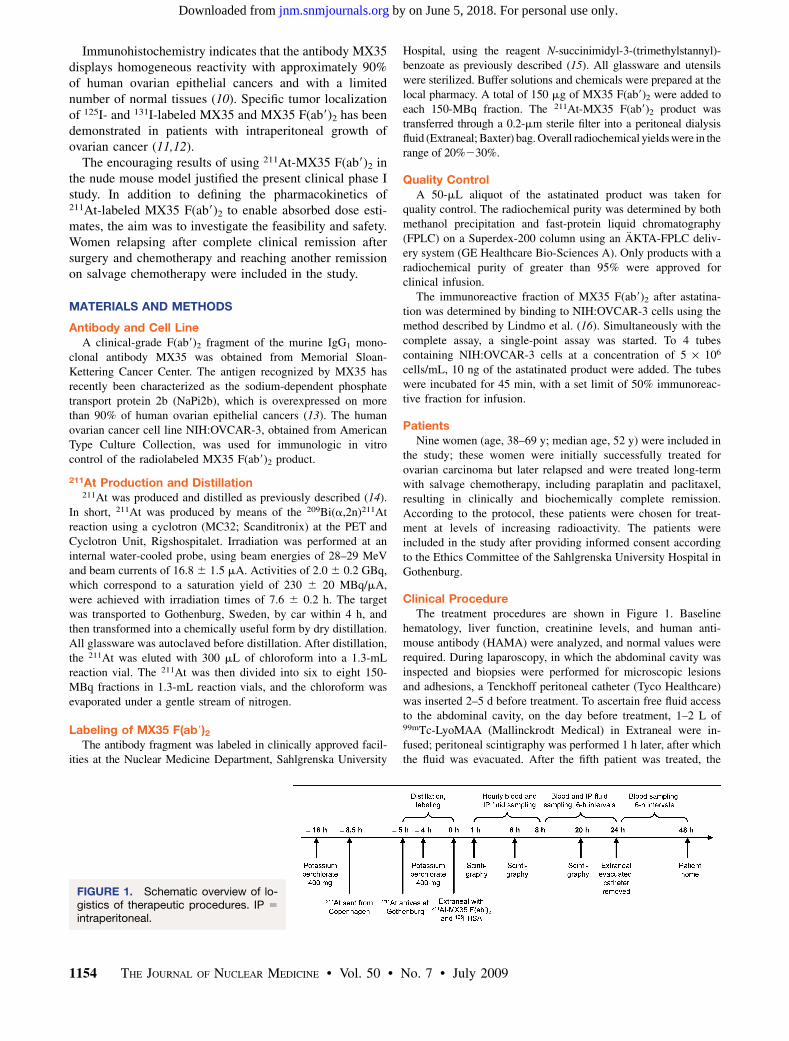
Clinical ProcedureThe treatment procedures are shown in Figure 1. Baseline
hematology, liver function, creatinine levels, and human anti-mouse antibody (HAMA) were analyzed, and normal values wererequired. During laparoscopy, in which the abdominal cavity wasinspected and biopsies were performed for microscopic lesionsand adhesions, a Tenckhoff peritoneal catheter (Tyco Healthcare)was inserted 2–5 d before treatment. To ascertain free fluid accessto the abdominal cavity, on the day before treatment, 1–2 L of99mTc-LyoMAA (Mallinckrodt Medical) in Extraneal were in-fused; peritoneal scintigraphy was performed 1 h later, after whichthe fluid was evacuated. After the fifth patient was treated, the
FIGURE 1. Schematic overview of lo-gistics of therapeutic procedures. IP 5
intraperitoneal.
1154 THE JOURNAL OF NUCLEAR MEDICINE • Vol. 50 • No. 7 • July 2009
by on June 5, 2018. For personal use only. jnm.snmjournals.org Downloaded from
protocol was amended, and the ensuing patients were givenpotassium perchlorate (custom-made tablets, 200 mg)—2 tabletsthe evening before and 2 the morning of the day of treatment—toblock uptake of astatine to the thyroid. The last patient (patient 9)was instead given potassium iodide (Recip tablets, 65 mg)—1tablet the evening before and 2 on the day of treatment.
211At-MX35 F(ab9)2 (22.4–101 MBq/L) in Extraneal (1–2 L,37�C) was infused via the peritoneal catheter over 30 min,together with 0.2 MBq of 125I-human serum albumin (HSA), areference for in vivo stability. Treatment specifications includingadministered activity concentration and volume are shown inTable 1. Twenty-four hours after infusion, the remaining perito-neal fluid was drained and collected from the catheter, which wasthen removed. Over the first 8 h after infusion, blood and intra-peritoneal fluid were sampled hourly. After this period, sampleswere drawn every 6 h until 48 h after infusion for blood and untildrainage (24 h) for intraperitoneal fluid. Urine was collectedat each voiding until 48 h. Scintigraphy was performed 1 h afterinfusion and every 6–12 h, totaling 3–5 scans. In addition, 6 hafter infusion, a SPECT or SPECT/CT study of the thoracic andabdominal area was conducted.
The patients were released from the hospital 48 h after infusionand were followed weekly for the next 8 wk at the outpatientdepartment. Follow-up consisted of the following tests: hematol-ogy together with biochemistry, including liver function, creati-nine, thyroid function, CA-125, and HAMA (i.e., the same testsperformed initially). Thereafter, the patients were followed ac-cording to the clinical routines.
Fluid Sample AnalysisBlood was taken in 2 separate tubes at each time point for
serum and whole-blood analysis. The serum was separated bycentrifugation at 500g for 10 min. From each blood, serum, andintraperitoneal fluid sample, a 1-mL aliquot was weighed andmeasured for the determination of 211At and 125I content. In afew patients, FPLC or methanol precipitation analyses wereperformed on serum or intraperitoneal fluid samples, to estimatethe protein-bound fraction of 211At. Reliable urine data wereobtained from only 4 patients. All urine was weighed, and asmall sample from each voiding was measured for 211At and125I content.
ScintigraphyPlanar g-camera scans were acquired using the polonium
x-rays inherent to the 211At decay for imaging. A 2-detector
g-camera system (Millenium VG; GE Healthcare) with a me-dium-energy collimator was used. The scans covered the areafrom the thyroid to the ankles. The scan speed was 10 cm/min,and the energy window was 79 keV 6 10%. A calibration factorof 7.65 counts/kBq for thyroid activity uptake was obtained forplanar images using a neck phantom. SPECT or SPECT/CT wasconducted using the same equipment and energy window. TheSPECT scan was acquired over 20 min in 360� and 120 projec-tions.
Radioactivity MeasurementsHigh activity (.100 kBq) was measured in an ionization
chamber (CRC-15 dose calibrator; Capintec), and low activity(,10 kBq) was measured with a thallium-doped sodium iodideg-counter (Wizard 1480; Wallac). Samples containing both 211Atand 125I were measured twice, the second time after decay of211At. Dual-energy window settings in the g-counter enabledspillover correction. The 2 devices were cross-calibrated for bothnuclides.
DosimetryA simple approach, in which all but the a-particle contribution
were disregarded, was used for the dosimetry. Because themicrodistribution of the labeled immunoconjugate in human tissueis not known in detail, a homogenous distribution of the radioac-tivity was assumed. The absorbed dose, Dx, for organ x wascalculated using Equation 1, in which the absorbed fraction of thea-particles, fa, was assumed to be 1, the mean energy releasedper 211At decay, Da, is 1.09 · 10212 J (Bq s)21, and the cumulatedactivity in organ x, Ax, was calculated from organ uptake datausing Equation 2:
Dx 5Ax
mxDafa Eq. 1
Ax 5
Z N
0
pxexp 2ltdt; l 5ln2
T1=2
: Eq. 2
Function px in Equation 2 was fitted to decay-corrected uptakedata for organ x. Different fit models were used for differentorgans. The integration was performed numerically to 48 h, cor-responding to approximately 6.6 half-lives of 211At (T1/2 5 7.21 h).The absorbed doses presented in this study were not weightedfor relative biologic effectiveness for 2 reasons: first, because
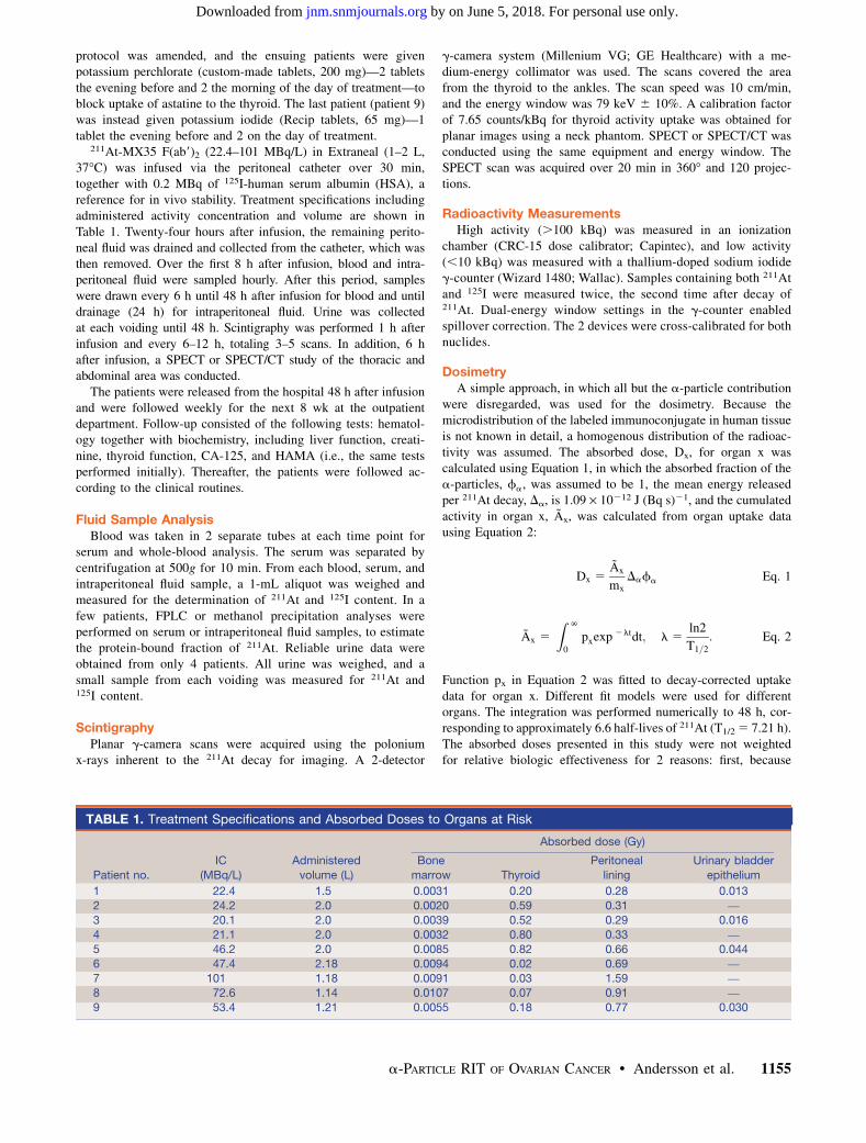
TABLE 1. Treatment Specifications and Absorbed Doses to Organs at Risk
Patient no.
IC
(MBq/L)
Administered
volume (L)
Absorbed dose (Gy)
Bone
marrow Thyroid
Peritoneal
lining
Urinary bladder
epithelium
1 22.4 1.5 0.0031 0.20 0.28 0.013
2 24.2 2.0 0.0020 0.59 0.31 —
3 20.1 2.0 0.0039 0.52 0.29 0.0164 21.1 2.0 0.0032 0.80 0.33 —
5 46.2 2.0 0.0085 0.82 0.66 0.044
6 47.4 2.18 0.0094 0.02 0.69 —
7 101 1.18 0.0091 0.03 1.59 —
8 72.6 1.14 0.0107 0.07 0.91 —
9 53.4 1.21 0.0055 0.18 0.77 0.030
a-PARTICLE RIT OF OVARIAN CANCER • Andersson et al. 1155
by on June 5, 2018. For personal use only. jnm.snmjournals.org Downloaded from
there is no formally adopted standard for the quantity (17), andsecond, because reliable relative biological effectiveness values arenot yet determined for all organs included.
The bone-marrow dosimetry was based on serum activityconcentration data. The activity concentration in red bone mar-row is directly proportional to the serum activity concentrationvia the red marrow extracellular fluid fraction (18), which wasestimated to be 0.19 in a study of the albumin distributionvolume in the femoral bone marrow of rabbits (19). Calculatedred bone–marrow concentration data were fitted to a third-degreepolynomial to be used in Equation 2. For the thyroid, a second-degree polynomial was fitted to decay-corrected uptake datacalculated from the planar g-camera scans. The thyroid locationwas not always easily found, especially in the blocked patients.In these cases, the location was estimated; if the content was lessthan background, it was set to zero. A standard thyroid mass of20 g was used for all patients. For the peritoneal lining andurinary bladder epithelium, the absorbed dose was calculated ashalf of the equilibrium absorbed dose to the fluid in the cavity.The intraperitoneal fluid activity concentration data were fitted toEquation 3:
pi:p:fluid 5 Ae2Bt 1 C: Eq. 3
The dose calculations were made for 0–48 h, even though thedata were limited to 24 h. This approach was justified by the factthat a fluid film will remain on the peritoneal surfaces after thedrainage at 24 h. The urine data were entered into Equation 2 as astepwise flat function, assuming constant activity concentrationbetween each voiding.
RESULTS
For this treatment modality, it is the activity concentrationand not the activity per se that determines the absorbed doseto tumor and to organs at risk. The absorbed doses weretherefore related to the initial activity concentration, IC(MBq/L), of the infused solution. All activities were decay-corrected to the time of infusion and normalized to thestarting concentration.
Fluid Sampling
The protein-bound fraction of 211At in intraperitonealfluid was evaluated in patient 1 by FPLC analysis of samplesdrawn 2, 8, and 25 h after infusion. At none of the time pointscould significant amounts of free 211At be found. This wasconfirmed by methanol precipitation of intraperitoneal fluidsamples from the same patient and from patient 2. Theprotein-bound fraction was 95% or higher. Methanol precip-itation of serum was performed on samples from patients1 and 9. The protein-bound fraction was approximately 80%in patient 1 and above 90% in patient 9 after 6 h.
A curve of the form A*exp(2Bt) 1 C was fitted to theintraperitoneal fluid concentration data for each patient.Extrapolation of the curve to t 5 0 yielded the individualIC, to which all other data were normalized. The initialconcentrations are listed in Table 1.
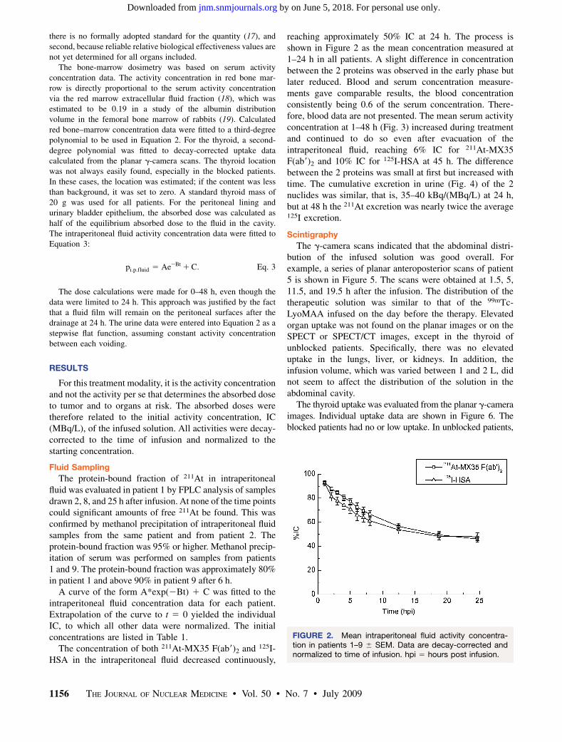
The concentration of both 211At-MX35 F(ab9)2 and 125I-HSA in the intraperitoneal fluid decreased continuously,
reaching approximately 50% IC at 24 h. The process isshown in Figure 2 as the mean concentration measured at1–24 h in all patients. A slight difference in concentrationbetween the 2 proteins was observed in the early phase butlater reduced. Blood and serum concentration measure-ments gave comparable results, the blood concentrationconsistently being 0.6 of the serum concentration. There-fore, blood data are not presented. The mean serum activityconcentration at 1–48 h (Fig. 3) increased during treatmentand continued to do so even after evacuation of theintraperitoneal fluid, reaching 6% IC for 211At-MX35F(ab9)2 and 10% IC for 125I-HSA at 45 h. The differencebetween the 2 proteins was small at first but increased withtime. The cumulative excretion in urine (Fig. 4) of the 2nuclides was similar, that is, 35–40 kBq/(MBq/L) at 24 h,but at 48 h the 211At excretion was nearly twice the average125I excretion.
Scintigraphy
The g-camera scans indicated that the abdominal distri-bution of the infused solution was good overall. Forexample, a series of planar anteroposterior scans of patient5 is shown in Figure 5. The scans were obtained at 1.5, 5,11.5, and 19.5 h after the infusion. The distribution of thetherapeutic solution was similar to that of the 99mTc-LyoMAA infused on the day before the therapy. Elevatedorgan uptake was not found on the planar images or on theSPECT or SPECT/CT images, except in the thyroid ofunblocked patients. Specifically, there was no elevateduptake in the lungs, liver, or kidneys. In addition, theinfusion volume, which was varied between 1 and 2 L, didnot seem to affect the distribution of the solution in theabdominal cavity.
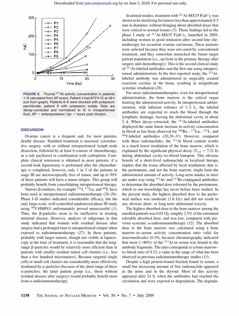
The thyroid uptake was evaluated from the planar g-cameraimages. Individual uptake data are shown in Figure 6. Theblocked patients had no or low uptake. In unblocked patients,
FIGURE 2. Mean intraperitoneal fluid activity concentra-tion in patients 1–9 6 SEM. Data are decay-corrected andnormalized to time of infusion. hpi 5 hours post infusion.
1156 THE JOURNAL OF NUCLEAR MEDICINE • Vol. 50 • No. 7 • July 2009
by on June 5, 2018. For personal use only. jnm.snmjournals.org Downloaded from
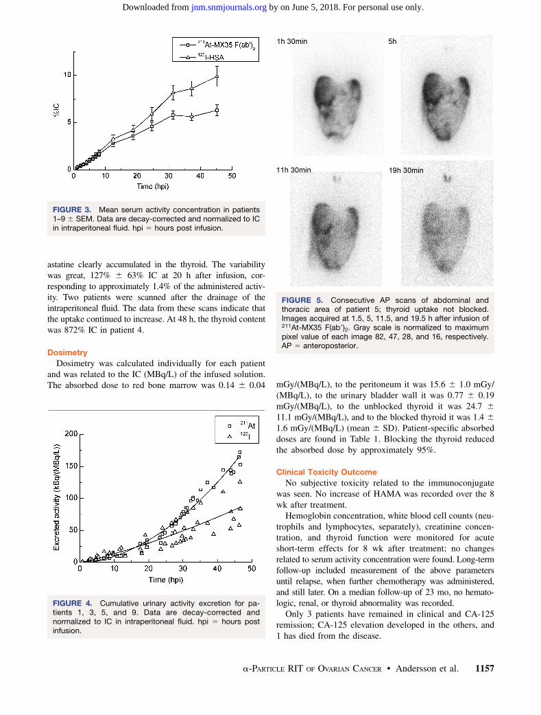
astatine clearly accumulated in the thyroid. The variabilitywas great, 127% 6 63% IC at 20 h after infusion, cor-responding to approximately 1.4% of the administered activ-ity. Two patients were scanned after the drainage of theintraperitoneal fluid. The data from these scans indicate thatthe uptake continued to increase. At 48 h, the thyroid contentwas 872% IC in patient 4.
Dosimetry
Dosimetry was calculated individually for each patientand was related to the IC (MBq/L) of the infused solution.The absorbed dose to red bone marrow was 0.14 6 0.04 mGy/(MBq/L), to the peritoneum it was 15.6 6 1.0 mGy/
(MBq/L), to the urinary bladder wall it was 0.77 6 0.19mGy/(MBq/L), to the unblocked thyroid it was 24.7 6
11.1 mGy/(MBq/L), and to the blocked thyroid it was 1.4 6
1.6 mGy/(MBq/L) (mean 6 SD). Patient-specific absorbeddoses are found in Table 1. Blocking the thyroid reducedthe absorbed dose by approximately 95%.
Clinical Toxicity Outcome
No subjective toxicity related to the immunoconjugatewas seen. No increase of HAMA was recorded over the 8wk after treatment.
Hemoglobin concentration, white blood cell counts (neu-trophils and lymphocytes, separately), creatinine concen-tration, and thyroid function were monitored for acuteshort-term effects for 8 wk after treatment; no changesrelated to serum activity concentration were found. Long-termfollow-up included measurement of the above parametersuntil relapse, when further chemotherapy was administered,and still later. On a median follow-up of 23 mo, no hemato-logic, renal, or thyroid abnormality was recorded.
Only 3 patients have remained in clinical and CA-125remission; CA-125 elevation developed in the others, and1 has died from the disease.
FIGURE 3. Mean serum activity concentration in patients1–9 6 SEM. Data are decay-corrected and normalized to ICin intraperitoneal fluid. hpi 5 hours post infusion.
FIGURE 4. Cumulative urinary activity excretion for pa-tients 1, 3, 5, and 9. Data are decay-corrected andnormalized to IC in intraperitoneal fluid. hpi 5 hours postinfusion.
FIGURE 5. Consecutive AP scans of abdominal andthoracic area of patient 5; thyroid uptake not blocked.Images acquired at 1.5, 5, 11.5, and 19.5 h after infusion of211At-MX35 F(ab9)2. Gray scale is normalized to maximumpixel value of each image 82, 47, 28, and 16, respectively.AP 5 anteroposterior.
a-PARTICLE RIT OF OVARIAN CANCER • Andersson et al. 1157
by on June 5, 2018. For personal use only. jnm.snmjournals.org Downloaded from
DISCUSSION
Ovarian cancer is a frequent and, for most patients,deadly disease. Standard treatment is maximal cytoreduc-tive surgery, with or without retroperitoneal lymph nodedissection, followed by at least 6 courses of chemotherapy,as a rule paclitaxel in combination with carboplatin. Com-plete clinical remission is obtained in most patients; if asecond-look laparotomy is performed after the chemother-apy is completed, however, only 1 in 3 of the patients instage III are microscopically free of tumor, and up to 50%of those patients will have a tumor relapse. This group willprobably benefit from consolidating intraperitoneal therapy.
Various b-emitters, for example 131I, 177Lu, and 90Y, havebeen used in intraperitoneal radioimmunotherapy (20–26).Phase I–II studies indicated considerable efficacy, but theonly large-scale, well-controlled randomized phase III studyusing 90Y-HMFG1 unfortunately proved unsuccessful (1).Thus, the b-particles seem to be ineffective in treatingminimal disease. However, analysis of subgroups in thatstudy indicated that women with residual disease aftersurgery had a prolonged time to intraperitoneal relapse whenexposed to radioimmunotherapy (27). In these patients,probably with larger tumors, though not visible at laparos-copy at the time of treatment, it is reasonable that the long-range b-particles would be relatively more efficient than inpatients with smaller residual tumor cell clusters (i.e., lessthan a few hundred micrometers). Because targeted singlecells or small cell clusters are considerably more effectivelyirradiated by a-particles (because of the short range of thesea-particles), the latter patient group (i.e., those withoutresidual disease after surgery) would probably benefit mostfrom a-radioimmunotherapy.
In animal studies, treatment with 211At-MX35 F(ab9)2 wasshown to be sterilizing for tumors less than approximately 0.5mm in diameter, without bringing about absorbed doses thatwere critical to normal tissues (5). Those findings led to thephase I study of 211At-MX35 F(ab9)2, launched in 2005,including women in good remission after second-line che-motherapy for recurrent ovarian carcinoma. These patientswere selected because they were not cured by conventionaltreatment, and they somewhat mimicked the future targetpatient population (i.e., up front in the primary therapy aftersurgery and chemotherapy). This is the second clinical studyof 211At-labeled antibodies and the first one using intraperi-toneal administration. In the first reported study, the 211At-labeled antibody was administered in surgically createdresection cavities in the brain, resulting in insignificantsystemic irradiation (28).
For most radioimmunotherapies, even for intraperitonealadministration, the bone marrow is the critical organlimiting the administered activity. In intraperitoneal admin-istration, with infusion volumes of 1–2 L, the labeledantibodies are expected to reach the blood through thelymphatic drainage, leaving the abdominal cavity in about2 d. When decay-corrected, the 211At-labeled antibodiesdisplayed the same linear increase in activity concentrationin blood as has been observed for 186Re-, 177Lu-, 131I-, and90Y-labeled antibodies (20,29–31). However, comparedwith these radionuclides, the 211At blood content resultsin a much lower irradiation of the bone marrow, which isexplained by the significant physical decay (T1/2 5 7.21 h)during abdominal cavity–to–blood transport. This obviousbenefit of a short-lived radionuclide in localized therapyimplies that the tissue affected by local irradiation, that is,the peritoneum, and not the bone marrow, might limit theadministered amount of activity. Long-term studies in miceare under way using 211At- and 213Bi-conjugated antibodiesto determine the absorbed dose tolerated by the peritoneum,which to our knowledge has never before been studied. Inthe present study, the highest absorbed dose to the perito-neal surface was moderate (1.6 Gy) and did not result inany obvious short- or long-term abdominal toxicity.
The highest absorbed dose to the bone marrow among theenrolled patients was 0.01 Gy, roughly 2.5% of the estimatedtolerable absorbed dose, and was low, compared with pre-vious systemic a-radioimmunotherapy (32). The absorbeddose to the bone marrow was calculated using a bonemarrow–to–serum activity concentration ratio valid formacromolecules (0.19), because chromatography indicatedthat most (.80%) of the 211At in serum was bound to theantibody fragments. The ratio corresponds to a bone marrow–to–blood ratio of 0.32, a value in the range of what has beenobserved in previous radioimmunotherapy studies (33).
Despite a high protein-bound fraction found in serum, asmall but increasing amount of free radionuclide appearedin the urine and in the thyroid. Most of this activityappeared after 24 h, when the antibodies had reached thecirculation and were exposed to degradation. The degrada-
FIGURE 6. Thyroid 211At activity concentration in patients1–9 calculated from AP scans. Patient 4 had 872% IC at 48 h(cut from graph). Patients 6–8 were blocked with potassiumperchlorate, patient 9 with potassium iodide. Data aredecay-corrected and normalized to IC in intraperitonealfluid. AP 5 anteroposterior; hpi 5 hours post infusion.
1158 THE JOURNAL OF NUCLEAR MEDICINE • Vol. 50 • No. 7 • July 2009
by on June 5, 2018. For personal use only. jnm.snmjournals.org Downloaded from
tion and subsequent release of free 211At was also indicatedby the increasing difference in plasma concentrations of211At-MX35 F(ab9)2 and 125I-HSA, the latter being themore stable conjugate, having a long half-life in blood andnot being filtered in the kidneys. If not blocked, theirradiation of the thyroid is significant. After the introduc-tion of a blocking agent, the thyroid uptake was signifi-cantly reduced, thereby reducing by approximately 95% theabsorbed dose to an acceptable level. No change in thyroid-stimulating hormone levels was seen in either unblocked,low-activity patients or in blocked, higher-activity ones.
The stomach is another organ known to accumulate free211At via the sodium/iodide symporter receptor, thoughneither planar nor SPECT images revealed any uptakedistinguishable from background activity. However, a sig-nificant uptake in the thin outspread mucosa could havebeen overlooked against the high abdominal backgroundactivity. It is likely that the thyroid blocking agent may alsoblock any potential uptake of free 211At in the stomach(34); the risk of significant radiation damage to stomachtissue was, therefore, considered low. The other majororgans of interest, that is, the liver, lungs, and kidneys,did not display any uptake. The MX35 antigen has beendetected in normal human tissues, including the epithelialcells of the normal bronchus, lungs, sweat glands, kidneycollecting ducts, thyroid, fallopian tubes, cervix, and uterus(10). Thus, although not detected by the g-camera, thesetissues might accumulate 211At-MX35 F(ab9)2. Because thesystemic irradiation in general is low, the irradiation ofthese tissues is probably well below the tolerable level.
The binding of the antibodies to the tumor cells inmicrometastases will most likely occur via the peritonealfluid and not via vascular flow. Thus, the activity concen-tration of 211At-MX35 F(ab9)2 in the peritoneal fluiddetermines the irradiation of the microscopic peritonealtumors. In this study, the decay-corrected fluid concentra-tion of radiolabeled antibody declined with time, contraryto previous studies finding a stable or increasing concen-tration over 24 h (29,31). This difference is likely explainedby the osmotic agent used in our study, Extraneal, whichresults in water influx into the abdominal cavity. Extranealwas used to guarantee a well-filled abdominal cavity duringthe time of irradiation, that is, for 24 h.
Animal data indicate that a concentration of approxi-mately 200 MBq/L is needed for high therapeutic efficacy(8). To achieve that concentration for our patients, weaimed at a maximum concentration of 400 MBq in 2 L.Because the availability of labeled product was restrictedby limited production capacity of 211At and moderatelabeling yields, the volume was reduced to 1 L during thestudy to enable escalation of activity concentration. Yet,only a concentration of 100 MBq/L was attained. Thereduced volume does not affect tumor irradiation as long asthe peritoneal surface is completely exposed throughout theirradiation time (i.e., over ;24 h), because the binding ofantibodies to tumor cells and unspecific irradiation of the
peritoneum are concentration-dependent. This criterion ismost likely also fulfilled with a volume of 1 L whenExtraneal is used. From the g-camera images, we coulddistinguish no difference in distribution between patientswho received different volumes.
We have chosen to report biokinetic data and absorbeddoses related to the initial peritoneal fluid activity concen-tration (IC). The common way to provide biokinetic data isto relate them to the administered activity, presented in theform of percentage injected dose per gram. This form is notapplicable in the present study. This seemingly odd state-ment is explained by the short half-life of 211At in relationto the abdominal fluid–to–blood transport time for infusionvolumes in the range of 1–2 L. Almost all 211At activity hasdecayed before the fluid in the abdominal cavity is emptied,suggesting that the total volume, and consequently the totalactivity, is of little importance. For a constant lymphaticflow draining the abdominal cavity of the 211At-labeledantibody, the time-dependent activity concentration inblood and various tissues depends on the activity con-centration in the peritoneal fluid. Therefore, maximumtolerated dosage should be stated in terms of activityconcentration.
The highest administered activity concentration in thisstudy, 100 MBq/L, did not result in any side effects. Thus,the concentration is safe. Furthermore, the organ dosesresulting from this concentration were much lower thanexpected tolerance levels. One exception could be theperitoneal surface, for which the margin of toxicity is yetunknown. Consequently, the maximum tolerated activityconcentration cannot be stated until the radiosensitivity ofthe peritoneal membrane has been investigated. Increasedactivity of 211At-labeled antibody is now available, becauselabeling yield has been considerably improved (35).
It is beyond the scope of the present study to validate thetherapeutic efficacy for the studied patients. Laparoscopicexamination of the peritoneum before treatment did notreveal any tumor growth in any of the patients, but mostlikely microscopic peritoneal tumors and retroperitoneallymph node invasion are present in this patient category.The highest administered concentration, 100 MBq/L, mightbe sterilizing for single cells and small cell clusters (7).However, the rate and amount of 211At-MX35 F(ab9)2
binding to the tumor cells is critically dependent on theantigen expression. Approximately 10% of ovarian cancersdo not express the MX35 antigen (10). The enrolledpatients were not tested for this antigen expression becausethe aim of the present phase I study was to clarify thepharmacokinetics of the conjugate and potential side ef-fects, which were not assumed to be dependent on thetumor cell antigen presentation.
CONCLUSION
The pharmacokinetic information obtained here indicatesthat therapeutic absorbed doses to micrometastases in the
a-PARTICLE RIT OF OVARIAN CANCER • Andersson et al. 1159
by on June 5, 2018. For personal use only. jnm.snmjournals.org Downloaded from
abdominal cavity may be delivered by the intraperitonealadministration of 211At-MX35 F(ab9)2 without observed orestimated toxicity. However, the maximum tolerated activ-ity concentration cannot yet be established because thetolerable absorbed dose to the peritoneum is unknown.
ACKNOWLEDGMENTS
Ann-Christine Bergh and Irma Nikadon are acknowledgedfor assistance with scintigraphy, Ingela Claesson for cellculturing, Borje Haraldsson for consultation on peritonealtransport mechanisms and help with the study design, SofiaFrost for help with the protocol and quality control, andPernilla Dahm-Kahler for laparoscopy and catheter insertion.This study was supported by grants from the SwedishResearch Council, the Swedish Cancer Society, and the KingGustaf V Jubilee Clinic Research Foundation in Gothenburg,Sweden.
REFERENCES
1. Verheijen RH, Massuger LF, Benigno BB, et al. Phase III trial of intraperitoneal
therapy with yttrium-90-labeled HMFG1 murine monoclonal antibody in
patients with epithelial ovarian cancer after a surgically defined complete
remission. J Clin Oncol. 2006;24:571–578.
2. Andersson H, Lindegren S, Back T, Jacobsson L, Leser G, Horvath G. The
curative and palliative potential of the monoclonal antibody MOv18 labelled
with 211At in nude mice with intraperitoneally growing ovarian cancer
xenografts: a long-term study. Acta Oncol. 2000;39:741–745.
3. Andersson H, Lindegren S, Back T, Jacobsson L, Leser G, Horvath G.
Radioimmunotherapy of nude mice with intraperitoneally growing ovarian
cancer xenograft utilizing 211At-labelled monoclonal antibody MOv18. Anti-
cancer Res. 2000;20(1A):459–462.
4. Andersson H, Palm S, Lindegren S, et al. Comparison of the therapeutic efficacy
of 211At- and 131I-labelled monoclonal antibody MOv18 in nude mice with
intraperitoneal growth of human ovarian cancer. Anticancer Res. 2001;21(1A):
409–412.
5. Elgqvist J, Andersson H, Back T, et al. a-radioimmunotherapy of intraperito-
neally growing OVCAR-3 tumors of variable dimensions: outcome related to
measured tumor size and mean absorbed dose. J Nucl Med. 2006;47:1342–
1350.
6. Elgqvist J, Andersson H, Back T, et al. Fractionated radioimmunotherapy of
intraperitoneally growing ovarian cancer in nude mice with 211At-MX35 F(ab9)2:
therapeutic efficacy and myelotoxicity. Nucl Med Biol. 2006;33:1065–1072.
7. Elgqvist J, Andersson H, Back T, et al. Therapeutic efficacy and tumor dose
estimations in radioimmunotherapy of intraperitoneally growing OVCAR-3 cells
in nude mice with 211At-labeled monoclonal antibody MX35. J Nucl Med.
2005;46:1907–1915.
8. Elgqvist J, Andersson H, Bernhardt P, et al. Administered activity and metastatic
cure probability during radioimmunotherapy of ovarian cancer in nude mice with211At-MX35 F(ab9)2. Int J Radiat Oncol Biol Phys. 2006;66:1228–1237.
9. Palm S, Back T, Claesson I, et al. Therapeutic efficacy of astatine-211-labeled
trastuzumab on radioresistant SKOV-3 tumors in nude mice. Int J Radiat Oncol
Biol Phys. 2007;69:572–579.
10. Mattes MJ, Look K, Furukawa K, et al. Mouse monoclonal antibodies to human
epithelial differentiation antigens expressed on the surface of ovarian carcinoma
ascites cells. Cancer Res. 1987;47:6741–6750.
11. Finstad CL, Lloyd KO, Federici MG, et al. Distribution of radiolabeled
monoclonal antibody MX35 F(ab9)2 in tissue samples by storage phosphor screen
image analysis: evaluation of antibody localization to micrometastatic disease in
epithelial ovarian cancer. Clin Cancer Res. 1997;3:1433–1442.
12. Rubin SC, Kostakoglu L, Divgi C, et al. Biodistribution and intraoperative
evaluation of radiolabeled monoclonal antibody MX35 in patients with epithelial
ovarian cancer. Gynecol Oncol. 1993;51:61–66.
13. Yin BW, Kiyamova R, Chua R, et al. Monoclonal antibody MX35 detects the
membrane transporter NaPi2b (SLC34A2) in human carcinomas. Cancer Immun.
2008;8:3–11.
14. Lindegren S, Back T, Jensen HJ. Dry-distillation of astatine-211 from irradiated
bismuth targets: a time-saving procedure with high recovery yields. Appl Radiat
Isot. 2001;55:157–160.
15. Lindegren S, Andersson H, Back T, Jacobsson L, Karlsson B, Skarnemark G.
High-efficiency astatination of antibodies using N-iodosuccinimide as the
oxidising agent in labelling of N-succinimidyl 3-(trimethylstannyl)benzoate.
Nucl Med Biol. 2001;28:33–39.
16. Lindmo T, Boven E, Cuttitta F, Fedorko J, Bunn PA Jr. Determination of the
immunoreactive fraction of radiolabeled monoclonal antibodies by linear
extrapolation to binding at infinite antigen excess. J Immunol Methods. 1984;
72:77–89.
17. Bolch WE, Eckerman KF, Sgouros G, Thomas SR. MIRD pamphlet no. 21: a
generalized schema for radiopharmaceutical dosimetry—standardization of
nomenclature. J Nucl Med. 2009;50:477–484.
18. Sgouros G. Bone marrow dosimetry for radioimmunotherapy: theoretical
considerations. J Nucl Med. 1993;34:689–694.
19. Michelsen K. Determination in inulin, albumin and erythrocyte spaces in the
bone marrow of rabbits. Acta Physiol Scand. 1969;77:28–35.
20. Alvarez RD, Partridge EE, Khazaeli MB, et al. Intraperitoneal radioimmuno-
therapy of ovarian cancer with 177Lu-CC49: a phase I/II study. Gynecol Oncol.
1997;65:94–101.
21. Crippa F, Bolis G, Seregni E, et al. Single-dose intraperitoneal radioimmuno-
therapy with the murine monoclonal antibody I-131 MOv18: clinical results in
patients with minimal residual disease of ovarian cancer. Eur J Cancer.
1995;31A:686–690.
22. Meredith RF, Partridge EE, Alvarez RD, et al. Intraperitoneal radioimmuno-
therapy of ovarian cancer with lutetium-177-CC49. J Nucl Med. 1996;37:1491–
1496.
23. Muto MG, Finkler NJ, Kassis AI, et al. Intraperitoneal radioimmunotherapy of
refractory ovarian carcinoma utilizing iodine-131-labeled monoclonal antibody
OC125. Gynecol Oncol. 1992;45:265–272.
24. Nicholson S, Gooden CS, Hird V, et al. Radioimmunotherapy after chemother-
apy compared to chemotherapy alone in the treatment of advanced ovarian
cancer: a matched analysis. Oncol Rep. 1998;5:223–226.
25. Stewart JS, Hird V, Snook D, et al. Intraperitoneal yttrium-90-labeled mono-
clonal antibody in ovarian cancer. J Clin Oncol. 1990;8:1941–1950.
26. Stewart JS, Hird V, Snook D, et al. Intraperitoneal radioimmunotherapy for
ovarian cancer: pharmacokinetics, toxicity, and efficacy of I-131 labeled
monoclonal antibodies. Int J Radiat Oncol Biol Phys. 1989;16:405–413.
27. Oei AL, Verheijen RH, Seiden MV, et al. Decreased intraperitoneal disease
recurrence in epithelial ovarian cancer patients receiving intraperitoneal con-
solidation treatment with yttrium-90-labeled murine HMFG1 without improve-
ment in overall survival. Int J Cancer. 2007;120:2710–2714.
28. Zalutsky MR, Reardon DA, Akabani G, et al. Clinical experience with
a-particle–emitting 211At: treatment of recurrent brain tumor patients with211At-labeled chimeric antitenascin monoclonal antibody 81C6. J Nucl Med.
2008;49:30–38.
29. Breitz HB, Durham JS, Fisher DR, et al. Pharmacokinetics and normal organ
dosimetry following intraperitoneal rhenium-186-labeled monoclonal antibody. J
Nucl Med. 1995;36:754–761.
30. Buijs WC, Tibben JG, Boerman OC, et al. Dosimetric analysis of chimeric
monoclonal antibody cMOv18 IgG in ovarian carcinoma patients after intraper-
itoneal and intravenous administration. Eur J Nucl Med. 1998;25:1552–1561.
31. Rosenblum MG, Verschraegen CF, Murray JL, et al. Phase I study of 90Y-labeled
B72.3 intraperitoneal administration in patients with ovarian cancer: effect of
dose and EDTA coadministration on pharmacokinetics and toxicity. Clin Cancer
Res. 1999;5:953–961.
32. Jurcic JG, Larson SM, Sgouros G, et al. Targeted a particle immunotherapy for
myeloid leukemia. Blood. 2002;100:1233–1239.
33. Hindorf C, Linden O, Tennvall J, Wingardh K, Strand SE. Time dependence of
the activity concentration ratio of red marrow to blood and implications for red
marrow dosimetry. Cancer. 2002;94(4, suppl):1235–1239.
34. Larsen RH, Slade S, Zalutsky MR. Blocking [211At]astatide accumulation in
normal tissues: preliminary evaluation of seven potential compounds. Nucl Med
Biol. 1998;25:351–357.
35. Lindegren S, Frost S, Back T, Haglund E, Elgqvist J, Jensen H. Direct procedure
for the production of 211At-labeled antibodies with an e-lysyl-3-(trimethylstannyl)-
benzamide immunoconjugate. J Nucl Med. 2008;49:1537–1545.
1160 THE JOURNAL OF NUCLEAR MEDICINE • Vol. 50 • No. 7 • July 2009
by on June 5, 2018. For personal use only. jnm.snmjournals.org Downloaded from
Doi: 10.2967/jnumed.109.062604Published online: June 12, 2009.
2009;50:1153-1160.J Nucl Med. Horvath, Lars Jacobsson, Holger Jensen, Sture Lindegren, Stig Palm and Ragnar HultbornHåkan Andersson, Elin Cederkrantz, Tom Bäck, Chaitanya Divgi, Jörgen Elgqvist, Jakob Himmelman, György
A Phase I Study−−2)′At-MX35 F(ab211Pharmacokinetics and Dosimetry of -Particle Radioimmunotherapy of Ovarian Cancer Patients:αIntraperitoneal
http://jnm.snmjournals.org/content/50/7/1153This article and updated information are available at:
http://jnm.snmjournals.org/site/subscriptions/online.xhtml
Information about subscriptions to JNM can be found at:
http://jnm.snmjournals.org/site/misc/permission.xhtmlInformation about reproducing figures, tables, or other portions of this article can be found online at:
(Print ISSN: 0161-5505, Online ISSN: 2159-662X)1850 Samuel Morse Drive, Reston, VA 20190.SNMMI | Society of Nuclear Medicine and Molecular Imaging
is published monthly.The Journal of Nuclear Medicine
© Copyright 2009 SNMMI; all rights reserved.
by on June 5, 2018. For personal use only. jnm.snmjournals.org Downloaded from


![Hyperpolarized [1-13C] pyruvate MR spectroscopy detect altered … · 2019-09-04 · Intraperitoneal insulin tolerance test (IPITT) The intraperitoneal insulin tolerance test was](https://img.pdfslide.us/doc/110x75/5e9660a450107a20a856158f/hyperpolarized-1-13c-pyruvate-mr-spectroscopy-detect-altered-2019-09-04-intraperitoneal.jpg)
![Effect of Intraperitoneal versus Intravenous Glucose ......[CANCER RESEARCH 49, 6313-6317, November 15, 1989] Effect of Intraperitoneal versus Intravenous Glucose Administration on](https://img.pdfslide.us/doc/110x75/608c4425e8245759bc65c336/effect-of-intraperitoneal-versus-intravenous-glucose-cancer-research-49.jpg)

![Specific Radioimmunotherapy Using ^Y-labeled Monoclonal … · [CANCER RESEARCH 47, 1905-1912, April l, 1987] Specific Radioimmunotherapy Using ^Y-labeled Monoclonal Antibody in Erythroleukemic](https://img.pdfslide.us/doc/110x75/60e2f870e605291ebe096d33/specific-radioimmunotherapy-using-y-labeled-monoclonal-cancer-research-47-1905-1912.jpg)