Embed Size (px)
Citation preview

A Pharmacokinetic Model forRadioimmunotherapy Delivered ThroughCerebrospinal Fluid for the Treatmentof Leptomeningeal Metastases
Yonggang Lv1, Nai-Kong V. Cheung2, and Bingmei M. Fu1
1Department of Biomedical Engineering, The City College of New York, New York, New York; and 2Department of Pediatrics,Memorial Sloan-Kettering Cancer Center, New York, New York
Radioimmunotherapy can effectively treat leptomeningeal me-tastases when radiolabeled antibodies are administered intothe cerebrospinal fluid (CSF). We developed a pharmacokineticmodel to evaluate the role of kinetic and transport parametersof radioimmunotherapy in maximizing the therapeutic ratio, theratio of the area under the curve for the concentration of thebound antibodies versus time (AUC[CIAR]), to that for unboundantibodies (AUC[CIA]). Methods: We simplified the CSF spaceas a single compartment and considered the binding of anti-bodies to antigens on tumor cells lining the surface of the CSFspace. Mass conservation was applied to set up the equationsfor CIAR, CIA, and other pharmacokinetic variables. A Runge–Kutta method was used to solve the equations. Results: Thismodel agreed with the measured data in 10 of 14 patients inthe phase I trial of intra-Ommaya radioimmunotherapy using131I-3F8. Using this model, we predicted that increasing the affin-ity of antibodies to antigens greatly increases AUC(CIAR) but notAUC(CIA); for the same amount of isotope administered, thesmaller antibody dose and the higher specific activity improvestherapeutic ratio. When the isotope half-life (t1/2-I) was 0.77 h, in-creasing the antibody association constant enhanced AUC(CIAR)much more than did decreasing the dissociation constant, evenif overall affinity was unchanged. When t1/2-I reached 240 h,decreasing the dissociation constant would slightly enhanceAUC(CIAR). Other predictions were that decreasing the CSF bulkflow rate would increase AUC(CIAR), with 3 mL/h being optimal;at the same amount of antibody administered by continuous in-fusion and by split administrations, compared with that by thesingle bolus administration, one could improve AUC(CIAR) byup to 1.8- and 1.7-fold, respectively; and for an antibody affinityof 1028 M, increasing t1/2-I from 0.77 up to 64 h could greatly en-hance the therapeutic ratio. Conclusion: The strong agreementbetween model predictions and patient data supports the validityof the assumptions and simplifications in our model. The predic-tions using this model are not intuitive and need to be validated infuture clinical trials. The improved therapeutic ratio by optimizedkinetic and transport parameters may enhance the clinical effi-cacy of this new treatment modality.
Key Words: kinetic model; radioimmunotherapy; intraventricularadministration; cerebrospinal fluid; 131I-3F8
J Nucl Med 2009; 50:1324–1331DOI: 10.2967/jnumed.108.060798
Tumor cells can invade the cerebrospinal fluid (CSF) anddisseminate throughout the neuroaxis by the constant flowof CSF, which travels from the ventricles to the spinal canaland over the cortical convexities. The involvement of theleptomeninges by any cancer is a serious complication withsignificant morbidity and mortality (1–3). Its frequency isincreasing as patients live longer and as neuroimaging mo-dalities improve, approaching 5% in solid tumors such asbreast cancer and lung cancer (2). Leptomeninges disease ismost common in patients with disseminated systemic disease(4,5) and is the initial manifestation in 5%210% of patients(6). Concurrent parenchymal brain metastases are not un-common (11%231% of patients) (5,7). Historically, the inci-dence of leptomeninges metastasis was often underestimatedbecause tumors were not apparent to gross inspection atautopsy and because leptomeninges seeding could be focal(e.g., spinal only) (8), microscopic, and clinically subtle (9).
Neuroblastoma is the most common extracranial tumor ofthe sympathetic nervous system, occurring predominantly inearly childhood and accounting for 6.7% of childhood cancer.With increasing periods of remission, CNS metastasis (bothparenchymal and leptomeninges)—though rare formerly(10)—has substantially increased in the past decade. Anti-body-based radioimmunotherapy administered through theCSF has clinical potential in the treatment of cancers met-astatic to the leptomeninges or brain. 131I-labeled monoclo-nal antibodies (mAbs) targeting GD2 (e.g., mAb 3F8) orB7H3 (e.g., mAb 8H9), when administered through anOmmaya reservoir, have proven safe in phase I clinical trials(11). Patients with relapsed neuroblastoma in the CNS (brainor leptomeninges), when treated with salvage regimens
Received Dec. 1, 2008; revision accepted May 4, 2009.For correspondence or reprints contact: Bingmei M. Fu, The City
College of New York, 138th St. at Convent Ave., T-402, New York, NY10031.
E-mail: [email protected] ª 2009 by the Society of Nuclear Medicine, Inc.
jnm060798-pm n 7/11/09
1324 THE JOURNAL OF NUCLEAR MEDICINE • Vol. 50 • No. 8 • August 2009
Journal of Nuclear Medicine, published on July 17, 2009 as doi:10.2967/jnumed.108.060798by on February 6, 2018. For personal use only. jnm.snmjournals.org Downloaded from
containing either intra-Ommaya 131I-3F8 or 131I-8H9, havesurvived for extended periods. Given the unique physiologyof the CSF compartment and the well-defined kinetic orradiochemical properties of mAbs, radioimmunotherapydelivered through the CSF can be optimized.
The CSF is secreted mainly by the choroid plexus in thewalls of the lateral ventricles and flows constantly andunidirectionally from the lateral ventricles through the inter-ventricular foramina into the third then fourth ventricles, andfinally into subarachnoid space before draining into lymphaticsand veins (12). Although the barrier between the blood andthe normal brain tissue is tight, antibodies can penetrate ab-normal tumor vessels and enter into tumor tissue, even whenit is in the brain. In addition, even though antibody penetra-tion into CSF is hampered by tight junctions in the blood–CSF barrier (13), the transfer of antibodies between the CSFin the subarachnoid space and the meninges and in theependyma-lined lateral ventricles (brain–CSF interface) isrelatively free (12). Because of these unique features, CSF ishighly suitable for radioimmunotherapy on metastatic tumorsto the leptomeninges. CSF compartmental radioimmuno-therapy (cRIT) avoids systemic toxicity (e.g., myelosuppres-sion) and neutralization of radiolabeled mAb by humanserum antibodies.
Although radioimmunotherapy through CSF administra-tion has been used to treat metastatic tumors to the CNS (14–16), a quantitative pharmacokinetic model is unavailable topredict the optimal conditions for radioimmunotherapy de-livered through the CSF. Over the last few decades, a series ofmodels has been developed for the distribution of antibodiesin humans and other animals (17–19). Unfortunately, all ofthese models considered only intravenous administration anddid not take account of the effect of isotope decay.
An optimization model will help explain the complexdynamics of antibody and radiation dose delivered to tumorcells and to the normal brain and provide a tool to definethe critical parameters to improve effectiveness and safetyof 131I-mAb radioimmunotherapy administered through theCSF. In addition, because different isotopes have distinctmicrodosimetric properties, their biologic effect in CSFradioimmunotherapy can be simulated and compared. Thesecritical parameters of CSF dynamics and mAb pharmacoki-netics can be manipulated by pharmacologic interventionsand genetic engineering, respectively, and be tested in vivousing rats or mice with leptomeninges xenografts. The long-term plan is to bring these concepts to human phase I and IIstudies.
cRIT using radioiodinated mAbs administered intrathe-cally results in favorable CSF–to–blood activity concentra-tions and radiation dose ratios and may be useful in thetreatment of leptomeninges disease (20–22). For example,cRIT using 131I-labeled murine antitenascin mAbs in pa-tients with malignant glioma was feasible and well toler-ated and improved survival (23–26).
We now report a pharmacokinetic model for the intraven-tricular (intra-Ommaya) administration of radiolabeled an-
tibodies. As a first validation, we note that model predictionsagreed almost perfectly with patient pharmacokinetic datafrom a phase I clinical study in which patients with metastaticneuroblastoma were treated with CSF radioimmunotherapy(11). For optimization of CSF radioimmunotherapy, wedefine thetherapeutic ratio by dividing the area under thecurve for CSF concentration versus time of the boundantibodies by that for the unbound antibodies. This modelwas used to evaluate the role of antibody association ordissociation rate constants, the antigen concentration, thespecific activity of radiolabeled antibody, the dose of injectedantibodies, the administration schedule, the half-life of theisotope, and the CSF bulk flow rate (CLCSF).
MATERIALS AND METHODS
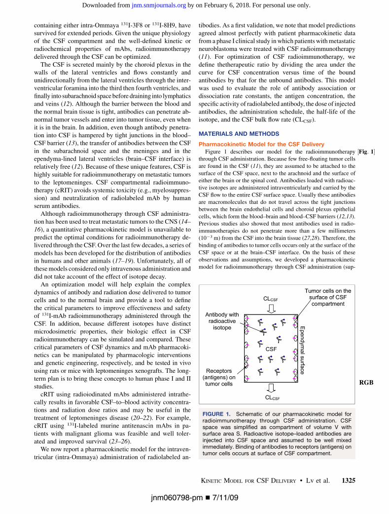
Pharmacokinetic Model for the CSF Delivery½Fig: 1�Figure 1 describes our model for the radioimmunotherapy
through CSF administration. Because few free-floating tumor cellsare found in the CSF (11), they are assumed to be attached to thesurface of the CSF space, next to the arachnoid and the surface ofeither the brain or the spinal cord. Antibodies loaded with radioac-tive isotopes are administered intraventricularly and carried by theCSF flow to the entire CSF surface space. Usually these antibodiesare macromolecules that do not travel across the tight junctionsbetween the brain endothelial cells and choroid plexus epithelialcells, which form the blood–brain and blood–CSF barriers (12,13).Previous studies also showed that most antibodies used in radio-immunotherapies do not penetrate more than a few millimeters(1023 m) from the CSF into the brain tissue (27,28). Therefore, thebinding of antibodies to tumor cells occurs only at the surface of theCSF space or at the brain–CSF interface. On the basis of theseobservations and assumptions, we developed a pharmacokineticmodel for radioimmunotherapy through CSF administration (sup-
FIGURE 1. Schematic of our pharmacokinetic model forradioimmunotherapy through CSF administration. CSFspace was simplified as compartment of volume V withsurface area S. Radioactive isotope–loaded antibodies areinjected into CSF space and assumed to be well mixedimmediately. Binding of antibodies to receptors (antigens) ontumor cells occurs at surface of CSF compartment.
RGB
jnm060798-pm n 7/11/09
KINETIC MODEL FOR CSF DELIVERY • Lv et al. 1325
by on February 6, 2018. For personal use only. jnm.snmjournals.org Downloaded from

plemental materials, which are available online only at http://jnm.snmjournals.org).
Therapeutic Ratio and Optimization CriteriaThe therapeutic amount of radioactive isotope–loaded antibodies
is determined by the time-dependent quantity of radioactivity onbound antibodies to the antigens on tumor cells (CIAR). This amountcan be represented by the area under the CIAR-versus-time curve,which is denoted as AUC(CIAR). The larger the AUC(CIAR), thebigger the radiation dose to tumor cells and the bigger the cytotoxiceffect. While maximizing AUC(CIAR) to achieve therapeutic ef-fects, the goal was to minimize bystander damage from radioactivityon free antibodies (CIA) in the CSF. This reduction of damage can beachieved if the area under the CIA-versus-time curve AUC(CIA) isminimized or the ratio of AUC(CIAR) to AUC(CIA) is maximized.
Mathematically, after obtaining CIAR(t) and CIA(t) by the nu-meric method described in the supplemental materials, we calcu-lated AUC(CIAR) and AUC(CIA) by the following integrations,
AUCðCIARÞ 5
Z N
0
CIARðtÞdt; and AUCðCIAÞ 5
Z N
0
CIAðtÞdt:
AUC(CIAR) and AUC(CIA) depend on the association and disso-ciation rate constants (kAR and k2AR, respectively) or the affinity ofthe antibody to the antigen Kd 5 k2AR
kAR, dose of the antibody admin-
istered, antibody administration schedules, tumor antigen concen-trations, specific activity of the isotope, isotope half-life (t1/2-I), andCLCSF.
Time–Activity RegistrationInjections of the 131I antibody were followed by pharmacokinetic
studies in the CSF and blood, in compliance with the institutionalreview board and hospital guidelines. These procedures wereperformed after informed written consent for all treatments wasreceived from guardians who understood the potential side effects ofeach agent and the possibility of unforeseen toxicities. CSF sampleswere obtained before infusion and at 5, 10, and 30 min and approxi-mately 1, 2, 5, 24, and 48 h after infusion. CSF (10 mL) was countedin a well scintillation counter (LKB; Wallac) alongside a 131Istandard. Counts per minute were entered into Excel (Microsoft)and converted to activity per gram decay-corrected to the time ofadministration of the dose.
Antibody LabelingAntibodies were initially labeled with the chloramine-T method
(29). Excess chloramine-T was neutralized with a 2 molar excess ofsodium metabisulphite before antibody purification using a G-25Sepharose and an anion-exchange column (GE Healthcare). Iodinewas greater than 95% bound by tricarboxylic acid, and immunore-activity was always greater than 50% by radioimmunoassay on GD2glycolipid. The iodination method was subsequently changed to theIODO-GEN method (Pierce), in which antibody was reacted withsolid-phase IODO-GEN before purification by G-25 Sepharose andanion exchange, to avoid soluble chemicals. Iodine remained greaterthan 95% bound by tricarboxylic acid, and immunoreactivity wasalways greater than 50% by radioimmunoassay on GD2 glycolipid.
Parameter ValuesThe CSF volume (V) was 140 mL, and its bulk flow rate (CLCSF)
was 20 mL/h (12,30). The surface area of the CSF space (S) was atleast 1,800 cm2 (27,31). The radioactive isotope used in the clinicaltrial, 131I, had a half-life of 193 h (32). The specific activity of
the 131I-3F8 was 185–370 MBq/mg, and the dose injected was370–1,480 MBq (11). The ranges of the kAR and k2AR for3F8 to GD2 were 3 · 103M21s21 # kAR# 3 · 105M21s21 and3 · 1025s21 # k2AR # 3 · 1023s21, respectively (33). All the abovevalues are summarized in Supplemental Table 1.
For the neuroblastoma, the antigen GD2 density per cell, NR,was approximately 105–107 (34). The tumor cell density at the
surface of the CSF space CSC 51
43p
DT
2
� �3 !
was estimated as 1.91
· 109 cells/mL if the tumor cell diameter DT was 10 mm and thesurface of the CSF space was completely covered by 1 layer oftumor cells. If assuming other coverage percentages (f), the tumorantigen concentration (CR0 5 f · CSC · NR) was estimated from1011 to 1014 antigens/mL. The estimation for the CR0 is detailed inSupplemental Table 2.
RESULTS
Agreement Between Model Predictionsand Patient Data
To validate our kinetic model for radioimmunotherapythrough CSF delivery, we compared the model predictionswith the clinical data obtained in the phase I clinical trial (11).In this study, 15 patients (14 completed the trial) receivedintra-Ommaya delivery of 131I-3F8 (;370–851 MBq). Theradioactivity of free antibodies CIA in the CSF was measuredby serial CSF samplings obtained before infusion and at 5,10, and 30 min and 1, 2, 5, 24, and 48 h after infusion. Ourmodel predictions fit well with the measured data for CIA in10 of 14 patients, whose CIAvalues were near the mean of themeasured data. The discrepancy existed when the value ofCIA was too high or too low. ½Fig: 2�Figure 2 shows the agreement ofour model predictions for 2 representative patients; the
FIGURE 2. Comparison of model predictions and clinicalresults for time-changing radioactivity of free antibodies CIA
in CSF compartment. Symbols are clinical results for 2representative patients, and curves are model predictions.CI0 was 185 MBq/mg-protein, V was 140 mL, CLCSF was 20mL/h, Kd was 1028 M (kAR 5 3 · 104 M21s21; k2AR 5
3 · 1024 s21), and t1/2-I was 193 h.
RGB
jnm060798-pm n 7/11/09
1326 THE JOURNAL OF NUCLEAR MEDICINE • Vol. 50 • No. 8 • August 2009
by on February 6, 2018. For personal use only. jnm.snmjournals.org Downloaded from

1 patient who received a lower dose may have had a lowertumor antigen concentration, and another who received ahigher dose may have had a higher tumor antigen concen-tration. Supplemental Table 3 summarizes the patient dataand model predictions for all 10 patients.
Effect of kAR and k2AR Rate Constants of Antibodies toAntigens on Therapeutic Ratios
Because the binding of antibodies to antigens plays animportant role in the radioactivity of bound antibodies (CIAR)and free antibodies (CIA) in the CSF, we used our model topredict this binding or dissociation effect. The predicted CIAR-versus-time and CIA-versus-time curves under various kAR
and k2AR are shown in½Fig: 3� Figures 3A and 3B. In Figure 3A, CIAR
increases transiently to a peak after the bolus infusion anddecays gradually. The peak value and the decay rate depend onthe quantity of kAR and k2AR, or the affinity Kd 5 k2AR
kAR. When
Kd was 1026 M, the peak value of CIAR was low and CIAR
decayed rapidly and reached equilibrium in approximately500 min. As the affinity increased, or Kd decreased, the peakvalue of CIAR increased and the decay rate decreased. Figure3B shows that CIA decayed quickly to almost zero in approx-imately 1,500 min after infusion. In contrast to CIAR, the decayrate and the time for reaching equilibrium of CIA were almostindependent of Kd. For these reasons, the AUC(CIAR) and theAUC(CIAR)/AUC(CIA) increase with the affinity. Figure 3Csummarizes the effect of Kd on AUC(CIAR) and AUC(CIAR)/AUC(CIA). To optimize the therapeutic effect of the radio-immunotherapy, we wanted to maximize AUC(CIAR) andAUC(CIAR)/AUC(CIA). Increasing the affinity of antibodiesto antigens allowed us to achieve this goal. However, im-provement was not uniform as Kd decreased. From 1026 to1027 M, the improvement in AUC(CIAR) and AUC(CIAR)/AUC(CIA) was 7.6-fold. The improvement reduced to 4.8-,2.7-, 2.4-, 1.5-, and 1.1-fold, respectively, as Kd decreasedfrom 1027 to 1028 M, 1028 to 1029 M, 1029 to 10210 M,10210 to 10211 M, and 10211 to 10212 M.
Effect of CLCSF on Therapeutic Ratio
CLCSF may change because of brain disorders (30) and canbe reduced using pharmacologic inhibitors (35–38). The
influence of CLCSF on AUC(CIAR) and AUC(CIAR)/AUC(CIA)predicted by our model is shown in ½Fig: 4�Figure 4. Reducing CLCSF
increases AUC(CIAR) and improves the therapeutic effect.However, reducing CLCSF decreases AUC(CIAR)/AUC(CIA)by increasing more in AUC(CIA). This may induce moretoxicity in normal tissues. Fortunately, the decreasing rate ofAUC(CIAR)/AUC(CIA) was slow, less than 3%/(mL/h) whenCLCSF decreased from 20 to 3 mL/h, whereas the increasingrate of AUC(CIAR) varied, from 8.7%/(mL/h) (CLCSF from20 to 10 mL/h) to 23.5%/(mL/h) (CLCSF from 5 to 3 mL/h).When CLCSF was from 3 to 0 mL/h, although the increasingrate of AUC(CIAR) was as high as 55.7%/(mL/h), the de-creasing rate for AUC(CIAR)/AUC(CIA) droped sharply to47.0%/(mL/h).
Effect of Specific Activity (CI0), Antibody Dose (DoseA),and Antigen Concentration (CR0) on Therapeutic Ratio
½Fig: 5�Figure 5 shows the model predictions for AUC(CIAR) andAUC(CIAR)/AUC(CIA) as a function of injected DoseA,
FIGURE 3. Time-changing radioactivity of bound antibodies CIAR (A) and free antibodies CIA (B) as function of kAR and k2AR
rate constants of antibodies to antigens. (C) AUC(CIAR) and AUC(CIAR)/AUC(CIA). Kd equaled k2AR
kAR, and V was 140 mL, CLCSF was
20 mL/h, tumor CR0 was 5.73 · 1014 antigens/mL, DoseA was 2 mg, CI0 was 370 MBq/mg, and t1/2-I was 193 h.
FIGURE 4. AUC(CIAR) and AUC(CIAR)/AUC(CIA) as functionof CLCSF. V was 140 mL, Kd was 1028 M (kAR 5
3 · 104 M21s21; k2AR 5 3 · 1024 s21), tumor CR0 was 5.73 ·1014 antigens/mL, DoseA was 2 mg, CI0 was 370 MBq/mg,and t1/2-I was 193 h.
jnm060798-pm n 7/11/09
KINETIC MODEL FOR CSF DELIVERY • Lv et al. 1327
by on February 6, 2018. For personal use only. jnm.snmjournals.org Downloaded from
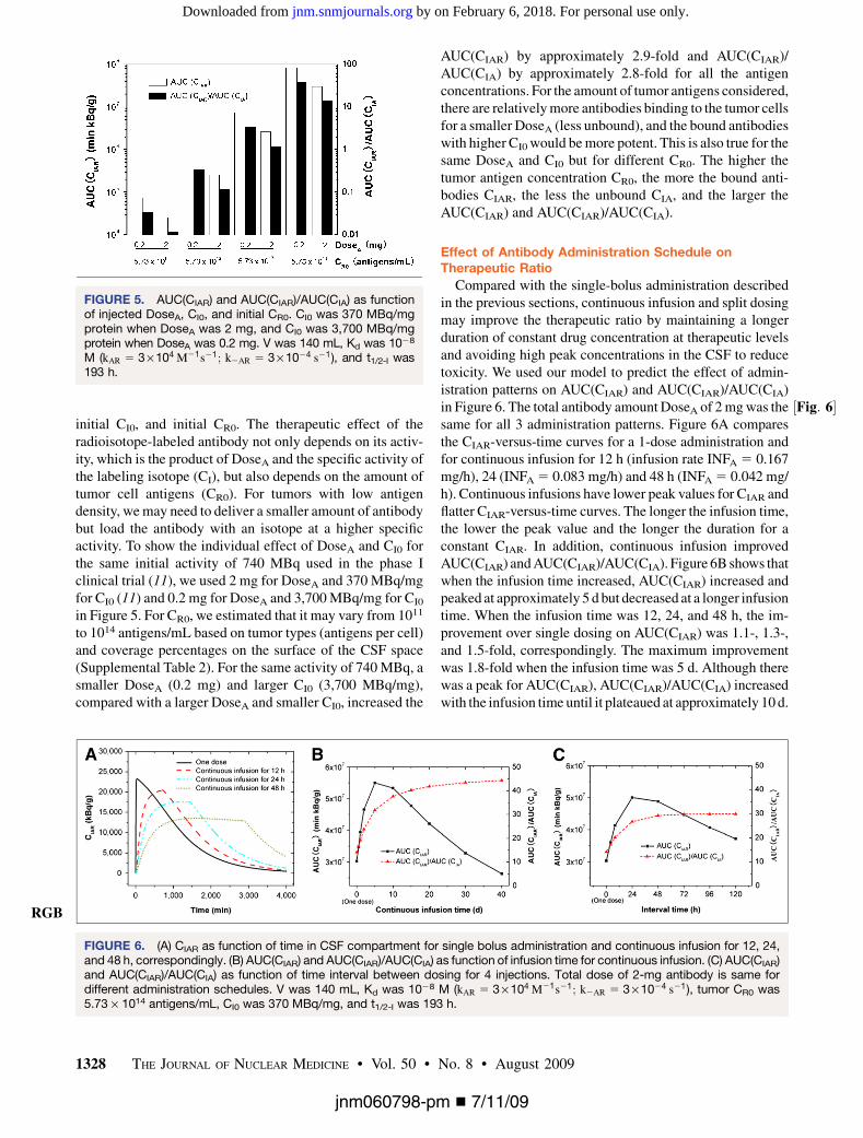
initial CI0, and initial CR0. The therapeutic effect of theradioisotope-labeled antibody not only depends on its activ-ity, which is the product of DoseA and the specific activity ofthe labeling isotope (CI), but also depends on the amount oftumor cell antigens (CR0). For tumors with low antigendensity, we may need to deliver a smaller amount of antibodybut load the antibody with an isotope at a higher specificactivity. To show the individual effect of DoseA and CI0 forthe same initial activity of 740 MBq used in the phase Iclinical trial (11), we used 2 mg for DoseA and 370 MBq/mgfor CI0 (11) and 0.2 mg for DoseA and 3,700 MBq/mg for CI0
in Figure 5. For CR0, we estimated that it may vary from 1011
to 1014 antigens/mL based on tumor types (antigens per cell)and coverage percentages on the surface of the CSF space(Supplemental Table 2). For the same activity of 740 MBq, asmaller DoseA (0.2 mg) and larger CI0 (3,700 MBq/mg),compared with a larger DoseA and smaller CI0, increased the
AUC(CIAR) by approximately 2.9-fold and AUC(CIAR)/AUC(CIA) by approximately 2.8-fold for all the antigenconcentrations. For the amount of tumor antigens considered,there are relatively more antibodies binding to the tumor cellsfor a smaller DoseA (less unbound), and the bound antibodieswith higher CI0 would be more potent. This is also true for thesame DoseA and CI0 but for different CR0. The higher thetumor antigen concentration CR0, the more the bound anti-bodies CIAR, the less the unbound CIA, and the larger theAUC(CIAR) and AUC(CIAR)/AUC(CIA).
Effect of Antibody Administration Schedule onTherapeutic Ratio
Compared with the single-bolus administration describedin the previous sections, continuous infusion and split dosingmay improve the therapeutic ratio by maintaining a longerduration of constant drug concentration at therapeutic levelsand avoiding high peak concentrations in the CSF to reducetoxicity. We used our model to predict the effect of admin-istration patterns on AUC(CIAR) and AUC(CIAR)/AUC(CIA)in ½Fig: 6�Figure 6. The total antibody amount DoseA of 2 mg was thesame for all 3 administration patterns. Figure 6A comparesthe CIAR-versus-time curves for a 1-dose administration andfor continuous infusion for 12 h (infusion rate INFA 5 0.167mg/h), 24 (INFA 5 0.083 mg/h) and 48 h (INFA 5 0.042 mg/h). Continuous infusions have lower peak values for CIAR andflatter CIAR-versus-time curves. The longer the infusion time,the lower the peak value and the longer the duration for aconstant CIAR. In addition, continuous infusion improvedAUC(CIAR) and AUC(CIAR)/AUC(CIA). Figure 6B shows thatwhen the infusion time increased, AUC(CIAR) increased andpeaked at approximately 5 d but decreased at a longer infusiontime. When the infusion time was 12, 24, and 48 h, the im-provement over single dosing on AUC(CIAR) was 1.1-, 1.3-,and 1.5-fold, correspondingly. The maximum improvementwas 1.8-fold when the infusion time was 5 d. Although therewas a peak for AUC(CIAR), AUC(CIAR)/AUC(CIA) increasedwith the infusion time until it plateaued at approximately 10 d.
FIGURE 5. AUC(CIAR) and AUC(CIAR)/AUC(CIA) as functionof injected DoseA, CI0, and initial CR0. CI0 was 370 MBq/mgprotein when DoseA was 2 mg, and CI0 was 3,700 MBq/mgprotein when DoseA was 0.2 mg. V was 140 mL, Kd was 1028
M (kAR 5 3 · 104 M21s21; k2AR 5 3 · 1024 s21), and t1/2-I was193 h.
FIGURE 6. (A) CIAR as function of time in CSF compartment for single bolus administration and continuous infusion for 12, 24,and 48 h, correspondingly. (B) AUC(CIAR) and AUC(CIAR)/AUC(CIA) as function of infusion time for continuous infusion. (C) AUC(CIAR)and AUC(CIAR)/AUC(CIA) as function of time interval between dosing for 4 injections. Total dose of 2-mg antibody is same fordifferent administration schedules. V was 140 mL, Kd was 1028 M (kAR 5 3 · 104 M21s21; k2AR 5 3 · 1024 s21), tumor CR0 was5.73 · 1014 antigens/mL, CI0 was 370 MBq/mg, and t1/2-I was 193 h.
RGB
jnm060798-pm n 7/11/09
1328 THE JOURNAL OF NUCLEAR MEDICINE • Vol. 50 • No. 8 • August 2009
by on February 6, 2018. For personal use only. jnm.snmjournals.org Downloaded from
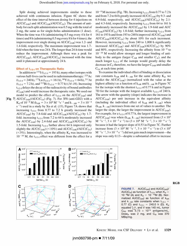
Split dosing achieved improvements similar to thoseachieved with continuous infusion. Figure 6C shows theeffect of the time interval between dosing for 4 injections onAUC(CIAR) and AUC(CIAR)/AUC(CIA). The amount of anti-body for each split administration was 0.5 mg with the total of2 mg, the same as for single-bolus administration (1 dose).When the time was 4 h (administering 0.5 mg every 4 h for 4times) and 8 h (administering 0.5 mg every 8 h for 4 times), theimprovement over single dosing on AUC(CIAR) was 1.2- and1.4-fold, respectively. The maximum improvement was 1.7-fold when the time was 24 h. The longer than 24-h time wouldreduce the improvement. Although there was a peak forAUC(CIAR), AUC(CIAR)/AUC(CIA) increased with the timeuntil it plateaued at approximately 24 h.
Effect of t1/2-I on Therapeutic Ratio
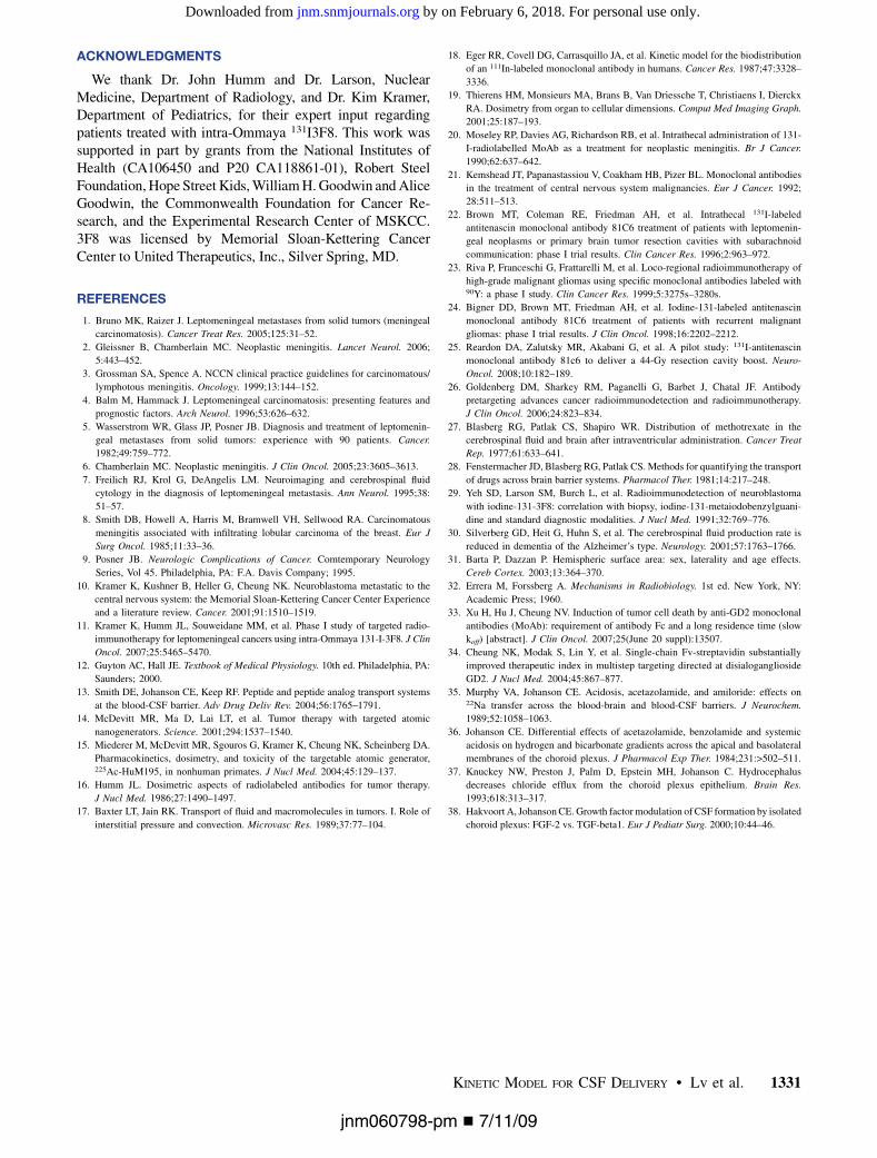
In addition to 131I (t1/2-I 5 193 h), many other isotopes withvarious half-lives can be used in radioimmunotherapy: 225Ac(t1/2-I 5 240 h), 172Lu (t1/2-I 5 161 h), 90Y (t1/2-I 5 64 h), 211At(t1/2-I 5 7.2 h), and 213Bi (t1/2-I 5 0.77 h) (14–16). Increasingt1/2-I delays the decay of the radioactivity of bound antibodies(CIAR) and would increase the therapeutic ratio. We used ourmodel to predict the effect of t1/2-I on the AUC(CIAR) andAUC(CIAR)/AUC(CIA) (½Fig: 7� Fig. 7). For 3F8 (anti-GD2) with aKd of 1028 M (kAR 5 3 · 104 M21s21 and k2AR 5 3 · 1024
s21) used in a study by Xu et al. (33), Figure 7A shows thatincreasing t1/2-I from 0.77 to 7.2 h greatly increased theAUC(CIAR) by 7.8-fold and AUC(CIAR)/AUC(CIA) by 1.7-fold; increasing t1/2-I from 7.2 to 64 h moderately increasedthe AUC(CIAR) by 2.4-fold and AUC(CIAR)/AUC(CIA) by1.5-fold. Increasing t1/2-I further above 64 h improved onlyslightly the AUC(CIAR) (,10%) and AUC(CIAR)/AUC(CIA)(,5%). Interestingly, when the affinity Kd was increased to10210 M, the t1/2-I effect was different from the effect for a
1028 M increase (Fig. 7B). Increasing t1/2-I from 0.77 to 7.2 hand from 7.2 to 64 h greatly increased AUC(CIAR) by 9.3- and6.9-fold, respectively, and AUC(CIAR)/AUC(CIA) by 2.1-and 4.3-fold, respectively. Increasing t1/2-I from 64 to 161 hmoderately increased the AUC(CIAR) by 1.9-fold and AUC(CIAR)/AUC(CIA) by 1.8-fold; further increasing t1/2-I from161 to 193 h and from 193 to 240 h improved AUC(CIAR) andAUC(CIAR)/AUC(CIA) by about 10% for each increment.However, increasing t1/2-I from 240 to 1,000 h significantlyincreased AUC(CIAR) and AUC(CIAR)/AUC(CIA) by 90%and 80%, respectively. Increasing the affinity from 1028 to10210 M would allow stronger and longer binding of anti-body to the antigen (larger CAR and smaller CA), and themuch longer t1/2-I of the isotope would greatly delay thedecrease in CI; therefore, we have the larger CIAR and smallerCIA at each time point.
To examine the individual effect of binding or dissociationrate constants kAR and k2AR for the same affinity Kd, wepredict the AUC(CIAR) (normalized with the value at thehighest affinity) as a function of kAR and k2AR in Figure 7Cfor the isotope with the shortest t1/2-I of 0.77 h and in Figure7D for the isotope with the longest available t1/2-I of 240 h.The arrow with the quantity (slope) indicates the increase inAUC(CIAR) per unit increase in the equivalent affinity(including the individual effect of kAR and k2AR) when(kAR, k2AR) increases from one set of values to another. Thelarger the slope, the larger the improvement in AUC(CIAR).For example, for a t1/2-I of 0.77 h, the largest improvement inAUC(CIAR) was when (kAR, k2AR) increased from (3 · 103
M21s21, 3 · 1023 s21) to (3 · 104 M21s21, 3 · 1024 s21)because it had the largest slope of 0.53 in Figure 7C. Furtherincrease from (3 · 104 M21s21, 3 · 1024 s21) to (3 · 105
M21s21, 3 · 1025 s21) did not gain much improvement—theslope was only 0.13—despite a substantial effort to increase
FIGURE 7. AUC(CIAR) and AUC(CIAR)/AUC(CIA) as function of t1/2-I when Kd 5
1028M (A) and Kd 5 10210M (B). Nor-malized AUC(CIAR) as function of kAR
and k2AR rate constants when t1/2-I 5
0.77 (C) and t1/2-I 5 240.0 h (D). Kd
equaled k2AR
kAR, and V was 140 mL, tumor
CR0 was 5.73 · 1014 antigens/mL,DoseA was 2 mg, and CI0 was 370MBq/mg.
RGB
jnm060798-pm n 7/11/09
KINETIC MODEL FOR CSF DELIVERY • Lv et al. 1329
by on February 6, 2018. For personal use only. jnm.snmjournals.org Downloaded from

Kd from 1028 to 10210 M. In contrast, for a t1/2-I of 240 h, themost improvement in AUC(CIAR) was when (kAR, k2AR)increased from (3 · 104 M21s21, 3 · 1024 s21) to (3 · 105
M21s21, 3 · 1025 s21) because the slope was 0.63, the largestamong others. So, it is worthwhile to increase Kd from 1028
to 10210 M for the antibody with a t1/2-I of 240 h.For a t1/2-I of 0.77 h, the plot of AUC(CIAR) is not
symmetric about the (kAR, k2AR) axes. The increase in kAR
led to more improvement in AUC(CIAR) than did the decreasein k2AR for the same Kd. For example, if (kAR, k2AR)increased from (3 · 103 M21s21, 3 · 1023 s21) to (3 · 105
M21s21, 3 · 1023 s21) along the kAR axis, the improvementin AUC(CIAR) was 12.5-fold, whereas from (3 · 103 M21s21,3 · 1023 s21) to (3 · 103 M21s21, 3 · 1025 s21) along thek2AR axis, the improvement was only 6.7-fold. For bothcases, Kd was from 1026 M to 1028 M. However, thisasymmetry was lost when the t1/2-I was 240 h. The decreasein k2AR instead of the increase in kAR could bring aboutslightly higher gain in AUC(CIAR), about 20% when Kd isfrom 1026 M to 1028 M.
DISCUSSION
The validity of any model rests on how well the data fit.We passed that test by showing near-perfect agreementbetween model predictions and patient pharmacokineticdata obtained in the phase I clinical trial (the correlationcoefficients were greater than 0.99). Several of the predic-tions from the model were intuitive, although not neces-sarily quantitative, and some of the predictions wereunexpected.
We expected that increasing the antibody affinity to theantigen (Kd) would enhance the AUC(CIAR). With a quanti-tative model, we now can predict how much enhancement toexpect with each increment of Kd and what the respectivecontribution is from kAR or k2AR. When the affinity Kd is im-proved from 1026 to 1027 M, the enhancement in AUC(CIAR)is 7.6-fold. This enhancement is reduced to 4.8-, 2.7-, 2.4-,1.5-, and 1.1-fold, respectively, as Kd is decreased from 1027
to 1028 M, 1028 to 1029 M, 1029 to 10210 M, 10210 to 10211
M, and 10211 to 10212 M. These predictions suggest thatincreasing the affinity up to 10210 M should bring about theoptimal enhancement in AUC(CIAR). Further increases inaffinity have diminishing returns, especially when one con-siders the technical difficulty in developing ligands with Kd
beyond 10210 M. Somewhat unexpected was the predictionthat for the same increase in the affinity, increasing kAR
provides much more benefit for an isotope with a short t1/2-I
(0.77 h); but when the t1/2-I increases to 240 h, decreasingk2AR would bring slightly more enhancement in AUC(CIAR).
The biologic half-life of 131I loaded to the antibody in theCSF was much faster (3–12.9 h) (11) than its physical t1/2-I
of 193 h. The half-life of 131I-3F8 in the CSF predicted byour model was 5.2 and 6.7 h, respectively, for 2 represen-tative patients. The strong agreement between the modelprediction and the measured patient data suggests that therapid decrease in the activity of the CSF 131I-3F8 is mainly
due to the clearance from the CLCSF, an assumption wemade in our model. If we reduce the CLCSF by acetazol-amide and furosemide (36), we would increase AUC(CIAR).However, reducing CLCSF would also reduce AUC(CIAR)/AUC(CIA) but at a much slower rate if CLCSF was above 3mL/h. For the CSF delivery of 131I-3F8, CLCSF of 3 mL/hwas an optimal condition. This quantitative conclusion wasalso unexpected because common sense may suggest thatzero CSF flow should be ideal after the antibody injection.For other compartments not through the CSF, such asintraperitoneal administration, a new model needs to bedeveloped to include clearances through peritoneal fluidcirculation and excretion through the kidney and liver.
In addition to the antibody clearance from CSF and thehalf-life of the isotope loaded onto antibodies, the strength(or half-life) of the bonding between the isotope andantibody (iodine bond or metal chelation) may be just asimportant. Halogen bonds (e.g., iodine or astatine) arebroken during systemic circulation (e.g., dehalogenationby the liver) but less so in the intrathecal space. Moreimportantly, unlike serum, CSF does not have the enzymesand proteins to destabilize these bonds; therefore, weexpect radioconjugate to be more stable in the CSF versusthe blood compartment. As predicted by our model, for thesame amount of isotope (total activity) CIA0 (DoseA · CI0),the smaller the antibody dose DoseA, the higher the specificactivity CI0, the larger the AUC(CIAR), and more radio-immunotherapy efficacy. However, it is difficult to producehigh-specific-activity CI0 before destroying the immunore-activity of mAb. Using a more potent radioisotope (e.g.,a-emitter 225Ac) may be an alternative.
Although continuous infusion and split dosing, comparedwith a single bolus administration, would improve theradioimmunotherapy efficacy by up to 1.8- and 1.7-fold inAUC(CIAR) it may not be clinically convenient. The maxi-mum enhancement of 1.8- or 1.7-fold requires a 5-d contin-uous infusion or 4 injections given every 24 h over 4 d.
Parameters in Equation A5 in the supplemental materials(i.e., CLCSF/V, S/V, CIA0, and CR0) are species-specific, anda different set of values will need to be used if one were toapply these equations to nonhuman primates or smallrodents. However, predictions from our model should bevalid irrespective of species. Before translating these pre-dictions to patient clinical trials, they can be further testedusing these preclinical animal systems.
CONCLUSION
The strong agreement between model predictions andpatient pharmacokinetic data obtained from patients in aclinical trial validates the assumptions and simplifications inour model. Using this model to optimize therapeutic ratio, wemade predictions on critical kinetic and transport parameters,which will require further clinical validation. We believe thatthis model can provide an efficient and cost-effective ap-proach in improving the clinical efficacy in this emergingtreatment modality.
jnm060798-pm n 7/11/09
1330 THE JOURNAL OF NUCLEAR MEDICINE • Vol. 50 • No. 8 • August 2009
by on February 6, 2018. For personal use only. jnm.snmjournals.org Downloaded from
ACKNOWLEDGMENTS
We thank Dr. John Humm and Dr. Larson, NuclearMedicine, Department of Radiology, and Dr. Kim Kramer,Department of Pediatrics, for their expert input regardingpatients treated with intra-Ommaya 131I3F8. This work wassupported in part by grants from the National Institutes ofHealth (CA106450 and P20 CA118861-01), Robert SteelFoundation, Hope Street Kids, William H. Goodwin and AliceGoodwin, the Commonwealth Foundation for Cancer Re-search, and the Experimental Research Center of MSKCC.3F8 was licensed by Memorial Sloan-Kettering CancerCenter to United Therapeutics, Inc., Silver Spring, MD.
REFERENCES
1. Bruno MK, Raizer J. Leptomeningeal metastases from solid tumors (meningeal
carcinomatosis). Cancer Treat Res. 2005;125:31–52.
2. Gleissner B, Chamberlain MC. Neoplastic meningitis. Lancet Neurol. 2006;
5:443–452.
3. Grossman SA, Spence A. NCCN clinical practice guidelines for carcinomatous/
lymphotous meningitis. Oncology. 1999;13:144–152.
4. Balm M, Hammack J. Leptomeningeal carcinomatosis: presenting features and
prognostic factors. Arch Neurol. 1996;53:626–632.
5. Wasserstrom WR, Glass JP, Posner JB. Diagnosis and treatment of leptomenin-
geal metastases from solid tumors: experience with 90 patients. Cancer.
1982;49:759–772.
6. Chamberlain MC. Neoplastic meningitis. J Clin Oncol. 2005;23:3605–3613.
7. Freilich RJ, Krol G, DeAngelis LM. Neuroimaging and cerebrospinal fluid
cytology in the diagnosis of leptomeningeal metastasis. Ann Neurol. 1995;38:
51–57.
8. Smith DB, Howell A, Harris M, Bramwell VH, Sellwood RA. Carcinomatous
meningitis associated with infiltrating lobular carcinoma of the breast. Eur J
Surg Oncol. 1985;11:33–36.
9. Posner JB. Neurologic Complications of Cancer. Comtemporary Neurology
Series, Vol 45. Philadelphia, PA: F.A. Davis Company; 1995.
10. Kramer K, Kushner B, Heller G, Cheung NK. Neuroblastoma metastatic to the
central nervous system: the Memorial Sloan-Kettering Cancer Center Experience
and a literature review. Cancer. 2001;91:1510–1519.
11. Kramer K, Humm JL, Souweidane MM, et al. Phase I study of targeted radio-
immunotherapy for leptomeningeal cancers using intra-Ommaya 131-I-3F8. J Clin
Oncol. 2007;25:5465–5470.
12. Guyton AC, Hall JE. Textbook of Medical Physiology. 10th ed. Philadelphia, PA:
Saunders; 2000.
13. Smith DE, Johanson CE, Keep RF. Peptide and peptide analog transport systems
at the blood-CSF barrier. Adv Drug Deliv Rev. 2004;56:1765–1791.
14. McDevitt MR, Ma D, Lai LT, et al. Tumor therapy with targeted atomic
nanogenerators. Science. 2001;294:1537–1540.
15. Miederer M, McDevitt MR, Sgouros G, Kramer K, Cheung NK, Scheinberg DA.
Pharmacokinetics, dosimetry, and toxicity of the targetable atomic generator,225Ac-HuM195, in nonhuman primates. J Nucl Med. 2004;45:129–137.
16. Humm JL. Dosimetric aspects of radiolabeled antibodies for tumor therapy.
J Nucl Med. 1986;27:1490–1497.
17. Baxter LT, Jain RK. Transport of fluid and macromolecules in tumors. I. Role of
interstitial pressure and convection. Microvasc Res. 1989;37:77–104.
18. Eger RR, Covell DG, Carrasquillo JA, et al. Kinetic model for the biodistribution
of an 111In-labeled monoclonal antibody in humans. Cancer Res. 1987;47:3328–
3336.
19. Thierens HM, Monsieurs MA, Brans B, Van Driessche T, Christiaens I, Dierckx
RA. Dosimetry from organ to cellular dimensions. Comput Med Imaging Graph.
2001;25:187–193.
20. Moseley RP, Davies AG, Richardson RB, et al. Intrathecal administration of 131-
I-radiolabelled MoAb as a treatment for neoplastic meningitis. Br J Cancer.
1990;62:637–642.
21. Kemshead JT, Papanastassiou V, Coakham HB, Pizer BL. Monoclonal antibodies
in the treatment of central nervous system malignancies. Eur J Cancer. 1992;
28:511–513.
22. Brown MT, Coleman RE, Friedman AH, et al. Intrathecal 131I-labeled
antitenascin monoclonal antibody 81C6 treatment of patients with leptomenin-
geal neoplasms or primary brain tumor resection cavities with subarachnoid
communication: phase I trial results. Clin Cancer Res. 1996;2:963–972.
23. Riva P, Franceschi G, Frattarelli M, et al. Loco-regional radioimmunotherapy of
high-grade malignant gliomas using specific monoclonal antibodies labeled with90Y: a phase I study. Clin Cancer Res. 1999;5:3275s–3280s.
24. Bigner DD, Brown MT, Friedman AH, et al. Iodine-131-labeled antitenascin
monoclonal antibody 81C6 treatment of patients with recurrent malignant
gliomas: phase I trial results. J Clin Oncol. 1998;16:2202–2212.
25. Reardon DA, Zalutsky MR, Akabani G, et al. A pilot study: 131I-antitenascin
monoclonal antibody 81c6 to deliver a 44-Gy resection cavity boost. Neuro-
Oncol. 2008;10:182–189.
26. Goldenberg DM, Sharkey RM, Paganelli G, Barbet J, Chatal JF. Antibody
pretargeting advances cancer radioimmunodetection and radioimmunotherapy.
J Clin Oncol. 2006;24:823–834.
27. Blasberg RG, Patlak CS, Shapiro WR. Distribution of methotrexate in the
cerebrospinal fluid and brain after intraventricular administration. Cancer Treat
Rep. 1977;61:633–641.
28. Fenstermacher JD, Blasberg RG, Patlak CS. Methods for quantifying the transport
of drugs across brain barrier systems. Pharmacol Ther. 1981;14:217–248.
29. Yeh SD, Larson SM, Burch L, et al. Radioimmunodetection of neuroblastoma
with iodine-131-3F8: correlation with biopsy, iodine-131-metaiodobenzylguani-
dine and standard diagnostic modalities. J Nucl Med. 1991;32:769–776.
30. Silverberg GD, Heit G, Huhn S, et al. The cerebrospinal fluid production rate is
reduced in dementia of the Alzheimer’s type. Neurology. 2001;57:1763–1766.
31. Barta P, Dazzan P. Hemispheric surface area: sex, laterality and age effects.
Cereb Cortex. 2003;13:364–370.
32. Errera M, Forssberg A. Mechanisms in Radiobiology. 1st ed. New York, NY:
Academic Press; 1960.
33. Xu H, Hu J, Cheung NV. Induction of tumor cell death by anti-GD2 monoclonal
antibodies (MoAb): requirement of antibody Fc and a long residence time (slow
koff) [abstract]. J Clin Oncol. 2007;25(June 20 suppl):13507.
34. Cheung NK, Modak S, Lin Y, et al. Single-chain Fv-streptavidin substantially
improved therapeutic index in multistep targeting directed at disialoganglioside
GD2. J Nucl Med. 2004;45:867–877.
35. Murphy VA, Johanson CE. Acidosis, acetazolamide, and amiloride: effects on22Na transfer across the blood-brain and blood-CSF barriers. J Neurochem.
1989;52:1058–1063.
36. Johanson CE. Differential effects of acetazolamide, benzolamide and systemic
acidosis on hydrogen and bicarbonate gradients across the apical and basolateral
membranes of the choroid plexus. J Pharmacol Exp Ther. 1984;231:>502–511.
37. Knuckey NW, Preston J, Palm D, Epstein MH, Johanson C. Hydrocephalus
decreases chloride efflux from the choroid plexus epithelium. Brain Res.
1993;618:313–317.
38. Hakvoort A, Johanson CE. Growth factor modulation of CSF formation by isolated
choroid plexus: FGF-2 vs. TGF-beta1. Eur J Pediatr Surg. 2000;10:44–46.
jnm060798-pm n 7/11/09
KINETIC MODEL FOR CSF DELIVERY • Lv et al. 1331
by on February 6, 2018. For personal use only. jnm.snmjournals.org Downloaded from

Doi: 10.2967/jnumed.108.060798Published online: July 17, 2009.JNM Yonggang Lv, Nai-Kong V. Cheung and Bingmei M. Fu Cerebrospinal Fluid for the Treatment of Leptomeningeal MetastasesA Pharmacokinetic Model for Radioimmunotherapy Delivered Through
http://jnm.snmjournals.org/content/early/2009/07/17/jnumed.108.060798.citationThis article and updated information are available at:
http://jnm.snmjournals.org/site/subscriptions/online.xhtml
Information about subscriptions to can be found at:
http://jnm.snmjournals.org/site/misc/permission.xhtmlInformation about reproducing figures, tables, or other portions of this article can be found online at:
the manuscript and the final, published version.typesetting, proofreading, and author review. This process may lead to differences between the accepted version of
ahead of print area, they will be prepared for print and online publication, which includes copyediting,JNMthe copyedited, nor have they appeared in a print or online issue of the journal. Once the accepted manuscripts appear in
. They have not beenJNM ahead of print articles have been peer reviewed and accepted for publication in JNM
(Print ISSN: 0161-5505, Online ISSN: 2159-662X)1850 Samuel Morse Drive, Reston, VA 20190.SNMMI | Society of Nuclear Medicine and Molecular Imaging
is published monthly.The Journal of Nuclear Medicine
© Copyright 2009 SNMMI; all rights reserved.
by on February 6, 2018. For personal use only. jnm.snmjournals.org Downloaded from