Embed Size (px)
Citation preview

Intracellular Calcium Oscillations in Astrocytes: A Highly Plastic,Bidirectional Form of Communication between Neurons andAstrocytes In Situ
Lucia Pasti,1 Andrea Volterra,2 Tullio Pozzan,1 and Giorgio Carmignoto1
1Department of Experimental Biomedical Sciences and Consiglio Nazionale delle Ricerche Center for Biomembranes,University of Padova, 35121 Padova, Italy, and 2Institute of Pharmacological Sciences, University of Milan, 20133Milan, Italy
The spatial–temporal characteristics of intracellular calcium([Ca21]i ) changes elicited in neurons and astrocytes by varioustypes of stimuli were investigated by means of confocal fluo-rescent microscopy in acute rat brain slices loaded with theCa21 indicator indo-1. Neurons and astrocytes from the visualcortex and CA1 hippocampal region were identified in situ onthe basis of their morphological, electrophysiological, and phar-macological features. We show here that stimulation of neuro-nal afferents triggered periodic [Ca21]i oscillations in astro-cytes. The frequency of these oscillations was under a dynamiccontrol by neuronal activity as it changed according to thepattern of stimulation. After repetitive episodes of neuronalstimulation as well as repetitive stimulation with a metabotropic
glutamate receptor agonist, astrocytes displayed a long-lastingincrease in [Ca21]i oscillation frequency. Oscillating astrocyteswere accompanied by repetitive [Ca21]i elevations in adjacentneurons, most likely because of the release of glutamate via atetanus toxin-resistant process. These results reveal that[Ca21]i oscillations in astrocytes represent a highly plastic sig-naling system that underlies the reciprocal communication be-tween neurons and astrocytes.
Key words: astrocytes; metabotropic glutamate receptor; in-tracellular calcium oscillations; synaptic plasticity; neurotrans-mitter release; hippocampus; visual cortex; tetanus toxin; con-focal microscopy
Changes in the intracellular calcium concentration ([Ca21]i )mediate a variety of biological responses in both excitable andnonexcitable cells. In the CNS the mechanism of calcium signal-ing has been investigated extensively in neurons (Ghosh andGreenberg, 1995), whereas less attention has been granted toother CNS cells such as glial cells (but see Barres, 1991). Recentstudies, however, revealed that various stimuli, including neuro-transmitters, induced in astrocytes [Ca 21]i oscillations and wavespropagating from astrocyte to astrocyte via gap junctions(Cornell-Bell et al., 1990; Glaum et al., 1990; Verkhratsky andKettenmann, 1996). Although the mechanism underlying [Ca 21]i
oscillations is relatively well defined (Berridge, 1993), little isknown about their physiological role. An interesting hypothesis isthat the action of Ca 21 as a second messenger in cells displaying[Ca21]i oscillations may be expressed via a frequency- rather thanan amplitude-dependent code (Woods et al., 1986; Jacobs, 1990).This mechanism ensures that the information carried by theintensity of the stimulus is preserved and converted into a definedfrequency of oscillations. In astrocytes, [Ca21]i increases proba-bly regulate features such as the glycogenolysis (Tsacopoulos andMagistretti, 1996) and the synthesis and/or release of arachidonic
acid (Seregi et al., 1987), neurotransmitters (Pin and Bockaert,1989; Szatkowski et al., 1990; Gallo et al., 1991), and neurotro-phins (Martin, 1992; Zafra et al., 1992).
[Ca21]i oscillations and waves in astrocytes can be triggered byneuronal activity in primary cortical cultures (Murphy et al.,1993) as well as in cultured brain slices (Dani et al., 1992),although they were not observed in acute brain slices (Porter andMc Carthy, 1996). After stimulation with bradykinin, culturedastrocytes can trigger significant elevations of the [Ca21]i inneurons (Parpura et al., 1994a). This action is proposed to bemediated by either a calcium-dependent release from astrocytes(Parpura et al., 1994a; Jeftinija et al., 1996) or gap junctioncommunication between astrocytes and neurons (Nedergaard,1994). Active interactions may, therefore, exist between neuronsand astrocytes, at least in culture (Pfrieger and Barres, 1996). Therole of astrocytes as passive cells has been disputed further by theobservation that cultured astrocytes display a surprising form ofcellular memory (Pasti et al., 1995). After repetitive episodes ofstimulation with glutamate, the frequency of [Ca21]i oscillationsincreased drastically (Pasti et al., 1995). The potentiation of theresponse was long-lasting and involved the activation of a metabo-tropic glutamate receptor (mGluR) subtype linked to the produc-tion of inositol trisphosphate.
Although these observations from cells in culture suggest anactive participation of astrocytes in brain functions, an under-standing of their role in the neural network requires experimentsthat are performed in intact tissue preparations. The aim of thisstudy was to investigate whether a communication system existsbetween neurons and astrocytes in the developing brain. Astro-cytes and neurons from hippocampal CA1 region and visualcortex were identified in acute brain slice preparations on the
Received May 19, 1997; revised July 14, 1997; accepted Aug. 6, 1997.This work was supported by Grants from Telethon number 845, the European
Union Programs—Human Capital and Mobility Network CHRXCT940500, theHuman Frontier Science Program RG520/95, the Italian University Ministry, FidiaResearch Laboratories, and Biotechnology Program BIO4CT960382. We thank Drs.Aldebaran Hofer and Rosario Rizzuto for critically reading this manuscript. Wealso thank Dr. Cesare Montecucco for the generous gift of the purified tetanus toxin.
Correspondence should be addressed to Dr. Giorgio Carmignoto, Department ofExperimental Biomedical Sciences, University of Padova, Viale G. Colombo 3,35121 Padova, Italy.Copyright © 1997 Society for Neuroscience 0270-6474/97/177817-14$05.00/0
The Journal of Neuroscience, October 15, 1997, 17(20):7817–7830

basis of their morphological, electrophysiological, and pharmaco-logical features. The spatial–temporal features of their [Ca21]i
changes were investigated after various stimuli, including neuro-nal stimulation. A confocal fluorescence microscope in conjunc-tion with the calcium indicator indo-1 was used to monitor[Ca21]i changes at the single-cell level.
We here demonstrate a long-term change in [Ca 21]i oscillationfrequency in astrocytes in response to repetitive episodes ofneuronal stimulation as well as to successive applications of themGluR agonist 1-aminocyclopentane-1,3-dicarboxylic acid (t-ACPD). In addition, we provide compelling, although indirect,evidence that activation of the mGluR triggers the release ofglutamate that, in turn, induces [Ca21]i oscillations in neighbor-ing neurons.
MATERIALS AND METHODSSlice preparation for confocal microscopy. Transverse brain slices (150–250 mm) from both the visual cortex and the hippocampus were preparedfrom Wistar rats at postnatal days 7–12, as previously described (Edwardset al., 1989; Carmignoto and Vicini, 1992). Slices were incubated inphysiological saline containing 20 mM indo-1/AM (Molecular Probes,Eugene, OR) and 0.02% pluronic acid at 37°C for 40–50 min undercontinuous mild stirring. Following the evidence that antioxidant agentscan protect neurons from degeneration (Rice et al., 1994), the physio-logical saline for slice incubation was as follows (in mM): NaCl 120, KCl3.1, NaH2PO4 1.25, NaHCO3 25, dextrose 4, MgCl2 2, CaCl2 1, Na-pyruvate 2, myo-inositol 0.5, and ascorbic acid 0.1 at pH 7.4 with5%CO2 /95%O2.
Ratio image acquisition. Recording sessions were performed at roomtemperature. After incubation with indo-1/AM, slices were mounted in achamber that was placed on the stage of a Nikon inverted microscope(Diaphot 300), equipped with a 403 water immersion objective, numer-ical aperture 1.1 (Nikon), connected with a real time confocal microscope(Nikon, RCM8000). The 351 nm band of the argon ion laser was used forexcitation, and the emitted light, separated into its two components (405and 485 nm) by a dichroic mirror, was collected by two separate photo-multipliers. The ratio of the intensity of the light emitted at the twowavelengths (405/485) was displayed as a pseudocolor scale. Time serieswere acquired with a frame interval of 1, 2, or 3 sec, and 16 images wereaveraged for each frame. During recordings, slices were perfused con-tinuously (3 ml/min) with physiological saline of the following composi-tion (in mM): NaCl 120, KCl 3.1, NaH2PO4 1.25, NaHCO3 25, dextrose5, MgCl2 1, and CaCl2 2 at pH 7.4 with 5%CO2 /95%O2. The R405/485in basal conditions was observed to vary little in different cells. Occa-sionally, a slight decrease was observed in R405/485 basal levels (see, forexample, Fig. 7A). Indeed, prolonged UV irradiation of indo-1 can causeoverall photobleaching and conversion to a fluorescent, but Ca 21-insensitive, species (Scheenen et al., 1996). In several experiments weused 100 mM Trolox, a vitamin E analog that inhibits the formation ofindo-1 photodegradation products (Scheenen et al., 1996). No substantialdifferences, however, were observed in our conditions. The frequency ofoscillations is expressed as the number of [Ca 21]i peaks per minute.
Stimulation protocols. To investigate the role of extracellular Ca 21, at5 min before the onset of the t-ACPD stimulation, we perfused sliceswith a Ca 21-free physiological saline supplemented with 1 mM EGTA.The stimulation with high K 1 extracellular solution was obtained byisosmotic replacement of Na 1 with K 1. Changes of [Ca 21]i because ofsynaptic activity were evoked by stimulus trains consisting of 50 msecpulses at 20–30 Hz for 100–200 msec delivered through an isolation unit(World Precision Instruments, Sarasota, FL) to a bipolar tungsten elec-trode (5 mm tip, Roboz, Rockville, MD) positioned either intracorticallyor at the white-matter/ layer VI border, in the case of the visual cortex,and at the stratum radiatum to stimulate the Schaffer collateral–com-missural pathway, in the case of the hippocampus. The electrode waspositioned at 150–500 mm from the cells of interest. To optimize theresponse, we often found it necessary to move the stimulating electrodein different positions, but once it was established, the electrode was notmoved further for the duration of the experiment. The stimulus wasapplied at various frequencies (0.1–1 Hz) and amplitudes (50–500 pA).
Electrophysiolog ical recordings. Brain slice preparation was performedas previously described (Carmignoto and Vicini, 1992). In the holdingchamber, slices were perfused continuously (3–5 ml/min) with the same
saline used for recording at the confocal microscope. The patch-clamptechnique (Edwards et al., 1989) was used in the whole-cell recordingconfiguration. Cells were viewed with an upright Zeiss Axioskop micro-scope equipped with differential interference contrast (DIC), Nomarskioptics (UEM, Zeiss, Oberkochen, Germany), and an electrically insu-lated water immersion 403 objective with a long working distance (2mm). Electrodes were pulled in two stages on a vertical pipette pullerfrom borosilicate glass capillaries (Wiretrol II, Drummond, Broomall,PA). Typical pipette resistance was 5–10 MV. Intracellular (patch pi-pette) solutions contained (in mM): KCl or K-gluconate 145, MgCl2 1,Mg-ATP 2.0, and HEPES 10 to pH 7.2 with KOH. Indo-1-free acid wasincluded in the patch pipette at 500 mM concentration. The intracellularsolution was filtered with a 0.22 pore size filter (Millipore, Yonezawa,Japan). Recordings for 3–5 min were sufficient to obtain the loading ofindo-1 in thin processes of both neurons and astrocytes. A rest of ;30min was allowed before visualization of the cell at the confocal micro-scope. Recordings were performed in current clamp and voltage clampwith a patch-clamp amplifier (EPC 7, List Electronics, Darmstadt, Ger-many), sampled at 10 or 20 kHz, filtered at 1.5 kHz (eight-pole low-passBessel filter; Frequency Devices, Haverhill, MA), and digitized by theinterface Digidata 1200A and pCLAMP-6 software (Axon Instruments,Foster City, CA). In the current-clamp mode depolarizing and hyperpo-larizing current pulses of increasing amplitude and 100–500 msec dura-tion were applied to elicit action potential firing from the recorded cells.The inhibitory action of tetanus neurotoxin (TeNT) on synaptic trans-mission was investigated in CA1 hippocampal region by recording EPSCsevoked by stimuli consisting of 50 msec pulses (50–200 mA at 0.2 Hz)applied through a bipolar tungsten electrode (5 mm tip, Roboz) to theSchaffer collaterals before and during slow perfusion (1 ml/min) with 100mM TeNT. Once the whole-cell configuration was achieved and beforethe onset of the TeNT perfusion, the intensity and duration of thestimulus eliciting the EPSC were adjusted to obtain a stable baseline ofsynaptic responses. In control neurons from slices perfused at 1 ml/min,EPSCs were still present after 30 min recordings. Origin (MicroCalSoftware, Northampton, MA) was used for data analysis and figurepreparation.
Drugs. Excitatory amino acid receptor antagonists 4-carboxyphenyl-glycine (4CPG), L(1)-2-amino-3-phosphonopropionic acid (L-AP3), 1-aminoindan-1,5-dicarboxylic acid (AIDA), a-methyl-4-carboxyphenyl-glycine (MCPG), 2-amino-5-phosphonopentanoic acid (D-AP5), 6-nitro-7-sulfamoylbenzo[f]quinoxaline-2,3-dione (NBQX), and t-ACPD wereobtained from Tocris Cookson (Buckhurst Hill, UK), dissolved in NaOHor DMSO, and diluted in the physiological saline used for recordings.Purified TeNT (Schiavo and Montecucco, 1995) was a gift from C. Mon-tecucco (Department of Experimental Biomedical Sciences, University ofPadova, Italy).
RESULTSActivation of mGluRs induces [Ca21]i increases indifferent cell types from developing hippocampus andvisual cortexAcute brain slices from the hippocampus and the visual cortexwere loaded with the fluorescent Ca21 indicator indo-1 by usingthe cell-permeant acetoxymethyl derivative and were analyzed bylaser scanning confocal microscopy. As illustrated in Figure 1A,pyramidal neurons from CA1 hippocampal region were wellloaded and can be distinguished clearly by their typical shapesand the large size of their somas. Small cells with a stellate shape,like cells labeled 1 and 2, also were well loaded with indo-1 butcould not be classified unambiguously solely on the basis of theirmorphological features. The series of pseudocolor images inFigure 1A illustrates the effects on the [Ca21]i induced on thesecells by application of the mGluR agonist t-ACPD (5 mM; Palmeret al., 1989). Small cells like cells 1 and 2, as well as two of thepyramidal neurons labeled 3 and 4, displayed a [Ca 21]i transienton t-ACPD addition. The kinetics of the [Ca21]i changes, asexpressed by the ratio between indo-1 emission at 405 and 485nm, on continuous exposure to t-ACPD are reported in Figure 1C(lef t) and reveal the presence of periodic [Ca 21]i oscillations insmall cells 1 and 2 ( green and red traces), whereas the pyramidal
7818 J. Neurosci., October 15, 1997, 17(20):7817–7830 Pasti et al. • Reciprocal Communication between Neurons and Astrocytes
neurons (cells 3 and 4) displayed a single transient peak (blacktraces). A delayed response also was observed in six additionalneurons in the field. In two of these the pattern of the responsewas oscillatory (data not shown). In a series of comparableexperiments from CA1 region, the pyramidal neurons responding
to t-ACPD (ranging from 2 to 10 mM in the various experiments)were 68.7 6 8.9% (mean 6 SE; range 17–100), and small cellswere 76.3 6 3.7% (range 52–100). In a few experiments from bothhippocampus and visual cortex, concentrations higher than 10 mM
were tested also. At t-ACPD concentrations of 50–100 mM the
Figure 1. Stimulation of mGluRs induces [Ca 21]i oscillations in hippocampal cells. A, Time series of pseudocolor images of the [Ca 21]i changesoccurring in indo-1-loaded cells from CA1 hippocampal region of a young rat (at postnatal day 8) after perfusion of the slice with 5 mM t-ACPD.The sequence shows the [Ca 21]i transient in two small-sized cells (labeled 1 and 2) and two pyramidal neurons (labeled 3 and 4 ). The R405/485 isdisplayed as a pseudocolor scale. Sampling rate, 3 sec; scale bar, 10 mm. B, Pseudocolor images (a–d) from the same field illustrating the early, sustained[Ca 21]i increase in neurons, including neurons labeled 3 and 4 in A, and the transient, delayed response in small cells, including cells labeled 1 and 2in A, after bath application of 60 mM KCl. Symbols and conditions are as in A. C, Kinetics of the [Ca 21]i changes in the cells labeled 1–4 after t-ACPDand KCl stimulation, as expressed by the ratio between indo-1 emission wavelength at 405 and 485 nm. Letters a–d correspond to images a–d in B.
Pasti et al. • Reciprocal Communication between Neurons and Astrocytes J. Neurosci., October 15, 1997, 17(20):7817–7830 7819
probability of observing [Ca21]i oscillations decreased, and mostof the cells displayed a [Ca21]i rise, followed by a slowly decreas-ing plateau, suggesting that [Ca 21]i oscillations were criticallydependent on the concentration of t-ACPD. In the experimentsdescribed below, for the analysis of [Ca21]i oscillations we usedt-ACPD concentrations between 2 and 10 mM.
Astrocyte identificationSmall cells displaying [Ca21]i oscillations from both visual cortexand hippocampal CA1 and stratum radiatum regions had anastrocyte-like morphology with a mean soma diameter of 5–10mm and numerous radiating processes. These cells could bedistinguished easily from pyramidal neurons. On the basis of puremorphological criteria, however, astrocytes hardly can be distin-guished from small nonpyramidal neurons with a stellate orbipolar shape that are present in both areas (Ramon y Cajal,1911). We first tried to identify astrocytes via glial fibrillary acidicprotein (GFAP) immunostaining performed at the end of therecording session at the confocal microscope. Several cells withastrocyte-like morphology that displayed [Ca21]i oscillations inresponse to t-ACPD could, indeed, be identified retrospectivelyas astrocytes by anti-GFAP immunostaining. Because of the vari-ability in the intensity of the GFAP staining in different slices andamong cells and to the overall alteration in the cell morphologyafter fixation and permeabilization procedures, in ,10% of thecells in the recording field was the identification unambiguous.An alternative approach thus was developed to distinguish func-tionally each astrocyte and neuron present in the recording field.The experiments described below refer to CA1 hippocampalregion, although identical results were obtained in the visualcortex. We took advantage of the observation that, in mixedneuron–astrocyte cultures, neurons, as identified by immunocy-tochemical criteria, responded promptly with a [Ca21]i increaseto depolarization induced by 60 mM K1, whereas none of theimmunocytochemically identifiable astrocytes was sensitive tothis treatment (data not shown). We thus applied the sameprotocol to brain slices to analyze whether neurons and presumedastrocytes responded differently, as in culture, to stimulation with60 mM K1. As shown in Figure 1B, in response to this treatmentall cells in the field showed large [Ca 21]i increases. The onset ofthe response from the two cell populations was, however, clearlydifferent. In the first population, mainly composed of cells with anevident pyramidal shape, the perfusion with high K1 induced aprompt [Ca 21]i increase (Fig. 1Bb). In the majority of neuronsthis first increase was followed by a further [Ca 21]i elevation(Fig. 1C, right). In the second population, composed of small-sized cells with astrocyte-like morphology, the [Ca 21]i increaseoccurred many seconds (13.3 6 1.6 sec; mean 6 SE; n 5 5) afterthat of pyramidal neurons at approximately the same time of thesecond [Ca21]i peak in neurons (Fig. 1Bc,C, right). A delay in theresponse to 60 mM K1 was never observed in cells having apyramidal shape. The kinetics of the recovery were also different:in cells that displayed the prompt response to depolarization, the[Ca 21]i remained elevated for several minutes and recovered tobasal levels quite slowly, whereas in cells that displayed thedelayed response the kinetics of the [Ca 21]i decrease were faster,
and basal [Ca 21]i levels were recovered in ;40 sec (Fig. 1Bd,C,right). Occasionally, these cells displayed repetitive [Ca21]i tran-sients that resemble the typical t-ACPD-induced [Ca21]i oscilla-tions (data not shown). A delayed response pattern is consistentwith a secondary response to glutamate massively released bydepolarized synaptic terminals. Indeed, the response of small-sized cells, but not that of pyramidal neurons, to the stimulationwith 60 mM K1 was blocked in slices incubated for 40 min inTeNT (100 mg/ml), a well known, potent inhibitor of neuronalexocytosis (data not shown; see Fig. 7E). Given that all neuronsexpress at least one subtype of functional voltage-gated Ca 21
channels, the delayed response to 60 mM K1 could be attributedonly to non-neuronal cells, such as astrocytes, that either lackthese channels or express voltage-gated Ca21 channels at suchlow density (Barres et al., 1990) to induce negligible [Ca 21]i
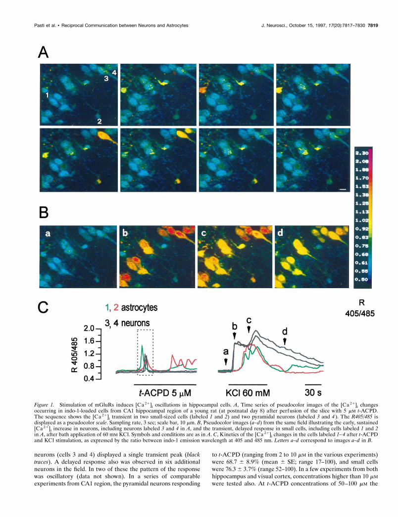
increases, at least under our experimental conditions. We alsostimulated slices with 100 mM NMDA. Given that NMDA recep-tors in astrocytes are most likely absent or nonpermeable to Ca21
(Muller et al., 1993), NMDA should induce a [Ca21]i increase inneurons, whereas astrocytes either should not respond or shoulddisplay a delayed [Ca 21]i increase because of a secondary releaseof glutamate. Stimulation with NMDA (100 mM) induced in thetwo populations a pattern of [Ca 21]i increase similar to thatobserved after perfusion with 60 mM K1 (Fig. 2). Interestingly,NMDA did not induce the biphasic response observed afterdepolarization with high K1. This latter finding supports thehypothesis that the second peak in the [Ca21]i increase inducedin neurons by high K1 stimulation is attributable to glutamatereleased by depolarized terminals.
To confirm that this type of response pattern can be used asa functional tool to identif y astrocytes in situ, we first distin-guished neurons and astrocytes on the basis of their biophys-ical properties by patch-clamp recording while indo-1 diffusedinto the cell through the patch pipette. Then the pipette wasgently withdrawn, and the [Ca 21]i response finally was ana-
Figure 2. Kinetics of [Ca 21]i in cells from the visual cortex in responseto NMDA. Presumed astrocytes (n 5 7; solid lines) from the visual cortexof a 5-d-old rat display a delayed [Ca 21]i increase to NMDA (100 mM)with respect to the prompt response of pyramidal neurons (n 5 5; dashedlines).
3
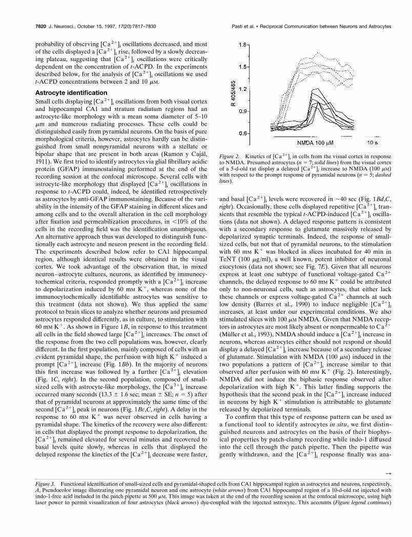
Figure 3. Functional identification of small-sized cells and pyramidal-shaped cells from CA1 hippocampal region as astrocytes and neurons, respectively.A, Pseudocolor image illustrating one pyramidal neuron and one astrocyte (white arrows) from CA1 hippocampal region of a 10-d-old rat injected withindo-1-free acid included in the patch pipette at 500 mM. This image was taken at the end of the recording session at the confocal microscope, using highlaser power to permit visualization of four astrocytes (black arrows) dye-coupled with the injected astrocyte. This accounts (Figure legend continues)
7820 J. Neurosci., October 15, 1997, 17(20):7817–7830 Pasti et al. • Reciprocal Communication between Neurons and Astrocytes
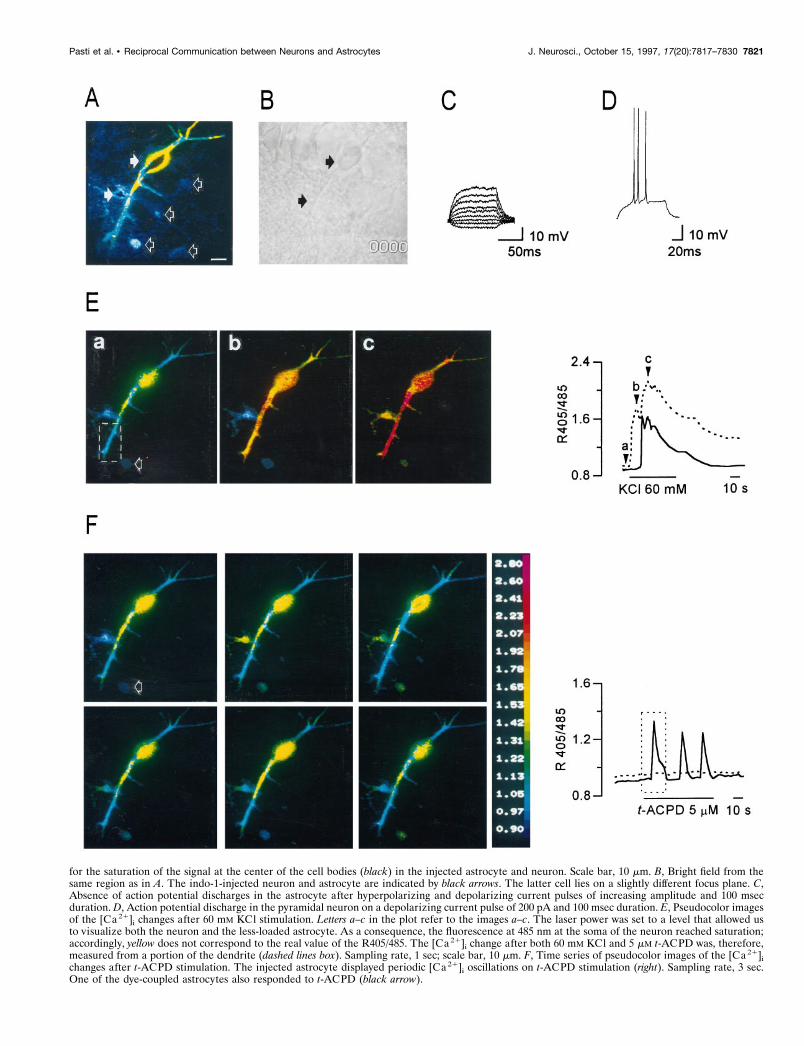
for the saturation of the signal at the center of the cell bodies (black) in the injected astrocyte and neuron. Scale bar, 10 mm. B, Bright field from thesame region as in A. The indo-1-injected neuron and astrocyte are indicated by black arrows. The latter cell lies on a slightly different focus plane. C,Absence of action potential discharges in the astrocyte after hyperpolarizing and depolarizing current pulses of increasing amplitude and 100 msecduration. D, Action potential discharge in the pyramidal neuron on a depolarizing current pulse of 200 pA and 100 msec duration. E, Pseudocolor imagesof the [Ca 21]i changes after 60 mM KCl stimulation. Letters a–c in the plot refer to the images a–c. The laser power was set to a level that allowed usto visualize both the neuron and the less-loaded astrocyte. As a consequence, the fluorescence at 485 nm at the soma of the neuron reached saturation;accordingly, yellow does not correspond to the real value of the R405/485. The [Ca 21]i change after both 60 mM KCl and 5 mM t-ACPD was, therefore,measured from a portion of the dendrite (dashed lines box). Sampling rate, 1 sec; scale bar, 10 mm. F, Time series of pseudocolor images of the [Ca 21]ichanges after t-ACPD stimulation. The injected astrocyte displayed periodic [Ca 21]i oscillations on t-ACPD stimulation (right). Sampling rate, 3 sec.One of the dye-coupled astrocytes also responded to t-ACPD (black arrow).
Pasti et al. • Reciprocal Communication between Neurons and Astrocytes J. Neurosci., October 15, 1997, 17(20):7817–7830 7821

lyzed at the confocal microscope. Figure 3A illustrates anastrocyte and a pyramidal neuron (closed arrows) after intra-cellular injection of indo-1 (bright field is shown in Fig. 3B).Neurons were identified electrophysiologically by their actionpotential discharges on depolarizing current pulses (Fig. 3D)and astrocytes by their absence (Fig. 3C) and highly negativeresting potentials (,75 mV). In addition, indo-1 staining wasobserved not only in the patched astrocyte but also in othersmall cells, like those indicated in Figure 3A by open arrows,indicating coupling via gap junctions. Small-sized cells werenever stained with indo-1 when only neurons were injected(n 5 12), excluding the existence of communication betweenthese two types of cells, at least in the brain regions that wereanalyzed. As illustrated in the pseudocolor images of Figure3Ea–c, after perfusion with 60 mM K 1 the neuron displayed aprompt [Ca 21]i increase (Fig. 3Eb), whereas the response ofthe astrocyte appeared several seconds after that of the neuron(Fig. 3Ec). A similar response was detected in another smallcell (open arrow) dye-coupled with the injected astrocyte. Thekinetics of the [Ca 21]i increase in the electrophysiologicallyclassified neuron (dashed line) and astrocyte (continuous line)are reported in Figure 3E (right). As previously observed incells from slices loaded with indo-1/AM, the pyramidal neurondisplayed a biphasic response to stimulation with 60 mM K 1,and the second [Ca 21]i increase occurred at the time of the[Ca 21]i increase in the astrocyte. After removal of KCl, theslice was perfused with t-ACPD (5 mM). The sequence ofimages in Figure 3F (lef t) and the kinetics of the [Ca 21]i
changes (Fig. 3F, right) demonstrate that the small cell re-sponded to t-ACPD with [Ca 21]i oscillations, whereas theneuron was unresponsive. Identical results were obtained fromtwo additional experiments.
In conclusion, on the basis of these observations, the delayedincrease in the response to 60 mM K1 reasonably can be used asa criterion to distinguish astrocytes from neurons in situ. In thevarious experiments that will be described below, at the end ofeach recording session astrocytes and neurons were identifiedaccording to the different kinetics of their response to stimulationwith 60 mM K1. It cannot be excluded that other non-neuronalcells such as oligodendrocytes might respond to the various stim-uli with a pattern similar to that of astrocytes, but their number in
the brain regions we analyzed is known to be much lower thanthat of the astrocytes.
Repetitive activation of the mGluR induces long-termchanges in [Ca21]i oscillations in astrocytesWe previously reported that astrocytes from the visual cortex inculture displayed a long-term modification of their response, i.e.,an increased frequency of [Ca21]i oscillations, on repetitive stim-ulation with L-glutamate (Pasti et al., 1995). We thus investigatedwhether repetitive stimulation with t-ACPD can induce a similarpotentiation in the response of astrocytes from acute brain slices.Oscillations in neuronal cells will be considered separately. Fig-ure 4A shows the oscillatory response from one astrocyte on threesuccessive 5 mM t-ACPD stimulations applied with a time intervalof 10 min. The second stimulation induced [Ca21]i oscillations ofincreased frequency, although their amplitude and shape did notchange significantly (Fig. 4A). A third stimulation resulted in afurther increase in oscillation frequency. The potentiation of theresponse was observed mainly in astrocytes displaying a lowfrequency of oscillations during the first challenge with t-ACPD,as in the case reported in Figure 4A, whereas those oscillatinginitially at high frequency (Fig. 4B) were, in general, not poten-tiated. Results are summarized in Figure 4C, which reports therelative change in oscillation frequency occurring in each cell onthe second ( filled symbols) and third (open symbols) t-ACPDstimulations as a function of the oscillation frequency on the firstt-ACPD stimulation. The average increase in oscillation fre-quency on repetitive stimulation obtained from a subpopulationof cells comprising astrocytes oscillating initially at a frequency#1 was more pronounced than that obtained from the wholepopulation of cells (Fig. 4D, Table 1). Similar results were ob-tained from visual cortical astrocytes (Table 1).
In contrast to the results obtained in culture, in calcium-freemedium astrocytes in situ failed to respond to t-ACPD with anoscillatory pattern and displayed, in most cases, a single [Ca21]i
rise (data not shown). Only a few cells (5 of 30) exhibited two orthree [Ca21]i transients under these conditions. The subsequentaddition in the perfusate of 2 mM Ca21 resulted in an immediateincrease in the [Ca 21]i that recovered to basal levels in a rela-tively short time. At this extracellular Ca21 concentration thenormal response to t-ACPD stimulation was restored (n 5 13).
Figure 4. Long-term changes of the astrocyte response tot-ACPD. A, Progressive increase in the frequency of [Ca 21]ioscillations on three successive stimulations with 5 mM t-ACPD in one astrocyte oscillating at low frequency after thefirst stimulation. The continuous line at the bottom of thetraces indicates the application of t-ACPD. The time intervalbetween stimulations was 10 min. B, The frequency of [Ca 21]ioscillations on three successive bath applications of 5 mMt-ACPD did not increase in one astrocyte oscillating at highfrequency during the first stimulation. Conditions and labelsare as in A. C, The frequency of oscillations in each cell, asmeasured during the first t-ACPD pulse, is plotted as afunction of the relative change in oscillation frequency in thesecond ( filled symbols) and third (open symbols) with respectto the first pulse. D, Average frequency of oscillations afterthe three t-ACPD stimulations (I, II, and III ) from all astro-cytes (open bars) and from a subpopulation of astrocytescomprising cells oscillating at a frequency #1 (striped bars);*p , 0.05; **p , 0.001 (paired t test). The frequency of[Ca 21]i oscillations in this as well as in the other figures isexpressed as the number of [Ca 21]i peaks per minute.
7822 J. Neurosci., October 15, 1997, 17(20):7817–7830 Pasti et al. • Reciprocal Communication between Neurons and Astrocytes
The change in oscillation frequency is a relatively long-lastingphenomenon. In fact, we observed a significant increase in oscil-lation frequency when the second t-ACPD stimulation was ap-plied after a time interval of 3 hr (see Table 1).
t-ACPD is known to activate all the mGluR subtypes, althoughwith different affinity. To identify the mGluR subtype that medi-ates [Ca21]i oscillations in astrocytes, we used several knownblockers of group 1 mGluRs. The competitive antagonist 4CPG(10–500 mM; Watkins and Collingridge, 1994) did not blockt-ACPD-induced oscillations in a total of 30 astrocytes from thehippocampus and 11 from the visual cortex. The mGluR1/5antagonist AIDA (50–200 mM; n 5 14, two experiments; Pellic-ciari et al., 1995), as well as the noncompetitive antagonist L-AP3(30 mM; n 5 11, two experiments), was also ineffective. In con-trast, the nonspecific mGluR antagonist MCPG at 1 mM concen-tration blocked t-ACPD-induced [Ca21]i increases.
Neuronal stimulation induces [Ca21]i oscillationsin astrocytesBy applying current pulses to afferent projections through abipolar tungsten electrode, we next investigated whether stimula-tion of neuronal afferents could trigger [Ca 21]i oscillations inastrocytes. The sequence of images in Figure 5A corresponds tothe portion of the trace highlighted by the dashed lines box inFigure 5B and illustrates the somatic [Ca 21]i transients of apyramidal neuron (cell 1) in response to stimulation at 0.16 Hz ofSchaffer collaterals. Between stimuli, [Ca21]i recovered to base-line levels (see also top trace in Fig. 5B). A [Ca 21]i rise out ofsynchrony with the stimulus was observed in cell 2 that wasidentified retrospectively as an astrocyte. On continuous neuronalstimulation this cell displayed repetitive transients with a rela-tively regular periodicity (Fig. 5B). The [Ca21]i increase in as-trocytes occurred, in general, with a delay of 10–16 sec withrespect to that of adjacent neurons, although a delay of ,2 secwas, in some cases, observed when the train of stimuli was appliedat a frequency of 1 Hz. In all cases, the [Ca 21]i oscillations in theastrocytes were clearly out of synchrony with respect to the timingof the electrical stimulation. Interestingly, [Ca21]i oscillationswith a frequency higher than that at the soma were observed atthe level of the astrocyte process labeled 3 in Figure 5A (seebottom trace in Fig. 5B). Figure 5, C and D, illustrates an addi-tional example. The sequence of images (Fig. 5C) shows that, onstimulation of Schaffer collaterals at 0.16 Hz, the [Ca21]i increaseinitially was restricted to the process only (arrow), whereas thesecond [Ca21]i increase spread through the whole cell body (seealso inset of Fig. 5D). The kinetics of the [Ca21]i changes fromthe process and the soma are compared in Figure 5D (the tracewithin the dashed lines box corresponds to the sequence of imagesin C). Oscillations triggered by stimulation of afferent fibers were
observed in a low number of astrocytes corresponding to ;15%of indo-1-loaded astrocytes.
These results suggest that glutamate released by synaptic ter-minals is responsible for the [Ca21]i increase in astrocytes. How-ever, the stimulation of presynaptic fibers can trigger actionpotential discharges in target neurons and results in a secondaryrelease of the neurotransmitter. The [Ca 21]i change in astrocytesmight, therefore, originate also from glutamate released at syn-apses that belong to intrinsic connections among target neurons.To investigate this point, we blocked the activation of postsynapticneurons by perfusing slices with the AMPAR antagonist NBQX(50 mM) and the NMDAR open channel blocker MK801 (50 mM;Watkins et al., 1990). Under these conditions the electrical stimulusfailed to produce any [Ca21]i change in neurons that were respon-sive before the application of the blockers, whereas astrocytes werestill responsive with a pattern of oscillations similar to that ob-served in the absence of glutamate receptor antagonists (Fig. 5B).On the contrary, perfusion with the Na1 channel blocker tetrodo-toxin (TTX; 5 mM) abolished the response from both types of cells(15 neurons and 7 astrocytes in CA1 region from two hippocampalslices; 10 neurons and 4 astrocytes from two visual cortical slices).The perfusion for 15 min with the mGluR antagonist MCPG (1mM) abolished the astrocyte response to electrical stimulation ofafferents, whereas that from neurons was unchanged (14 neuronsand 8 astrocytes from four hippocampal slices).
Neuronal stimulation modulates the frequency ofoscillations in astrocytesHaving demonstrated that neuronal activity can trigger [Ca 21]i
oscillations in astrocytes from both visual cortex and hippocam-pus, we next investigated whether the frequency of oscillationscould change according to the firing rates of neuronal afferents.We analyzed the oscillatory response of astrocytes after a train ofstimuli applied first at low frequency (0.1–0.2 Hz) and low inten-sity (50–100 pA) and then at either increased frequency (0.3–1Hz) or higher intensity (200–500 pA). Figure 6 shows that anincrease in either the frequency (A) or intensity (B) of thestimulus resulted in a clear increase in the frequency of [Ca 21]i
oscillations in astrocytes. The mean frequency before and afterthe change in the stimulus is reported in Figure 6, A and B (right).When the frequency of the stimulus was increased, the averagefrequency of oscillations (6 SE) changed from 1.41 6 0.23 to2.16 6 0.24 peaks/min. When the percentage of increase inoscillation frequency in the second with respect to the first stim-ulation from each individual astrocyte was taken into account, theaverage increase (6 SE) corresponded to 115 6 44.1%. A similarincrease was obtained when the stimulus was applied at higherintensity (mean 6 SE; 106 6 27.5%, n 5 20). It is noteworthy thatchanges in the pattern of the electrical stimulus induced either an
Table 1. Frequency of [Ca21] oscillations and its relative change in astrocytes after three consecutive t-ACPD applications
Number ofastrocytes
Oscillation frequencymean 6 SEI pulse
Change in frequency (%)mean 6 SEII pulse
Change in frequency (%)mean 6 SEIII pulse
CA1 132 (20) 1.55 6 0.11 128.4 6 5.6 144.7 6 8.7CA1 subpopulation 57 (20) 0.66 6 0.02 146.2 6 7.9 198.8 6 13.7Visual cortex 15 (4) 1.05 6 0.13 147.4 6 13.1 153 6 7.41CA1 (3 hr interval) 19 (2) 0.80 6 0.11 162.7 6 13.4 ND
The time interval between stimuli, unless specified, was 10 min. Values in the second and third pulse column indicate the average increase in oscillation frequency in the secondand third pulse, respectively, as compared with the oscillation frequency in the first pulse. Numbers in parentheses indicate the number of experiments. ND, Not determined.
Pasti et al. • Reciprocal Communication between Neurons and Astrocytes J. Neurosci., October 15, 1997, 17(20):7817–7830 7823

increased amplitude of the [Ca21]i rise in neurons that werealready responsive, as in the case of the two neurons in A and B,or the appearance of [Ca 21]i increased in neurons and astrocytesthat were initially unresponsive (data not shown), suggesting therecruitment of additional presynaptic inputs.
In the experiment presented in Figure 4, we showed thatsuccessive t-ACPD stimulations resulted in an increase in thefrequency of [Ca 21]i oscillations in astrocytes. The questionthen arises as to whether a similar form of potentiation inastrocytes can be elicited by repetitive stimulation of afferentfibers. Figure 6C shows the response from a single astrocyte forwhich the frequency of [Ca 21]i oscillations changed from 1.0 atthe first to 2.1 at the second series of pulses. Similar to whatwas observed with repetitive t-ACPD stimulation, the poten-tiation of the response was observed mainly in cells having alow frequency of oscillations at the first pulse (Fig. 6D), al-though the average increase in oscillation frequency (65.4 618.3%; Fig. 6 D, right) was more pronounced than that observedon two successive t-ACPD applications (see Table 1). The
higher efficacy of neuronal stimulation in inducing the poten-tiation of the astrocyte response was investigated further. Theexperimental protocol was as follows: after the first and beforethe second t-ACPD stimulation, we applied a series of 0.5–1Hz stimuli to CA1 region afferent projections. As discussedabove, only a percentage of the astrocytes displayed oscilla-tions after neuronal stimulation. We thus had the ability tocompare in the same slice the increase in oscillations frequencythat follows the second t-ACPD stimulation in two astrocytepopulations: the first composed of astrocytes that displayedoscillations on neuronal stimulation between the two succes-sive t-ACPD applications and the second composed of astro-cytes that were not responsive to neuronal stimulation. Figure6 E reports the relative change in frequency in each astrocyte(lef t) and the mean values from the two subpopulations (right).The increase in the frequency of [Ca 21]i oscillations after thesecond t-ACPD application was higher in those astrocytes thatshowed oscillations on neuronal stimulation (Fig. 6 E, closedsymbols and bars) with respect to that of astrocytes that did not
Figure 5. Neuronal activity-dependent [Ca 21]i oscillations in astrocytes. A, Time series of pseudocolor images illustrating the [Ca 21]i changes in onepyramidal neuron (labeled 1) and one adjacent astrocyte (labeled 2) from CA1 hippocampal region of a 8-d-old rat after neuronal stimulation at 0.16Hz, i.e., a series of six pulses at 30 Hz applied every 6 sec. Label 3 indicates one of the astrocyte processes. The sequence of images (time interval, 2sec) corresponds to the portion of the traces shown in B and is highlighted by the dashed lines box. Because the pyramidal neuron and the astrocyte werelocalized at a different depth and the plane of focus was set to visualize the astrocyte, the neuron looks smaller than the astrocyte. The real meandiameter of the neuron was 16.9 mm, whereas that of the astrocyte was 10 mm. Scale bar, 10 mm. B, Kinetics of the [Ca 21]i changes in the cells and theprocess shown in A after two successive episodes of neuronal stimulation applied with 5 min intervals. The second episode of stimulation was performedin the presence of MK801 and NBQX, both at 50 mM. C, Time series of pseudocolor images illustrating the [Ca 21]i changes in an astrocyte as measuredat the level of one process ( filled arrow) and the cell body after neuronal stimulation. The sequence of images (time interval, 2 sec) corresponds to theportion of the trace highlighted by the dashed lines box in D. Scale bar, 10 mm. D, Kinetics of the [Ca 21]i oscillations in the process and the soma of theastrocyte shown in C during neuronal stimulation at 0.16 Hz. In the inset, the sequence of points representing the R405/485 values at the process ( filledsymbols) and the soma (open symbols) corresponds to the sequence of images in C.
7824 J. Neurosci., October 15, 1997, 17(20):7817–7830 Pasti et al. • Reciprocal Communication between Neurons and Astrocytes
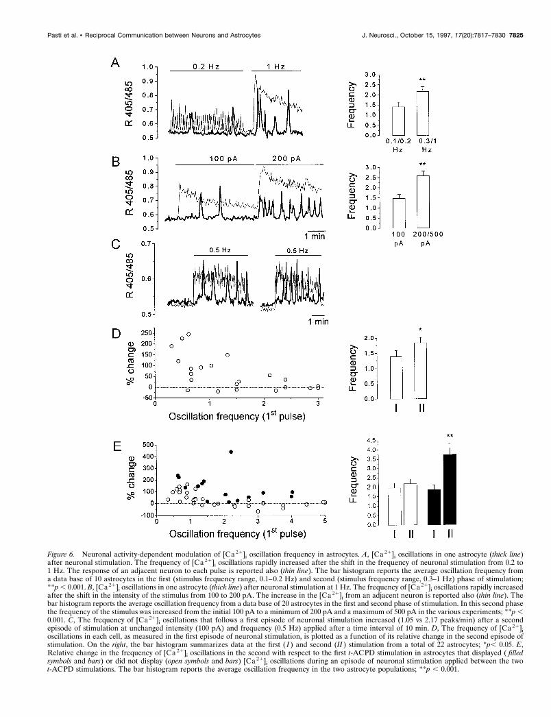
Figure 6. Neuronal activity-dependent modulation of [Ca 21]i oscillation frequency in astrocytes. A, [Ca 21]i oscillations in one astrocyte (thick line)after neuronal stimulation. The frequency of [Ca 21]i oscillations rapidly increased after the shift in the frequency of neuronal stimulation from 0.2 to1 Hz. The response of an adjacent neuron to each pulse is reported also (thin line). The bar histogram reports the average oscillation frequency froma data base of 10 astrocytes in the first (stimulus frequency range, 0.1–0.2 Hz) and second (stimulus frequency range, 0.3–1 Hz) phase of stimulation;**p , 0.001. B, [Ca 21]i oscillations in one astrocyte (thick line) after neuronal stimulation at 1 Hz. The frequency of [Ca 21]i oscillations rapidly increasedafter the shift in the intensity of the stimulus from 100 to 200 pA. The increase in the [Ca 21]i from an adjacent neuron is reported also (thin line). Thebar histogram reports the average oscillation frequency from a data base of 20 astrocytes in the first and second phase of stimulation. In this second phasethe frequency of the stimulus was increased from the initial 100 pA to a minimum of 200 pA and a maximum of 500 pA in the various experiments; **p ,0.001. C, The frequency of [Ca 21]i oscillations that follows a first episode of neuronal stimulation increased (1.05 vs 2.17 peaks/min) after a secondepisode of stimulation at unchanged intensity (100 pA) and frequency (0.5 Hz) applied after a time interval of 10 min. D, The frequency of [Ca 21]ioscillations in each cell, as measured in the first episode of neuronal stimulation, is plotted as a function of its relative change in the second episode ofstimulation. On the right, the bar histogram summarizes data at the first ( I ) and second (II ) stimulation from a total of 22 astrocytes; *p, 0.05. E,Relative change in the frequency of [Ca 21]i oscillations in the second with respect to the first t-ACPD stimulation in astrocytes that displayed ( filledsymbols and bars) or did not display (open symbols and bars) [Ca 21]i oscillations during an episode of neuronal stimulation applied between the twot-ACPD stimulations. The bar histogram reports the average oscillation frequency in the two astrocyte populations; **p , 0.001.
Pasti et al. • Reciprocal Communication between Neurons and Astrocytes J. Neurosci., October 15, 1997, 17(20):7817–7830 7825

respond to this latter challenge (open symbols and bars). It isnoteworthy that the average increase at the second t-ACPDapplication in astrocytes responsive to neuronal stimulationwas higher than that observed after three successive t-ACPDapplications (114% vs 44.7%; Table 1).
[Ca21]i oscillations in astrocytes are followed by[Ca21]i oscillations in neuronsBesides the effects on astrocytes, t-ACPD induced in a number ofCA1 pyramidal neurons either a single [Ca 21]i transient or[Ca21]i oscillations. These [Ca21]i changes could be attributableto (1) direct stimulation of neuronal mGluRs (Stratton et al.,1990) and (2) t-ACPD-induced release of glutamate from astro-cytes with activation of neuronal ionotropic glutamate receptors(iGluR), i.e., AMPA and NMDARs. Indeed, cultured astrocyteshave been reported to release glutamate and excite adjacentneurons (Parpura et al., 1994a; Jeftinija et al., 1996). Figure 7, Aand B, shows a typical oscillatory response induced by 10 mM
t-ACPD in one neuron (A) and one adjacent astrocyte (B). Onwashout of the antagonist, the [Ca 21]i returned to basal levels inboth cell types. A second challenge with t-ACPD (10 mM) wasapplied in the presence of NBQX and D-AP5 (both at 50 mM),specific antagonists of AMPAR and NMDARs, respectively.Under these conditions, in this as well as in a number of otherneurons (14 of 49), the response to t-ACPD was inhibited com-pletely but recovered on washout of the antagonists (Fig. 7A),whereas the astrocyte response was not changed significantly (Fig.7B). In the remaining 35 neurons, the response to the secondt-ACPD stimulation, performed in the presence of NBQX andD-AP5, either was decreased significantly in amplitude, as in thecase reported in Figure 7C, or was unchanged. As a mean, theamplitude of the [Ca21]i elevation induced in neurons by 10 mM
t-ACPD applied in the presence of NBQX/D-AP5 was reducedsignificantly (Fig. 7D). The relative change in the amplitude of the[Ca21]i increase in each neuron at the second (in the presence ofthe AMPAR and NMDAR blockers) with respect to the firstt-ACPD stimulation is reported in Figure 7D. It is noteworthythat no reductions of the response after two successive t-ACPDstimulations were observed in control neurons (n 5 32; Fig. 7D).These results indicate that [Ca21]i elevations in neurons can beelicited, partially or totally, by glutamate released in response tothe t-ACPD challenge. To clarify whether glutamate releasedafter t-ACPD stimulation derived from afferent fibers, neurons,or astrocytes, we incubated the slice with TeNT (100 mg/ml).TeNT is known to be a highly specific blocker of neurotransmittersecretion in neurons (Calabresi et al., 1989; Schiavo et al., 1992).Its action is exerted on the vesicle-associated membrane proteinVAMP/synaptobrevin (Schiavo et al., 1992), one of the compo-nents of the neuroexocytosis apparatus, after cell internalizationprobably via the recycling of synaptic vesicles (Matteoli et al.,1996). Although VAMP/synaptobrevin is expressed in many celltypes besides neurons, the lack of TeNT effects on other celltypes probably depends on the absence of a receptor for the
neurotoxin (Parpura et al., 1994b; Rossetto et al., 1996). Byrecording EPSCs from CA1 neurons in response to stimulation ofthe Schaffer collaterals before and after perfusion of the slicewith TeNT (100 mg/ml), we obtained evidence for the inhibitoryeffects of TeNT on synaptic transmission. As shown in Figure 7E,the EPSC first was reduced in amplitude and then abolished after20 min of TeNT treatment. Despite changing the position of thestimulating electrode and increasing the stimulus intensity, weobtained no response from this and three other neurons of thesame slice, as well as from neurons of an additional slice, testedafter 40 min of incubation with TeNT. In toxin-treated slices theeffects of t-ACPD on both neurons and astrocytes was unchanged,as compared with controls: it stimulated [Ca21]i oscillations inastrocytes and [Ca21]i elevations in neurons. In 6 of 35 t-ACPD-responsive neurons, the response was inhibited completely byNBQX/D-AP5 application, whereas in the remaining 29 neuronsthe amplitude was either reduced significantly or unchanged. Asa mean, the reduction of the response of neurons to t-ACPD byNBQX and D-AP5 in toxin-treated slices was not significantlydifferent as compared with that obtained from neurons in toxin-untreated slices (Fig. 7D, right). The relative change in the am-plitude of the [Ca21]i increase in each neuron after the secondwith respect to the first t-ACPD stimulation also is reported inFigure 7D (right).
The inhibitory effect of TeNT on neurotransmitter exocytosiswas confirmed in each slice used at the confocal microscope by thefollowing experimental observations (data not shown in figures):(1) electrical stimulation of Schaffer collaterals failed to produce[Ca21]i increases in both neurons and astrocytes; (2) small cells,i.e., presumed astrocytes, did not display any [Ca21]i increaseafter 60 mM K1 stimulation; and (3) pyramidal neurons stillresponded to K1-induced depolarization with an early [Ca21]i
increase, but their response did not display the biphasic patternobserved in controls (see Figs. 1C, 3E) and recovered to [Ca21]i
basal levels much faster than in neurons from toxin-untreatedslices.
In the great majority of neurons (16 of 20) from untreated andTeNT-treated slices for which the [Ca 21]i increase was entirelyattributable to the t-ACPD-induced release of glutamate, thepattern of the response was clearly oscillatory (see, for example,Fig. 7A).
When a second t-ACPD challenge was applied in the presenceof the mGluR antagonist MCPG (1 mM), the [Ca 21]i elevationsobserved after the first t-ACPD stimulation were abolished in allresponsive astrocytes (n 5 8) and neurons (n 5 12).
DISCUSSIONLong-term changes in oscillation frequency mediatedby t-ACPDWe previously reported that successive stimulations of themGluR in cultured astrocytes induced a long-lasting increase in[Ca21]i oscillation frequency (Pasti et al., 1995). Here we dem-
3
Figure 7. Astrocyte oscillations mediate repetitive [Ca 21]i increases in neurons. A, [Ca 21]i repetitive increases in one pyramidal hippocampal neuronafter three consecutive stimulations with 10 mM t-ACPD. The response to t-ACPD was abolished by NBQX/D-AP5. Before the second t-ACPDapplication, the slice was perfused for 10 min with NBQX and D-AP5, both at 50 mM. Before the third t-ACPD stimulation, the slice was perfused withnormal saline for 20 min. B, [Ca 21]i oscillations in one astrocyte adjacent to the neuron in A after the three t-ACPD stimulations. C, Reduction byNBQX and D-AP5, both at 50 mM, in the amplitude of the t-ACPD-induced [Ca 21]i increase after the second episode of stimulation from a hippocampalpyramidal neuron and its recovery in the third t-ACPD stimulation performed in the absence of the iGluR blockers after a time interval of 20 min. D,Relative change in the amplitude of the [Ca 21]i increase in each neuron after the second t-ACPD stimulation performed in the absence (control ) orpresence of NBQX/AP5, as compared with the first t-ACPD stimulation. Filled symbols represent the values of the mean 6 SE. (Figure legend continues)
7826 J. Neurosci., October 15, 1997, 17(20):7817–7830 Pasti et al. • Reciprocal Communication between Neurons and Astrocytes

The mean change in the response from NBQX/D-AP5 neurons was significantly different from that from control neurons; **p , 0.0001; t test. E,Whole-cell recordings of EPSCs evoked on a CA1 pyramidal neuron by stimulation of Schaffer collaterals at 0.2 Hz. Three consecutive EPSCs and theaverage trace from eight consecutive EPSCs (bottom traces) before and after the TeNT application are shown. Despite increasing the intensity of thestimulus (note the increased amplitude of the stimulus artifact), we recorded no EPSCs 20 min after TeNT. F, Relative change in the amplitude of the[Ca 21]i increase after the second t-ACPD stimulation, as compared with the first t-ACPD stimulation, in each neuron from slices incubated for 40–60min with TeNT. The second t-ACPD stimulation was performed in the presence of NBQX/D-AP5, both at 50 mM. The mean change in the responsefrom these neurons was significantly different with respect to that from control neurons (**p , 0.0001; t test), but not with respect to that fromNBQX/D-AP5 neurons from slices not treated with TeNT. Symbols are as in D.
Pasti et al. • Reciprocal Communication between Neurons and Astrocytes J. Neurosci., October 15, 1997, 17(20):7817–7830 7827

onstrate that repetitive activations of mGluRs by t-ACPD inducea similar potentiation in the [Ca21]i response of astrocytes inacute brain slices from both CA1 hippocampal region and visualcortex. The potentiation was induced rapidly and was relativelylong-lasting. In contrast to the results obtained in cultured astro-cytes (Pasti et al., 1995), in the absence of extracellular Ca21,astrocytes either failed to oscillate or displayed rapidly fadingoscillations. The effects of t-ACPD on astrocytes probably aremediated by the mGluR5 subtype (Romano et al., 1995). Antag-onists of group 1 mGluRs with a preference specificity formGluR1, failed, however, to affect t-ACPD-induced oscillations,whereas MCPG, a nonspecific mGluR antagonist, blocked theresponse. The mGluR subtype responsible for [Ca21]i oscilla-tions thus remains to be established.
Modulation of the astrocyte response byneuronal activityOne of the most striking observations of this study is that astro-cytes are extremely sensitive to synaptic activity. Indeed, thepattern of their [Ca21]i oscillations in response to neuronalstimulation changed according to the level of synaptic activity;when the frequency or intensity of the stimulus applied to pre-synaptic afferents was increased, the frequency of astrocyte oscil-lations was increased also. It is noteworthy that the increase in thestimulus rate determines a higher firing rate of neuronal afferents,whereas the increase in intensity may result in the recruitment ofadditional fibers that were not stimulated initially. The change instimulus intensity or frequency probably causes an increasedglutamate concentration in the extrasynaptic space and/or theactivation of a higher number of localized [Ca21]i transientsalong processes of individual astrocyte that may account for theincreased oscillation frequency in the astrocytes. These resultsprovide a mechanism for a highly regulated and dynamic controlon [Ca21]i oscillation frequency that depends on the integrationof the Ca 21 signal deriving from these multiple sites ofactivation.
The inhibition of astrocyte [Ca21]i oscillations by TTX ex-cludes that the response of astrocytes after the application of thestimulus to neuronal afferents could be attributable to a directmechanical or electrical stimulation of the glial cells (Charles etal., 1991; Nedergaard, 1994). On the other hand, the finding thatthe mGluR antagonist MCPG blocked the astrocyte response toneuronal stimulation provides convincing evidence that synapti-cally released glutamate is responsible for astrocyte [Ca21]i os-cillations. Further support for the synaptic origin of the stimulusinducing the [Ca21]i changes in astrocytes derives from theobservation that in the presence of iGluR blockers the electricalstimulus failed to produce any response from neurons, whereas itdid induce an oscillatory response in astrocytes. This result alsosuggests that activation of afferent fibers is sufficient for inducingastrocyte oscillations, thereby excluding the hypothesis that ef-fects secondary to postsynaptic neuron activation are critical forastrocyte responsiveness.
Oscillations in astrocytes occurred with a delay with respect tothe [Ca 21]i increase observed in neurons. One possible explana-tion for this delay may be the time required for the diffusion ofglutamate away from the synaptic cleft. With respect to thereceptors at the neuronal postsynaptic membrane, those at theastrocyte membrane are relatively far away from the site ofneurotransmitter release and are probably activated only whenthe concentration of glutamate in the perisynaptic space reachesa threshold level. Theoretical models, however, suggest that glu-
tamate can diffuse from the site of release for several micrometersand reaches concentrations of .10 mM in the perisynaptic spacewithin 0.5–5 msec (Clements, 1996). This explanation can, there-fore, hardly account for the delay observed in the astrocyteresponse. The delay may be attributable to the time necessary forthe [Ca21]i change to spread from the site of activation, presum-ably in proximity of the synaptic cleft, to the cell body and to theoccurrence of a spatial–temporal integration of the Ca21 signal.Indeed, astrocyte membranes have been identified within 1 mmfrom the spine synapses of the cerebellum (Chaudhry et al.,1995), and a similar anatomical pattern has been described inCA1 hippocampal region (Rothstein et al., 1994).
Long-term changes in [Ca21]i oscillation frequencymediated by neuronal activityA second intriguing property of astrocytes is the plasticity of theirresponse to neuronal stimulation. When successive stimulationswere applied to neuronal afferents, astrocytes adjacent to stimu-lated neurons displayed an increased oscillation frequency rathersimilar to that observed after repetitive t-ACPD stimulation.Apparently, the astrocyte response can be potentiated accordingto previous episodes of activity occurring at synapses in closeproximity. This represents, therefore, an activity-dependentchange previously considered an exclusive feature of neuronalcells. It is reminiscent of the activity-dependent increase in syn-aptic efficacy of excitatory synaptic transmission, the so-calledlong-term potentiation (LTP) (Bliss and Lomo, 1973). LTP isbelieved to represent, at the cellular level, certain aspects oflearning and memory phenomena (Bliss and Collingridge, 1993).Although many questions still remain unresolved, substantialunderstanding exists on the cellular and molecular basis of LTP(Bliss and Collingridge, 1993; Kullmann and Singelbaum, 1995).In contrast, very little is known about the long-term change in theastrocyte response. Thus it may seem premature to compare thetwo phenomena. Nevertheless, besides the fact that glutamate isthe principal mediator of both events, the potentiation of theastrocyte response appears to share with neurons at least two ofthe formal properties that characterize LTP, such as saturationand persistency. As to the first, we observed the failure of induc-ing potentiation in cells oscillating at high frequency at the verybeginning. As to the second property, i.e., persistency, we dem-onstrated that the potentiation of the astrocyte response persistedfor at least 3 hr. In contrast to these common aspects, the kineticsof the induction mechanism are very different: LTP occurs withinmilliseconds, whereas the potentiation in astrocytes appearsmuch slower. This latter observation suggests that the plasticity inthe astrocyte response likely is involved in the slow modulation ofthe neuron–astrocyte network actions. The slowness of a signal-ing system, based on [Ca21]i oscillations and waves with respectto the rapidity of synaptic transmission, has been suggested todenote the modulatory role of astrocytes in brain function (Daniet al., 1992; Smith, 1994).
On the functional role of [Ca21]i oscillationsin astrocytesThe final and key question is the functional role of [Ca21]i
oscillations in astrocytes and the possible significance of theirpotentiation in response to repetitive episodes of neuronal activ-ity. Our observation that [Ca21]i oscillations in astrocytes areaccompanied by [Ca21]i elevations in adjacent neurons, togetherwith the finding that this response could be blocked, at least in anumber of neurons, by iGluR antagonists, suggests that astrocytes
7828 J. Neurosci., October 15, 1997, 17(20):7817–7830 Pasti et al. • Reciprocal Communication between Neurons and Astrocytes

in situ can release glutamate or a glutamate analog efficiently.Indeed, in a number of neurons the [Ca21]i increase induced byt-ACPD was blocked by NBQX and D-AP5, indicating that thiseffect was totally dependent on the activation of AMPA andNMDA receptors. The insensitivity to TeNT of t-ACPD-induced[Ca21]i increase in these neurons indicates that the neurotrans-mitter is released by neither presynaptic terminals nor otherneurons and thus points to the astrocytes as the cells responsiblefor this release. In other neurons, the activation of AMPARs andNMDARs by glutamate as well as the direct simulation by t-ACPD of mGluRs expressed at the neuronal membrane areresponsible for the [Ca 21]i elevation induced by t-ACPD. Activesynaptic terminals may, therefore, not only rapidly excite postsyn-aptic target neurons but, by triggering [Ca 21]i oscillations inastrocytes and governing their frequency, modulate the excitabil-ity of other neurons that are not activated synaptically but laywithin the domain underlined by the actions of responsive astro-cytes. Signaling transduction systems that are based on [Ca 21]i
elevations as well as typical manifestations of neuronal plasticitysuch as LTP, which critically depend on both the degree ofactivity of presynaptic afferents and the [Ca 21]i rise in thepostsynaptic neurons (Bliss and Collingridge, 1993), thus may bedeeply modified by the activity of astrocytes. Interestingly, inGFAP mutant mice the induction of long-term depression ofsynaptic transmission in the cerebellum is impaired (Shibuki etal., 1996).
Stimuli that increase the [Ca21]i in cultured astrocytes havebeen demonstrated to cause a Ca 21-dependent release of gluta-mate from astrocytes that can affect the [Ca21]i in adjacentneurons (Parpura et al., 1994a). Our results demonstrated thatglutamate-mediated [Ca21]i elevations were repetitive in most ofthe neurons (80%), thus suggesting that they may derive from apulsating release of the neurotransmitter by astrocytes. The hy-pothesis could be proposed that each of the [Ca21]i transients inoscillating astrocytes in situ could trigger an episode of release. Insuch a case the increase in the frequency of [Ca 21]i oscillations inastrocytes after repetitive episodes of neuronal stimulation ulti-mately might result in a higher glutamate release and thus in ahigher or more extensive influence of astrocytes on neuronalexcitability.
Interestingly, the increase in oscillation frequency was higherafter repetitive episodes of neuronal stimulation than after repet-itive stimulation with t-ACPD. The possibility that the physiolog-ical stimulus is more effective than the confined activation ofmGluRs is supported by the observation that a higher potentia-tion in the response of the astrocytes on the second t-ACPDapplication was observed in astrocytes displaying [Ca 21]i oscil-lations after neuronal stimulation applied between the two t-ACPD applications. Apparently, glutamate is a more powerfulagent than t-ACPD in inducing the potentiation of the astrocyteresponse. The activation of other receptors besides the metabo-tropic GluR that probably follows the synaptic release of gluta-mate or a factor co-released with glutamate by synaptic terminalsor derived from postsynaptic neurons, such as nitric oxide (Schu-man and Madison, 1993), may account for this difference. Theobservation that in cultured astrocytes the application of nitricoxide synthase inhibitors inhibited the plasticity of the astrocyteresponse (Pasti et al., 1995) is in favor of this latter hypothesis.
The critical dependence of the [Ca 21]i oscillation frequency inastrocytes on the pattern of neuronal activity, their long-lastingfrequency change after repetitive stimulation, and the [Ca21]i
increases in neurons that follow astrocyte activation suggest the
existence of a glutamate-mediated bidirectional communicationbetween neurons and astrocytes that may uncover unexpectedroles of astrocyte [Ca21]i oscillations in synaptic transmission.
REFERENCESBarres BA (1991) New roles for glia. J Neurosci 11:3685–3694.Barres BA, Koroshetz WJ, Chun L, Corey D (1990) Ion channel expres-
sion by white matter glia: the type-1 astrocyte. Neuron 5:527–544.Berridge MJ (1993) Inositol trisphosphate and calcium signalling. Na-
ture 361:315–325.Bliss TVP, Collingridge GL (1993) A synaptic model of memory: long-
term potentiation in the hippocampus. Nature 361:31–39.Bliss TVP, Lomo T (1973) Long-term potentiation of synaptic transmis-
sion in the dentate area of anaesthetized rabbit following stimulation ofthe perforant path. J Physiol (Lond) 232:331–356.
Calabresi P, Benedetti M, Mercuri NB, Bernardi G (1989) Selectivedepression of synaptic transmission by tetanus toxin: a comparativestudy on hippocampal and neostriatal slices. Neuroscience 30:663–670.
Carmignoto G, Vicini S (1992) Activity-dependent decrease in NMDAreceptor responses during development of the visual cortex. Science258:1007–1011.
Charles AC, Merrill JE, Dirksen ER, Sanderson MJ (1991) Intracellularsignalling in glial cells: calcium waves and oscillations in response tomechanical stimulation. Neuron 6:983–992.
Chaudhry FA, Lehre KP, van Lookeren Campagne M, Ottersen OP,Danbolt NC, Storm-Mathisen J (1995) Glutamate transporter in glialplasma membranes: highly differentiated localizations revealed byquantitative ultrastructural immunocytochemistry. Neuron 15:711–720.
Clements JD (1996) Transmitter time course in the synaptic cleft: its rolein central synaptic function. Trends Neurosci 19:163–171.
Cornell-Bell AH, Finkbeiner SM, Cooper MS, Smith SJ (1990) Gluta-mate induces calcium waves in cultured astrocytes: long-range glialsignaling. Science 247:470–473.
Dani JW, Chernjavsky A, Smith SJ (1992) Neuronal activity triggerscalcium waves in hippocampal astrocyte network. Neuron 8:429–440.
Edwards FA, Konnerth A, Sakmann B, Takahashi T (1989) A thin slicepreparation for patch-clamp recordings from synaptically connectedneurones of the mammalian central nervous system. Pflugers Arch414:600–612.
Gallo V, Patrizio M, Levi G (1991) GABA release triggered by theactivation of neuron-like non-NMDA receptors in cultured type 2astrocytes is carrier-mediated. Glia 4:245–255.
Ghosh A, Greenberg ME (1995) Calcium signaling in neurons: molec-ular mechanisms and cellular consequences. Science 268:239–247.
Glaum SR, Holzwarth JA, Miller RJ (1990) Glutamate receptors acti-vate Ca 21 mobilization and Ca 21 influx into astrocytes. Proc NatlAcad Sci USA 86:3454–3458.
Jacob R (1990) Calcium oscillations in electrically non-excitable cells.Biochim Biophys Acta 1052:427–238.
Jeftinija SD, Jeftinija KF, Stefanovic G, Liu F (1996) Neuroligands-evoked calcium-dependent release of excitatory amino acids from cul-tured astrocytes. J Neurochem 66:676–684.
Kullmann DM, Singelbaum SA (1995) The site of expression of NMDAreceptor-dependent LTP: new fuel for an old fire. Neuron 15:997–1002.
Martin DL (1992) Synthesis and release of neuroactive substances byglia cells. Glia 5:81–94.
Matteoli M, Verderio C, Rossetto O, Iezzi N, Cocco S, Schiavo G,Montecucco C (1996) Synaptic vesicle endocytosis mediates the entryof tetanus toxin into hippocampal neurons. Proc Natl Acad Sci USA93:13310–13315.
Muller T, Grosche J, Ohlemeyer C, Kettenmann H (1993) NMDA-activated currents in glial Bergmann cells. NeuroReport 4:671–674.
Murphy TH, Blatter LA, Wier WG, Baraban JM (1993) Rapid commu-nication between neurons and astrocytes in primary cortical cultures.J Neurosci 13:2672–2679.
Nedergaard M (1994) Direct signaling from astrocytes to neurons incultures of mammalian brain cells. Science 263:1768–1771.
Palmer E, Monaghan DT, Cotman CW (1989) Trans-ACPD, a selectiveagonist of the phosphoinositide-coupled excitatory amino acid recep-tor. Eur J Pharmacol 166:585–587.
Parpura V, Basarky TA, Liu F, Jeftinija K, Jeftinija S, Haydon PG(1994a) Glutamate-mediated astrocyte–neuron signalling. Nature369:744–747.
Parpura V, Fang Y, Basarky TA, Jahn R, Haydon PG (1994b) Expres-
Pasti et al. • Reciprocal Communication between Neurons and Astrocytes J. Neurosci., October 15, 1997, 17(20):7817–7830 7829

sion of synaptobrevin II, cellubrevin, and syntaxin but not SNAP-25 incultured astrocytes. FEBS Lett 377:489–492.
Pasti L, Pozzan T, Carmignoto G (1995) Long-lasting changes of cal-cium oscillations in astrocytes. A new form of glutamate-mediatedplasticity. J Biol Chem 25:15203–15210.
Pellicciari R, Luneia R, Costantino G, Marinozzi M, Natalini B, JakobsenP, Kanstrup A, Lombardi G, Moroni F, Thomsen C (1995) 1-Aminoindan-1,5-dicarboxylic acid: a novel antagonist at phospholipaseC-linked metabotropic glutamate receptors. J Med Chem38:3717–3719.
Pfrieger FW, Barres BA (1996) New view on synapse–glia interactions.Curr Opin Neurobiol 6:615–621.
Pin JP, Bockaert J (1989) Two distinct mechanisms, differentially af-fected by excitatory amino acids, trigger GABA release from fetalmouse striatal neurons in primary culture. J Neurosci 9:648–656.
Porter JA, McCarthy KD (1996) Hippocampal astrocytes in situ respondto glutamate released from synaptic terminals. J Neurosci 16:5073–5081.
Ramon y Cajal S (1911) Histology of the nervous system (Americantranslation, 1995). Oxford: Oxford UP.
Rice ME, Perez-Pinzon MA, Lee EJK (1994) Ascorbic acid, but notglutathione, is taken up by brain slices and preserves cell morphology.J Neurophysiol 71:1591–1560.
Romano C, Sesma MA, McDonald C, O’Malley K, van den Pol AN,Olney JW (1995) Distribution of metabotropic glutamate receptormGluR5 immunoreactivity in the rat brain. J Comp Neurol355:455–469.
Rossetto O, Gorza L, Schiavo G, Schiavo N, Scheller RH, Montecucco C(1996) Vamp/synaptobrevin isoforms 1 and 2 are widely and differen-tially expressed in non-neuronal tissues. J Cell Biol 132:167–179.
Rothstein JD, Martin L, Levey AI, Dykes-Hoberg M, Jin L, Wu D, NashN, Kuncl RW (1994) Localization of neuronal and glial glutamatetransporter. Neuron 13:713–725.
Scheenen WJ, Makings LR, Gross LR, Pozzan T, Tsien RY (1996)Photodegradation of indo-1 and its effects on apparent Ca 21 concen-tration. Chem Biol 3:765–774.
Schiavo G, Montecucco C (1995) Tetanus and botulinum neurotoxins:isolation and assay. Methods Enzymol 248:643–652.
Schiavo G, Benfenati F, Puolain B, Rossetto O, Polverino de Laureto P,
DasGupta BR, Montecucco C (1992) Tetanus and botulinum-B neu-rotoxins block neurotransmitter release by proteolytic cleavage of syn-aptobrevin. Nature 359:832–835.
Schuman EM, Madison DV (1993) Nitric oxide as an intercellular signalin long-term potentiation. Semin Neurosci 5:207–215.
Seregi A, Keller M, Hertting G (1987) Are cerebral prostanoids ofastroglial origin? Studies on the prostanoid forming system in develop-ing rat brain and primary cultures of rat astrocytes. Brain Res404:113–120.
Shibuki K, Gomi H, Chen I, Bao S, Kim JJ, Wakatsuki H, Fujisaki T,Fujimoto K, Katoh A, Ikeda T, Chen C, Thompson RF, Itohara S(1996) Deficient cerebellar long-term depression, impaired eyeblinkconditioning, and normal motor coordination in GFAP mutant mice.Neuron 16:587–599.
Smith SJ (1994) Neuromodulatory astrocytes. Curr Biol 4:807–810.Stratton KR, Worley PF, Baraban JM (1990) Pharmacological charac-
terization of phosphoinositide-linked glutamate receptor excitation ofhippocampal neurons. Eur J Pharmacol 186:357–361.
Szatkowski M, Barbour B, Atwell D (1990) Non-vesicular release ofglutamate from glial cells by reversed electrogenic glutamate uptake.Nature 348:443–446.
Tsacopoulos M, Magistretti PJ (1996) Metabolic coupling between gliaand neurons. J Neurosci 16:877–885.
Verkhratsky A, Kettenmann H (1996) Calcium signaling in glial cells.Trends Neurosci 19:346–352.
Watkins JC, Collingridge GL (1994) Phenylglycine derivatives as antag-onists of metabotropic glutamate receptors. Trends Pharmacol Sci15:333–342.
Watkins JC, Krogsgaard-Larsen P, Honore T (1990) Structure–activityrelationships in the development of excitatory amino acid receptoragonists and competitive antagonists. Trends Pharmacol Sci 11:25–33.
Woods NM, Cuthbertson KSR, Cobbold PH (1986) Repetitive transientrises in cytoplasmic free calcium in hormone-stimulated hepatocytes.Nature 319:600–602.
Zafra F, Lindholm D, Castren E, Hartikka J, Thoenen H (1992) Reg-ulation of brain-derived neurotrophic factor and nerve growth factormRNA in primary cultures of hippocampal neurons and astrocytes.J Neurosci 12:4793–4799.
7830 J. Neurosci., October 15, 1997, 17(20):7817–7830 Pasti et al. • Reciprocal Communication between Neurons and Astrocytes