Embed Size (px)
Citation preview
Pathologic findings in Western gray squirrels (Sciurus griseus) from anotoedric mange epidemic in the San Bernardino Mountains, California q
Nicole Stephenson a,⇑, Pam Swift b, Jeffrey T. Villepique c, Deana L. Clifford b, Akinyi Nyaoke d,Alfonso De la Mora d, Janet Moore d, Janet Foley a
a Department of Medicine and Epidemiology, School of Veterinary Medicine, University of California, Davis, CA 95616, USAb California Department of Fish and Wildlife, Wildlife Investigations Laboratory, 1701 Nimbus Road, Rancho Cordova, CA 95670, USAc California Department of Fish and Wildlife, P.O. Box 3222, Big Bear City, CA 92314, USAd California Animal Health and Food Safety Laboratory System, 105 West Central Avenue, San Bernardino, CA 92408, USA
a r t i c l e i n f o
Article history:Received 12 July 2013Revised 5 September 2013Accepted 6 September 2013
Keywords:DermatitisInternal transcribed spacerITS-2Notoedres centriferaSciurid
a b s t r a c t
Notoedric mange, caused by the contagious, burrowing mite Notoedres centrifera, has been associatedwith several large-scale population declines of western gray squirrels (Sciurus griseus) and has been a sig-nificant obstacle to population recovery in Washington State where the species is listed as threatened. In2009, residents and wildlife rehabilitators in the isolated San Bernardino Mountains of southern Califor-nia reported a dramatic die-off of western gray squirrels, in what had been a previously dense and robustpopulation. Individuals were observed suffering from abnormal neurologic behaviors (ataxia and obtun-dation) and severe skin disease. Full necropsy of five squirrels from the epidemic showed that all hadmoderate to severe infestation with mange mites and severe dermatitis characterized by hyperkeratosis,acanthosis, intralesional mites, intracorneal pustules and superficial bacteria. Mites from affected squir-rels were evaluated by light and electron microscopy and identified as N. centrifera based on morphologiccriteria. Additionally, the internal transcribed spacer-2 region of the mite was cloned, sequenced andaccessioned in GenBank. The cause for the abnormal neurologic behavior was not confirmed on post-mortem examination. However, we hypothesize that mange can cause incoordination and obtundationas a result of malnutrition and dehydration, and intense pruritis may induce abnormal or erratic behaviorthat could be mistaken for neurologic signs. While we have characterized the severe impact this diseasecan have on individual animals, more work is needed to understand the impact on squirrel populations,particularly in view of the anecdotal reports of dramatic population declines that may take decades torecover.
! 2013 The Authors. Published by Elsevier Ltd. All rights reserved.
1. Introduction
The western gray squirrel (Sciurus griseus) is a native arborealsquirrel that inhabits oak and conifer forest ranging from southernCalifornia to northern Washington (Verts and Carraway, 1998).There are three subspecies; S. griseus anthonyi in the mountainsof southern California, S. griseus nigripes in the central Californiacoast, and S. griseus in northern California ranging from the SierraNevada Mountains north up throughout Oregon and Washington(Ingles, 1947). Major threats to western gray squirrel populationsinclude habitat degradation/fragmentation, predation, disease
and competition with native and non-native species of squirrelsthat are expanding in range including the eastern gray squirrel(S. carolinensis), the fox squirrel (S. niger) and the California groundsquirrel (Otospermophilus beecheyi; Linders and Stinson, 2006).
Notoedric mange is a parasitic skin disease caused by the sar-coptiform mite Notoedres centrifera, formerly N. douglasi (Lavoipi-erre, 1964; Klompen, 1992). Infestation with these mites cancause alopecia and crusting of the skin and lead to secondary bac-terial infection, emaciation and death, though spontaneous recov-ery can occur (Carlson et al., 1982; Nebraska Game and ParksCommission, 1991; Cornish et al., 2001). Notoedres centrifera hasbeen reported in the western gray squirrel, the eastern gray squir-rel, the fox squirrel, the southern flying squirrel (Glaucomys volans),the eastern chipmunk (Tamias striatus) and the black giant squirrel(Ratufa bicolor; Carlson et al., 1982; Klompen, 1992; Cornish et al.,2001). Notoedric mange is the most important known disease ofthe western gray squirrel, contributing to at least four documentedlarge scale die-offs in California, Oregon and Washington since its
2213-2244/$ - see front matter ! 2013 The Authors. Published by Elsevier Ltd. All rights reserved.http://dx.doi.org/10.1016/j.ijppaw.2013.09.004
q This is an open-access article distributed under the terms of the CreativeCommons Attribution-NonCommercial-ShareAlike License, which permits non-commercial use, distribution, and reproduction in any medium, provided theoriginal author and source are credited.⇑ Corresponding author. Tel.: +1 530 7549740; fax: +1 530 7520414.
E-mail address: [email protected] (N. Stephenson).
International Journal for Parasitology: Parasites and Wildlife 2 (2013) 266–270
Contents lists available at ScienceDirect
International Journal for Parasitology:Parasites and Wildlife
journal homepage: www.elsevier .com/locate/ i jppaw
earliest reports in the 1920s (Bryant, 1921, 1926; Payne, 1940; Cor-nish et al., 2001; Linders and Stinson, 2006; Vander Haegen et al.,2007). Notoedric mange caused the near extinction of a populationin the Yosemite Valley, taking over 20 years to recover (Bryant,1926; Michael, 1940; Payne, 1940). Notoedric mange has also con-tributed to population declines of Western gray squirrels in Wash-ington State, where the squirrel is listed as threatened (Cornishet al., 2001). In contrast, notoedric mange may occur at a low prev-alence in some squirrel populations without causing large die-offs(Asserson, 1974) and is probably endemic in some squirrelpopulations.
The San Bernardino Mountains, located in southern California,are considered a ‘‘sky island’’ whose plant and animal communitydiffers dramatically from the surrounding semi-arid habitat (Grin-nell, 1908). In 2009, residents and wildlife rehabilitators in the SanBernardino Mountains reported a dramatic die-off of Western graysquirrels. Residents reported dead squirrels at the base of trees, livesquirrels exhibiting unusual and erratic behavior, and squirrelswith hair loss and wounds. Although prospective surveillancewas not performed, cases were initially identified at the westernend of the San Bernardino Mountains, followed by reports pro-gressing eastward to Lake Arrowhead and later Big Bear Lake, ur-ban areas at 1550 m and 2050 m elevations, respectively, thathad high densities of Western gray squirrels (Villepique, unpub-lished data).
In this report, we confirm the cause of the outbreak as themange mite N. centrifera and report the full necropsy findings fromfive squirrels with moderate to severe mange. Lastly, we publishthe first genetic sequence for the internal transcribed spacer-2(ITS-2) region for mite identification by PCR, which will aid futureresearch and diagnostics.
2. Materials and methods
2.1. Study area
The San Bernardino Mountains (34.1256" N, 116.8764" W) arelocated in southern California approximately 150 km east of LosAngeles, CA, USA. Elevation ranges from 800 to 3500 m. The cli-mate is Mediterranean with most precipitation falling as snowabove 2000 m and as rain below this elevation (Minnich, 1988).The foothill regions are primarily chaparral, with a transition toforest composed of Jeffery pine (Pinus jeffreyi), Ponderosa pine (Pi-nus ponderosa) and incense cedar (Calocedrus decurrens) above1800 m (Grinnell, 1908). There are several communities withinthe mountain range, including Lake Arrowhead and the Big BearLake area, which are year-round tourist destinations, and have per-manent residences, vacation homes and campgrounds. Thoughthere have been no recent studies on local Western gray squirrelsnumbers, squirrels were reportedly dense in those mountain com-munities, as evidenced by the presence of sheet metal ‘‘squirrelguards’’ on trees and bird feeders at many residences (Stephensonand Villepique, unpublished data).
2.2. Animal collection and necropsy
Between April and June 2011, we examined five squirrels fromthe Big Bear Lake area that were observed by local residents, wild-life rehabilitators and biologists and subsequently reported to theCalifornia Department of Fish and Wildlife. All animals were ob-served pre-mortem and exhibited abnormal neurologic signs rang-ing from erratic behavior to obtundation. Animals were eithereuthanized (n = 3) or died shortly after capture (n = 2), and all ap-peared to be suffering from severe skin disease. Veterinary pathol-ogists performed full post-mortem examinations including
necropsy and histopathologic examination on all animals at theCalifornia Animal Health and Food Safety Laboratory, San Bernar-dino, CA, USA (CAHFS). Tissue sample collection and tests per-formed varied at the discretion of the pathologists. Tissuescollected included skin, skeletal muscle, brain, spinal cord, trachea,lung, heart, liver, kidney, spleen, esophagus, stomach, small andlarge intestines, testicle or ovary, thyroid, thymus (when present),and adrenal gland. Grading of the skin lesions as moderate to se-vere on gross and microscopic examination was loosely adaptedfrom the classification of Pence et al. (1983). Criteria used on grossexamination were extent of lesion distribution and degree ofencrustation and thickening of the skin. Criteria on microscopicexamination were presence or absence of mites/mite density notedat 100! magnification in up to four randomly selected fields, andextent and severity of associated inflammatory lesions. Additionaltesting performed at CAHFS included West Nile Virus qRT PCR onthree squirrels, Salmonella PCR on liver and/or intestinal tissue onfour squirrels, aerobic bacterial culture on liver, lung and occasion-ally other tissues on all squirrels, and screening for heavy metaland selenium levels by inductively coupled plasma-atomic emis-sion spectrometry on liver tissue from four squirrels. Virus isola-tion was performed on tissue pools from two squirrels at theVeterinary Medical Diagnostic Laboratory, College of VeterinaryMedicine, University of Missouri-Columbia, Columbia, MO, USA.Fluorescent antibody testing for rabies virus on fresh brain tissuefrom four of the squirrels was performed at San Bernardino CountyDepartment of Public Health Laboratory, San Bernardino, CA, USA.
2.3. Mite collection and identification
Mites were collected from the skin of all five squirrels post-mortem by scraping the skin on the head or forelimb with a sterilesurgical blade. Scraped material was examined microscopically un-der 400! magnification. In order to visualize the mites, specimenswere fixed with Karnovsky’s fixative in 0.1 M sodium phosphatebuffer (Sorenson’s), and then washed using 0.l M sodium phos-phate. Dehydration was accomplished in increasing concentrationsof ethanol through 100% ethanol followed by critical point dryingin a Tousimis 931.GL Autosamdri critical point dryer (Tousimis Re-search Corp, Rockville, MD, USA). The mites were mounted on alu-minum stubs and sputter-coated with gold using a PELCO SC-7coater (Ted Pella, Redding, CA, USA). The samples were viewedon an FEI XL30 TMP scanning electron microscope (Eindhoven,The Netherlands). Mites were identified as N. centrifera based onbody size, absence of dorsal scale-like formations (as seen on N.cati and N. musculi), the pattern and location of dorsal striae, andmorphology of dorsal setae (Lavoipierre, 1964; Klompen, 1992).
2.4. PCR, cloning and sequencing
A small section of affected skin tissue from three of the squirrelswas used for DNA extraction using a Qiagen DNeasy Blood and Tis-sue Kit (Qiagen, Valencia, CA, USA) following the animal tissues(spin-column) protocol with overnight incubation with proteinaseK at 56 "C. Amplification of the ITS-2 gene for sarcoptid mites wasperformed using external primers RIB-18 (50- GGG CTG CAG TATCCG ATG GCT TCG T-30) and RIB-3 (50- CGG GAT CCT TC(A,G) CTCGCC G(C,T)T ACT-30) yielding an approximately 450 bp product(Zahler et al., 1999). PCR reactions were performed using GoTaqGreen Master Mix (Promega, Madison, WI, USA) per manufacturerinstructions. The final 25 ll reaction volume contained 1.0 M ofeach primer, 4.5 ll water and 3 ll of DNA. Thermal cycling condi-tions were 92 "C for 3 min; then 45 cycles at 92 "C for 60 s, 64 "C for60 s, and 72 "C for 90 s; followed by 7 min at 72 "C. Water-contain-ing negative control reactions were included in each run. Results ofPCR were assessed by electrophoresis and UV-transillumination of
N. Stephenson et al. / International Journal for Parasitology: Parasites and Wildlife 2 (2013) 266–270 267
GelStar (Lonza, Rockland, ME, USA) stained 1% agarose gels. Bandsof the expected size were excised and cleaned with a Qiagen gelextraction kit per manufacturer instructions. Three gel-extractedamplicons were then cloned using the pGEM-T easy vector system(Promega). Cloned products were sequenced in both forward andreverse directions in an ABI 3730 sequencer (Davis Sequencing, Da-vis, CA, USA). In silico analysis of the sequences was performedusing BLAST search of GenBank (NCBI, National Institutes of Health,Bethesda, MD, USA). The sequences were then submitted to Gen-Bank for accession.
3. Results
3.1. Necropsy
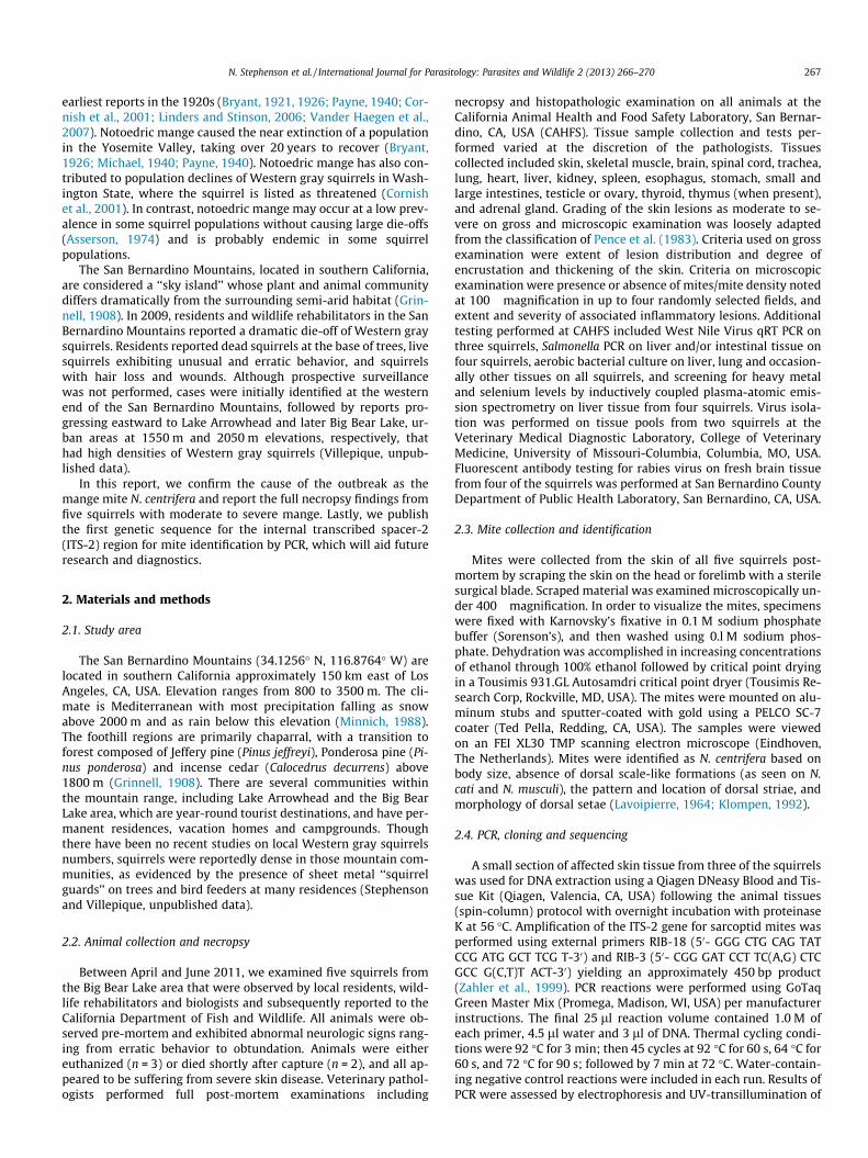
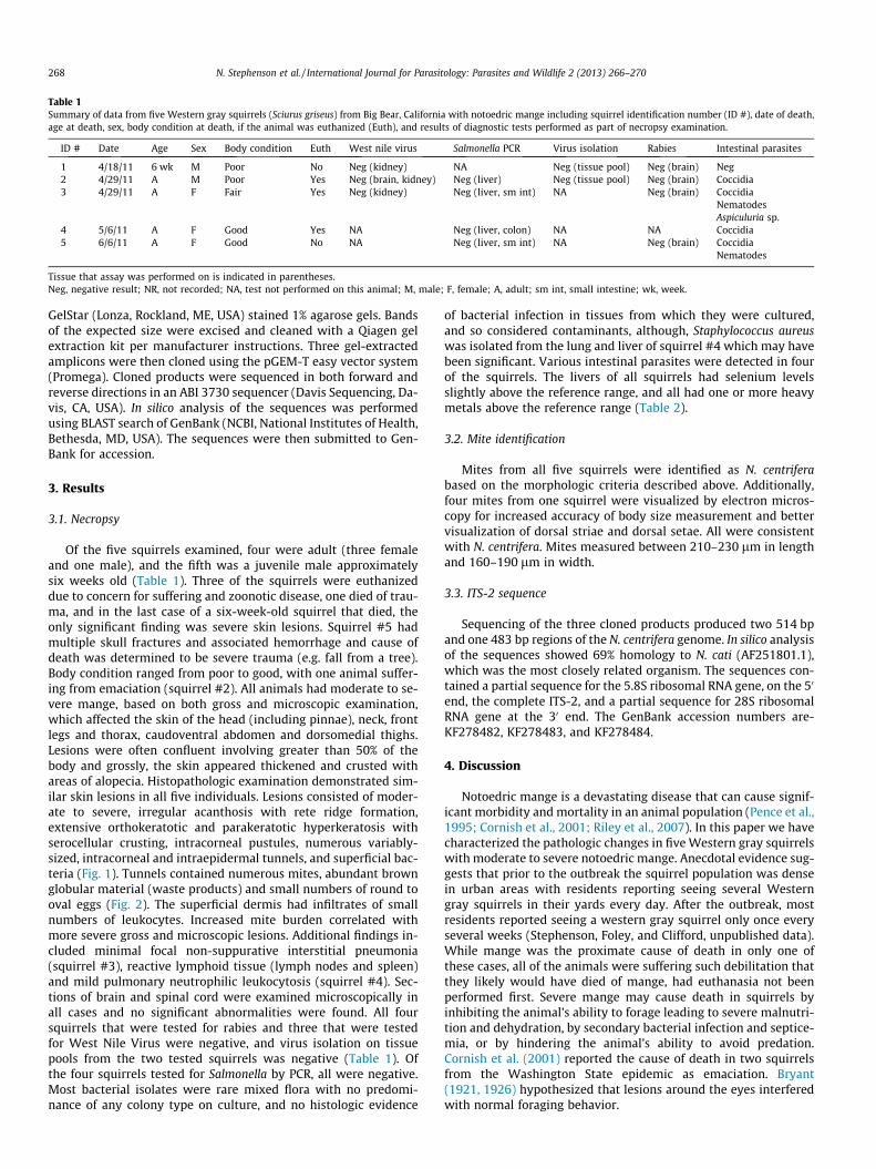
Of the five squirrels examined, four were adult (three femaleand one male), and the fifth was a juvenile male approximatelysix weeks old (Table 1). Three of the squirrels were euthanizeddue to concern for suffering and zoonotic disease, one died of trau-ma, and in the last case of a six-week-old squirrel that died, theonly significant finding was severe skin lesions. Squirrel #5 hadmultiple skull fractures and associated hemorrhage and cause ofdeath was determined to be severe trauma (e.g. fall from a tree).Body condition ranged from poor to good, with one animal suffer-ing from emaciation (squirrel #2). All animals had moderate to se-vere mange, based on both gross and microscopic examination,which affected the skin of the head (including pinnae), neck, frontlegs and thorax, caudoventral abdomen and dorsomedial thighs.Lesions were often confluent involving greater than 50% of thebody and grossly, the skin appeared thickened and crusted withareas of alopecia. Histopathologic examination demonstrated sim-ilar skin lesions in all five individuals. Lesions consisted of moder-ate to severe, irregular acanthosis with rete ridge formation,extensive orthokeratotic and parakeratotic hyperkeratosis withserocellular crusting, intracorneal pustules, numerous variably-sized, intracorneal and intraepidermal tunnels, and superficial bac-teria (Fig. 1). Tunnels contained numerous mites, abundant brownglobular material (waste products) and small numbers of round tooval eggs (Fig. 2). The superficial dermis had infiltrates of smallnumbers of leukocytes. Increased mite burden correlated withmore severe gross and microscopic lesions. Additional findings in-cluded minimal focal non-suppurative interstitial pneumonia(squirrel #3), reactive lymphoid tissue (lymph nodes and spleen)and mild pulmonary neutrophilic leukocytosis (squirrel #4). Sec-tions of brain and spinal cord were examined microscopically inall cases and no significant abnormalities were found. All foursquirrels that were tested for rabies and three that were testedfor West Nile Virus were negative, and virus isolation on tissuepools from the two tested squirrels was negative (Table 1). Ofthe four squirrels tested for Salmonella by PCR, all were negative.Most bacterial isolates were rare mixed flora with no predomi-nance of any colony type on culture, and no histologic evidence
of bacterial infection in tissues from which they were cultured,and so considered contaminants, although, Staphylococcus aureuswas isolated from the lung and liver of squirrel #4 which may havebeen significant. Various intestinal parasites were detected in fourof the squirrels. The livers of all squirrels had selenium levelsslightly above the reference range, and all had one or more heavymetals above the reference range (Table 2).
3.2. Mite identification
Mites from all five squirrels were identified as N. centriferabased on the morphologic criteria described above. Additionally,four mites from one squirrel were visualized by electron micros-copy for increased accuracy of body size measurement and bettervisualization of dorsal striae and dorsal setae. All were consistentwith N. centrifera. Mites measured between 210–230 lm in lengthand 160–190 lm in width.
3.3. ITS-2 sequence
Sequencing of the three cloned products produced two 514 bpand one 483 bp regions of the N. centrifera genome. In silico analysisof the sequences showed 69% homology to N. cati (AF251801.1),which was the most closely related organism. The sequences con-tained a partial sequence for the 5.8S ribosomal RNA gene, on the 50
end, the complete ITS-2, and a partial sequence for 28S ribosomalRNA gene at the 30 end. The GenBank accession numbers are-KF278482, KF278483, and KF278484.
4. Discussion
Notoedric mange is a devastating disease that can cause signif-icant morbidity and mortality in an animal population (Pence et al.,1995; Cornish et al., 2001; Riley et al., 2007). In this paper we havecharacterized the pathologic changes in five Western gray squirrelswith moderate to severe notoedric mange. Anecdotal evidence sug-gests that prior to the outbreak the squirrel population was densein urban areas with residents reporting seeing several Westerngray squirrels in their yards every day. After the outbreak, mostresidents reported seeing a western gray squirrel only once everyseveral weeks (Stephenson, Foley, and Clifford, unpublished data).While mange was the proximate cause of death in only one ofthese cases, all of the animals were suffering such debilitation thatthey likely would have died of mange, had euthanasia not beenperformed first. Severe mange may cause death in squirrels byinhibiting the animal’s ability to forage leading to severe malnutri-tion and dehydration, by secondary bacterial infection and septice-mia, or by hindering the animal’s ability to avoid predation.Cornish et al. (2001) reported the cause of death in two squirrelsfrom the Washington State epidemic as emaciation. Bryant(1921, 1926) hypothesized that lesions around the eyes interferedwith normal foraging behavior.
Table 1Summary of data from five Western gray squirrels (Sciurus griseus) from Big Bear, California with notoedric mange including squirrel identification number (ID #), date of death,age at death, sex, body condition at death, if the animal was euthanized (Euth), and results of diagnostic tests performed as part of necropsy examination.
ID # Date Age Sex Body condition Euth West nile virus Salmonella PCR Virus isolation Rabies Intestinal parasites
1 4/18/11 6 wk M Poor No Neg (kidney) NA Neg (tissue pool) Neg (brain) Neg2 4/29/11 A M Poor Yes Neg (brain, kidney) Neg (liver) Neg (tissue pool) Neg (brain) Coccidia3 4/29/11 A F Fair Yes Neg (kidney) Neg (liver, sm int) NA Neg (brain) Coccidia
NematodesAspiculuria sp.
4 5/6/11 A F Good Yes NA Neg (liver, colon) NA NA Coccidia5 6/6/11 A F Good No NA Neg (liver, sm int) NA Neg (brain) Coccidia
Nematodes
Tissue that assay was performed on is indicated in parentheses.Neg, negative result; NR, not recorded; NA, test not performed on this animal; M, male; F, female; A, adult; sm int, small intestine; wk, week.
268 N. Stephenson et al. / International Journal for Parasitology: Parasites and Wildlife 2 (2013) 266–270
In the present study, all of the squirrels were observed pre-mor-tem and all were reported to be displaying abnormal neurologicbehaviors, ranging from ataxia and erratic behavior to obtundation.The cause of these neurologic signs was not determined by post-mortem examination. Some of the animals were tested for WestNile Virus and rabies as possible causes of neurologic signs insquirrels, but all were negative (Table 1). Virus isolation performedon tissue pools from two animals failed to identify any viral ele-ments. Other causes of neurologic behavior could include head
trauma, other viral encephalitides, bacterial, fungal or parasiticcentral nervous system infections, toxins or hypoglycemia (Schuel-er, 1973; Kiupel et al., 2003; Heinz-Taheny et al., 2004; Carrascoet al., 2006). In this group of animals no lesions were noted onmicroscopic examination of the brain and spinal cord. Severemange can cause intense pruritis, malnutrition and weakness, theclinical signs of which may be interpreted as neurologic disease,and are considered to be the most likely explanation for the abnor-mal behavior of the observed squirrels.
Fig. 1. (a). Histologic sections of skin of free-ranging Western gray squirrels (Sciurus griseus). (a) Severe notoedric mange characterized by, irregular acanthosis with rete ridgeformation, extensive orthokeratotic and parakeratotic hyperkeratosis with serocellular crusting, intracorneal pustules, and numerous variably-sized, intracorneal andintraepidermal tunnels. H&E stain. Bar = 1000 lm. (b) Unaffected skin for comparison. H & E stain. Bar = 500 lm. [Brace = epidermis; star = dermis.]
Fig. 2. Histologic section of skin of a free-ranging western gray squirrel (Sciurus griseus) with notoedric mange. (a) Intraepidermal tunnels containing numerous mites[arrows]. H&E stain. Bar = 500 lm. [Brace = epidermis; star = dermis.] (b) High magnification demonstrating intralesional mites [arrows] and small numbers of round to ovaleggs [arrowheads]. H&E stain. Bar = 100 lm.
Table 2Results of heavy metal and selenium screening on liver tissue from four Western gray squirrels (Sciurus griseus) with notoedric mange from Big Bear, California.
ID # Lead Manganese Iron Mercury Arsenic Zinc Copper Cadmium Selenium(<2) (160–230) (63–120) (<0.2) (<0.5) (26–40) (3.0–6.0) (<0.1–1.2) (0.2–0.52)
1 ND 2.8 90 ND ND 60 29 ND 1.202 ND 3.3 1200 ND ND 84 6.4 3.7 0.793 ND 3.5 260 ND ND 36 6.7 ND 0.664 ND 2.2 220 ND ND 36 3.6 ND 0.64
All concentrations measured in parts per million. Reference ranges given in parentheses below analyte (Puls, 1988).Bold font indicates that the concentration is above the reference range.ID #, squirrel identification number; ND, not detected.
N. Stephenson et al. / International Journal for Parasitology: Parasites and Wildlife 2 (2013) 266–270 269
The cause of death for squirrel #5 was a fractured skull result-ing from blunt trauma, possibly resulting from a fall from a tree.This is a common finding during notoedric mange outbreaks (Bry-ant, 1921; Cornish et al., 2001) and may be the result of incoordi-nation or weakness secondary to mange. While mange may lead tosecondary bacterial infection and septicemia, this was not thelikely cause for neurologic signs in most cases, as there was evi-dence of possible septicemia in only one squirrel. In this animalS. aureus cultured from the liver and lung may represent terminalsepticemia secondary to severe dermatitis. All four squirrels thatwere tested were above the normal reference range for seleniumand at least one heavy metal, although most of the elevations wereslight, and likely of very little if any clinical significance (Table 2).
Notoedric mange can have significant impacts at the individuallevel, but also at the population level as well. As with other types ofparasitism, pregnant animals are likely more susceptible to infesta-tion with mange, especially in late gestation and early lactationwhen nutritional requirements are prioritized for reproduction,rather than immune function (Coop and Kyriazakis, 1999; Fthena-kis et al., 2001). Pregnant and lactating animals have increasedmite burdens, which are often passed onto the highly vulnerableyoung. Mite infestation in neonatal and juvenile production ani-mals causes lower growth rates, higher nutritional requirementsand higher mortality (Arends et al., 1990; Soulsbury et al., 2007).A study of sarcoptic mange in coyotes showed that females withsevere mange had lower ovulation and pregnancy rates than unin-fested coyotes (Pence and Windberg, 1994). By decreasing repro-duction rates and increasing juvenile mortality, notoedric mangecould have a large population impact. Western gray squirrels his-torically were very common throughout their range but since the1920s have undergone significant declines (Bryant, 1921; Payne,1940; Cornish et al., 2001; Vander Haegen et al., 2007). While not-oedric mange is likely a contributor to these declines, there areother factors including degradation and fragmentation of habitatdue to increased urbanization, competition with the non-nativeeastern gray and fox squirrel, predation and possibly other diseases(Ingles, 1947; Linders and Stinson, 2006). Anecdotal reports sug-gest that large die-offs might follow mast crop failure leading tonutritional stress, which could cause both increased transmissionand increased mortality (Cornish et al., 2001). Moreover, squirreldensity may increase transiently around sparse food resources,facilitating disease transmission. Notoedric mange may representa form of top-down population regulation in squirrel species (Cor-nish et al., 2001; Linders and Stinson, 2006; Vander Haegen et al.,2007). Large die-offs caused by notoedric mange are of particularconcern in the native western gray squirrel especially in Washing-ton State where the populations have suffered dramatic declinesand range reductions (Linders and Stinson, 2006).
In this paper we identify N. centrifera as the etiologic agent of adisease in five squirrels from an outbreak in Big Bear, Californiaand we show the marked effects that notoedric mange can have onindividual squirrels. We also report sequences of the ITS-2 regionof N. centrifera for future identification by PCR and genetic studies.While we have shown the significant effects that notoedric mangecan cause in individuals, further research is needed to fully under-stand the population impact since anecdotal reports suggest thatthis disease can cause dramatic die-offs which may take the popula-tion decades to recover (Bryant, 1921; Payne, 1940; Cornish et al.,2001).
Note
Nucleotide sequence data reported in this paper are available inthe GenBank databases under the accession numbers KF278482,KF278483, and KF278484.
Acknowledgements
We thank Joy Worth, Kevin Keel, Chandre Borwick, Craig Lassenand VCA Lakeside Animal Hospital, and Noelle Thomas and BigBear Alpine Zoo. Funding for this project was provided by the Cal-ifornia Department of Fish and Wildlife.
References
Arends, J., Stanislaw, C., Gerdon, D., 1990. Effects of sarcoptic mange on lactatingswine and growing pigs. J. Anim. Sci. 68, 1495–1499.
Asserson, W.C., 1974. Western Gray Squirrel Studies in Kern County, California.California Department of Fish and Game, Administrative Report No. 74-1,Sacramento, CA, USA.
Bryant, H.C., 1921. Tree squirrels infested with scabies. Calif. Fish Game 7, 128.Bryant, H.C., 1926. Gray squirrel disease still exists. Calif. Fish Game 11, 205–
206.Carlson, B.L., Daniel, P.R., Svend, W.N., 1982. Notoedric mange in gray squirrels
(Sciurus carolinensis). J. Wildl. Dis. 18, 347–348.Carrasco, L., Raya, A., Nunez, A., Gomez-Laguna, J., Hernandez, S., Dubey, J., 2006.
Fatal toxoplasmosis and concurrent Calodium hepaticum infection in Koreansquirrels (Tanias sibericus). Vet. Parasitol. 137, 180–183.
Coop, R., Kyriazakis, I., 1999. Nutrition–parasite interaction. Vet. Parasitol. 84, 187–204.
Cornish, T.E., Linders, M.J., Little, S.E., Vander Haegen, W.M., 2001. Notoedric mangein Western gray squirrels from Washington. J. Wildl. Dis. 37, 630–633.
Fthenakis, G., Karagiannidis, A., Alexopoulos, C., Brozos, C., Papadopoulos, E., 2001.Effects of sarcoptic mange on the reproductive performance of ewes andtransmission of Sarcoptes scabiei to newborn lambs. Vet. Parasitol. 95, 63–71.
Grinnell, J., 1908. The Biota of the San Bernardino Mountains. The University Press,Berkley, CA, USA.
Heinz-Taheny, K.M., Andrews, J.J., Kinsel, M.J., Pessier, A.P., Pinkerton, M.E.,Lemberger, K.Y., Novak, R.J., Dizikes, G.J., Edwards, E., Komar, N., 2004. WestNile virus infection in free-ranging squirrels in Illinois. J. Vet. Diagn. Invest. 16,186–190.
Ingles, L.G., 1947. Ecology and life history of the California grey squirrel. Calif. FishGame 33, 139–157.
Kiupel, M., Simmons, H., Fitzgerald, S., Wise, A., Sikarskie, J., Cooley, T., Hollamby, S.,Maes, R., 2003. West Nile virus infection in eastern fox squirrels (Sciurus niger).Vet. Pathol. Online 40, 703–707.
Klompen, J.S.H., 1992. Phylogenetic Relationships in the Mite Family Sarcoptidae(Acari: Astigmata). Museum of Zoology. The University of Michigan, Ann Arbor,MI, USA.
Lavoipierre, M.M.J., 1964. Mange mites of the genus Notoedres (Acari: Sarcoptidae)with descriptions of two new species and remarks on notoedric mange in thesquirrel and the vole. J. Med. Entomol. 1, 5–17.
Linders, M.J., Stinson, D.W., 2006. Draft Washington State Recovery Plan for theWestern Gray Squirrel. Washington Department of Fish and Wildlife, Olympia,WA, USA.
Michael, E., 1940. California gray squirrels coming back to Yosemite. YosemiteNature Notes 19, 37–38.
Minnich, R.A., 1988. The Biogeography of Fire in the San Bernardino Mountains ofCalifornia: A Historical Study. University of California Press, Berkley, CA, USA.
Nebraska Game and Parks Commission, 1991. Fox Squirrel (Scuirus niger). NebraskaGame and Parks Commission Publications. Available from: <http://digitalcommons.unl.edu/nebgamepubs/9>. Accessed March 2012.
Payne, E.A., 1940. The return of the California gray squirrel. Yosemite Nat. Notes 19,1–3.
Pence, D.B., Windberg, L.A., Pence, B.C., Sprowls, R., 1983. The epizootiology andpathology of sarcoptic mange in coyotes, Canis latrans, from South Texas. J.Parasitol., 1100–1115.
Pence, D.B., Windberg, L.A., 1994. Impact of a sarcoptic mange epizootic on a coyotepopulation. J. Wildl. Manag. 58, 624–633.
Pence, D.B., Tewes, M.E., Shindle, D.B., Dunn, D.M., 1995. Notoedric mange in anocelot (Felis pardalis) from southern Texas. J. Wildl. Dis. 31, 558–561.
Puls, R., 1988. Mineral Levels in Animal Health. Diagnostic Data, second ed. SherpaInternational, Clearbrook, BC, Canada.
Riley, S.P.D., Bromley, C., Poppenga, R.H., Uzal, F.A., Whited, L., Sauvajot, R.M., 2007.Anticoagulant exposure and notoedric mange in bobcats and mountain lions inurban southern California. J. Wildl. Manag. 71, 1874–1884.
Schueler, R.L., 1973. Cerebral nematodiasis in a red squirrel. J. Wildl. Dis. 9, 58–60.Soulsbury, C.D., Iossa, G., Baker, P.J., Cole, N.I.K.C., Funk, S.M., Harris, S., 2007. The
impact of sarcoptic mange Sarcoptes scabiei on the British fox Vulpes vulpespopulation. Mammal Rev. 37, 278–296.
Vander Haegen, W.M., Gregory, S.C., Linders, M.J., 2007. Implementation plan foraugmentation of the Western Gray Squirrel Population, Fort Lewis, Washington.Washington Department of Fish and Wildlife, Olympia, WA, USA.
Verts, B.J., Carraway, L.N., 1998. Land mammals of Oregon. University of CaliforniaPress, Berkley, CA, USA.
Zahler, M., Essig, A., Gothe, R., Rinder, H., 1999. Molecular analyses suggestmonospecificity of the genus Sarcoptes (Acari: Sarcoptidae). Int. J. Parasitol. 29,759–766.
270 N. Stephenson et al. / International Journal for Parasitology: Parasites and Wildlife 2 (2013) 266–270