Embed Size (px)
Citation preview
JOURNAL OF VIROLOGY,0022-538X/00/$04.0010
Oct. 2000, p. 9655–9667 Vol. 74, No. 20
Copyright © 2000, American Society for Microbiology. All Rights Reserved.
Internalization of Adenovirus by Alveolar Macrophages InitiatesEarly Proinflammatory Signaling during Acute
Respiratory Tract InfectionZSUZSANNA ZSENGELLER,1† KAZUHISA OTAKE,1‡ SHAIKH-ABU HOSSAIN,1
PIERRE-YVES BERCLAZ,2 AND BRUCE C. TRAPNELL1*
Division of Pulmonary Biology1 and Division of Pulmonary Medicine,2 Children’s HospitalMedical Center, Cincinnati, Ohio 45229
Received 30 March 2000/Accepted 13 July 2000
Adenovirus is a common respiratory pathogen which causes a broad range of distinct clinical syndromes andhas recently received attention for its potential for in vivo gene delivery. Although adenovirus respiratory tractinfection (ARTI) results in dose-dependent, local inflammation, the pathogenesis of this remains unclear. Wehypothesized that alveolar macrophages (AMf) rapidly internalize adenovirus following in vivo pulmonaryadministration and then initiate inflammatory signaling within the lung. To evaluate the role of AMf in theinduction of lung inflammation during ARTI in vivo, we directly assessed adenovirus uptake by murine AMfand correlated uptake with the initiation of proinflammatory gene expression. Stimulation of cytokine (tumornecrosis factor alpha [TNF-a], interleukin-6 [IL-6], macrophage inflammatory protein-2 [MIP-2], and MIP-1a) expression in the lung was evaluated at the level of mRNA (by reverse transcription-PCR [RT-PCR]) andprotein (by enzyme-linked immunosorbent assay) and by identification of cells expressing TNF-a and IL-6mRNA in lung tissues (by in situ hybridization) and isolated lung lavage cells (by RT-PCR). Adenovirus,labeled with the fluorescent dye (Cy3), was rapidly and widely distributed on epithelial surfaces of airways andalveoli and was very rapidly (;1 min) localized within AMf. At 30 min after infection AMf but not airwayepithelial or vascular endothelial cells expressed mRNA for TNF-a and IL-6, thus identifying AMf as the cellsource of initial cytokine signaling. IL-6, TNF-a, MIP-2, and MIP-1a levels progressively increased in bron-choalveolar lavage fluid after pulmonary adenovirus infection, and all were significantly elevated at 6 h (P <0.05). To begin to define the molecular mechanism(s) by which adenovirus initiates the inflammatory signalingin macrophages, TNF-a expression from adenovirus-infected RAW264.7 macrophages was evaluated in vitro.TNF-a expression was readily detected in adenovirus-infected RAW cell supernatant with kinetics similar toAMf during in vivo infection. Blockage of virus uptake at specific cellular sites, including internalization (bywortmannin), endosome acidification and/or lysis (by chloroquine) or by Ca21 chelation (by BAPTA) com-pletely blocked TNF-a expression. In conclusion, results showed that during ARTI, (i) AMf rapidly internal-ized adenovirus, (ii) expression of inflammatory mediators was initiated within AMf and not airway epithelialor other cells, and (iii) the initiation of inflammatory signaling was linked to virion uptake by macrophagesoccurring at a point after vesicle acidification. These results have implications for our understanding of therole of the AMf in the initiation of inflammation following adenovirus infection and adenovirus-mediated genetransfer to the lung.
Adenovirus, a nonenveloped DNA virus with at least 45serotypes, is an important respiratory pathogen affecting indi-viduals of all ages and accounting for 7 to 10% of all respira-tory illnesses in infants and children, with an incidence ofbetween 5 to 10 million infections annually in the UnitedStates alone (13, 21). Adenovirus respiratory tract infection(ARTI) occurs sporadically, epidemically, and nosocomially,presents in a wide spectrum of distinct clinical syndromes rang-ing from self-limited acute pharyngitis to fatal pneumonia, andhas been identified as an etiological factor associated withexacerbations in individuals with chronic obstructive lung dis-eases (13). Despite the frequency and broad range of clinical
presentations, the pathogenesis of inflammation in ARTI ispoorly understood. Early information regarding host responsesto adenovirus was derived from efforts to develop human ad-enovirus vaccines (42) or to understand the pathology of fataladenoviral pneumonia (4). Recently, ARTI has been studied ina variety of animal models, including mice (14, 37, 58), Cottonrats (Sigmodon hispidus) (15, 39, 59, 60), and primates (8, 44,55, 61) as part of preclinical toxicology studies for human genetherapy clinical trials for cystic fibrosis (reviewed in references7 and 48). Studies in humans showed that administration ofreplication-deficient adenovirus vectors to the respiratory tractcan cause dose-dependent local inflammation (12, 26, 33).
Inflammatory responses to ARTI have been best studied inrodent models. In the Cotton rat, a permissive host for repli-cation of human adenovirus, pulmonary histopathology con-sists of early and late phases similar to that seen in humanARTI (15, 39). The early phase consists primarily of accumu-lation of neutrophils, macrophages, and monocytes and devel-ops within the first 24 h (60). The late phase, consisting mainlyof lymphocytes, is apparent by day 5 (39, 60). In mice, a non-permissive host for human adenovirus, adenovirus early gene
* Corresponding author. Mailing address: Children’s Hospital Med-ical Center, Division of Pulmonary Biology, 3333 Burnet Ave., Cincin-nati, OH 45229. Phone: (513) 636-6361. Fax: (513) 636-3723. E-mail:[email protected].
† Present address: Department of Pediatrics, Harvard MedicalSchool, Boston, MA 02115.
‡ Present address: Department of Laboratory Medicine, YamagataUniversity School of Medicine, Yamagata 990-23, Japan.
9655
on March 24, 2018 by guest
http://jvi.asm.org/
Dow
nloaded from
expression occurs in the absence of viral replication or lategene expression (14). Importantly, the histopathologic re-sponse is similar to that observed in Cotton rats (14, 37, 39, 60)and humans (4) and inoculation of the respiratory tract withsufficient doses of either wild-type or replication-deficient ad-enovirus results in significant pulmonary inflammation (14,37). The earliest histopathological abnormality, the influx ofpolymorphonuclear neutrophils (PMN), is first noted 6 h afterinfection (37), peaks by 24 h, and evolves over the course ofseveral days into a mononuclear cell infiltrate due to influx ofboth CD41 and CD81 lymphocytes and expansion of themononuclear phagocyte pool (14, 37, 58). ARTI is accompaniedby an evolving pattern of elevated levels of proinflammatorycytokines (tumor necrosis factor alpha [TNF-a], interleukin-6[IL-6], IL-1, and interferon gamma [IFN-g] and chemotacticchemokines (macrophage inflammatory protein-1a [MIP-1a],MIP-2, and macrophage chemotactic protein-1 [MCP-1]), thechronology of which has been partially characterized in miceover the period from 6 h to several weeks (14, 32, 37). TNF-a,IL-6, MIP-1a, and MIP-2 are among the earliest proinflam-matory molecular mediators detected and are significantly el-evated in lung lavage fluid at 6 h, while MCP-1, IL-1, andIFN-g are elevated by 24 h (37). The pattern of molecular andcellular inflammation is similar in both normal and immuno-deficient (athymic) mice (37), demonstrating that initiation ofinflammatory signaling is independent of specific (adaptive)immunity. However, neither the precise location nor the mech-anism of the initiation of inflammatory signaling during ARTIis known. Such signalling might occur in epithelial cells (whichare tropic for the virus) or alveolar macrophages or as a con-sequence of recruited natural killer or other cells.
Alveolar macrophages (AMf) can provide a barrier to pul-monary infection through both intrinsic and extrinsic resis-tance pathways, the former by their ability to accumulatepathogenic organisms by phagocytosis and/or endocytosis andto degrade or restrict the replication of the organism and thelatter by the recruitment and activation of other inflammatorycells and by their ability to act as accessory cells in adaptiveimmune responses (6, 53). Macrophages (Mf) play a centralrole in the acute-phase response and intrinsic resistance tosome viruses, including vesicular stomatitis virus, encephalo-myocarditis virus, and influenza virus, while failing to providea barrier and instead providing a reservoir for latent infectionfor other viruses such as human immunodeficiency virus (re-viewed in reference 53). Adenovirus is cleared from the mouselung in a biphasic pattern consisting of an early-phase rapidelimination of approximately 60% of the adenovirus DNAwithin the first 24 h, followed by a late-phase slower elimina-tion of remaining adenovirus DNA over the course of severaldays to weeks (56). While the late-phase elimination is knownto be due to the destruction of infected epithelial cells byadenovirus-specific cytotoxic T lymphocytes (58), the veryrapid onset of the early phase within the first day suggests amechanism independent of adaptive immunity (56). This wasconfirmed by the finding of similar early-phase eliminationkinetics in normal and immunodeficient (athymic) mice (56).Pretreatment of the lungs with clodronate-laden liposomes todeplete phagocytic cells significantly impaired early-phaseclearance, suggesting that lung macrophages may mediate therapid adenovirus clearance. However, because rapid and sig-nificant PMN influx occurs during the first 24 h of infection(37), thus overlapping the early phase of adenovirus elimina-tion (56), PMN-mediated clearance cannot be excluded as animportant mediator of viral clearance in ARTI.
The mechanism by which phagocytic cells such as AMfmight internalize adenovirus in vivo is not known and might
involve endocytosis or phagocytosis and may also involve otherfactors in the local milieu. The in vitro uptake of adenovirus byhighly susceptible epithelial-like cells has been well studied(reviewed in reference 17), occurs by receptor-mediated endo-cytosis (31), and can be summarized as follows: (i) high-affinitybinding of the virion to the cell mediated by attachment of theadenovirus fiber knob to its 46-kDa cell surface receptor, CAR(5); (ii) receptor clustering and rapid virion internalization viaa clathrin-coated vesicle mediated by interaction of the ade-novirus penton base with integrins avb5 or avb3 (28–30, 52, 54);(iii) release of clathrin to generate an endocytotic vesicle; (iv)endosome acidification mediated by an endogenous vesicularmembrane proton pump (38); (v) penetration of the endosomemembrane (endosome lysis) and release of the virion into thecytoplasm mediated by the TVD motif-containing cytoplasmictail portion of integrin b5 (51); (vi) virion translocation to thenuclear membrane mediated by microtubules (28, 45); (vii)virion binding to the nuclear pore (16); (viii) capsid disassem-bly at the nuclear pore (17); and (ix) translocation of viralchromatin into the nucleus through the nuclear pore (16). Theability of specific blocking agents to interrupt virion uptake ateach of these sites has provided the means to study the inter-nalization mechanism in detail (17). In contrast to highly sus-ceptible CAR1 cells, in vitro studies show that hematopoieticlineage cells, including AMf, monocytes, and related cell lines,which do not express CAR, internalize adenovirus about 100-to 1,000-fold less well (22, 23, 25, 49). Despite the very slowuptake kinetics in vitro, internalization of adenovirus by suchcells in vitro requires cell surface av integrin, similar to CAR1
epithelial cells, and upregulation of avb5 and avb3 on humanmonocytes facilitates their infection by adenovirus (22).
In this study, we hypothesized that AMf rapidly internalizeadenovirus in vivo very early during ARTI and then directlyinitiate inflammatory signaling. To test this hypothesis, we ex-amined the immediate pulmonary distribution of adenovirusand internalization by AMf during acute ARTI in vivo. Virioninternalization was correlated with the kinetics and cellularorigin of the earliest detectable stimulated proinflammatorycytokine responses. Adenovirus was very rapidly internalizedby AMf, and the earliest detectable cytokine responses oc-curred in AMf and not airway epithelial cells as detected by insitu hybridization. To begin to define the molecular mecha-nism(s) whereby adenovirus stimulates inflammatory signalingin AMf during internalization of the virion, we correlatedvirion uptake with TNF-a release from adenovirus-infectedmacrophages after interrupting virion internalization at vari-ous sites with specific blockers. The data show that TNF-aexpression in adenovirus-exposed AMf is initiated by a mo-lecular mechanism activated during or subsequent to acidifi-cation of the adenovirus-containing vesicle.
MATERIALS AND METHODS
Adenovirus. The adenovirus used in this study was a human serotype 5 ade-novirus carrying a 2,936-bp deletion in E1, a 1,875-bp deletion in E3, and a3,932-bp lacZ marker gene within the E1 deletion (for convenience referred toas adenovirus hereafter except where otherwise specified) and has been previ-ously described (47). For the transduction experiment, Av1GFP, an adenovirusof identical structure except expressing a mammalianized green fluorescent pro-tein (GFP) instead of the lacZ marker was used. Viral growth in 293 cells,purification, and storage were done as previously described (41) except thatinfection was carried out in roller bottles (Falcon no. 3069) containing 12 3 107
cells/bottle and approximately five virions/cell in a Wheaton roller bottle appa-ratus with continuous slow rotation in a humidified atmosphere containing 5%CO2 at 37°C. All cell culture media, medium supplements, and solutions used invirus preparation were supplied routinely or by special arrangement as endotox-in-free materials (BioWhittaker, Inc., Walkersville, Md.). Adenovirus was pre-pared under conditions which have previously been shown to yield undetectablelevels of endotoxin using the Limulus amebocyte assay (BioWhittaker, Inc.) (55).The concentration of adenovirus virions was determined from the optical density
9656 ZSENGELLER ET AL. J. VIROL.
on March 24, 2018 by guest
http://jvi.asm.org/
Dow
nloaded from
of the purified virus at 260 nm and is expressed as optical particle units (OPU)as previously described (35).
Mice. Mice (BALB/c) were maintained in a “barrier” facility in microisolationcages with filtered air under controlled ventilation and constant temperature andhumidity. Bedding, food, and water were all sterilized before use, and food andwater were administered ad libitum. Sterilely gowned and gloved operatorsperformed cage changes and animal handling in a laminar flow biosafety cabinet.Sentinel mice were tested monthly to ensure a pathogen-free environment.
Localization of adenovirus within the lung after in vivo infection. To permitvisualization of adenovirus virions during in vivo infection, purified adenoviruswas labeled by covalent linking of the fluorescent dye, FluoroLink Cy3 (Amer-sham, Inc.) essentially as described by Leopold et al. (28). Virus labeling in thisway results in retention of greater than 90% virus infectivity (28). A molecularsieve chromatography step using PG10 columns (Bio-Rad, Inc.) was substitutedfor the final dialysis step to increase the purity of the labeled virions and toeliminate traces of unlinked dye from the fluorescently labeled adenovirus (Ad-Cy3) preparations. Ad-Cy3 (4 3 1010 OPU) was administered to lungs of normalmice by transtracheal instillation (63). At subsequent times indicated in thefigures, mice were anesthetized with pentobarbital and either bronchoalveolarlavage (BAL; three times with 1 ml of Hank’s balanced salt solution (HBSS)containing 20 mM EDTA) was done to collect cells or the lungs were fixed in situat 25 cm of H2O, embedded in paraffin, and sections (5 mm) were prepared.Specimens were evaluated by fluorescence microscopy (Nikon FXTA micro-scope) or by confocal microscopy. Differential counts were done for cells recov-ered by BAL after cytocentrifugation and Diff-Quick (Dade Diagnostics, Inc.)staining as previously described (37).
Evaluation of cytokine protein levels by ELISA. Quantification of cytokineexpression in murine lung following adenovirus infection was done as previouslydescribed (37). Briefly, adenovirus (4 3 1010 OPU) was administered by oralendotracheal delivery, and mice were sacrificed at various subsequent times (0.5,3, and 6 h) by pentobarbital injection followed by exsanguination. BAL fluid wasthen collected as described above, cleared of cells by centrifugation (200 3 g, 5min, 4°C), and stored at 280°C until use. Quantification of cytokine and che-mokine levels in unconcentrated BAL fluid was accomplished by enzyme-linkedimmunoassay (ELISA; Quantikine M kits; R&D Systems, Minneapolis, Minn.)under conditions specified by the manufacturer.
Evaluation of cytokine mRNA expression by RT-PCR. To evaluate cytokinemRNA expression during ARTI, adenovirus (4 3 1010 OPU) was administeredas described above. Mice were sacrificed at subsequent times (0.5, 3, and 6 h) asdescribed above, and lungs were removed en bloc. Total RNA was purified by theguanidine-acid phenol-guanidinium method, subjected to reverse transcription-polymerase chain reaction (RT-PCR) amplification using cytokine gene-specificoligonucleotide primers (1), and evaluated by agarose gel electrophoresis aspreviously described (37). To evaluate cytokine and chemokine mRNA expres-sion in recovered murine AMf, mice were infected and sacrificed as describedabove. AMf were recovered by BAL and collected by centrifugation (200 3 g, 5min, 4°C), total RNA was purified with a Micro-Scale RNA Prep Kit (5 Prime-3Prime, Inc.), and RT-PCR was carried out as described elsewhere (37).
Localization of in vivo mRNA expression by in situ hybridization. To localizeexpression of proinflammatory cytokine mRNA to specific cell types within thelung, mice were infected with adenovirus (4 3 1010 OPU/mouse). At subsequenttimes (30 min, 3 h, and 6 h), mice were sacrificed, and lung tissue was processedand evaluated by in situ hybridization with 35S-labeled cRNA antisense and sense(control) probes as previously described (62). Briefly, IL-6 cRNA riboprobesynthesis was performed using a 664-bp murine IL-6 cDNA (kindly provided byKeiko Yamauchi Takihara, Osaka, Japan) subcloned into pSP72 (Promega,Inc.). TNF-a cRNA riboprobe synthesis was performed with a 1,101-bp murineTNF-a cDNA subcloned into pGEM7Z1 (Promega, Inc.). Both antisense (formRNA detection) and sense (as a control for nonspecific hybridization)[35S]UTP (specific activity, 1,000 to 1,500 Ci/mmol)-labeled cRNA probes weresynthesized in vitro from opposing transcriptional promoters using the Ribo-probe Gemini Core System II transcription kit under conditions specified by themanufacturer (Promega, Inc.). Tissue sections were incubated at 42°C (IL-6) or55°C (TNF-a) overnight in hybridization buffer (15 ml) containing 106 cpm ofpurified probe and 50% formamide. Following hybridization, tissues werewashed to a final stringency of 0.13 SSC (13 SSC is 0.15 M NaCl plus 0.015 Msodium citrate), and autoradiography was performed using Kodak NTB2 emul-sion at 4°C for periods of 3 to 8 weeks. Development was done with Kodak D19developer as previously described (62). Representative examples of hybridizationresults were photographed with dark-field illumination and then counterstainedwith hematoxylin and eosin for bright-field or phase microscopy using a NikonMicrophot-FXA microscope.
Evaluation of adenovirus-induced cytokine induction in murine macrophages.To further explore the adenovirus-macrophage interactions that lead to induc-tion of proinflammatory cytokine expression, in vitro studies were conductedwith a murine macrophage cell line, RAW264.7 (American Type Culture Col-lection) (34). These cells have phagocytic properties and the capacity to secretecytokines in response to adenovirus infection similar to that of primary AMf.Cells were maintained in Dulbecco modified Eagle medium (DMEM) supple-mented with 10% heat-inactivated (56°C, 30 min) fetal bovine serum, 2 mMglutamine, 100 U of penicillin per ml, and 100 mg of streptomycin per ml(DMEM-10) in a humidified atmosphere containing 5% CO2 at 37°C. The
culture medium was changed every 2 to 3 days, and cells were passed using brieftrypsin digestion just prior to reaching confluence. To evaluate adenovirus-stimulated macrophage TNF-a expression, cells were seeded at 106 cells per wellin six-well plates and incubated (16 h, humidified atmosphere, 5% CO2, 37°C).Cells were then briefly exposed to adenovirus (5 3 1010 OPU/well, 30 min), afterwhich the medium was aspirated and replaced with DMEM-10 without virus.TNF-a release into the medium was then quantified by removing small aliquots(100 ml) of supernatant at subsequent times and quantitative determination ofTNF-a concentration by ELISA. To determine at which step of adenovirusuptake TNF-a signaling is initiated, virion uptake was blocked at one of severaldistinct cellular sites, and then TNF-a release was quantified in supernatant 2 hafter the start of infection. To evaluate the effect of blocking adenovirus inter-nalization at the stage of phagosome closure, wortmannin, in various concentra-tions (0 to 300 mM) was preincubated (15 min prior to infection) with cells,followed by adenovirus infection and evaluation of TNF-a release as describedabove. As an additional approach to block virion entry at the cell membrane,cells were incubated at 4°C for 10 min prior to and during adenovirus infection.At subsequent times (1 to 6 h), cells were warmed to 37°C (removal of “coldblock”), incubation was continued, and the TNF-a assay was done as describedabove. In experiments designed to evaluate the role of Ca21 signaling in ade-novirus-stimulated TNF-a release, cells were preincubated (30 min prior toadenovirus infection) in the absence or presence of 100 mM bis-(o-aminophe-noxy)ethane-N,N,N9,N9-tetra-acetic acid (BAPTA) to block intracellular Ca21
flux. Adenovirus infection and TNF-a expression were evaluated as describedabove. Chloroquine, which blocks the cellular pathway of adenovirus uptake atthe endosome by blocking endosome acidification (17, 43), was used to evaluatethe effect of blocking virion entry at the stage of endosome lysis. Chloroquine (50or 100 mM) was preincubated (10 min prior to infection) with cells, followed byadenovirus exposure and evaluation of TNF-a release as above. Trypan blueexclusion was used to assess the potential for toxicity from each of the inhibitorsunder each of the conditions used: in every condition, cell viability was greaterthan 97% at the end of the experimental period (2 h after the start of infection).
Statistical method. Numerical data are presented as the mean 6 the standarderror of the mean. Statistical comparisons were made by using the Student’s t testusing Sigma Plot (v4.0) software on an IBM-compatible microcomputer.
RESULTS
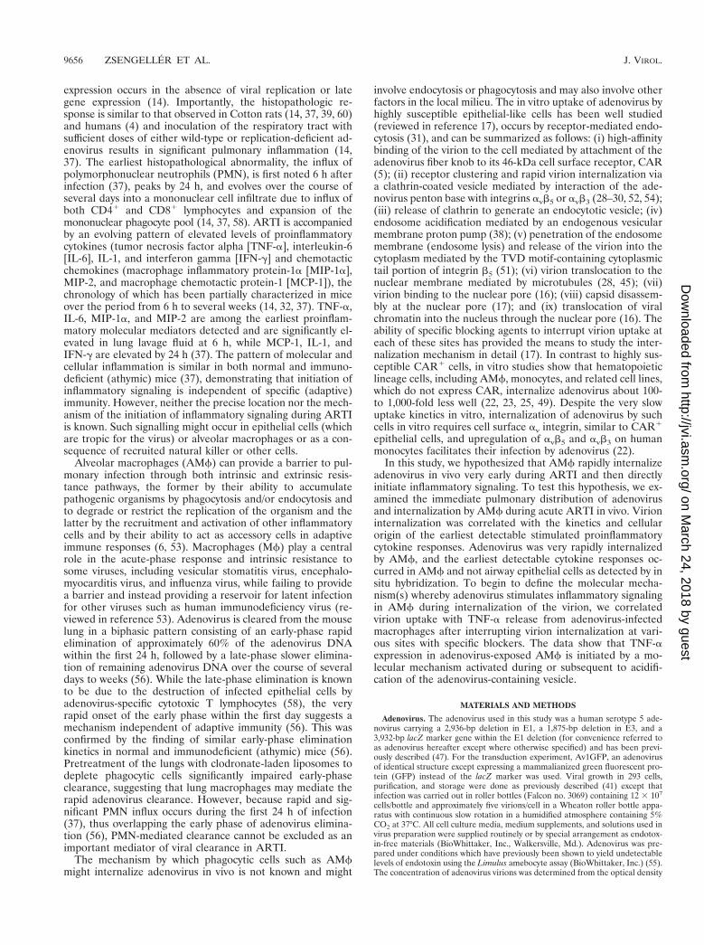
Intrapulmonary distribution and fate of virions during earlyadenovirus lung infection. To directly determine the distribu-tion and immediate fate of adenovirus following intrapulmo-nary administration, adenovirus labeled with the fluorescentdye, Cy3 (Ad-Cy3) was administered (4 3 1010 OPU/mouse)by tracheal instillation. Localization of Ad-Cy3 by fluorescencemicroscopy of unstained, paraffin-embedded sections revealedthat adenovirus was rapidly and widely distributed throughoutthe respiratory tract (Fig. 1). Within medium and larger air-ways, virus was seen along the airway epithelial surfaces atapproximately 1 min with little penetration into the lung pa-renchyma (top left panels). By 10 min, virus was observed in apunctate pattern in association with macrophages in the tissuenear the terminal airways (top right panels). Fluorescence re-mained associated with the airway lumen and within nearbymacrophages at later times (3 and 6 h; data not shown). Inalveolar spaces, Ad-Cy3 was initially (;1 min) distributed pre-dominantly in a “point-like” pattern, suggesting fine dispersionof the virions (middle left panels). By 10 min, adenovirus wasfound in a more punctate pattern associated with AMf (mid-dle right panels). The punctate pattern of macrophage-associ-ated fluorescence persisted at subsequent times (3 and 6 h) andbecame more pronounced, suggesting further accumulation inAMf (data not shown). Cells obtained by BAL revealed ac-cumulation of Ad-Cy3 as early as 1 min after adenovirus in-stillation (bottom left panels). Accumulation was pronouncedby 10 min (bottom right panels) and progressed substantiallythrough the 6-h period of observation (30 min and 3 and 6 h;data not shown). BAL cells from adenovirus-infected micecontained greater than 95% macrophages at all times afterinfection up to 6 h as determined by Diff-Quick staining anddifferential counting (P . 0.36 for all comparisons of AMfvalues at 30 min, 3 h, and 6 h; Table 1). However, neutrophilswere occasionally seen at 30 min and began to increase innumber at 3 and 6 h (without reaching statistical significance
VOL. 74, 2000 INFLAMMATION IN ADENOVIRUS LUNG INFECTION 9657
on March 24, 2018 by guest
http://jvi.asm.org/
Dow
nloaded from
[P . 0.6, all comparisons]), a result consistent with the patchyhistological evidence of neutrophil infiltration at 6 h as previ-ously observed (37). Confocal microscopy revealed the pres-ence of Ad-Cy3 within AMf recovered by BAL and, interest-ingly, also within occasional PMN recovered at 6 h (data notshown). These data show that during ARTI in mice, adenovi-
rus is initially widely distributed throughout the respiratorytract and that adenovirus rapidly accumulates within AMf.
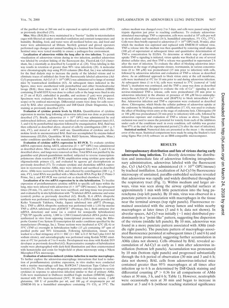
Early molecular inflammation induced by adenovirus lunginfection. To identify some of the molecular mediators thatinitiate the cascade of pulmonary inflammation that occursduring ARTI, we measured the level of several importantproinflammatory and chemotactic molecular mediators in un-concentrated BAL fluid obtained 0.5, 3, and 6 h after adeno-virus administration to the lung (Fig. 2). IL-6, TNF-a, MIP-2,and MIP-1a were all significantly elevated in unconcentratedBAL fluid obtained from adenovirus-exposed mice by 6 h afterinfection (P , 0.05, all comparisons to controls; Fig. 2). TNFa,MIP-2, and MIP-1a, but not IL-6, were also elevated at 3 h.None of these cytokines were yet detectable 30 min after in-fection. As a sham administration control, mice exposed toHBSS did not show detectable cytokine or chemokine activityat any time (Fig. 2). To determine if adenovirus infectionincreased cytokine levels by stimulating gene expression,mRNA levels for these cytokines were measured in whole lungtotal RNA by RT-PCR as previously described (37). CytokinemRNA transcripts were easily detected in the lungs of mice 3and 6 h after adenovirus exposure and variably detected 30 minafter adenovirus exposure (data not shown, but see below [Fig.6] for evaluation of expression in isolated AMf). In contrast,mice exposed to HBSS as a sham control did not have detect-able mRNA for IL-6, TNF-a, MIP-2, and MIP-1a at any time(0, 30 min, 3 h, or 6 h; data not shown). These results show thatinitiation of the inflammatory cascade occurs very early during
TABLE 1. Differential cell counts for BAL cells recovered atvarious times from mice receiving intrapulmonary administration
of adenovirusa
Time (h) Administration% Cells 6 SEM
Macrophages Neutrophils Lymphocytes
0 Nothing 96.0 6 0.5 2.2 6 0.9 1.8 6 0.4
0.5 Virus 97.2 6 0.31 0.53 6 0.29 2.3 6 0.07HBSS 99.0 6 0.30 0 1.0 6 0.3
3 Virus 96.8 6 1.2 2.1 6 1.0 1.1 6 0.24HBSS 99.2 6 0.29 0.44 6 0.29 0.33 6 0
6 Virus 95.7 6 1.4 3.5 6 1.1 0.82 6 0.32HBSS 98.3 6 0.38 0.77 6 0.28 0.9 6 0.1
a Normal (BALB/c) mice (n 5 3/group) were exposed to adenovirus and at thesubsequent times indicated were sacrificed and subjected to BAL. Cells wererecovered from BAL fluid by centrifugation and evaluated in cytocentrifugepreparations by routine cytological analysis. At least 200 cells were counted foreach determination.
FIG. 1. Distribution of adenovirus during acute respiratory tract infection. Infectious, fluorescently labeled adenovirus (Ad-Cy3) or HBSS, as a sham control, wasadministered by intratracheal instillation into the lungs of mice. Mice were then sacrificed after either 1 min (left panels) or 10 min (right panels), and the lungs wereremoved and processed for tissue sections or the mice were subjected to BAL, followed by recovery and cytospin preparation of cells as described in Materials andMethods. Shown are fluorescence and corresponding phase photomicrographs for tissue sections (top and middle panels, 3114) and fluorescence and bright-fieldphotomicrographs for BAL cells (bottom panels, 3232). For BAL cells, separate slides were prepared for fluorescence and bright-field photomicroscopy becauseDiff-Quick staining partially quenched Cy3 fluorescence.
9658 ZSENGELLER ET AL. J. VIROL.
on March 24, 2018 by guest
http://jvi.asm.org/
Dow
nloaded from
ARTI and is associated with activation of cytokine gene ex-pression.
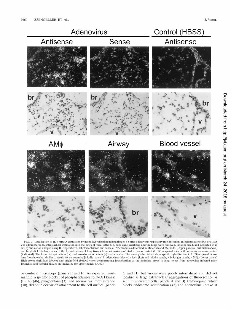
Localization of proinflammatory cytokine mRNA expressionduring early adenovirus lung infection. To determine a cellsource of adenovirus-stimulated proinflammatory cytokinegene expression, expression of cytokine (IL-6, TNF-a) mRNAwas evaluated by in situ hybridization. Six hours after infection,hybridization was detected in lung parenchyma with a 32P-labeled IL-6 cRNA antisense probe (Fig. 3, upper left panels).As a negative control for nonspecific probe binding, a 32P-labeled IL-6 cRNA sense probe did not show hybridization inlungs of virus-infected mice (upper middle panels). As a neg-ative sham administration control, the antisense probe did notshow hybridization in the lungs of mice exposed to HBSSinstead of adenovirus (upper right panels). Detailed evaluationof adenovirus-infected lungs revealed intense hybridization ofthe IL-6 antisense probe in AMf (lower left panels) but notairway epithelial cells (lower middle panels) or vascular endo-thelial cells (lower right panels). A similar pattern of expres-sion was observed for TNF-a 6 h after infection (Fig. 4).Specific hybridization was observed only to a 32P-labeledTNF-a cRNA antisense probe in the lung parenchyma of virus-infected mice (left panels). As a negative control for nonspe-cific probe binding, no hybridization was observed when a32P-labeled TNF-a cRNA sense probe was incubated with lungparenchyma from virus-infected mice (middle panels) or withthe antisense probe in sham HBSS administration control mice(right panels). Detailed evaluation of adenovirus-infected lungtissues showed hybridization of the TNF-a antisense probe inAMf but in not the airway epithelium or vascular endothe-lium. IL-6 and TNF-a antisense probes both showed specifichybridization to AMf within lung parenchyma obtained 30min after virus administration (Fig. 5). Neither IL-6 nor TNF-aantisense probes hybridized to airway epithelium or vascularendothelium of virus-infected mice at 30 min. As negativecontrols for nonspecific probe binding, neither IL-6 nor TNF-asense probes showed hybridization to lung tissues of virus-
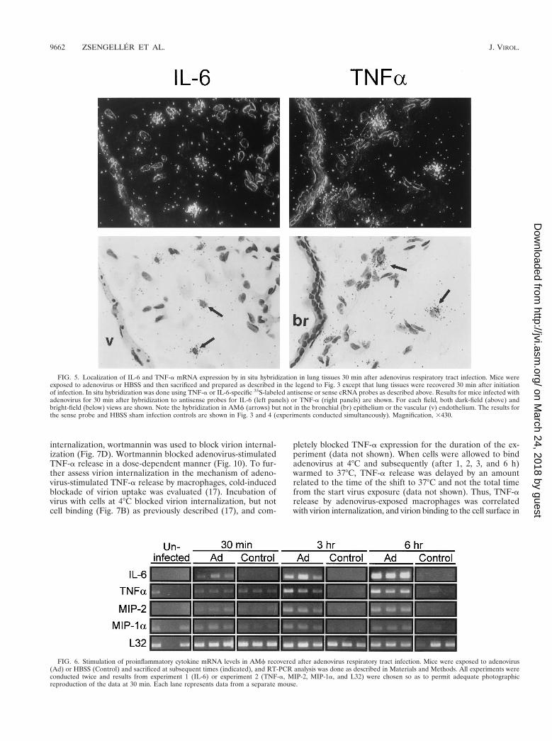
infected mice at 30 min (data not shown but similar to thatpresented in Fig. 3 and 4, upper middle panels). Neither IL-6nor TNF-a antisense probes hybridized to negative sham ad-ministration controls taken at 30 min (data not shown butsimilar to that presented in Fig. 3 and 4, upper right panels).Similar results were obtained with these probes using lungparenchyma taken at 3 h (data not shown). These results dem-onstrate readily detectable expression of proinflammatorygenes (e.g., IL-6 and TNF-a) by 30 min and further localizethis expression to AMf.
Proinflammatory cytokine mRNA expression in AMf recov-ered early after adenovirus lung infection. To confirm thatAMf were a source of stimulated expression of proinflamma-tory cytokines and chemotactic chemokines in the lungs ofadenovirus-exposed mice, BAL cells were recovered after in-fection and evaluated by RT-PCR. At 3 and 6 h after adeno-virus administration to the lung, IL-6, TNF-a, MIP-2, andMIP-1a mRNA transcripts were consistently detected at ele-vated levels in BAL cells from adenovirus-infected mice butnot in sham, HBBS-exposed controls (Fig. 6). TNF-a, IL-6,MIP-2, and MIP-1a mRNA transcripts were also detectable inBAL cells recovered from virus-infected mice after only 30 min(Fig. 6). Although consistently detected at later times, at 30min expression was variably detectable in independent exper-iments, suggesting that 30 min is approximately the time of theinitiation of expression. Since AMf comprised more than 95%of the recovered BAL cells for up to 6 h after adenovirusinfection in this study (Table 1), the RT-PCR data confirm thatAMf were the likely source of the rapidly upregulated proin-flammatory cytokine and chemotactic chemokine mRNA lev-els during ARTI. Taken together, these data demonstrate thatAMf begin to take up adenovirus immediately after in vivolung infection, upregulate cytokine and chemokine mRNA lev-els within minutes, and are the source of the earliest detectablecytokine signals in the initiation of the pulmonary inflamma-tory cascade during ARTI.
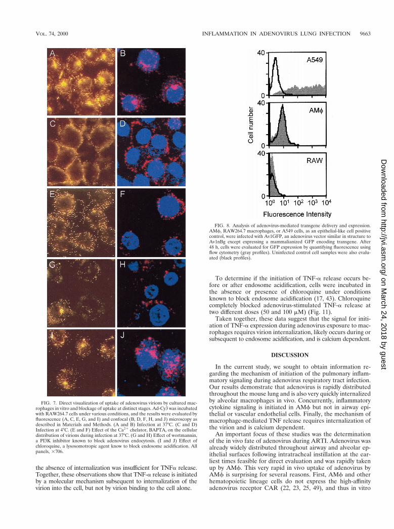
Mechanism of adenovirus-stimulated initiation of macro-phage proinflammatory cytokine stimulation. To begin to de-fine the molecular mechanism(s) whereby clearance of adeno-virus by macrophages stimulates acute-phase, proinflammatorycytokine expression, adenovirus-stimulated TNF-a expressionfrom the macrophage cell line, RAW264.7, was evaluated invitro. Preliminary experiments showed that these cells wereable to internalize adenovirus and rapidly responded by secre-tion of TNF-a with kinetics similar to those of AMf. Further,the in vitro approach permitted precise control of the experi-mental conditions required to block virion uptake into cells atvarious sites (17). We hypothesized that by blocking virionuptake, the molecular stimulus that initiates inflammatory cy-tokine signaling (e.g., TNF-a) could also be blocked. Ad-Cy3was used to verify, by direct visualization, that adenovirus entrycould be blocked at various specific cellular sites, as expectedbased on studies of adenovirus uptake and uncoating in epi-thelial cells (16, 17, 22, 23, 29, 30, 38, 43) (Fig. 7). Exposure ofRAW264.7 cells to Ad-Cy3 in vitro resulted in a pattern offluorescence demonstrating significant virus internalization,cytoplasmic translocation, and prominent extranuclear aggre-gation (Fig. 7A). As expected (16), incubation of virus and cellsat 4°C prevented virion internalization, resulting in a pericel-lular rim of Ad-Cy3 fluorescence (panel C). Evaluation byconfocal microscopy confirmed that virion entry into the cellwas blocked at 4°C but not at 37°C (panels B and D). Theintracellular calcium chelator BAPTA did not prevent virionattachment or internalization. However, little or no transloca-tion and no significant extranuclear aggregation of fluores-cence was noted in BAPTA-treated cells by either fluorescence
FIG. 2. Stimulation of proinflammatory cytokine and chemotactic chemo-kine levels in lung during acute adenovirus respiratory tract infection. Infectiousadenovirus (1Ad) or HBSS (Control) was administered by intratracheal instil-lation into the lungs of mice (n 5 3/time point). At the subsequent timesindicated, mice were sacrificed and the lung epithelial lining fluid was recoveredby BAL. Proinflammatory cytokine (IL-6 and TNFa) and chemoattractive che-mokine (MIP-2 and MIP-1a) levels were measured in unconcentrated BAL fluidby ELISA. The entire experiment was performed twice, and representative datafrom one experiment are shown.
VOL. 74, 2000 INFLAMMATION IN ADENOVIRUS LUNG INFECTION 9659
on March 24, 2018 by guest
http://jvi.asm.org/
Dow
nloaded from
or confocal microscopy (panels E and F). As expected, wort-mannin, a specific blocker of phosphatidylinositol 3-OH kinase(PI3K) (46), phagocytosis (3), and adenovirus internalization(30), did not block virion attachment to the cell surface (panels
G and H), but virions were poorly internalized and did notlocalize as large extranuclear aggregations of fluorescence asseen in untreated cells (panels A and B). Chloroquine, whichblocks endosome acidification (43) and adenovirus uptake at
FIG. 3. Localization of IL-6 mRNA expression by in situ hybridization in lung tissues 6 h after adenovirus respiratory tract infection. Infectious adenovirus or HBSSwas administered by intratracheal instillation into the lungs of mice. After 6 h, mice were sacrificed, and the lungs were removed, inflation fixed, and subjected to insitu hybridization analysis using IL-6-specific 35S-labeled antisense and sense cRNA probes as described in Materials and Methods. (Upper panels) Dark-field (above)and bright-field (below) views of the hybridizations of lung tissues from adenovirus-infected or sham control (HBSS)-exposed mice with antisense or sense probes(indicated). The bronchial epithelium (br) and vascular endothelium (v) are indicated. The sense probe did not show specific hybridization in HBSS-exposed mouselung (not shown but similar to results for sense probe [middle panels] in adenovirus-infected mice). (Left and middle panels, 3143; right panels, 3286). (Lower panels)High-power dark-field (above) and bright-field (below) views demonstrating hybridization of the antisense probe to lung tissues from adenovirus-infected mice.Bronchial and vascular tissues are indicated for upper panels (3343).
9660 ZSENGELLER ET AL. J. VIROL.
on March 24, 2018 by guest
http://jvi.asm.org/
Dow
nloaded from

the doses used here (17), did not prevent virus attachment orinternalization but did reduce the extranuclear aggregation offluorescence (panels I and J).
To assess the fate of adenovirus in macrophages subsequentto extranuclear aggregation, we evaluated adenovirus-medi-ated nuclear gene transfer and expression in AMf, RAW264.7macrophages and, as an epithelial-like cell positive control,A549 cells using Av1GFP. As expected, A549 cells were veryefficiently transduced, and nearly all cells expressed GFP whenevaluated 48 h after infection by using flow cytometry to quan-tify GFP-positive cells (Fig. 8, A549). In contrast, transductionof murine AMf was very inefficient, with only 0.2% of cellsshowing an increase in mean fluorescence by flow cytometry(Fig. 8, AMf). Similarly, RAW264.7 macrophages were alsopoorly transduced, with only 0.02% of cells showing an in-crease in mean fluorescence (Fig. 8, RAW). Evaluation oftransduced cells by fluorescence microscopy demonstratedthat, while nearly all A549 cells expressed GFP, none of theinfected alveolar or RAW264.7 macrophages expressed GFP(data not shown). These results show that the ultimate fate ofinternalized adenovirus is different in macrophages and thenonphagocytic A549 cells and is consistent with inefficientnuclear delivery of the adenovirus genome in Mf.
To assess the mechanism of adenovirus-stimulated macro-phage cytokine expression, studies were conducted usingRAW264.7 macrophages infected in vitro by adenovirus in theabsence or presence of various conditions known to blockvirion entry into cells at various sites along the pathway (17)(30). Adenovirus exposure produced a brisk, reproduciblestimulation of TNF-a expression in RAW264.7 cells, resulting
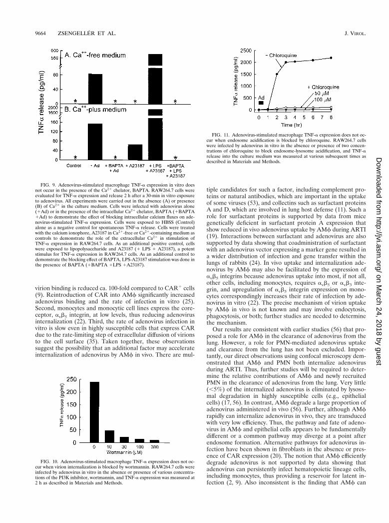
in easily detectable levels of TNF-a in the media at 2 h (Fig. 9;see Fig. 11 below for the time course of expression). In Ca21-free medium, adenovirus exposure resulted in marked TNF-arelease at 2 h, while unexposed controls did not release anydetectable TNF-a (panel A, 1Ad). Results were similar inCa21-containing medium except that TNF-a release was in-creased (panel B, 1Ad). To investigate the role of Ca21 in themechanism of adenovirus-stimulated TNF-a expression,RAW264.7 cells were infected in the absence or presence ofthe intracellular Ca21 chelator BAPTA. Preincubation of cellswith BAPTA completely blocked TNF-a release from adeno-virus-exposed cells in both Ca21-free (panel A, 1BAPTA1Ad) and Ca21-containing (panel B, 1BAPTA 1Ad) me-dium. As a positive control, to demonstrate the role of extra-cellular Ca21 influx in TNF-a expression by RAW264.7 cells,the Ca21 ionophore, A23187 stimulated a large TNF-a releasein Ca21-containing medium (panel B, 1A23187). As a nega-tive control for effect of this Ca21 ionophore on TNF-a ex-pression, as expected, TNF-a release did not occur in responseto A23187 in Ca21-free media (panel A, 1A23187). As apositive control for the blocking effects of BAPTA, the markedrelease of TNF-a occurring in cells exposed to a known potentTNF-a-releasing stimulus (lipopolysaccharide plus A23187)was blocked completely by BAPTA in both Ca21-free andCa21-containing medium (1BAPTA 1LPS 1A23187). Theseobservations show that adenovirus-stimulated TNF-a releaseby RAW264.7 macrophages is dependent on Ca21 and thatthis flux can derive from both intra- and extracellular sources.
To determine if the signal for TNF-a release during adeno-virus uptake by RAW264.7 cells occurred before or after virion
FIG. 4. Localization of TNF-a mRNA expression by in situ hybridization in lung tissues 6 h after adenovirus respiratory tract infection. Mice were exposed toadenovirus or HBSS and then sacrificed and prepared for in situ hybridization as described in the legend to Fig. 3. Tissues were hybridized with TNF-a-specific35S-labeled antisense and sense cRNA probes. Dark-field (above) and bright-field (below) views of the hybridizations of lung tissues from adenovirus-infected or shamcontrol (HBSS)-exposed mice with antisense or sense probes (indicated) are shown. The bronchial epithelium (br) and vascular endothelium (v) are indicated. Thesense probe did not show specific hybridization in HBSS-exposed mouse lung (not shown but similar to sense probe [middle panel] in adenovirus-infected mice). (Leftand middle panels, 3143; right panels, 3286).
VOL. 74, 2000 INFLAMMATION IN ADENOVIRUS LUNG INFECTION 9661
on March 24, 2018 by guest
http://jvi.asm.org/
Dow
nloaded from
internalization, wortmannin was used to block virion internal-ization (Fig. 7D). Wortmannin blocked adenovirus-stimulatedTNF-a release in a dose-dependent manner (Fig. 10). To fur-ther assess virion internalization in the mechanism of adeno-virus-stimulated TNF-a release by macrophages, cold-inducedblockade of virion uptake was evaluated (17). Incubation ofvirus with cells at 4°C blocked virion internalization, but notcell binding (Fig. 7B) as previously described (17), and com-
pletely blocked TNF-a expression for the duration of the ex-periment (data not shown). When cells were allowed to bindadenovirus at 4°C and subsequently (after 1, 2, 3, and 6 h)warmed to 37°C, TNF-a release was delayed by an amountrelated to the time of the shift to 37°C and not the total timefrom the start virus exposure (data not shown). Thus, TNF-arelease by adenovirus-exposed macrophages was correlatedwith virion internalization, and virion binding to the cell surface in
FIG. 5. Localization of IL-6 and TNF-a mRNA expression by in situ hybridization in lung tissues 30 min after adenovirus respiratory tract infection. Mice wereexposed to adenovirus or HBSS and then sacrificed and prepared as described in the legend to Fig. 3 except that lung tissues were recovered 30 min after initiationof infection. In situ hybridization was done using TNF-a or IL-6-specific 35S-labeled antisense or sense cRNA probes as described above. Results for mice infected withadenovirus for 30 min after hybridization to antisense probes for IL-6 (left panels) or TNF-a (right panels) are shown. For each field, both dark-field (above) andbright-field (below) views are shown. Note the hybridization in AMf (arrows) but not in the bronchial (br) epithelium or the vascular (v) endothelium. The results forthe sense probe and HBSS sham infection controls are shown in Fig. 3 and 4 (experiments conducted simultaneously). Magnification, 3430.
FIG. 6. Stimulation of proinflammatory cytokine mRNA levels in AMf recovered after adenovirus respiratory tract infection. Mice were exposed to adenovirus(Ad) or HBSS (Control) and sacrificed at subsequent times (indicated), and RT-PCR analysis was done as described in Materials and Methods. All experiments wereconducted twice and results from experiment 1 (IL-6) or experiment 2 (TNF-a, MIP-2, MIP-1a, and L32) were chosen so as to permit adequate photographicreproduction of the data at 30 min. Each lane represents data from a separate mouse.
9662 ZSENGELLER ET AL. J. VIROL.
on March 24, 2018 by guest
http://jvi.asm.org/
Dow
nloaded from
the absence of internalization was insufficient for TNFa release.Together, these observations show that TNF-a release is initiatedby a molecular mechanism subsequent to internalization of thevirion into the cell, but not by virion binding to the cell alone.
To determine if the initiation of TNF-a release occurs be-fore or after endosome acidification, cells were incubated inthe absence or presence of chloroquine under conditionsknown to block endosome acidification (17, 43). Chloroquinecompletely blocked adenovirus-stimulated TNF-a release attwo different doses (50 and 100 mM) (Fig. 11).
Taken together, these data suggest that the signal for initi-ation of TNF-a expression during adenovirus exposure to mac-rophages requires virion internalization, likely occurs during orsubsequent to endosome acidification, and is calcium dependent.
DISCUSSION
In the current study, we sought to obtain information re-garding the mechanism of initiation of the pulmonary inflam-matory signaling during adenovirus respiratory tract infection.Our results demonstrate that adenovirus is rapidly distributedthroughout the mouse lung and is also very quickly internalizedby alveolar macrophages in vivo. Concurrently, inflammatorycytokine signaling is initiated in AMf but not in airway epi-thelial or vascular endothelial cells. Finally, the mechanism ofmacrophage-mediated TNF release requires internalization ofthe virion and is calcium dependent.
An important focus of these studies was the determinationof the in vivo fate of adenovirus during ARTI. Adenovirus wasalready widely distributed throughout airway and alveolar ep-ithelial surfaces following intratracheal instillation at the ear-liest times feasible for direct evaluation and was rapidly takenup by AMf. This very rapid in vivo uptake of adenovirus byAMf is surprising for several reasons. First, AMf and otherhematopoietic lineage cells do not express the high-affinityadenovirus receptor CAR (22, 23, 25, 49), and thus in vitro
FIG. 7. Direct visualization of uptake of adenovirus virions by cultured mac-rophages in vitro and blockage of uptake at distinct stages. Ad-Cy3 was incubatedwith RAW264.7 cells under various conditions, and the results were evaluated byfluorescence (A, C, E, G, and I) and confocal (B, D, F, H, and J) microscopy asdescribed in Materials and Methods. (A and B) Infection at 37°C. (C and D)Infection at 4°C. (E and F) Effect of the Ca21 chelator, BAPTA, on the cellulardistribution of virions during infection at 37°C. (G and H) Effect of wortmannin,a PI3K inhibitor known to block adenovirus endocytosis. (I and J) Effect ofchloroquine, a lysosomotropic agent know to block endosome acidification. Allpanels, 3706.
FIG. 8. Analysis of adenovirus-mediated transgene delivery and expression.AMf, RAW264.7 macrophages, or A549 cells, as an epithelial-like cell positivecontrol, were infected with Av1GFP, an adenovirus vector similar in structure toAv1nBg except expressing a mammalianized GFP encoding transgene. After48 h, cells were evaluated for GFP expression by quantifying fluorescence usingflow cytometry (gray profiles). Uninfected control cell samples were also evalu-ated (black profiles).
VOL. 74, 2000 INFLAMMATION IN ADENOVIRUS LUNG INFECTION 9663
on March 24, 2018 by guest
http://jvi.asm.org/
Dow
nloaded from
virion binding is reduced ca. 100-fold compared to CAR1 cells(9). Reintroduction of CAR into AMf significantly increasedadenovirus binding and the rate of infection in vitro (25).Second, monocytes and monocytic cell lines express the core-ceptor, avb5 integrin, at low levels, thus reducing adenovirusinternalization (22). Third, the rate of adenovirus infection invitro is slow even in highly susceptible cells that express CARdue to the rate-limiting step of extracellular diffusion of virionsto the cell surface (35). Taken together, these observationssuggest the possibility that an additional factor may accelerateinternalization of adenovirus by AMf in vivo. There are mul-
tiple candidates for such a factor, including complement pro-teins or natural antibodies, which are important in the uptakeof some viruses (53), and collectins such as surfactant proteinsA and D, which are involved in lung host defense (11). Such arole for surfactant proteins is supported by data from micegenetically deficient in surfactant protein A expression thatshow reduced in vivo adenovirus uptake by AMf during ARTI(19). Interactions between surfactant and adenovirus are alsosupported by data showing that coadministration of surfactantwith an adenovirus vector expressing a marker gene resulted ina wider distribution of infection and gene transfer within thelungs of rabbits (24). In vivo uptake and internalization ade-novirus by AMf may also be facilitated by the expression ofavb5 integrins because adenovirus uptake into most, if not all,other cells, including monocytes, requires avb5 or avb3 inte-grin, and upregulation of avb5 integrin expression on mono-cytes correspondingly increases their rate of infection by ade-novirus in vitro (22). The precise mechanism of virion uptakeby AMf in vivo is not known and may involve endocytosis,phagocytosis, or both; further studies are needed to determinethe mechanism.
Our results are consistent with earlier studies (56) that pro-posed a role for AMf in the clearance of adenovirus from thelung. However, a role for PMN-mediated adenovirus uptakeand clearance from the lung has not been excluded. Impor-tantly, our direct observations using confocal microscopy dem-onstrated that AMf and PMN both internalize adenovirusduring ARTI. Thus, further studies will be required to deter-mine the relative contributions of AMf and newly recruitedPMN in the clearance of adenovirus from the lung. Very little(,5%) of the internalized adenovirus is eliminated by lysoso-mal degradation in highly susceptible cells (e.g., epithelialcells) (17, 56). In contrast, AMf degrade a large proportion ofadenovirus administered in vivo (56). Further, although AMfrapidly can internalize adenovirus in vivo, they are transducedwith very low efficiency. Thus, the pathway and fate of adeno-virus in AMf and epithelial cells appears to be fundamentallydifferent or a common pathway may diverge at a point afterendosome formation. Alternative pathways for adenovirus in-fection have been shown in fibroblasts in the absence or pres-ence of CAR expression (20). The notion that AMf efficientlydegrade adenovirus is not supported by data showing thatadenovirus can persistently infect hematopoietic lineage cells,including monocytes, thus providing a reservoir for latent in-fection (2, 9). Also inconsistent is the finding that AMf can
FIG. 9. Adenovirus-stimulated macrophage TNF-a expression in vitro doesnot occur in the presence of the Ca21 chelator, BAPTA. RAW264.7 cells wereevaluated for TNF-a expression and release 2 h after a 30-min in vitro exposureto adenovirus. All experiments were carried out in the absence (A) or presence(B) of Ca21 in the culture medium. Cells were infected with adenovirus alone(1Ad) or in the presence of the intracellular Ca21 chelator, BAPTA (1BAPTA1Ad) to demonstrate the effect of blocking intracellular calcium fluxes on ade-novirus-stimulated TNF-a expression. Cells were exposed to HBSS (Control)alone as a negative control for spontaneous TNF-a release. Cells were treatedwith the calcium ionophore, A23187 in Ca21-free or Ca21-containing medium ascontrols to demonstrate the role of the extracellular Ca21 in stimulation ofTNF-a expression in RAW264.7 cells. As an additional positive control, cellswere exposed to lipopolysaccharide and A23187 (1 LPS 1 A23187), a potentstimulus for TNF-a expression in RAW264.7 cells. As an additional control todemonstrate the blocking effect of BAPTA, LPS-A23187 stimulation was done inthe presence of BAPTA (1BAPTA 1LPS 1A23187).
FIG. 10. Adenovirus-stimulated macrophage TNF-a expression does not oc-cur when virion internalization is blocked by wortmannin. RAW264.7 cells wereinfected by adenovirus in vitro in the absence or presence of various concentra-tions of the PI3K inhibitor, wortmannin, and TNF-a expression was measured at2 h as described in Materials and Methods.
FIG. 11. Adenovirus-stimulated macrophage TNF-a expression does not oc-cur when endosome acidification is blocked by chloroquine. RAW264.7 cellswere infected by adenovirus in vitro in the absence or presence of two concen-trations of chloroquine to block endosome-lysosome acidification, and TNF-arelease into the culture medium was measured at various subsequent times asdescribed in Materials and Methods.
9664 ZSENGELLER ET AL. J. VIROL.
on March 24, 2018 by guest
http://jvi.asm.org/
Dow
nloaded from
harbor replication-deficient adenovirus vectors for up to 5weeks in immunocompetent mice in vivo (57).
The major focus of the present study was to obtain informa-tion regarding the mechanism of activation of the pulmonaryinflammatory signaling during ARTI. The sequence of inflam-matory events at late times following adenovirus infection ofthe lung has been best studied in animal models and consists ofan evolving cascade of molecular and cellular mediators (14,37, 44, 55, 58, 60, 63). In mice, cellular infiltration by PMN isnoted in the lung as early as 6 h after adenovirus administra-tion (37). This increases in magnitude, eventually giving way toa predominantly mononuclear cell infiltrate over the course ofseveral days. Elevated levels of several proinflammatory cyto-kines and chemokines have also been observed at intermediatetimes during ARTI. For example, elevated lung cytokine levelswere detected during ARTI for TNF-a, IL-6, MIP-1a, andMIP-2 in mice (37), IL-8 in nonhuman primates (55), and IL-6in humans (33). In humans, exposure of the adenovirus toAMf was postulated as the cause of elevated IL-6 levels (33).Consistent with this concept, following reduction of the volumeof adenovirus inoculum to prevent alveolar spread of virus(thus reducing exposure to AMf), subsequently treated pa-tients did not show elevated IL-6 levels (33). In the presentstudy, TNF-a was detectable 3 h after adenovirus infection,and IL-6 was detectable at 6 h. Importantly, in situ hybridiza-tion demonstrated elevated mRNA levels for both cytokines inmacrophages at 30 min, suggesting a common, very early acti-vation signal. NF-kB could provide such a signaling mecha-nism, and this factor has been reported to be required forstimulation of TNF-a expression in AMf (27). Nonetheless,no mRNA expression for either cytokine was observed in air-way or vascular endothelia, thus clearly identifying AMf as thesite of initiation of the pulmonary inflammatory cytokine cas-cade. Our results are consistent with in vitro data that showedthat cultured human bronchial epithelial cells do not secretecytokines after adenovirus exposure even though TNF-a, as apositive control, stimulated IL-6 and IL-8 expression (36).
Several lines of evidence suggest that inflammation duringARTI is initiated within AMf during virion uptake and/ordegradation rather than as a response of natural killer or othercells to infected epithelial cells or by events within epithelial orvascular cells. In vivo, adenovirus infection increased levels ofTNF-a mRNA in AMf by 30 min and protein in BAL by 3 h.RAW264.7 macrophages infected at high multiplicity in vitroshowed similar TNF-a expression kinetics and demonstratedthat macrophages alone are sufficient for initiation of cytokinesignaling. Further, TNF-a expression was completely abro-gated when virion internalization was interrupted at one ofseveral distinct cellular sites. Previous studies have demon-strated that receptor-mediated adenovirus internalization byepithelial cells is blocked by wortmannin or infection at 4°C(17, 30). Both of these conditions completely blocked adeno-virus internalization and TNF-a expression. Blocking intracel-lular Ca21 flux completely abrogated TNF-a expression byRAW264.7 cells. Although BAPTA did not affect virion inter-nalization, it prevented late extranuclear aggregation of ade-novirus. Ca21 is known to be involved in adenovirus uptake inCAR-expressing epithelial cells at early and late times (16).Although the precise point of the early involvement is notknown, the later involvement occurs during virion binding tothe nuclear pore (16). It is interesting that BAPTA enhancedadenovirus-mediated gene transfer to airway epithelium dur-ing in vivo administration to the lung (50). This enhancementwas attributed to an effect on airway cell tight junction perme-ability, although potential effects on macrophage virion uptakewere not evaluated. Chloroquine, at the doses used here, has
been shown to block endosome-lysosome acidification (38) andalso adenovirus-mediated endosome lysis and cellular infection(17). At lower doses (5 to 10 mM) which reduce the toxicityseen at late times (48 to 72 h) after infection, chloroquine wasreported to block endosome acidification but did not affectadenovirus infection of HeLa cells (40). Our data demon-strated that chloroquine completely blocked TNF-a expressionin RAW264.7 macrophages and appears to have altered theintracellular distribution of adenovirus, reducing the extranu-clear aggregation. Our results with chloroquine and the variousinhibitors in these short-term experiments cannot be explainedon the basis of toxicity since .97% of cells were viable underall conditions evaluated as determined by trypan blue exclu-sion. Expression of adenoviral early genes such as those of theE1 region has been postulated as important in stimulating theinflammatory cascade during ARTI in the mouse model (15).The adenovirus mutant used here was devoid of E1 genes andalso E3 region genes known to modulate inflammatory re-sponses (18) but still rapidly initiated inflammation duringARTI. Thus, our data do not support a requirement for ade-novirus early gene expression in the initiation of pulmonaryinflammation. Taken together, our data suggest that the mo-lecular event that initiates cytokine gene expression in AMfduring virus clearance occurs during or subsequent to endo-some acidification.
These observations have implications for the developmentof adenovirus vectors for human gene therapy for cystic fibrosisand lung disorders. Inflammation has been observed in multi-ple clinical trials where replication-deficient adenovirus vectorshave been administered to the respiratory tract (7). Impor-tantly, inflammation occurred after vector administration tonasal and bronchial epithelium. Thus, vector delivery to thealveolar surface cannot completely explain the inflammatoryhost response as previously proposed (33). Nasal inflammatoryresponses to adenovirus appear not to be explained by anepithelial cell response because cultured primary airway epi-thelial cells do not stimulate cytokines following in vitro ade-novirus infection (36). These prior findings may be reconciledby the fact that macrophages are found on the epithelial sur-face distributed throughout the respiratory tract with a densityin proportion to the surface area (6, 10) and our observationsthat macrophages initiate the inflammatory response duringadenovirus infection.
ACKNOWLEDGMENTS
We thank Keiko Takihara for the generous gift of the murine IL-6cDNA, Kim Wilmer for help with animal husbandry, and Jeff Whitsettfor critical reading of the manuscript.
This work was supported by the Cystic Fibrosis Foundation (S887)and the Children’s Hospital Research Foundation, Cincinnati, Ohio.
REFERENCES
1. Allen, R. D., T. A. Staley, and C. L. Sidman. 1993. Differential cytokineexpression in acute and chronic murine graft- versus-host-disease. Eur. J. Im-munol. 23:333–337.
2. Andiman, W. A., and G. Miller. 1982. Persistent infection with adenovirustypes 5 and 6 in lymphoid cells from humans and woolly monkeys. J. Infect.Dis. 145:83–88.
3. Araki, N., M. T. Johnson, and J. A. Swanson. 1996. A role for phosphoino-sitide 3-OH kinase in the completion of macropinocytosis and phagocytosisby macrophages. J. Cell Biol. 135:1249–1260.
4. Becroft, D. M. 1967. Histopathology of fatal adenovirus infection of therespiratory tract in young children. J. Clin. Pathol. 20:561–569.
5. Bergelson, J. M., A. Krithivas, L. Celi, G. Droguett, M. S. Horwitz, T.Wickham, R. L. Crowell, and R. W. Finberg. 1998. The murine CAR ho-molog is a receptor for coxsackie B viruses and adenoviruses. J. Virol.72:415–419.
6. Bezdicek, P., and R. G. Crystal. 1997. Pulmonary macrophages, p. 859–875.In R. G. Crystal, P. J. Barnes, J. B. West, and E. R. Weibel (ed.), The lung:
VOL. 74, 2000 INFLAMMATION IN ADENOVIRUS LUNG INFECTION 9665
on March 24, 2018 by guest
http://jvi.asm.org/
Dow
nloaded from
scientific foundations, 2nd ed., vol. 1. Lippincott-Raven, Philadelphia, Pa.7. Boucher, R. C. 1999. Status of gene therapy for cystic fibrosis lung disease.
J. Clin. Investig. 103:441–445.8. Brody, S. L., M. Metzger, C. Danel, M. A. Rosenfeld, and R. G. Crystal. 1994.
Acute responses of non-human primates to airway delivery of an adenovirusvector containing the human cystic fibrosis transmembrane conductanceregulator cDNA. Hum. Gene Ther. 5:821–836.
9. Chu, Y., K. Sperber, L. Mayer, and M. T. Hsu. 1992. Persistent infection ofhuman adenovirus type 5 in human monocyte cell lines. Virology 188:793–800.
10. Crapo, J. D., B. E. Barry, P. Gehr, M. Bachofen, and E. R. Weibel. 1982. Cellnumber and cell characteristics of the normal human lung. Am. Rev. Respir.Dis. 126:332–337.
11. Crouch, E. C. 1998. Collectins and pulmonary host defense. Am. J. Respir.Cell Mol. Biol. 19:177–201.
12. Crystal, R. G., N. G. McElvaney, M. A. Rosenfeld, C. S. Chu, A. Mastrangeli,J. G. Hay, S. L. Brody, H. A. Jaffe, N. T. Eissa, and C. Danel. 1994. Admin-istration of an adenovirus containing the human CFTR cDNA to the respi-ratory tract of individuals with cystic fibrosis. Nat. Genet. 8:42–51.
13. Fraser, R. S., N. L. Muller, N. Colman, and P. D. Pare. 1999. Viralrespiratory diseases, p. 994–996. In R. S. Fraser and P. D. Pare’s (ed.),Diagnosis of diseases of the chest, 4th ed., vol. 2. W. B. Saunders Co.,Philadelphia, Pa.
14. Ginsberg, H. S., L. L. Moldawer, P. B. Sehgal, M. Redington, P. L. Kilian,R. M. Chanock, and G. A. Prince. 1991. A mouse model for investigating themolecular pathogenesis of adenovirus pneumonia. Proc. Natl. Acad. Sci.USA 88:1651–1655.
15. Ginsberg, H. S., and G. A. Prince. 1994. The molecular basis of adenoviruspathogenesis. Infect. Agents Dis. 3:1–8.
16. Greber, U. F., M. Suomalainen, R. P. Stidwill, K. Boucke, M. W. Ebersold,and A. Helenius. 1997. The role of the nuclear pore complex in adenovirusDNA entry. EMBO J. 16:5998–6007.
17. Greber, U. F., M. Willetts, P. Webster, and A. Helenius. 1993. Stepwisedismantling of adenovirus 2 during entry into cells. Cell 75:477–486.
18. Harrod, K. S., T. W. Hermiston, B. C. Trapnell, W. S. Wold, and J. A.Whitsett. 1998. Lung-specific expression of adenovirus E3-14.7K in trans-genic mice attenuates adenoviral vector-mediated lung inflammation andenhances transgene expression. Hum. Gene Ther. 9:1885–1898.
19. Harrod, K. S., B. C. Trapnell, K. Otake, T. R. Korfhagen, and J. A. Whitsett.1999. SP-A enhances viral clearance and inhibits inflammation after pulmo-nary adenoviral infection. Am. J. Physiol. 277:L580–L588.
20. Hidaka, C., E. Milano, P. L. Leopold, J. M. Bergelson, N. R. Hackett, R. W.Finberg, T. J. Wickham, I. Kovesdi, P. Roelvink, and R. G. Crystal. 1999.CAR-dependent and CAR-independent pathways of adenovirus vector-me-diated gene transfer and expression in human fibroblasts. J. Clin. Investig.103:579–587.
21. Horowitz, M. S. 1996. Adenoviruses, p. 2149–2171. In B. N. Fields, D. M.Knipe, and P. M. Howley (ed.), Fields virology, 3rd ed., vol. 2. Lippincott-Raven Publishers, New York, N.Y.
22. Huang, S., R. I. Endo, and G. R. Nemerow. 1995. Upregulation of integrinsavb3 and avb5 on human monocytes and T lymphocytes facilitates adenovi-rus-mediated gene delivery. J. Virol. 69:2257–2263.
23. Huang, S., T. Kamata, Y. Takada, Z. M. Ruggeri, and G. R. Nemerow. 1996.Adenovirus interaction with distinct integrins mediates separate events incell entry and gene delivery to hematopoietic cells. J. Virol. 70:4502–4508.
24. Jobe, A. H., T. Ueda, J. A. Whitsett, B. C. Trapnell, and M. Ikegami. 1996.Surfactant enhances adenovirus-mediated gene expression in rabbit lungs.Gene Ther. 3:775–779.
25. Kaner, R. J., S. Worgall, P. L. Leopold, E. Stolze, E. Milano, C. Hidaka, R.Ramalingam, N. R. Hackett, R. Singh, J. Bergelson, R. Finberg, E. Falck-Pedersen, and R. G. Crystal. 1999. Modification of the genetic program ofhuman alveolar macrophages by adenovirus vectors in vitro is feasible butinefficient, limited in part by the low level of expression of the coxsackie/adenovirus receptor. Am. J. Respir. Cell Mol. Biol. 20:361–370.
26. Knowles, M. R., K. W. Hohneker, Z. Zhou, J. C. Olsen, T. L. Noah, P. C. Hu,M. W. Leigh, J. F. Engelhardt, L. J. Edwards, K. R. Jones, et al. 1995. Acontrolled study of adenoviral-vector-mediated gene transfer in the nasalepithelium of patients with cystic fibrosis. N. Engl. J. Med. 333:823–831.
27. Lentsch, A. B., B. J. Czermak, N. M. Bless, N. Van Rooijen, and P. A. Ward.1999. Essential role of alveolar macrophages in intrapulmonary activation ofNF-kappaB. Am. J. Respir. Cell Mol. Biol. 20:692–698.
28. Leopold, P. L., B. Ferris, I. Grinberg, S. Worgall, N. R. Hackett, and R. G.Crystal. 1998. Fluorescent virions: dynamic tracking of the pathway of ad-enoviral gene transfer vectors in living cells. Hum. Gene Ther. 9:367–378.
29. Li, E., D. Stupack, G. M. Bokoch, and G. R. Nemerow. 1998. Adenovirusendocytosis requires actin cytoskeleton reorganization mediated by Rhofamily GTPases. J. Virol. 72:8806–8812.
30. Li, E., D. Stupack, R. Klemke, D. A. Cheresh, and G. R. Nemerow. 1998.Adenovirus endocytosis via av integrins requires phosphoinositide-3-OHkinase. J. Virol. 72:2055–2061.
31. Marsh, M., and H. T. McMahon. 1999. The structural era of endocytosis.Science 285:215–220.
32. McCoy, R. D., B. L. Davidson, B. J. Roessler, G. B. Huffnagle, S. L. Janich,T. J. Laing, and R. H. Simon. 1995. Pulmonary inflammation induced byincomplete or inactivated adenoviral particles. Hum. Gene Ther. 6:1553–1560.
33. McElvaney, N. G., and R. G. Crystal. 1995. IL-6 release and airway admin-istration of human CFR cDNA adenovirus vector. Nat. Med. 1:182–184.
34. McKernan, L. N., and M. T. Largen. 1983. Identification of multiple-molec-ular-weight forms of thymocyte comitogenic activity from the monocyte/macrophage cell line RAW 264.7. Cell. Immunol. 80:84–96.
35. Mittereder, N., K. L. March, and B. C. Trapnell. 1996. Evaluation of theconcentration and bioactivity of adenovirus vectors for gene therapy. J. Vi-rol. 70:7498–7509.
36. Noah, T. L., I. A. Wortman, P. C. Hu, M. W. Leigh, and R. C. Boucher. 1996.Cytokine production by cultured human bronchial epithelial cells infectedwith a replication-deficient adenoviral gene transfer vector or wild-type ad-enovirus type 5. Am. J. Respir. Cell Mol. Biol. 14:417–424.
37. Otake, K., D. L. Ennist, K. Harrod, and B. C. Trapnell. 1998. Nonspecificinflammation inhibits adenovirus-mediated pulmonary gene transfer andexpression independent of specific acquired immune responses. Hum. GeneTher. 9:2207–2222.
38. Perez, L., and L. Carrasco. 1994. Involvement of the vacuolar H(1)-ATPasein animal virus entry. J. Gen. Virol. 75:2595–606.
39. Prince, G. A., D. D. Porter, A. B. Jenson, R. L. Horswood, R. M. Chanock,and H. S. Ginsberg. 1993. Pathogenesis of adenovirus type 5 pneumonia incotton rats (Sigmodon hispidus). J. Virol. 67:101–111.
40. Rodriguez, E., and E. Everitt. 1996. Adenovirus uncoating and nuclear es-tablishment are not affected by weak base amines. J. Virol. 70:3470–3477.
41. Rosenfeld, M. A., K. Yoshimura, B. C. Trapnell, K. Yoneyama, E. R.Rosenthal, W. Dalemans, M. Fukayama, J. Bargon, L. E. Stier, L. Stratford-Perricaudet, et al. 1992. In vivo transfer of the human cystic fibrosis trans-membrane conductance regulator gene to the airway epithelium. Cell 68:143–155.
42. Rubin, B. A., and L. B. Rorke. 1988. Adenovirus vaccines, p. 492–512. InS. A. Plotkin and J. Y. Mortimer (ed.), Vaccines. W. B. Saunders, Philadel-phia, Pa.
43. Seglen, P. O., B. Grinde, and A. E. Solheim. 1979. Inhibition of the lysosomalpathway of protein degradation in isolated rat hepatocytes by ammonia,methylamine, chloroquine and leupeptin. Eur. J. Biochem. 95:215–225.
44. Simon, R. H., J. F. Engelhardt, Y. Yang, M. Zepeda, S. Weber-Pendleton, M.Grossman, and J. M. Wilson. 1993. Adenovirus-mediated transfer of theCFTR gene to lung of nonhuman primates: toxicity study. Hum. Gene Ther.4:771–780.
45. Suomalainen, M., M. Y. Nakano, S. Keller, K. Boucke, R. P. Stidwill, andU. F. Greber. 1999. Microtubule-dependent plus and minus end-directedmotilities are competing processes for nuclear targeting of adenovirus.J. Cell Biol. 144:657–672.
46. Swanson, J. A., M. T. Johnson, K. Beningo, P. Post, M. Mooseker, and N.Araki. 1999. A contractile activity that closes phagosomes in macrophages.J. Cell Sci. 112:307–316.
47. Trapnell, B. C. 1993. Adenoviral vectors for gene transfer. Adv. Drug De-livery Rev. 12:185–199.
48. Trapnell, B. C. 1997. Gene therapy for cystic fibrosis lung disease. In R. W.Wilmott (ed.), The pediatric lung. Birkhauser Verlag, Basel, Switzerland.
49. Von Seggern, D. J., C. Y. Chiu, S. K. Fleck, P. L. Stewart, and G. R.Nemerow. 1999. A helper-independent adenovirus vector with E1, E3, andfiber deleted: structure and infectivity of fiberless particles. J. Virol. 73:1601–1608.
50. Wang, G., J. Zabner, C. Deering, J. Launspach, J. Shao, M. Bodner, D. J.Jolly, B. L. Davidson, and P. McCray. 2000. Increasing epithelial junctionpermeability enhances gene transfer to airway epithelia In vivo. Am. J.Respir. Cell Mol. Biol. 22:129–138.
51. Wang, K., T. Guan, D. A. Cheresh, and G. R. Nemerow. 2000. Regulation ofadenovirus membrane penetration by the cytoplasmic tail of integrin beta 5.J. Virol. 74:2731–2739.
52. Wang, K., S. Huang, A. Kapoor-Munshi, and G. Nemerow. 1998. Adenovirusinternalization and infection require dynamin. J. Virol. 72:3455–3458.
53. Welsh, R. M., and G. C. Sen. 1997. Nonspecific host responses to viralinfections, p. 109–141. In N. Nathason (ed.), Viral pathogenesis. Lippincott-Raven Publishers, Philadelphia, Pa.
54. Wickham, T. J., P. Mathias, D. A. Cheresh, and G. R. Nemerow. 1993.Integrins avb3 and avb5 promote adenovirus internalization but not virusattachment. Cell 73:309–319.
55. Wilmott, R. W., R. S. Amin, C. R. Perez, S. E. Wert, G. Keller, G. P. Boivin,R. Hirsch, J. De Inocencio, P. Lu, S. F. Reising, S. Yei, J. A. Whitsett, andB. C. Trapnell. 1996. Safety of adenovirus-mediated transfer of the humancystic fibrosis transmembrane conductance regulator cDNA to the lungs ofnonhuman primates. Hum. Gene Ther. 7:301–318.
56. Worgall, S., P. L. Leopold, G. Wolff, B. Ferris, N. Van Roijen, and R. G.Crystal. 1997. Role of alveolar macrophages in rapid elimination of adeno-virus vectors administered to the epithelial surface of the respiratory tract.Hum. Gene Ther. 8:1675–1684.
57. Worgall, S., R. Singh, P. L. Leopold, R. J. Kaner, N. R. Hackett, N. Topf,
9666 ZSENGELLER ET AL. J. VIROL.
on March 24, 2018 by guest
http://jvi.asm.org/
Dow
nloaded from
M. A. Moore, and R. G. Crystal. 1999. Selective expansion of alveolarmacrophages in vivo by adenovirus-mediated transfer of the murine gran-ulocyte-macrophage colony-stimulating factor cDNA. Blood 93:655–666.
58. Yang, Y., Q. Li, H. C. Ertl, and J. M. Wilson. 1995. Cellular and humoralimmune responses to viral antigens create barriers to lung-directed genetherapy with recombinant adenoviruses. J. Virol. 69:2004–2015.
59. Yei, S., N. Mittereder, K. Tang, C. O’Sullivan, and B. C. Trapnell. 1994.Adenovirus-mediated gene transfer for cystic fibrosis: quantitative evalua-tion of repeated in vivo vector administration to the lung. Gene Ther.1:192–200.
60. Yei, S., N. Mittereder, S. Wert, J. A. Whitsett, R. W. Wilmott, and B. C.Trapnell. 1994. In vivo evaluation of the safety of adenovirus-mediatedtransfer of the human cystic fibrosis transmembrane conductance regulator
cDNA to the lung. Hum. Gene Ther. 5:731–744.61. Zabner, J., D. M. Petersen, A. P. Puga, S. M. Graham, L. A. Couture,
L. D. Keyes, M. J. Lukason, J. A. St. George, R. J. Gregory, A. E. Smith,et al. 1994. Safety and efficacy of repetitive adenovirus-mediated transferof CFTR cDNA to airway epithelia of primates and cotton rats. Nat.Genet. 6:75–83.
62. Zsengeller, Z. K., S. E. Wert, C. J. Bachurski, K. L. Kirwin, B. C. Trapnell,and J. A. Whitsett. 1997. Recombinant adenoviral vector disrupts surfactanthomeostasis in mouse lung. Hum. Gene Ther. 8:1331–1344.
63. Zsengeller, Z. K., S. E. Wert, W. M. Hull, X. Hu, S. Yei, B. C. Trapnell, andJ. A. Whitsett. 1995. Persistence of replication-deficient adenovirus-medi-ated gene transfer in lungs of immune-deficient (nu/nu) mice. Hum. GeneTher. 6:457–467.
VOL. 74, 2000 INFLAMMATION IN ADENOVIRUS LUNG INFECTION 9667
on March 24, 2018 by guest
http://jvi.asm.org/
Dow
nloaded from