Embed Size (px)
Citation preview
METHODOLOGY ARTICLE Open Access
Interference chromatography: a novelapproach to optimizing chromatographicselectivity and separation performance forvirus purificationLisa A. Santry1†, Renaud Jacquemart2,3†, Melissa Vandersluis2†, Mochao Zhao2†, Jake M. Domm1†,Thomas M. McAusland1, Xiaojiao Shang2, Pierre M. Major4, James G. Stout2,3† and Sarah K. Wootton1*†
Abstract
Background: Oncolytic viruses are playing an increasingly important role in cancer immunotherapy applications.Given the preclinical and clinical efficacy of these virus-based therapeutics, there is a need for fast, simple, andinexpensive downstream processing methodologies to purify biologically active viral agents that meet theincreasingly higher safety standards stipulated by regulatory authorities like the Food and Drug Administration andthe European Agency for the Evaluation of Medicinal Products. However, the production of virus materials forclinical dosing of oncolytic virotherapies is currently limited—in quantity, quality, and timeliness—by currentpurification technologies. Adsorption of virus particles to solid phases provides a convenient and practical choicefor large-scale fractionation and recovery of viruses from cell and media contaminants. Indeed, chromatography hasbeen deemed the most promising technology for large-scale purification of viruses for biomedical applications. Theimplementation of new chromatography media has improved process performance, but low yields and longprocessing times required to reach the desired purity are still limiting.
Results: Here we report the development of an interference chromatography-based process for purifying high titer,clinical grade oncolytic Newcastle disease virus using NatriFlo® HD-Q membrane technology. This novel approachto optimizing chromatographic performance utilizes differences in molecular bonding interactions to achieve highpurity in a single ion exchange step.
Conclusions: When used in conjunction with membrane chromatography, this high yield method based oninterference chromatography has the potential to deliver efficient, scalable processes to enable viable production ofoncolytic virotherapies.
Keywords: Interference chromatography, Anion exchange membrane, Single-use bioprocessing, Oncolyticvirotherapy, Newcastle disease virus, Virus purification
© The Author(s). 2020 Open Access This article is licensed under a Creative Commons Attribution 4.0 International License,which permits use, sharing, adaptation, distribution and reproduction in any medium or format, as long as you giveappropriate credit to the original author(s) and the source, provide a link to the Creative Commons licence, and indicate ifchanges were made. The images or other third party material in this article are included in the article's Creative Commonslicence, unless indicated otherwise in a credit line to the material. If material is not included in the article's Creative Commonslicence and your intended use is not permitted by statutory regulation or exceeds the permitted use, you will need to obtainpermission directly from the copyright holder. To view a copy of this licence, visit http://creativecommons.org/licenses/by/4.0/.The Creative Commons Public Domain Dedication waiver (http://creativecommons.org/publicdomain/zero/1.0/) applies to thedata made available in this article, unless otherwise stated in a credit line to the data.
* Correspondence: [email protected]†Lisa A. Santry, Renaud Jacquemart, Melissa Vandersluis, Mochao Zhao, JakeM. Domm, James G. Stout and Sarah K. Wootton contributed equally to thiswork.1Department of Pathobiology, University of Guelph, Guelph, Ontario N1G2W1, CanadaFull list of author information is available at the end of the article
Santry et al. BMC Biotechnology (2020) 20:32 https://doi.org/10.1186/s12896-020-00627-w

BackgroundA major challenge in virus production for gene andoncolytic virotherapies is the time-efficient purificationof large quantities of clinical grade material [1, 2]. Invaccine manufacturing, limitations in virus purificationsuch as low productivity, high capital expenditure, scal-ability challenges, and low overall process yields arealready experienced with conventional technologies in-cluding ultracentrifugation, tangential flow filtration(TFF), size exclusion chromatography (also known as gelfiltration), and DNase treatments [3]. These limitationsare magnified in the production of gene and oncolyticvirotherapies due to even higher product quality require-ments which must meet a variety of specific criteria forpurity (endotoxin, contaminating host cell DNA), po-tency, identity (endpoint PCR, sequencing, restrictionanalysis), stability, and product characterization (e.g. ra-tio of total virus particles to infectious particles) prior totheir use in the clinic [4]. Moreover, there is a need formuch larger quantities of viruses to treat patients, par-ticularly for oncolytic viruses (OVs), many of which areadministered intravenously and therefore must meetstrict purity standards. This often needs to be achievedin a short period of time and sometimes in a small foot-print facility located at point-of-care.Thanks to its improved productivity, reproducibility,
cost, and yield, chromatography methods have gained inpopularity and have been developed for manycommonly-used viral vectors [5], including adenovirus[6–9], adeno-associated virus [10, 11], and lentivirus vec-tors [12–14], as well as oncolytic viruses such as reovirus[9], herpesvirus [15, 16] and vaccinia virus [17, 18].These chromatography techniques can be broadly classi-fied into affinity chromatography, ion-exchange chroma-tography, hydrophobic interaction chromatography, andsize-exclusion chromatography. Affinity chromatographymethods exploit the natural ability of some viruses tobind cell surface molecules such as heparin or sialicacids, or make use of antibodies to enrich specific vi-ruses and lead to high product purity in a single stepthereby reducing the number of downstream process(DSP) operations [19]. Ionic exchange resins can select-ively enrich for viruses based on unique net surfacecharge. Ion exchange (IEX) chromatography has gainedin popularity throughout the OV community (IEX isused for T-Vec (Imlygic®) and Reolysin® [20]), but thestrategy can still be improved to deliver purificationschemes compatible with the needs of these new typesof viral therapies. For instance, the implementation oftechnologies such as membrane chromatography ormonoliths has improved the productivity and cost pro-files when compared to IEX resins [21, 22]. Hydrophobicinteraction chromatography separates biomoleculesbased on their degree of surface hydrophobicity and has
been used to purify a wide range of viruses includingfoot-and-mouth disease virus, influenza virus and vac-cinia virus [17, 23–25]. Finally, given the large size of vi-ruses compared to most cellular factors, size exclusionchromatography is frequently applied as a singular- orcomplementary step during industrial virus purification[26, 27]. Chromatography separation methods thereforeoffer a growing alternative to gradient ultracentrifuga-tion for producing high purity virus in an industrialsetting.In this paper, we present interference chromatography,
a novel approach to optimizing chromatographic select-ivity and separation performance for virus production.Interference chromatography involves the addition ofinterfering agents to the sample and mobile phase thatwill modify the molecular interactions between the sam-ple and chromatographic matrix. Altering these interac-tions can improve the selectivity, purification, andthroughput of the target molecule separation.Newcastle disease virus (NDV) was chosen as the
model virus for this proof of concept study because ofits potent anti-neoplastic properties [28, 29]. The ob-served antitumor effect of NDV is attributed not only toits tumor-selective replication leading to significantoncolytic activity [30–33], but also to its potent immunestimulatory properties [29, 34–36], which together re-sults in tumor regression and systemic tumor-specificimmunity [37–39]. Additionally, NDV has the longesthistory of use in clinical applications (> 50 years) of anyOV, with reportedly low side effects and a high safetyprofile [40]. The absence of recombination (genomic sta-bility), its exclusive cytoplasmic replication cycle (inabil-ity to integrate into host cell genome), and the mild sideeffects experienced by cancer patients further promoteNDV as an attractive virotherapeutic agent.Given the myriad of promising pre-clinical and human
clinical data demonstrating the efficacy of NDV as anoncolytic immunostimulatory agent [41–44], scalablemanufacturing methods for purifying NDV for the pur-pose of human clinical testing are greatly needed. Stocksof the virus are prepared by growth in either specificpathogen free embryonated chicken eggs or in cell cul-ture; however, growth in embryonated eggs is currentlythe only method for producing high titer NDV stocks(average titers ranging from 107 to 108 TCID50/mL), asto date, no cell lines able to produce comparable titersof NDV have been identified [45]. There are a numberof challenges associated with clinical grade productionof NDV including the removal of host cell proteins andgenetic material from the allantoic fluid, variation inNDV particle size and morphology [46, 47], susceptibil-ity of the virus to shearing [48], and the requirement forupwards of 1013 PFU per patient [41] (a total of 1014
PFU would be required for a Phase I-II trial) to be
Santry et al. BMC Biotechnology (2020) 20:32 Page 2 of 15
delivered intravenously. Current virus purification pro-cesses employ multiple steps [45, 49], which are typicallycentrifugation-based; leading to slow, labor-intensiveprocesses with low yields and high failure rates in amanufacturing environment, highlighting the need fornew purification strategies that deliver ultra-pure prod-ucts, with short development and production times andat low manufacturing costs. Here we describe an opti-mized, scalable interference chromatography method forthe purification of high titer, ultra clean NDV from al-lantoic fluid.
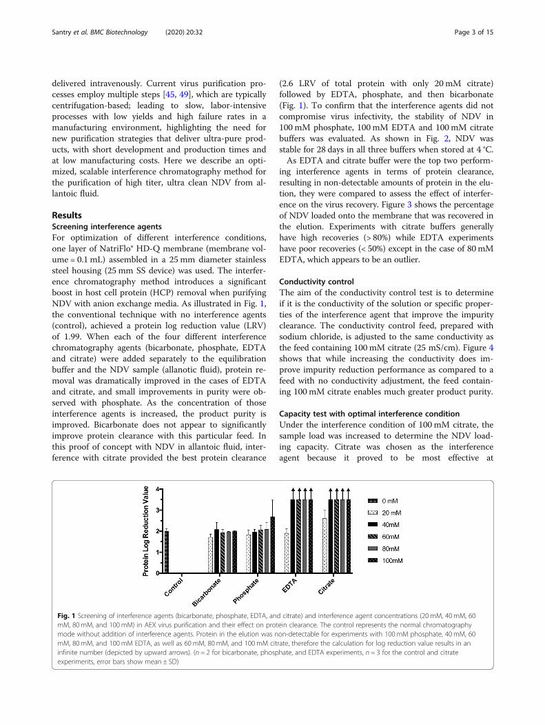
ResultsScreening interference agentsFor optimization of different interference conditions,one layer of NatriFlo® HD-Q membrane (membrane vol-ume = 0.1 mL) assembled in a 25 mm diameter stainlesssteel housing (25 mm SS device) was used. The interfer-ence chromatography method introduces a significantboost in host cell protein (HCP) removal when purifyingNDV with anion exchange media. As illustrated in Fig. 1,the conventional technique with no interference agents(control), achieved a protein log reduction value (LRV)of 1.99. When each of the four different interferencechromatography agents (bicarbonate, phosphate, EDTAand citrate) were added separately to the equilibrationbuffer and the NDV sample (allanotic fluid), protein re-moval was dramatically improved in the cases of EDTAand citrate, and small improvements in purity were ob-served with phosphate. As the concentration of thoseinterference agents is increased, the product purity isimproved. Bicarbonate does not appear to significantlyimprove protein clearance with this particular feed. Inthis proof of concept with NDV in allantoic fluid, inter-ference with citrate provided the best protein clearance
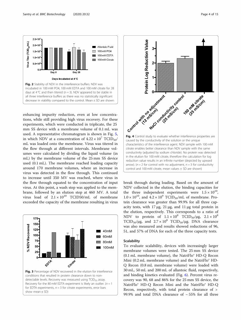
(2.6 LRV of total protein with only 20 mM citrate)followed by EDTA, phosphate, and then bicarbonate(Fig. 1). To confirm that the interference agents did notcompromise virus infectivity, the stability of NDV in100 mM phosphate, 100mM EDTA and 100mM citratebuffers was evaluated. As shown in Fig. 2, NDV wasstable for 28 days in all three buffers when stored at 4 °C.As EDTA and citrate buffer were the top two perform-
ing interference agents in terms of protein clearance,resulting in non-detectable amounts of protein in the elu-tion, they were compared to assess the effect of interfer-ence on the virus recovery. Figure 3 shows the percentageof NDV loaded onto the membrane that was recovered inthe elution. Experiments with citrate buffers generallyhave high recoveries (> 80%) while EDTA experimentshave poor recoveries (< 50%) except in the case of 80mMEDTA, which appears to be an outlier.
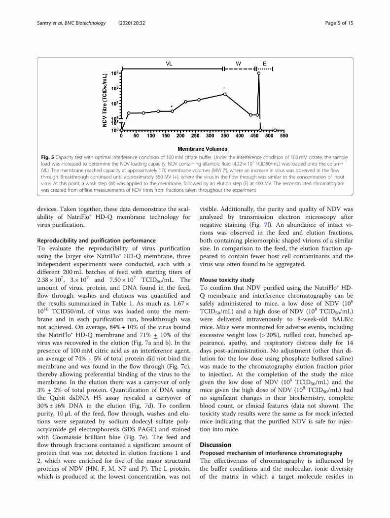
Conductivity controlThe aim of the conductivity control test is to determineif it is the conductivity of the solution or specific proper-ties of the interference agent that improve the impurityclearance. The conductivity control feed, prepared withsodium chloride, is adjusted to the same conductivity asthe feed containing 100 mM citrate (25 mS/cm). Figure 4shows that while increasing the conductivity does im-prove impurity reduction performance as compared to afeed with no conductivity adjustment, the feed contain-ing 100 mM citrate enables much greater product purity.
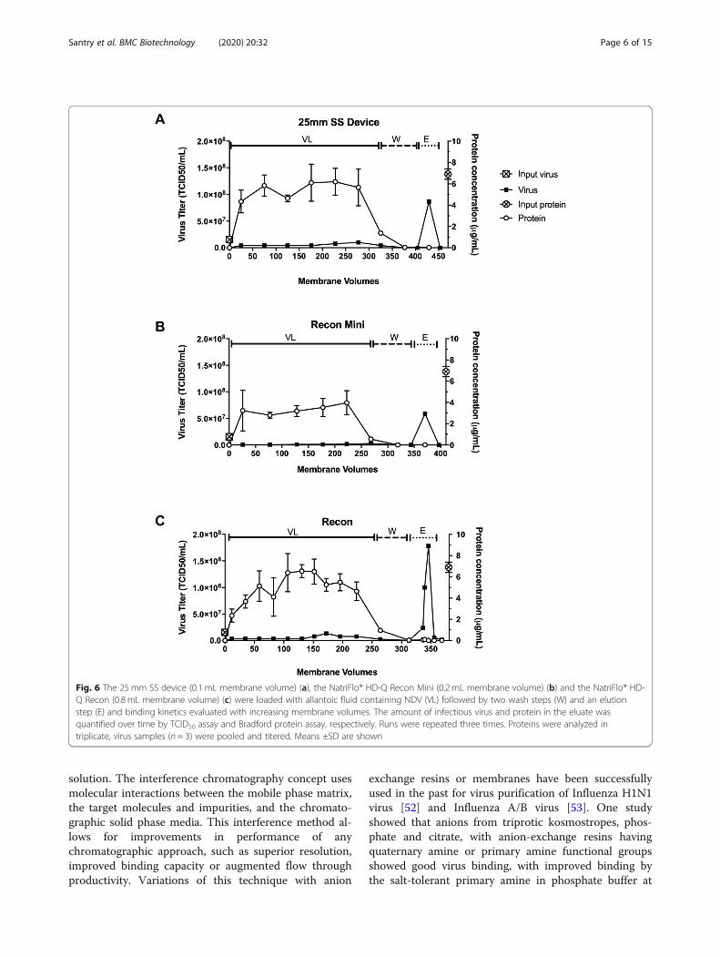
Capacity test with optimal interference conditionUnder the interference condition of 100 mM citrate, thesample load was increased to determine the NDV load-ing capacity. Citrate was chosen as the interferenceagent because it proved to be most effective at
Fig. 1 Screening of interference agents (bicarbonate, phosphate, EDTA, and citrate) and interference agent concentrations (20 mM, 40mM, 60mM, 80 mM, and 100mM) in AEX virus purification and their effect on protein clearance. The control represents the normal chromatographymode without addition of interference agents. Protein in the elution was non-detectable for experiments with 100 mM phosphate, 40 mM, 60mM, 80 mM, and 100mM EDTA, as well as 60 mM, 80 mM, and 100mM citrate, therefore the calculation for log reduction value results in aninfinite number (depicted by upward arrows). (n = 2 for bicarbonate, phosphate, and EDTA experiments, n = 3 for the control and citrateexperiments, error bars show mean ± SD)
Santry et al. BMC Biotechnology (2020) 20:32 Page 3 of 15
enhancing impurity reduction, even at low concentra-tions, while still providing high virus recovery. For theseexperiments, which were conducted in triplicate, the 25mm SS device with a membrane volume of 0.1 mL wasused. A representative chromatogram is shown in Fig. 5,in which NDV at a concentration of 4.22 × 10⁷ TCID50/mL was loaded onto the membrane. Virus was titered inthe flow through at different intervals. Membrane vol-umes were calculated by dividing the liquid volume (inmL) by the membrane volume of the 25mm SS deviceused (0.1 mL). The membrane reached loading capacityaround 170 membrane volumes, where an increase invirus was detected in the flow through. This continuedto increase until 350 MV was reached, where virus inthe flow through equated to the concentration of inputvirus. At this point, a wash step was applied to the mem-brane, followed by an elution step at 460 MV. A totalvirus load of 2.1 × 10¹⁰ TCID50/mL of membraneexceeded the capacity of the membrane resulting in virus
break through during loading. Based on the amount ofNDV collected in the elution, the binding capacities forthe three independent experiments were 1.5 × 1010,1.0 × 1010, and 4.2 × 10⁹ TCID50/mL of membrane. Pro-tein clearance was greater than 99.9% for all three cap-acity tests, with 17 μg, 21 μg, and 11 μg total protein inthe elution, respectively. This corresponds to a ratio ofNDV to protein of 1.1 × 10⁸ TCID50/μg, 2.1 × 10⁸
TCID50/μg, and 2.7 × 10⁸ TCID50/μg. DNA clearancewas also measured and results showed reductions of 96,51, and 57% of DNA for each of the three capacity tests.
ScalabilityTo evaluate scalability, devices with increasingly largermembrane volumes were tested. The 25mm SS device(0.1 mL membrane volume), the NatriFlo® HD-Q ReconMini (0.2 mL membrane volume) and the NatriFlo® HD-Q Recon (0.8 mL membrane volume) were loaded with30mL, 50 mL and 200 mL of allantoic fluid, respectively,and binding kinetics evaluated (Fig. 6). Percent virus re-covery was 90, 68 and 86% for the 25 mm SS device, theNatriFlo® HD-Q Recon Mini and the NatriFlo® HD-QRecon, respectively, with total protein clearance of >99.9% and total DNA clearance of ~ 55% for all three
Fig. 2 Stability of NDV in the interference buffers. NDV wasincubated in 100mM PO4, 100 mM EDTA and 100mM citrate for 28days at 4 °C and then titered (n = 3). NDV appeared to be stable inall three interference buffers as there was no statistically significantdecrease in viability compared to the control. Mean ± SD are shown
Fig. 3 Percentage of NDV recovered in the elution for interferenceconditions that resulted in protein clearance down to non-detectable levels. Recovery was measured using TCID50 assay.Recovery for the 80mM EDTA experiment is likely an outlier. (n = 1for EDTA experiments, n = 3 for citrate experiments, error barsshow mean ± SD)
Fig. 4 Control study to evaluate whether interference properties arecaused by the conductivity of the solution or the uniquecharacteristics of the interference agent. NDV sample with 100 mMcitrate enables better clearance than NDV sample with the sameconductivity (adjusted by sodium chloride). No protein was detectedin the elution for 100 mM citrate, therefore the calculation for logreduction value results in an infinite number (depicted by upwardarrow). (n = 2 for control with no adjustment, n = 3 for conductivitycontrol and 100mM citrate, mean values ± SD are shown)
Santry et al. BMC Biotechnology (2020) 20:32 Page 4 of 15
devices. Taken together, these data demonstrate the scal-ability of NatriFlo® HD-Q membrane technology forvirus purification.
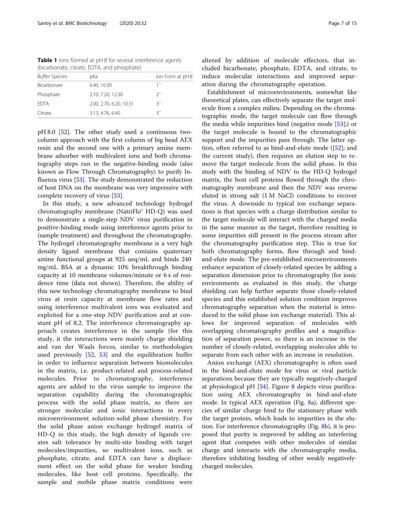
Reproducibility and purification performanceTo evaluate the reproducibility of virus purificationusing the larger size NatriFlo® HD-Q membrane, threeindependent experiments were conducted, each with adifferent 200 mL batches of feed with starting titers of2.38 × 107, 3.× 107 and 7.50 × 10⁷ TCID50/mL. Theamount of virus, protein, and DNA found in the feed,flow through, washes and elutions was quantified andthe results summarized in Table 1. As much as, 1.67 ×10¹⁰ TCID50/mL of virus was loaded onto the mem-brane and in each purification run, breakthrough wasnot achieved. On average, 84% + 10% of the virus boundthe NatriFlo® HD-Q membrane and 71% + 10% of thevirus was recovered in the elution (Fig. 7a and b). In thepresence of 100 mM citric acid as an interference agent,an average of 74% + 5% of total protein did not bind themembrane and was found in the flow through (Fig. 7c),thereby allowing preferential binding of the virus to themembrane. In the elution there was a carryover of only3% + 2% of total protein. Quantification of DNA usingthe Qubit dsDNA HS assay revealed a carryover of30% ± 16% DNA in the elution (Fig. 7d). To confirmpurity, 10 μL of the feed, flow through, washes and elu-tions were separated by sodium dodecyl sulfate poly-acrylamide gel electrophoresis (SDS PAGE) and stainedwith Coomassie brilliant blue (Fig. 7e). The feed andflow through fractions contained a significant amount ofprotein that was not detected in elution fractions 1 and2, which were enriched for five of the major structuralproteins of NDV (HN, F, M, NP and P). The L protein,which is produced at the lowest concentration, was not
visible. Additionally, the purity and quality of NDV wasanalyzed by transmission electron microscopy afternegative staining (Fig. 7f). An abundance of intact vi-rions was observed in the feed and elution fractions,both containing pleiomorphic shaped virions of a similarsize. In comparison to the feed, the elution fraction ap-peared to contain fewer host cell contaminants and thevirus was often found to be aggregated.
Mouse toxicity studyTo confirm that NDV purified using the NatriFlo® HD-Q membrane and interference chromatography can besafely administered to mice, a low dose of NDV (106
TCID50/mL) and a high dose of NDV (108 TCID50/mL)were delivered intravenously to 8-week-old BALB/cmice. Mice were monitored for adverse events, includingexcessive weight loss (> 20%), ruffled coat, hunched ap-pearance, apathy, and respiratory distress daily for 14days post-administration. No adjustment (other than di-lution for the low dose using phosphate buffered saline)was made to the chromatography elution fraction priorto injection. At the completion of the study the micegiven the low dose of NDV (106 TCID50/mL) and themice given the high dose of NDV (108 TCID50/mL) hadno significant changes in their biochemistry, completeblood count, or clinical features (data not shown). Thetoxicity study results were the same as for mock infectedmice indicating that the purified NDV is safe for injec-tion into mice.
DiscussionProposed mechanism of interference chromatographyThe effectiveness of chromatography is influenced bythe buffer conditions and the molecular, ionic diversityof the matrix in which a target molecule resides in
Fig. 5 Capacity test with optimal interference condition of 100 mM citrate buffer. Under the interference condition of 100 mM citrate, the sampleload was increased to determine the NDV loading capacity. NDV containing allantoic fluid (4.22 × 10⁷ TCID50/mL) was loaded onto the column(VL). The membrane reached capacity at approximately 170 membrane volumes (MV) (*), where an increase in virus was observed in the flowthrough. Breakthrough continued until approximately 350 MV (+), where the virus in the flow through was similar to the concentration of inputvirus. At this point, a wash step (W) was applied to the membrane, followed by an elution step (E) at 460 MV. The reconstructed chromatogramwas created from offline measurements of NDV titres from fractions taken throughout the experiment
Santry et al. BMC Biotechnology (2020) 20:32 Page 5 of 15
solution. The interference chromatography concept usesmolecular interactions between the mobile phase matrix,the target molecules and impurities, and the chromato-graphic solid phase media. This interference method al-lows for improvements in performance of anychromatographic approach, such as superior resolution,improved binding capacity or augmented flow throughproductivity. Variations of this technique with anion
exchange resins or membranes have been successfullyused in the past for virus purification of Influenza H1N1virus [52] and Influenza A/B virus [53]. One studyshowed that anions from triprotic kosmostropes, phos-phate and citrate, with anion-exchange resins havingquaternary amine or primary amine functional groupsshowed good virus binding, with improved binding bythe salt-tolerant primary amine in phosphate buffer at
Fig. 6 The 25mm SS device (0.1 mL membrane volume) (a), the NatriFlo® HD-Q Recon Mini (0.2 mL membrane volume) (b) and the NatriFlo® HD-Q Recon (0.8 mL membrane volume) (c) were loaded with allantoic fluid containing NDV (VL) followed by two wash steps (W) and an elutionstep (E) and binding kinetics evaluated with increasing membrane volumes. The amount of infectious virus and protein in the eluate wasquantified over time by TCID50 assay and Bradford protein assay, respectively. Runs were repeated three times. Proteins were analyzed intriplicate, virus samples (n = 3) were pooled and titered. Means ±SD are shown
Santry et al. BMC Biotechnology (2020) 20:32 Page 6 of 15
pH 8.0 [52]. The other study used a continuous two-column approach with the first column of big bead AEXresin and the second one with a primary amine mem-brane adsorber with multivalent ions and both chroma-tography steps run in the negative-binding mode (alsoknown as Flow Through Chromatography) to purify In-fluenza virus [53]. The study demonstrated the reductionof host DNA on the membrane was very impressive withcomplete recovery of virus [53].In this study, a new advanced technology hydrogel
chromatography membrane (NatriFlo® HD-Q) was usedto demonstrate a single-step NDV virus purification inpositive-binding mode using interference agents prior to(sample treatment) and throughout the chromatography.The hydrogel chromatography membrane is a very highdensity ligand membrane that contains quaternaryamine functional groups at 925 ueq/mL and binds 240mg/mL BSA at a dynamic 10% breakthrough bindingcapacity at 10 membrane volumes/minute or 6 s of resi-dence time (data not shown). Therefore, the ability ofthis new technology chromatography membrane to bindvirus at resin capacity at membrane flow rates andusing interference multivalent ions was evaluated andexploited for a one-step NDV purification and at con-stant pH of 8.2. The interference chromatography ap-proach creates interference in the sample (for thisstudy, it the interactions were mainly charge shieldingand van der Waals forces, similar to methodologiesused previously [52, 53] and the equilibration bufferin order to influence separation between biomoleculesin the matrix, i.e. product-related and process-relatedmolecules. Prior to chromatography, interferenceagents are added to the virus sample to improve theseparation capability during the chromatographicprocess with the solid phase matrix, so there arestronger molecular and ionic interactions in everymicroenvironment solution-solid phase chemistry. Forthe solid phase anion exchange hydrogel matrix ofHD-Q in this study, the high density of ligands cre-ates salt tolerance by multi-site binding with targetmolecules/impurities, so multivalent ions, such asphosphate, citrate, and EDTA can have a displace-ment effect on the solid phase for weaker bindingmolecules, like host cell proteins. Specifically, thesample and mobile phase matrix conditions were
altered by addition of molecule effectors, that in-cluded bicarbonate, phosphate, EDTA, and citrate, toinduce molecular interactions and improved separ-ation during the chromatography operation.Establishment of microenvironments, somewhat like
theoretical plates, can effectively separate the target mol-ecule from a complex milieu. Depending on the chroma-tographic mode, the target molecule can flow throughthe media while impurities bind (negative mode [53];) orthe target molecule is bound to the chromatographicsupport and the impurities pass through. The latter op-tion, often referred to as bind-and-elute mode ([52]; andthe current study), then requires an elution step to re-move the target molecule from the solid phase. In thisstudy with the binding of NDV to the HD-Q hydrogelmatrix, the host cell proteins flowed through the chro-matography membrane and then the NDV was reverseeluted in strong salt (1 M NaCl) conditions to recoverthe virus. A downside to typical ion exchange separa-tions is that species with a charge distribution similar tothe target molecule will interact with the charged mediain the same manner as the target, therefore resulting insome impurities still present in the process stream afterthe chromatography purification step. This is true forboth chromatography forms, flow through and bind-and-elute mode. The pre-established microenvironmentsenhance separation of closely-related species by adding aseparation dimension prior to chromatography (for ionicenvironments as evaluated in this study, the chargeshielding can help further separate those closely-relatedspecies and this established solution condition improveschromatography separation when the material is intro-duced to the solid phase ion exchange material). This al-lows for improved separation of molecules withoverlapping chromatography profiles and a magnifica-tion of separation power, so there is an increase in thenumber of closely-related, overlapping molecules able toseparate from each other with an increase in resolution.Anion exchange (AEX) chromatography is often used
in the bind-and-elute mode for virus or viral particleseparations because they are typically negatively-chargedat physiological pH [54]. Figure 8 depicts virus purifica-tion using AEX chromatography in bind-and-elutemode. In typical AEX operation (Fig. 8a), different spe-cies of similar charge bind to the stationary phase withthe target protein, which leads to impurities in the elu-tion. For interference chromatography (Fig. 8b), it is pro-posed that purity is improved by adding an interferingagent that competes with other molecules of similarcharge and interacts with the chromatography media,therefore inhibiting binding of other weakly negatively-charged molecules.
Table 1 Ions formed at pH 8 for several interference agents(bicarbonate, citrate, EDTA, and phosphate)
Buffer Species pKa Ion Form at pH 8
Bicarbonate 6.40, 10.30 1−
Phosphate 2.10, 7.20, 12.30 2−
EDTA 2.00, 2.70, 6.20, 10.31 3−
Citrate 3.13, 4.76, 6.40 3−
Santry et al. BMC Biotechnology (2020) 20:32 Page 7 of 15
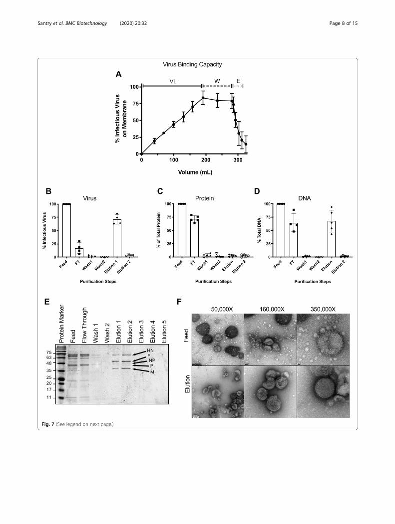
Fig. 7 (See legend on next page.)
Santry et al. BMC Biotechnology (2020) 20:32 Page 8 of 15
(See figure on previous page.)Fig. 7 Virus binding kinetics and analysis of large-scale production runs. A total of 200 mL of allantoic fluid (VL) was loaded onto the NatriFlo®HD-Q Recon A membrane followed by two wash steps (W) of 45 mL each and a 30 mL elution step. a Virus binding kinetics showing averageamount of infectious virus bound to the membrane throughout the three purification runs.. The experiment was conducted on three separateoccasions using three different batches of feed. Percent infectious virus (b), protein (c) and DNA (d) in the feed, flow through (FT), wash andelution fractions (n = 4–5, means ±SD are shown). e Representative SDS-PAGE analysis of samples (10 μL) taken from the feed, flow through, washand elution steps. Proteins were separated on a 12% Tris-glycine polyacrylamide gel and stained with Coomassie blue. The elution fractionappeared to be enriched for viral proteins HN (Hemagglutinin Neuraminidase; 62.8 kDa), F (Fusion protein; 59 kDa), NP (Nucleocapsid protein; 53.4kDa), P (Phosphate protein; 41.9 kDa), and M (Matrix protein; 39.8 kDa) based on their predicted molecular weights [50, 51]. f Transmissionelectron micrographs of negatively stained feed and elution fractions. Samples were adsorbed to 200 mesh formvar–carbon copper grids,negatively stained with 2% uranyl acetate and viewed on a FEI Tecnai G2 F20 at 200 kV scanning transmission electron microscope. The fuzzysurface suggests the presence of a glycoprotein layer on the outside of the virion. Scale bars denote 100 nm (left panel), 50 nm (center panel),and 20 nm (right panel). Images were recorded at magnifications of 50,000x (left panel), 160,000x (center panel), and 350,000x (right panel)
Fig. 8 Illustration of anion exchange chromatography using bind-and-elute mode for virus purification. a Conventional operation of anionexchange chromatography for virus purification. b Proposed mechanism of interference chromatography in which an interference agent (citrate)reduces the binding of impurities, therefore improving the purity of virus
Santry et al. BMC Biotechnology (2020) 20:32 Page 9 of 15
Selection of interference agent and its concentrationSelecting appropriate interference agents and their con-centration requires careful examination of the agents’ ef-fect on impurity removal and virus recovery. Certainagents such as citrate and EDTA create stronger inter-ference compared to others like bicarbonate and phos-phate, as illustrated in Fig. 1. A possible explanation forthese different behaviors is the difference in the valenceof the ions and the ion forms present at the loading pH,shown in Table 2. In the case of interference chromatog-raphy with NDV, more negatively-charged interferenceagents can induce a stronger weakening effect on the in-teractions between the HD-Q membrane and total pro-tein, leading to better impurity clearance. Since eachfamily of virus will require a specific condition to pre-serve stability and infectivity, selection of the appropriateinterference agent and its concentration needs to beevaluated on an individual basis. In the case with thisNDV purification work, 100 mM citrate was selected ascitrate provides better impurity removal than phosphateand more effective infectivity preservation than EDTA.
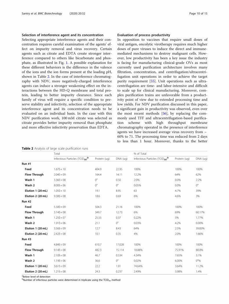
Evaluation of process productivityIn opposition to vaccines that require small doses ofviral antigen, oncolytic virotherapy requires much higherdoses of pure viruses to induce the direct and immune-mediated mechanisms to destroy malignant cells. How-ever, low productivity has been a key issue the industryis facing for manufacturing clinical-grade OVs as mostcurrently used purification architecture involves manyfiltration, concentration, and centrifugation/ultracentri-fugation unit operations in order to achieve the targetpurity requirement [55]. Unit operations such as ultra-centrifugation are time- and labor-intensive and difficultto scale up for clinical manufacturing. Moreover, com-plex purification trains are unfavorable from a product-ivity point of view due to extended processing time andlow yields. For NDV purification discussed in this paper,a significant gain in productivity was observed, even overthe most recent methods [56], by replacing the com-monly used TFF and ultracentrifugation-based purifica-tion scheme with high throughput membranechromatography operated in the presence of interferenceagents we have increased average virus recovery from ~60% to 71. The processing time was reduced from 2 daysto less than 1 hour. Moreover, thanks to the better
Table 2 Analysis of large scale purification runs
Total % of Total
Infectious Particles (TCID50)b Protein (ug) DNA (ug) Infectious Particles (TCID50)
b Protein (ug) DNA (ug)
Run #1
Feed 1.67E+ 10 404.9 22.95 100% 100% 100%
Flow Through 2.04E+ 09 164.4 14.11 12.2% 64% 62%
Wash 1 3.36E+ 08 0a 0.50 2.0% 0.0% 2.2%
Wash 2 8.00E+ 06 0a 0a 0.05% 0.0% 0a
Elution 1 (20mL) 1.05E+ 10 19.1 8.95 63 4.7% 39%
Elution 2 (20mL) 9.38E+ 08 18.6 0.69 6% 4.6% 3%
Run #2
Feed 5.58E+ 09 504.3 21.16 100% 100% 100%
Flow Through 3.14E+ 08 349.7 12.73 6% 69% 60.17%
Wash 1 1.25E+ 07 25.55 0.37 0.22% 5% 1.77%
Wash 2 1.91E+ 06 21.1 0a 0.03% 4.2% 0.00%
Elution 1 (20mL) 3.56E+ 09 12.7 8.43 64% 2.5% 39.83%
Elution 2 (20mL) 2.42E+ 08 10.1 0.35 4% 2.0% 1.66%
Run #3
Feed 4.84E+ 09 610.7 17.028 100% 100% 100%
Flow Through 9.14E+ 08 482.3 15.114 18.88% 75.91% 88.8%
Wash 1 2.10E+ 08 46.7 0.534 4.34% 7.65% 3.1%
Wash 2 1.19E+ 06 36.6 0a 0.02% 6.00% 0a%
Elution 1 (20mL) 3.61E+ 09 22.2 1.91 74.64% 3.64% 11.2%
Elution 2 (20mL) 1.21E+ 08 24.3 0.237 2.49% 3.98% 1.4%aBelow level of detectionbNumber of infectious particles were determined in triplicate using the TCID50 method
Santry et al. BMC Biotechnology (2020) 20:32 Page 10 of 15
overall recovery in the membrane process (90–100% ver-sus 60–65%), upstream production (i.e. production ineggs) can be downsized. With the current loading cap-acity, one run on a commercially available membranecolumn with 460 mL membrane volume will be suffi-cient to support 3–5 treatments of a Phase I study [41].On average, 71% of the virus was eluted, leaving ap-
proximately 14% of bound virus unaccounted for. Lossof infectious virus could be due to a variety of reasonsincluding changes in pH which inactivate the virus,strong adherence of the virus to the membrane, orshearing of the virus due to pressure build up. Addition-ally, it is possible that virus aggregation may have led toan underestimation of the true titer. While a significantamount of work was done to recover as much NDV aspossible, there are other options that can be explored tofurther optimize this process.We observed larger differences in DNA removal for
the trial runs, in which three different sizes of mem-branes were used. This could potentially be due to batchto batch variability in the feed or differences in themembranes. When we scaled up production and usedthe same sized membrane, we observed less variability inDNA removal (70 ± 16% DNA).
Future perspectives of interference chromatographyIn the case of NDV purification, further studies can bedone to improve upon the process that has been de-scribed. It is proposed that adding an interference agentto the elution would enable a reduction in the strengthof the elution species needed to remove the target fromthe chromatography media (which is synonymous withdisplacement chromatography). The current elution con-dition requires high NaCl concentration (minimum 1M)to achieve good virus recovery (see Figure 1S in Supple-mental Data). Mild elution conditions are beneficial forlabile viruses and proteins and may enable elution col-lection that can be used directly for therapeutic use, sim-plifying or eliminating a buffer exchange step beforeformulation. Exploring various buffer conditions mayalso improve the DNA removal. The optimal elutioncondition would completely elute the virus, but leave theDNA bound to the membrane which can subsequentlybe disposed of if a single-use strategy is implemented. Ina review by Ungerechts et al., 2016 [20], which providesan overview of clinical-grade manufacturing proceduresfor OVs, nuclease treatment to degrade host cell nucleicacid prior to chromatography was common to all OVpurification processes, and thus would likely be imple-mented prior to purification of clinical grade NDV usinginterference chromatography.A challenge with interference chromatography is that
the process development method requires investigation,characterization, and may not conform to standard
process platforms. The method utilizes knowledge of thesample matrix, products/variants, and impurity levelsand requires an empirical study for optimum separationand purity. This methodology necessitates increased de-velopment times and costs due to extensive design of ex-periment (DOE) testing. It is largely unknown howinterference agents will interact with different target spe-cies such as viruses, virus-like-particles (VLPs), anti-bodies, or proteins. NDV used as a model, achievedsingle-step purification of proteins and dramatically re-duced processing time, but this is not necessarily truefor all targets. Interference chromatography may need tobe complemented with other polishing methods (for ex-ample, ultracentrifugation to resolve empty from fullvirus particles). Moreover, further steps would be neededin order to generate clinical grade virus. The virus wouldhave to be subjected to dialysis or diafiltration to ex-change it into a more physiologically balanced bufferand electrolyte solution as well as to improve storage.Once established, however, the interference technique
should be very reproducible and could separate moredifficult molecules, like viruses or labile proteins wherestandard chromatographic methods do not perform ad-equately. The interference chromatography method isexpected to be easily scalable since the interferenceagent concentration is independent of the purificationscale. Depending on the chromatography media, theconsumables and processing equipment can also be eas-ily scaled up to increase purification efficiency. It is an-ticipated that interference chromatography can beapplied in either positive or negative chromatographymodes. Currently traditional IEX chromatography isoften used in the manufacturing of several vaccines in-cluding hepatitis A (EP 0538142 A2, 1994), hepatitis B(WO 02/122887 A1, 2002), polio [57], adenoviral vectors[58], Ebola virus glycoproteins [59], and influenza VLPs[60]. Introducing interference methods to these AEXprocesses may produce purer product in less DSP stepsand higher yields and therefore lower production costs.
ConclusionThe interference chromatography method is a powerfulchromatography method for the separation of impuritiesfrom product molecules and the separation of closely-related product variants. This chromatographic methodestablishes an in-solution gradient of molecule interac-tions using simple interfering agents that enhance or in-hibit in-solution matrix interactions with the targetproduct. The combination of interfering agents andchromatographic separation actually mimics a quasi,multi-dimensional chromatographic separation, but in asingle chromatography step. The impact of the interfer-ence method is a more powerful (stronger interactionsof targeted product molecules or removal of unwanted
Santry et al. BMC Biotechnology (2020) 20:32 Page 11 of 15
impurities), combinatorial chromatography operationthat increases separation efficiency and simplifies down-stream processing with fewer steps. This provides bene-fits in purification speed and yield that enables costeffective therapeutics to be delivered to the patient fas-ter. It was demonstrated here that NDV was able to bepurified from allantoic fluid in a single-step using inter-ference agent, multi-valent citrate, and HD-Q membranechromatography, which reduced protein by > 97% andDNA up to 70% with good recovery of virus > 70%.
MethodsCell cultureDF-1 chicken embryo fibroblast cells (obtained from theAmerican Type Culture Collection; ATCC® CRL-12203)were maintained in Dulbecco’s modified Eagle’s medium,supplemented with 10% bovine calf serum (BCS), 2 mML-glutamine, 100 units/mL penicillin and 100 μg/mLstreptomycin at 37 °C in 5% CO2/air atmosphere.
Virus productionThe NDV-F3aa-GFP genome and helper plasmids werepurified with the GenElute HP Plasmid Maxiprep Kit. Re-combinant NDV-F3aa-GFP was rescued and propagatedin specific pathogen free eggs as described [56]. Allantoicfluid was harvested at 50 h post-inoculation and clarifiedby centrifugation (1,500 x g for 10min at 4 °C). To con-firm the presence of NDV in the allantoic fluid, ahemagglutination assay (HA) was performed as described[56]. The average virus titer of pooled allantoic fluid was1 × 108 TCID50/mL. Virus was stored at − 80 °C.
ChromatographyFor all experiments, virus containing allantoic fluid wasthawed at 4 °C overnight, equilibrated to roomtemperature, and clarified by centrifugation (1,500 x g for10min) followed by the addition of 60% sucrose to a finalconcentration of 2.5%. It is important to note that usingcold feed (i.e. 4 °C) can cause a spike in pressure, poten-tially due to aggregate formation, and may impede loadingof the virus onto the membrane and subsequent elution.Therefore, it is recommended that once the virus feed issupplemented with interference agent and sucrose, it beallowed to reach room temperature before 0.45 μm filtra-tion and left at room temperature for the duration of therun. For screening tests, an appropriate volume of concen-trated interfering salt solution [1M monobasic sodiumphosphate, 1M citric acid, 0.77M sodium bicarbonate, or0.24M ethylenediaminetetraacetic acid (EDTA)] wasadded to harvested allantoic fluid to achieve the desiredinterfering agent concentration (20mM, 40mM, 60mM,80mM or 100mM). For control tests, there was no ad-justment to allantoic fluid besides the addition of sucrose.For salt comparison tests, the conductivity of allantoic
fluid was adjusted with NaCl to 25ms/cm, to normalizeall samples to the conductivity of the feed with 100mMcitrate. All feeds were filtered with a 0.45 μm PES bottletop filter. All experiments were performed with 1 layer ofNatriFlo® HD-Q membrane (membrane volume = 0.1mL)assembled in a 25mm diameter stainless steel housing(25mm SS device). All experiments were performed on aKDS 220 Multi-Syringe Infusion Pump with a flow rate of20 membrane volumes (MV) per minute. Pressure waskept under 15 psi, as NDV is known to be sensitive toshearing at high pressure. The membrane was first equili-brated with equilibration buffer (25mM Tris with appro-priate interfering agent concentration, pH 8.2) for 5mL.After sample loading (5mL for screening, control, andNaCl conductivity control, and 13mL to 37mL for cap-acity test) the membrane washed with 5mL of equilibra-tion buffer followed by a second wash with low salt buffer(5mL of 25mM Tris, 100mM NaCl, pH 8.2). The flowdirection was then reversed for elution to reduce shearingand ensure good recovery. Step elution conditions (25mM Tris with 0.5M NaCl, 1M NaCl, 1.5M NaCl, 2MNaCl, and 2.5M NaCl, pH 8.2) were used for all screeningtests as well as control and salt comparison tests. One-step elution with 25mM Tris, 1M NaCl, pH 8.2 was usedfor loading capacity test. This elution was chosen becauseit was the optimal balance between NDV recovery and re-duced NaCl concentration (see supplementary Figure S1for further details).
Scale upScale up was performed as described above with someminor changes. Briefly, the interference agent 100 mMcitric acid (pH 8.2) was added as a 10x buffer to the feed(equilibrated to room temperature), followed by theaddition of 60% sucrose to achieve a final concentrationof 2.5% sucrose. The feed was then 0.45 μm filtered im-mediately prior to being loaded onto the membrane. Allexperiments were performed with a Masterflex L/S peri-staltic pump (Cole Parmer, USA) and Masterflex L/S 14BioPharm Plus Platinum-Cured Silicone Pump tubing.Membranes were first equilibrated with 40mL of equili-bration buffer (25 mM Tris with 100mM citrate as inter-fering agent, pH 8.2). Once the feed was loaded onto themembrane, this was followed by wash 1 with 40mL ofequilibration buffer and wash 2 with 40mL of low saltbuffer (25 mM Tris, 100 mM NaCl, pH 8.2). The flowdirection was then reversed for elution to reduce shear-ing and to ensure good recovery. Elution was performedusing 40 mL of 25 mM Tris, 1.0M NaCl buffer, in two20mL fractions.
Total protein quantificationTotal protein was quantified in triplicate using the Bio-Rad Protein Assay kit (Bio-Rad Laboratories), which is
Santry et al. BMC Biotechnology (2020) 20:32 Page 12 of 15
based on the method of Bradford, in a flat bottom 96-well microtiter plate according to the manufacturer’s in-structions. Bovine serum albumin (BSA) (New EnglandBiolabs) was used to generate a standard curve, whichranged from 0 to 20 μg/mL BSA.
Characterization of virus in the elution solutionSamples (3 μL) of the feed and elutions from the threelarge scale purification experiments were adsorbed to200 mesh formvar–carbon copper grids for 2 min atroom temperature and excess liquid was removed. Thegrids were then negatively stained with 2% uranyl acetateand viewed on a FEI Tecnai G2 F20 at 200 kV scanningtransmission electron microscope. Images were recordedat a magnification of 50, 000X, 160,000X and 350,000Xwith a Gatan Ultrascan 4 k × 4 k CCD camera (Gatan,Pleasanton, CA, USA) and all data processing and ana-lysis was performed using the Gatan Digital Micrographsoftware (Molecular and Cellular Imaging Facility at theUniversity of Guelph).Sodium dodecyl sulfate polyacrylamide gel electro-
phoresis (SDS-PAGE) was performed to visualize proteincontent in the feed, flow through, wash and elution fromthe three large scale purification experiments. Briefly,10 μL of feed, flow through, wash 1, wash 2 and elutions1–5 were denatured at 100 °C for 5 min in 1× SDSPAGE sample buffer containing 143 mM β-Mercaptoethanol, separated by SDS–PAGE on 12%Tris–glycine gels and stained with Coomassie Blue R250.
TCID50 assayNDV-GFP titer was determined by 50% tissue culture infect-ive dose (TCID50) assay and expressed as TCID50/mL. NDVsamples were serially diluted 10-fold with 1x Dulbecco’sphosphate-buffered saline (DPBS) from 10− 1 up to 10− 8 and10 μL were added to DF-1 chicken fibroblast cells grown inDMEM (supplemented with 4mML-glutamine, 7% bovinecalf serum) seeded at 70–80% confluency in a 96-well cellculture plate (n= 8). After 96 h, green fluorescence was ob-served using a Carl Zeiss Axio® 154 Observer A1 invertedfluorescence microscope and titer was calculated accordingto the Spearman-Kärber method (Ramakrishnan, 2016) [61].
Total DNA quantificationTotal DNA from feed, flow through and elution frac-tions were purified using Qiagen DNeasy Blood & TissueKit, including the RNase A (Invitrogen, Canada) step, ac-cording to the manufacturer’s protocol. Fluorometricquantification of DNA was performed using a high sen-sitivity dsDNA Qubit® assay using a Qubit® 2.0Fluorometer (Genomics Facility, Advanced AnalysisCenter, The University of Guelph, Canada).
Toxicity testingMouse toxicity experiments were performed in compli-ance with the guidelines set forth by the CanadianCouncil on Animal Care. The Animal Care Committeeat the University of Guelph approved all methods. Ran-domly allocated groups of six eight-week old femaleBALB/c mice purchased from Charles River Laboratories(Wilmington, MA) were housed at the University ofGuelph in a specific pathogen-free isolation facility. Micewere housed in groups of four and food (Teklad Global14% Protein Rodent Maintenance Diet, Indianapolis,USA) and water (tap) were provided ad libitum. Micewere acclimated to the environment for 7 days prior tostudy initiation. Mice received 100 μl injections intraven-ously of 1M NaCl (control), 1 × 10⁶ TCID50 units ofpurified NDV in 1M NaCl (low-dose) or 1 × 10⁸ TCID50
units of purified NDV in 1M NaCl (high dose). Micewere observed for changes in body weight or behaviorover a 2-week period. On day 14 mice were euthanizedby anesthetizing with isoflurane prior to cervical disloca-tion and blood was drawn for complete blood count andbiochemical analysis, performed at the Animal HealthLaboratory, the University of Guelph.
Supplementary informationSupplementary information accompanies this paper at https://doi.org/10.1186/s12896-020-00627-w.
Additional file 1.
Additional file 2.
AbbreviationsTFF: Tangential flow filtration; OVs: Oncolytic viruses; IEX: Ion exchange;NDV: Newcastle disease virus; HCP: Host cell protein; LRV: Log reductionvalue; MV: Membrane volume; AEX: Anion exchange; DOE: Design ofexperiment; VLP: Virus like particle; BSA: Bovine serum albumin;EDTA: Ethylenediaminetetraacetic acid
AcknowledgementsWe thank all those who participated in the care of the animals at theUniversity of Guelph Animal Isolation Unit. We also thank Bob Harris(Molecular and Cellular Imaging Facility at the University of Guelph), forpreparing and imaging the samples for electron microscopy.
Authors’ contributionsConceived and designed the experiments: LAS, MZ, RJ, PMM, JGS, SKW.Performed the experiments: LAS, MV, MZ, JMD, TMM and XS. Analyzed thedata: LAS, RJ, MV, MZ and JMD. Contributed reagents and materials: SKW,PMM and JMD. Wrote the paper: LAS, MV, MZ, SKW and JMD. All authorsread and approved the final manuscript.
FundingThis work was funded by the Natural Sciences and Engineering ResearchCouncil of Canada (NSERC) Engage (499834–16) and Engage Plus (EGP2515222–17) grants to SKW. This work was supported in part through theCanadian Scientific Research and Experimental Development (SRED)program. The funders had no role in study design, data collection andanalysis, decision to publish, or preparation of the manuscript.
Santry et al. BMC Biotechnology (2020) 20:32 Page 13 of 15
Availability of data and materialsAll data are included with the manuscript. All data generated or analyzedduring this study are included in this published article and its supplementaryand additional information files.
Ethics approval and consent to participateAnimal experiments were performed in compliance with the guidelines setforth by the Canadian Council on Animal Care (CCAC). The Animal CareCommittee at the University of Guelph approved all methods (AUP#3827).
Consent for publicationNot applicable.
Competing interestsThe authors declare no competing interests.
Author details1Department of Pathobiology, University of Guelph, Guelph, Ontario N1G2W1, Canada. 2MilliporeSigma, 5295 John Lucas Drive, Burlington, OntarioL7L 6A8, Canada. 3Present Address: BioVectra Inc., 24 Ivey Lane, PO Box 766,Windsor, Nova Scotia B0N 2T0, Canada. 4Juravinski Cancer Centre, 699Concession Street, Hamilton, ON L8V 5C2, Canada.
Received: 30 August 2019 Accepted: 10 June 2020
References1. Working PK, Lin A, Borellini F. Meeting product development challenges in
manufacturing clinical grade oncolytic adenoviruses. Oncogene. 2005;24(52):7792–801.
2. Yamaguchi T, Uchida E. Regulatory aspects of oncolytic virus products. CurrCancer Drug Targets. 2007;7(2):203–8.
3. Akerblom A, Bergvall P. Constraints on vaccine production. BioProcess Int.2012:64–6 http://www.bioprocessintl.com/wp-content/uploads/2014/05/BPI_A_121007AR23_O_182745a.pdf.
4. Husain SR, Han J, Au P, Shannon K, Puri RK. Gene therapy for cancer: regulatoryconsiderations for approval. Cancer Gene Ther. 2015;22(12):554–63.
5. Segura MM, Kamen AA, Garnier A. Overview of current scalable methods forpurification of viral vectors. Methods Mol Biol. 2011;737:89–116.
6. Lusky M. Good manufacturing practice production of adenoviral vectors forclinical trials. Hum Gene Ther. 2005;16(3):281–91.
7. Altaras NE, Aunins JG, Evans RK, Kamen A, Konz JO, Wolf JJ. Production andformulation of adenovirus vectors. Adv Biochem Eng Biotechnol. 2005;99:193–260.
8. Moleirinho MG, Rosa S, Carrondo MJT, Silva RJS, Hagner-McWhirter Å, AhlénG, Lundgren M, Alves PM, Peixoto C. Clinical-grade Oncolytic adenoviruspurification using Polysorbate 20 as an alternative for cell Lysis. Curr GeneTher. 2018;18(6):366–74.
9. James KT, Cooney B, Agopsowicz K, Trevors MA, Mohamed A, Stoltz D, HittM, Shmulevitz M. Novel high-throughput approach for purification ofinfectious Virions. Sci Rep. 2016;6:36826.
10. Clément N, Grieger JC. Manufacturing of recombinant adeno-associatedviral vectors for clinical trials. Mol Ther Methods Clin Dev. 2016;3:16002.
11. Penaud-Budloo M, François A, Clément N, Ayuso E. Pharmacology ofrecombinant Adeno-associated virus production. Mol Ther Methods ClinDev. 2018;8:166–80.
12. Merten OW, Hebben M, Bovolenta C. Production of lentiviral vectors. MolTher Methods Clin Dev. 2016;3:16017.
13. Schweizer M, Merten OW. Large-scale production means for themanufacturing of lentiviral vectors. Curr Gene Ther. 2010;10(6):474–86.
14. Segura MM, Kamen A, Garnier A. Downstream processing of oncoretroviraland lentiviral gene therapy vectors. Biotechnol Adv. 2006;24(3):321–37.
15. Jiang C, Glorioso JC, Ataai M: Presence of imidazole in loading bufferprevents formation of free radical in immobilized metal affinitychromatography and dramatically improves the recovery of herpes simplexvirus type 1 gene therapy vectors. J Chromatogr A 2006, 1121(1):40–45.
16. Jiang C, Wechuck JB, Goins WF, Krisky DM, Wolfe D, Ataai MM, Glorioso JC.Immobilized cobalt affinity chromatography provides a novel, efficientmethod for herpes simplex virus type 1 gene vector purification. J Virol.2004;78(17):8994–9006.
17. Wolff MW, Siewert C, Hansen SP, Faber R, Reichl U. Purification of cellculture-derived modified vaccinia Ankara virus by pseudo-affinitymembrane adsorbers and hydrophobic interaction chromatography.Biotechnol Bioeng. 2010;107(2):312–20.
18. Wolff MW, Siewert C, Lehmann S, Hansen SP, Djurup R, Faber R, Reichl U.Capturing of cell culture-derived modified Vaccinia Ankara virus by ionexchange and pseudo-affinity membrane adsorbers. Biotechnol Bioeng.2010;105(4):761–9.
19. Zhao M, Vandersluis M, Stout J, Haupts U, Sanders M, Jacquemart R. Affinitychromatography for vaccines manufacturing: finally ready for prime time?Vaccine. 2018;37:5491–503.
20. Ungerechts G, Bossow S, Leuchs B, Holm PS, Rommelaere J, Coffey M, CoffinR, Bell J, Nettelbeck DM. Moving oncolytic viruses into the clinic: clinical-grade production, purification, and characterization of diverse oncolyticviruses. Mol Ther Methods Clin Dev. 2016;3:16018.
21. Hou Y, Brower M, Pollard D, Kanani D, Jacquemart R, Kachuik B, Stout J.Advective hydrogel membrane chromatography for monoclonal antibodypurification in bioprocessing. Biotechnol Prog. 2015;31(4):974–82.
22. Jacquemart R, Vandersluis M, Zhao M, Sukhija K, Sidhu N, Stout J. A single-use strategy to enable manufacturing of affordable biologics. ComputStruct Biotechnol J. 2016;14:309–18.
23. Sviben D, Forcic D, Ivancic-Jelecki J, Halassy B, Brgles M. Recovery ofinfective virus particles in ion-exchange and hydrophobic interactionmonolith chromatography is influenced by particle charge and total-to-infective particle ratio. J Chromatogr B Analyt Technol Biomed Life Sci. 2017;1054:10–9.
24. Li H, Yang Y, Zhang Y, Zhang S, Zhao Q, Zhu Y, Zou X, Yu M, Ma G, Su Z. Ahydrophobic interaction chromatography strategy for purification ofinactivated foot-and-mouth disease virus. Protein Expr Purif. 2015;113:23–9.
25. Weigel T, Soliman R, Wolff MW, Reichl U. Hydrophobic-interactionchromatography for purification of influenza a and B virus. J Chromatogr BAnalyt Technol Biomed Life Sci. 2019;1117:103–17.
26. Nestola P, Peixoto C, Silva RR, Alves PM, Mota JP, Carrondo MJ. Improvedvirus purification processes for vaccines and gene therapy. BiotechnolBioeng. 2015;112(5):843–57.
27. Moleirinho MG, Silva RJS, Alves PM, Carrondo MJT, Peixoto C. Currentchallenges in biotherapeutic particles manufacturing. Expert Opin Biol Ther.2019;20:1–15.
28. Tayeb S, Zakay-Rones Z, Panet A. Therapeutic potential of oncolyticNewcastle disease virus: a critical review. Oncolytic Virother. 2015;4:49–62.
29. Zhao L, Liu H. Newcastle disease virus: a promising agent for tumourimmunotherapy. Clin Exp Pharmacol Physiol. 2012;39(8):725–30.
30. Ahlert T, Schirrmacher V. Isolation of a human melanoma adaptedNewcastle disease virus mutant with highly selective replication patterns.Cancer Res. 1990;50(18):5962–8.
31. Fiola C, Peeters B, Fournier P, Arnold A, Bucur M, Schirrmacher V. Tumorselective replication of Newcastle disease virus: association with defects oftumor cells in antiviral defence. Int J Cancer. 2006;119(2):328–38.
32. Krishnamurthy S, Takimoto T, Scroggs RA, Portner A. Differentially regulatedinterferon response determines the outcome of Newcastle disease virusinfection in normal and tumor cell lines. J Virol. 2006;80(11):5145–55.
33. Mansour M, Palese P, Zamarin D. Oncolytic specificity of Newcastle diseasevirus is mediated by selectivity for apoptosis-resistant cells. J Virol. 2011;85(12):6015–23.
34. Haas C, Ertel C, Gerhards R, Schirrmacher V. Introduction of adhesive andcostimulatory immune functions into tumor cells by infection withNewcastle disease virus. Int J Oncol. 1998;13(6):1105–15.
35. Washburn B, Schirrmacher V. Human tumor cell infection by Newcastledisease virus leads to upregulation of HLA and cell adhesion molecules andto induction of interferons, chemokines and finally apoptosis. Int J Oncol.2002;21(1):85–93.
36. Koks CA, Garg AD, Ehrhardt M, Riva M, Vandenberk L, Boon L, DeVleeschouwer S, Agostinis P, Graf N, Van Gool SW. Newcastle diseasevirotherapy induces long-term survival and tumor-specific immune memoryin orthotopic glioma through the induction of immunogenic cell death. IntJ Cancer. 2015;136(5):E313–25.
37. Ricca JM, Oseledchyk A, Walther T, Liu C, Mangarin L, Merghoub T, WolchokJD, Zamarin D. Pre-existing immunity to Oncolytic virus potentiates itsimmunotherapeutic efficacy. Mol Ther. 2018;26(4):1008–19.
38. Zamarin D, Holmgaard RB, Ricca J, Plitt T, Palese P, Sharma P, Merghoub T,Wolchok JD, Allison JP. Intratumoral modulation of the inducible co-
Santry et al. BMC Biotechnology (2020) 20:32 Page 14 of 15
stimulator ICOS by recombinant oncolytic virus promotes systemic anti-tumour immunity. Nat Commun. 2017;8:14340.
39. Zamarin D, Holmgaard RB, Subudhi SK, Park JS, Mansour M, Palese P,Merghoub T, Wolchok JD, Allison JP. Localized oncolytic virotherapyovercomes systemic tumor resistance to immune checkpoint blockadeimmunotherapy. Sci Transl Med. 2014;6(226):226ra232.
40. Schirrmacher V. Fifty years of clinical application of newcastle disease virus:time to celebrate! Biomedicines. 2016;4(3):16.
41. Hotte SJ, Lorence RM, Hirte HW, Polawski SR, Bamat MK, O'Neil JD, RobertsMS, Groene WS, Major PP. An optimized clinical regimen for the oncolyticvirus PV701. Clin Cancer Res. 2007;13(3):977–85.
42. Lam HY, Yeap SK, Rasoli M, Omar AR, Yusoff K, Suraini AA, Alitheen NB.Safety and clinical usage of Newcastle disease virus in cancer therapy. JBiomed Biotechnol. 2011;2011:718710.
43. Lorence RM, Pecora AL, Major PP, Hotte SJ, Laurie SA, Roberts MS,Groene WS, Bamat MK. Overview of phase I studies of intravenousadministration of PV701, an oncolytic virus. Curr Opin Mol Ther. 2003;5(6):618–24.
44. Schirrmacher V. Oncolytic Newcastle disease virus as a prospective anti-cancer therapy. A biologic agent with potential to break therapy resistance.Expert Opin Biol Ther. 2015;15(12):1757–71.
45. McGinnes LW, Pantua H, Reitter J, Morrison TG. Newcastle diseasevirus: propagation, quantification, and storage. Curr Protoc Microbiol.2006;15:12.
46. DiNapoli JM, Yang L, Suguitan A, Elankumaran S, Dorward DW, Murphy BR,Samal SK, Collins PL, Bukreyev A. Immunization of primates with aNewcastle disease virus-vectored vaccine via the respiratory tract induces ahigh titer of serum neutralizing antibodies against highly pathogenic avianinfluenza virus. J Virol. 2007;81(21):11560–8.
47. Samson ACR. Newcastle Disease, vol. 8. USA: Springer; 1988. https://doi.org/10.1007/978-1-4613-1759-3.
48. Langfield KK, Walker HJ, Gregory LC, Federspiel MJ. Manufacture of measlesviruses. Methods Mol Biol. 2011;737:345–66.
49. Freeman AI, Zakay-Rones Z, Gomori JM, Linetsky E, Rasooly L, Greenbaum E,Rozenman-Yair S, Panet A, Libson E, Irving CS, et al. Phase I/II trial ofintravenous NDV-HUJ oncolytic virus in recurrent glioblastoma multiforme.Mol Ther. 2006;13(1):221–8.
50. Ren X, Xue C, Kong Q, Zhang C, Bi Y, Cao Y. Proteomic analysis of purifiedNewcastle disease virus particles. Proteome Sci. 2012;10(1):32.
51. Linde AM, Munir M, Zohari S, Ståhl K, Baule C, Renström L, Berg M.Complete genome characterisation of a Newcastle disease virus isolatedduring an outbreak in Sweden in 1997. Virus Genes. 2010;41(2):165–73.
52. Vajda J, Weber D, Brekel D, Hundt B, Müller E. Size distribution analysis ofinfluenza virus particles using size exclusion chromatography. J ChromatogrA. 2016;1465:117–25.
53. Iyer G, Ramaswamy S, Cheng KS, Sisowath N, Mehta U, Leahy A, Chung F,Asher D. Flow-through purification of viruses- a novel approach to vaccinepurification. Procedia Vaccinol. 2012;6:106–12.
54. Michen B, Graule T. Isoelectric points of viruses. J Appl Microbiol. 2010;109(2):388–97.
55. Vandersluis M, Jacquemart R, Zhao M, Stout JG, Wootton S. Achievingintensification and flexibility in virus purification with next-generationchromatography tools; 2017. p. 39–44.
56. Santry LA, McAusland TM, Susta L, Wood GA, Major PP, Petrik JJ, Bridle BW,Wootton SK. Production and purification of high-titer Newcastle diseasevirus for use in preclinical mouse models of Cancer. Mol Ther Methods ClinDev. 2018;9:181–91.
57. Thomassen YE, van t’ Oever AG, Vinke M, Spiekstra A, Wijffels RH, van derPol LA, Bakker WA. Scale-down of the inactivated polio vaccine productionprocess. Biotechnol Bioeng. 2013;110(5):1354–65.
58. Eglon MN, Duffy AM, O’Brien T, Strappe PM. Purification of adenoviralvectors by combined anion exchange and gel filtration chromatography. JGene Med. 2009;11(11):978–89.
59. Hahn TJ, Webb B, Kutney J, Fis E, Nidel N, Wong J, Jendrek D, Smith GE.Rapid manufacture and release of a GMP batch of Zaire Ebolavirusglycoprotein vaccine made using recombinant Baculovirus-Sf9 insect cellculture technology. BioProcess J. 2015;14:6–15.
60. Hahn TJ, Courbron D, Hamer M, Masoud M, Wong J, Taylor K,Hatch J, Sowers M, Shane E, Nathan M, Jiang H, Wei Z, Higgins J,Roh K, Burd J, Chinchilla-Olszar D, Malou-Williams M, Baskind DP,Smith GE. Rapid manufacture and release of a gmp batch of avian
influenza a(h7n9) virus-like particle vaccine made using recombinantbaculovirus-sf9 insect cell culture technology. BioProcess J. 2013;12:4–17.
61. Ramakrishnan MA. Determination of 50% endpoint titer using a simpleformula. World J Virol. 2016;5(2):85–6.
Publisher’s NoteSpringer Nature remains neutral with regard to jurisdictional claims inpublished maps and institutional affiliations.
Santry et al. BMC Biotechnology (2020) 20:32 Page 15 of 15


![Journal of Chromatography Atomas.cajka.sweb.cz/doc/JCA_VOL1262_P8-18.pdf · chromatography–mass spectrometry (GC–MS) [11–14]. Since the interference has commonly different exact](https://img.pdfslide.us/doc/110x75/60cac64882280b59e523e358/journal-of-chromatography-chromatographyamass-spectrometry-gcams-11a14.jpg)