Embed Size (px)
Citation preview
ORIGINAL ARTICLE
Interaction between Cucumber mosaic virus 2b protein and plantcatalase induces a specific necrosis in association with proteasomeactivity
Katsunori Murota1 • Hanako Shimura1 • Minoru Takeshita2 • Chikara Masuta1
Received: 12 July 2016 / Accepted: 6 September 2016 / Published online: 22 September 2016
� The Author(s) 2016. This article is published with open access at Springerlink.com
Abstract
Key message Cucumber mosaic virus (CMV) can induce
a specific necrosis on Arabidopsis through the interac-
tion between the CMV 2b protein and host catalase, in
which the ubiquitin–proteasome pathway may be
involved.
Abstract We previously reported that the CMV 2b protein,
the viral RNA silencing suppressor, interactedwith theH2O2
scavenger catalase (CAT3), leading to necrosis on CMV-
inoculatedArabidopsis leaves.We here confirmed that CMV
could more abundantly accumulate in the CAT3-knockout
mutant (cat3), and that CAT3makes host plants a little more
tolerant to CMV. We also found that the necrosis severity is
not simply explained by a high level of H2O2 given by the
lack of CAT3, because the recombinant CMV, CMV-N,
induced much milder necrosis in cat3 than in the wild type,
suggesting some specific mechanism for the necrosis
induction. To further characterize the 2b-inducing necrosis
in relation to its binding to CAT3, we conducted the
agroinfiltration experiments to overexpress CAT3 and 2b in
N. benthamiana leaves. The accumulation levels of CAT3
were higher when co-expressed with the CMV-N 2b (N2b)
than with CMV-Y 2b (Y2b). We infer that N2b made a more
stable complexwithCAT3 thanY2b did, and the longevity of
the 2b–CAT3 complex seemed to be important to induce
necrosis. By immunoprecipitation (IP) with an anti-ubiquitin
antibody followed by the detection with anti-CAT3 anti-
bodies, we detected a higher molecular-weight smear and
several breakdown products ofCAT3 among the IP-proteins.
In addition, the proteasome inhibitor MG132 treatment
could actually increase the accumulation levels of CAT3.
This study suggests that the host proteasome pathway is, at
least partially, responsible for the degradation of CAT3,
which is manifested in CMV-infected tissues.
Keywords Cucumber mosaic virus � Catalase � Ubiquitin–proteasome � 2b Protein Arabidopsis thaliana � Necrosis
Abbreviations
CAT Catalase
CMV Cucumber mosaic virus
H2O2 Hydrogen peroxide
HR Hypersensitive reaction
PCD Programmed cell death
ROS Reactive oxygen species
Introduction
Reactive oxygen species (ROS), such as hydrogen peroxide
(H2O2) and O2-, are generated during numerous physio-
logical processes, including photosynthesis, plant devel-
opment, and resistance responses against pathogens. H2O2
serves as an important molecular messenger to induce a
form of programmed cell death (PCD) and especially
Communicated by H. Ebinuma.
Electronic supplementary material The online version of thisarticle (doi:10.1007/s00299-016-2055-2) contains supplementarymaterial, which is available to authorized users.
& Hanako Shimura
& Chikara Masuta
1 Research Faculty of Agriculture, Hokkaido University,
Kita-ku kita 9, Nishi 9, Sapporo 060-8589, Japan
2 Laboratory of Plant Pathology, Faculty of Agriculture,
University of Miyazaki, Miyazaki 889-2192, Japan
123
Plant Cell Rep (2017) 36:37–47
DOI 10.1007/s00299-016-2055-2
called as the hypersensitive reaction (HR) in plant–patho-
gen interactions. Catalase is one of the most important
antioxidant enzymes that catalyze the decomposition of
H2O2, thus playing a role in protecting cells from H2O2
toxicity. Arabidopsis has three catalase enzymes (Frugoli
et al. 1996), and catalase 3 (CAT3) is the most abundantly
expressed and controlled by a circadian rhythm. CAT3
expression is enhanced with plant age and is accompanied
by H2O2 accumulation in vascular bundles (Zimmermann
et al. 2006; Hu et al. 2010). CAT3 has been found to
interact with several proteins, such as nucleoside diphos-
phate kinase, NDK1 (Fukamatsu et al. 2003), class 3
sucrose-nonfermenting 1-related kinase, SOS2 (Verslues
et al. 2007), and LESION SIMULATING DISEASE1,
LSD1 (Li et al. 2013), and even a viral protein, the 2b
protein (2b) of Cucumber mosaic virus (CMV) (Inaba et al.
2011). The interactions between catalase and other proteins
may cause the diverse effects on catalase’s function. For
example, Zou et al. (2015) demonstrated that the interac-
tion between CAT3 and calcium-dependent protein kinase
8 (CPK8) enhanced CAT3 activity to maintain H2O2
homeostasis in response to drought stress. On the other
hand, some interactions cause functional disturbance of
catalase resulting in the accumulation of H2O2 and subse-
quent cell death (i.e., necrosis); the interactions of CAT3
with LSD1 and with 2b have been reported to be involved
in necrosis on Arabidopsis (Inaba et al. 2011; Li et al.
2013).
CMV, the type member of the genus Cucumovirus, has a
broad host range of more than 1000 plant species. It has a
tripartite genome consisting of RNAs 1–3. RNAs 1 and 2
encode viral helicase and replicase, respectively, for viral
replication, and RNA 3 encodes the viral movement protein
3a. RNA 4, a subgenomic RNA derived from the 30 half ofRNA 3, is the mRNA for the coat protein, while RNA 4A, a
subgenomic RNA from RNA 2, encodes 2b (Ding et al.
1994). 2b is known as an RNA silencing suppressor (RSS)
and also functions in viral cell-to-cell and long-distance
movement (Ding et al. 1995; Ji and Ding 2001; Soards
et al. 2002; Shi et al. 2003; Goto et al. 2007). In addition,
2b contains nuclear localization signals (NLSs) that are
required for the manifestation of viral symptoms and for
RSS activity (Lucy et al. 2000; Lewsey et al. 2009). We
previously found a protein–protein interaction between 2b
and Arabidopsis CAT3, which apparently causes H2O2
accumulation and subsequent necrosis in infected Ara-
bidopsis leaves. The interaction between 2b and CAT3 also
dramatically changes the localization of CAT3, which is
normally localized in the cytoplasm; CAT3 was translo-
cated to the nucleus in the presence of 2b (Inaba et al.
2011; Masuta et al. 2012).
Although catalase is well known to play an important
role in regulating HR through the decomposition of H2O2
during plant–pathogen interactions, there are not many
reports that describe the molecular details of the catalase-
mediated pathways against viruses. For CMV in pepper
plants, catalase activity was important for determining the
degree of host susceptibility to CMV (Petrova et al. 2009).
In addition, it was shown that CMV infection significantly
induced catalase expression in squash plants (Havelda and
Maule 2000). We also observed that 2b’s RSS activities
were cancelled by a high level of CAT3 expression in the
protoplast experiment (Inaba et al. 2011), and that CAT3-
overexpressing transgenic Col-0 lines showed the sup-
pression of CMV multiplication until 7 dpi, although the
levels of CMV reached those of the nontransgenic control
plants at 14 dpi. Therefore, CAT3 seems to play an antiviral
role in CMV infection, but a role of CAT3 in CMV toler-
ance of Arabidopsis still remains unknown. For the
necrosis induction, we reasoned that CAT3 and 2b were
important, but we did not have any answer to explain the
phenomenon that the necrosis severity greatly varied
depending on the CMV strains and Arabidopsis ecotypes.
Here, we further investigated the mechanism underlying
the manifestation of necrosis symptoms observed in Ara-
bidopsis infected with CMV. Our results of this study
suggest that the stability of the CAT3-2b complex is
important for the necrosis, and that the proteasome system
is involved in degrading CAT and regulating the induction
of necrosis in Arabidopsis.
Materials and methods
Plant materials and viruses
For Arabidopsis thaliana, ecotype Col-0 and the CAT3-
knockout Col-0 mutant (cat3) were used in this study.
Arabidopsis was grown in a growth chamber at 21 �C with
12 h photoperiod (150 lmol/m2/s). Nicotiana benthami-
ana, which were used for agroinfiltration, was grown at
23 �C with 16 h light/8 h dark. To create CAT3-comple-
mented plants (CAT3/cat3), the homozygous cat3 plant
(cat3/cat3, T-DNA insertion line) was transformed with the
wild-type CAT3 cDNA. We first PCR-amplified the cDNA
covering the ORF using a primer pair (the forward primer,
50-GGACTAGTATGGATCCTTACAAGTATCGTCC-30
and the reverse primer, 50-GCGGAGCTCCTAGATGCTTGGCCTGACGTTCAG-30) based on the sequence in
GenBank (accession no. NM_001035996). The cDNA was
then inserted in the plant expression vector, pIG121-Hm, to
create pIG21-CAT3. cat3 plants were transformed with
pIG21-CAT3 by the conventional floral dip method. T1
plants were selected for resistance to hygromycin and used
for the subsequent inoculation experiments. CMV-Y
infectious clones (pCY1, pCY2, and pCY3) (Suzuki et al.
38 Plant Cell Rep (2017) 36:37–47
123
1991) and the CMV-Y-based vectors of CMV-A1 (Otagaki
et al. 2006) and CMV-H1 (Matsuo et al. 2007) were pre-
viously constructed. A1Ds and H1Ds were created by
inserting the Ds-Red2 gene into CMV-A1 or CMV-H1,
respectively (Takeshita et al. 2012). CMV-N has the CMV-
Y backbone, but it contains a different C-terminal of the 2b
protein; CMV-N was coincidentally created by inserting a
100-bp DNA fragment into the CMV-A1 vector.
Viral inoculation and fluorescence microscopy
The plasmids containing the full-length cDNAs of RNAs of
CMVwere transcribed in vitro. Leaves of 6-week-old plants of
N. benthamiana were dusted with carborundum and rub-inoc-
ulated with the in vitro-transcribed RNAs. For Arabidopsis,
4-week-old plants were inoculated with the sap of infected
tissues. Ds-Red2 fluorescence images were taken essentially
according toTakeshita et al. (2012). Inbrief, redfluorescenceof
inoculated leaves of Arabidopsis Col-0 and cat3 was acquired
using SMZ1500 (Nikon) with Ds-Red2 filter sets. For each
inoculum, a set of four plants was used. Leaves of different
plants were removed and used for imaging at 11 dpi.
BiFC assay
The BiFC plasmid vectors for transient expression (Singh
et al. 2009) were kindly supplied by Dr. S. Mano, National
Institute for Basic Biology, Japan. The full-length cDNA of
the CAT3 gene of Arabidopsis was cloned in either
pGWnG or pnGGW, while the 2b gene was cloned in either
pcCGGW or pGWcCG. All constructs were created by the
Gateway Technology (Invitrogen). To amplify the
designed fusion genes from the constructs containing the
inserts, PCR was conducted using the forward primer (T7
promoter sequence ? the 50 end sequence of the ORF for
the N-terminal protein) and the reverse primer (oligo-dT of
66 T residues ? the sequence of the 30 nontranslated
region just before the terminator). Capped RNAs were then
in vitro-transcribed from the PCR products and subse-
quently co-transfected into N. benthamiana protoplasts as
essentially described before (Shimura et al. 2008a, b).
MG132 treatment
For the MG132 treatment, either healthy Col-0 leaves or
CMV-Y-inoculated leaves at 2 dpi were detached from the
basal part of the petiole, and the leaves were then trans-
ferred to glass tubes containing 50 lM MG132 (Sigma),
which was originally dissolved in DMSO, and incubated at
21 �C with 12 h photoperiod for 3 days before protein
extraction.
Quantitative RT-PCR
Total RNA was isolated using the Trizol reagent (Invitro-
gen) essentially as described before (Kim et al. 2008).
Total RNA (100 ng) was used for the first-strand cDNA
synthesis by AMV reverse transcriptase (Nippon Gene).
Quantitative PCR was performed using Universal SYBR
Select Master Mix (Applied Biosystems) in a StepOne
Real-Time PCR System (Applied Biosystems). The Ara-
bidopsis tubulin gene (AtTub) was used as an internal
control. Primer sets for each gene amplification were as
follows: 50-GAGGGAGCCATTGACAACATCTT-30 and
50-GCGAACAGTTCACAGCTATGTTCA-30 (for AtTub),50-GCGCGTCGACGTTGACGTCGAGCACCAAC-30 and50-CCATCGATTGGTCTCCTTTTGGAGGCC-30 (for
CMV).
Agroinfiltration experiments
The plasmid construct of pBE2113:CAT3-FLAG with a
FLAG tag sequence at the 30 end has been already
described (Inaba et al. 2011). In addition, the FLAG-
CAT3 with a FLAG tag at the 50 end and the 2b gene of
CMV-N (N2b) were inserted in the Ti plasmid vector
pBE2113 in this study. The agroinfiltration was conducted
according to Goto et al. (2007). Agrobacterium (KYRT1)
culture containing each construct (FLAG-CAT3, CAT3-
FLAG, Y2b, and N2b) was prepared to an optimal density
(OD) at 600 of 1.0 and infiltrated in N. benthamiana
leaves using a 1-ml syringe. Total proteins were extracted
3 days postinfiltration (dpi) and then subjected to Western
blot analysis.
Immunoprecipitation and Western blot analysis
Total protein was extracted from the inoculated leaves
essentially as described before (Masuta et al. 1995).
Immunoprecipitation was performed using Dynabeads
protein G (Life Technologies) with anti-ubiquitin anti-
body (Abcam) according to the method essentially
described by He and Kermode (2010). Western blots were
probed using either anti-FLAG (Sigma) antibody or anti-
CAT3 antibodies (Inaba et al. 2011). The anti-CAT3
antibodies can recognize the catalase(s) in N. benthami-
ana (at least NbCAT1) as well as Arabidopsis CAT3,
because CAT3 and NbCAT1 share 94 % amino-acid
sequence similarity.
Statistical analysis
Data were evaluated using Student’s t test. A P value of
\0.05 was considered to be significant.
Plant Cell Rep (2017) 36:37–47 39
123
Results
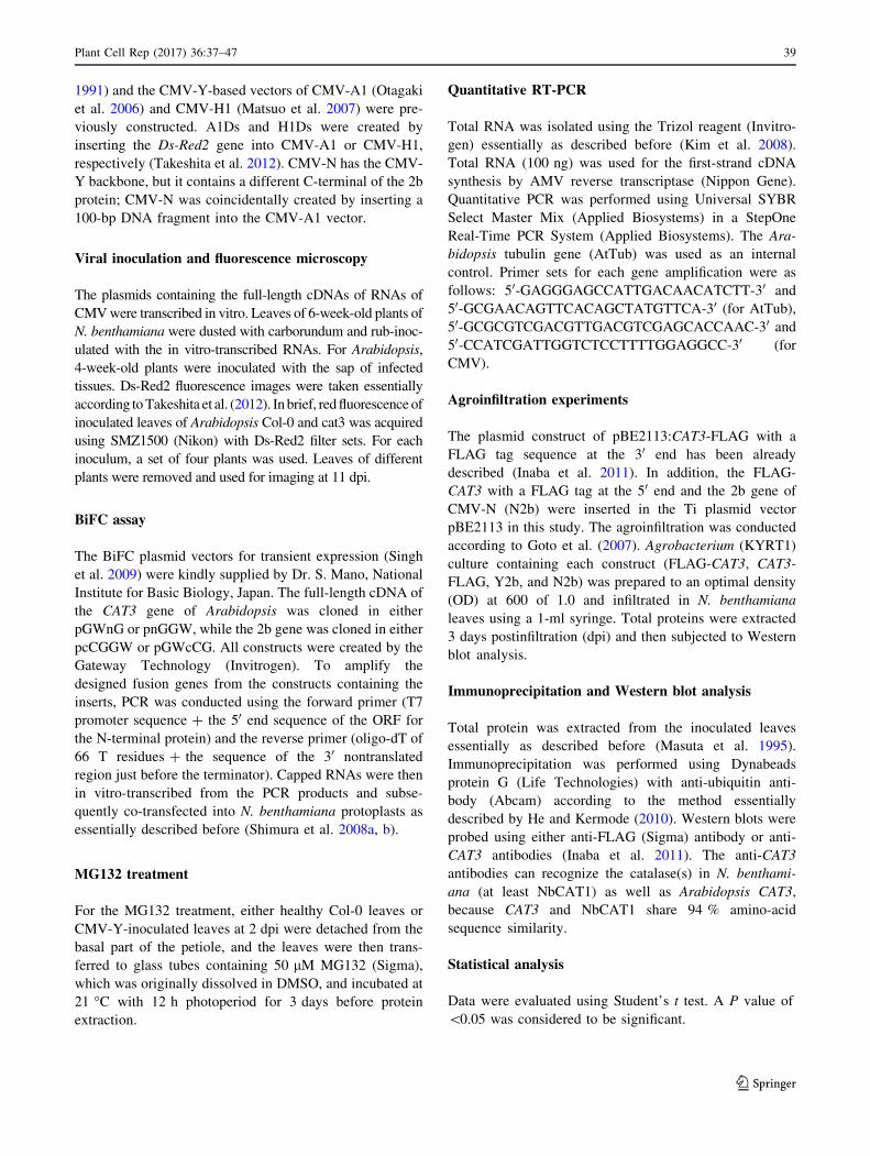
CMV symptom, accumulation, and movement
in CAT3-knockout mutant (cat3)
We previously reported that the interaction between CAT3
and CMV 2b causes necrosis accompanied with H2O2
accumulation, suggesting that the malfunction of CAT3
induced H2O2-mediated cell death. We here examined the
effects of depletion of CAT3 from Arabidopsis on CMV
symptom and accumulation using CAT3-knockout plants of
Col-0 (cat3). The T-DNA insertion knockout mutant
(SALK-092911) was obtained from the Arabidopsis Bio-
logical Resource Center (ABRC). We identified homozy-
gous T-DNA insertion line from T4 plants using PCR.
Compared with the wild-type control plants, the cat3 plants
infected with CMV-Y at 14 dpi were somewhatmore stunted
(Fig. 1a) and showed weak necrosis; there was a little dif-
ference in visible necrotic appearance between wild type
(Col-0) and cat3. We examined the catalase activities and
found that the activities were one-third of the levels in the
control even after CMV infection (Supplementary Fig. 1).
These results suggest that the depletion of CAT3 did not
facilitate necrosis symptoms, and that 2b alone has an ability
to essentially induce weak necrosis on Arabidopsis leaves.
We previously reported that CAT3 can weaken the RSS
activity of 2b, and that CMV levels were reduced at least
until 7 dpi in CAT3-overexpressing Col-0 (Inaba et al.
2011). In this study, we observed that CMV accumulation
levels were significantly higher in cat3 plants at 14 dpi
(Fig. 1b). To confirm that the susceptibility of cat3 to CMV
was due to disruption of the CAT3 gene, we complemented
cat3/cat3 with the wild-type CAT3 gene. We selected
several hygromicin-resistant T1 plants, which produce
certain levels of the CAT3 gene transcript (Supplementary
Fig. 2). When those T1 transformants were inoculated with
CMV, there was no difference in CMV accumulation
between WT and the CAT3-complemented cat3/cat3
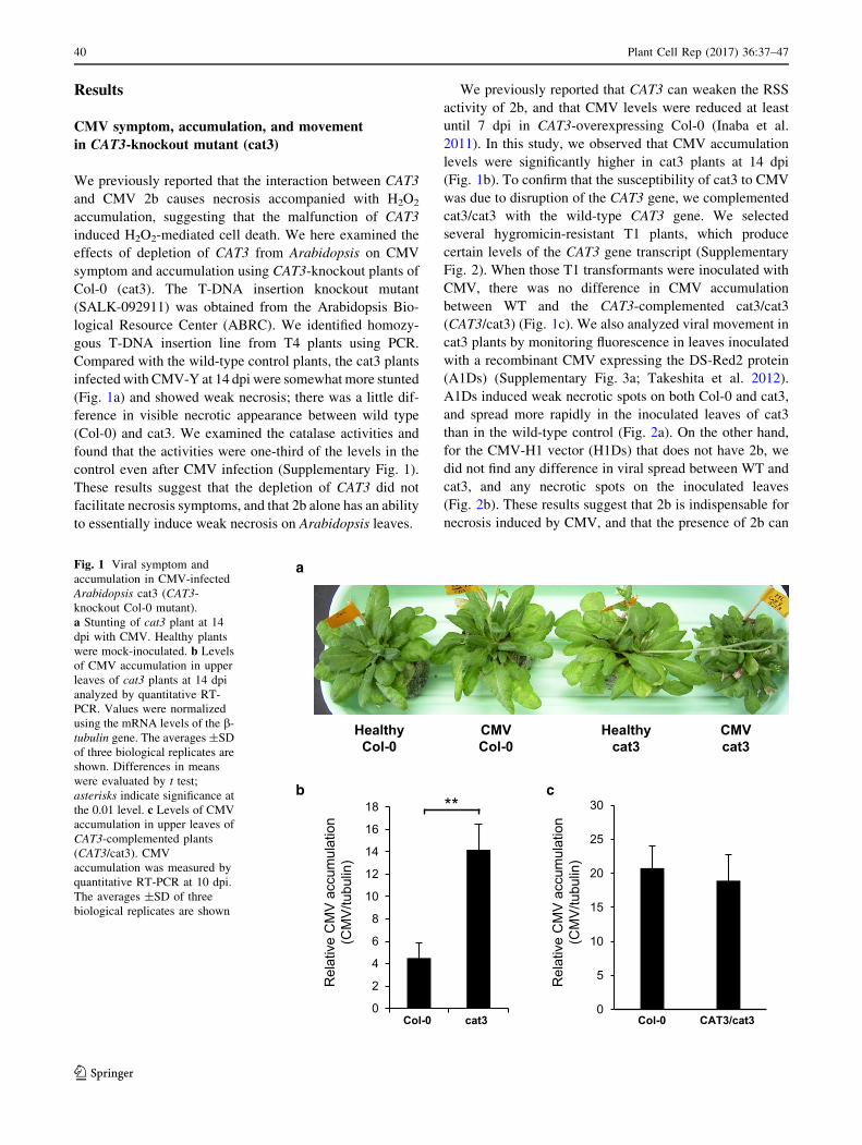
(CAT3/cat3) (Fig. 1c). We also analyzed viral movement in
cat3 plants by monitoring fluorescence in leaves inoculated
with a recombinant CMV expressing the DS-Red2 protein
(A1Ds) (Supplementary Fig. 3a; Takeshita et al. 2012).
A1Ds induced weak necrotic spots on both Col-0 and cat3,
and spread more rapidly in the inoculated leaves of cat3
than in the wild-type control (Fig. 2a). On the other hand,
for the CMV-H1 vector (H1Ds) that does not have 2b, we
did not find any difference in viral spread between WT and
cat3, and any necrotic spots on the inoculated leaves
(Fig. 2b). These results suggest that 2b is indispensable for
necrosis induced by CMV, and that the presence of 2b can
HealthyCol-0
CMVCol-0
Healthycat3
CMVcat3
a
b
Rel
ativ
e C
MV
acc
umul
atio
n (C
MV
/tubu
lin)
0
2
4
6
8
10
12
14
16
18 **
Col-0 cat30
5
10
15
20
25
30
Col-0 CAT3/cat3
c
Rel
ativ
e C
MV
acc
umul
atio
n (C
MV
/tubu
lin)
Fig. 1 Viral symptom and
accumulation in CMV-infected
Arabidopsis cat3 (CAT3-
knockout Col-0 mutant).
a Stunting of cat3 plant at 14
dpi with CMV. Healthy plants
were mock-inoculated. b Levels
of CMV accumulation in upper
leaves of cat3 plants at 14 dpi
analyzed by quantitative RT-
PCR. Values were normalized
using the mRNA levels of the b-tubulin gene. The averages ±SD
of three biological replicates are
shown. Differences in means
were evaluated by t test;
asterisks indicate significance at
the 0.01 level. c Levels of CMV
accumulation in upper leaves of
CAT3-complemented plants
(CAT3/cat3). CMV
accumulation was measured by
quantitative RT-PCR at 10 dpi.
The averages ±SD of three
biological replicates are shown
40 Plant Cell Rep (2017) 36:37–47
123
make CMV move more rapidly in cat3 than in wild-type.
Taken together, to some extent, CAT3 contributes to
inhibiting CMV movement.
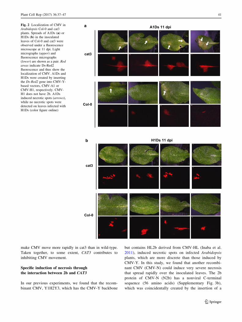
Specific induction of necrosis through
the interaction between 2b and CAT3
In our previous experiments, we found that the recom-
binant CMV, Y1H2Y3, which has the CMV-Y backbone
but contains HL2b derived from CMV-HL (Inaba et al.
2011), induced necrotic spots on infected Arabidopsis
plants, which are more discrete than those induced by
CMV-Y. In this study, we found that another recombi-
nant CMV (CMV-N) could induce very severe necrosis
that spread rapidly over the inoculated leaves. The 2b
protein of CMV-N (N2b) has a nonviral C-terminal
sequence (56 amino acids) (Supplementary Fig. 3b),
which was coincidentally created by the insertion of a
a
cat3
Col-0
A1Ds 11 dpi
b H1Ds 11 dpi
cat3
Col-0
Fig. 2 Localization of CMV in
Arabidopsis Col-0 and cat3
plants. Spreads of A1Ds (a) orH1Ds (b) in the inoculated
leaves of Col-0 and cat3 were
observed under a fluorescence
microscope at 11 dpi. Light
micrographs (upper) and
fluorescence micrographs
(lower) are shown as a pair. Red
areas indicate Ds-Red2
fluorescence and thus show the
localization of CMV. A1Ds and
H1Ds were created by inserting
the Ds-Red2 gene into CMV-Y-
based vectors, CMV-A1 or
CMV-H1, respectively. CMV-
H1 does not have 2b. A1Ds
induced necrotic spots (arrows),
while no necrotic spots were
detected on leaves infected with
H1Ds (color figure online)
Plant Cell Rep (2017) 36:37–47 41
123
100 bp-foreign fragment in the CMV-A1 vector. For
Arabidopsis, CMV-N induced a very distinct, extensive
necrosis on inoculated Col-0 leaves (Fig. 3a, b). How-
ever, when we inoculated CMV-N onto cat3 plants, we
observed much milder necrosis (Fig. 3a), suggesting that
the necrosis severity on CMV-inoculated Arabidopsis
leaves is determined by the presence of CAT3; CAT3
would be a modulator of the 2b-inducing necrosis. In
addition to Arabidopsis, CMV-N induced not only severe
necrosis on the inoculated leaves but also systemic lethal
necrosis in N. benthamiana (Fig. 3c). To confirm whe-
ther N2b still has an ability to interact with CAT3, the
BiFC assay was conducted by co-transfecting N. ben-
thamiana protoplasts with two combinations of the
N-terminal and C-terminal GFP constructs for BiFC. As
shown in Fig. 4, we observed distinct GFP fluorescence
derived from the interactions between N2b and CAT3, as
is the case in the interaction between Y2b and CAT3
(Inaba et al. 2011). In this assay, we noticed that GFP
fluorescence might be localized possibly in the nucleus
and to a lesser extent in the cytosol. These observations
suggest that CAT3 can interact with N2b like Y2b, and
that 2b-inducing necrosis feature is determined by a
specific combination of 2b and CAT3.
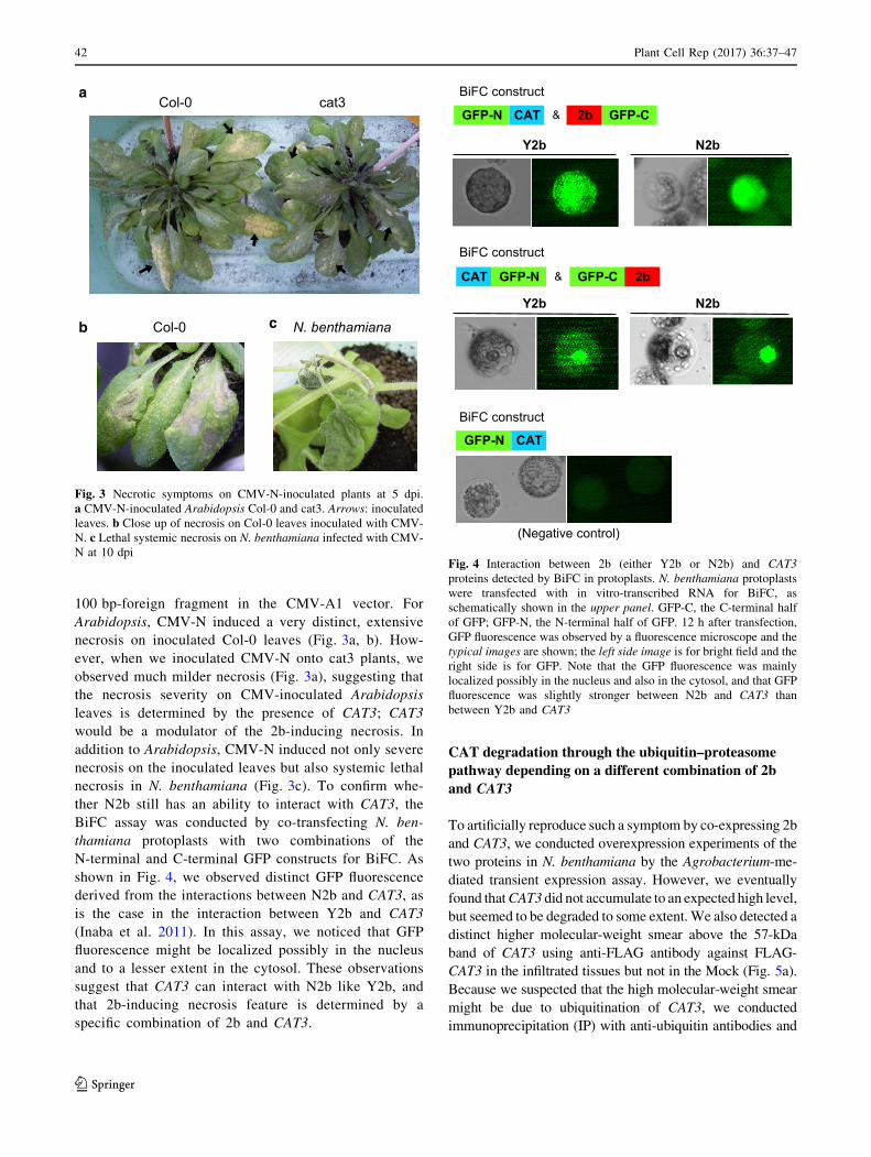
CAT degradation through the ubiquitin–proteasome
pathway depending on a different combination of 2b
and CAT3
To artificially reproduce such a symptom by co-expressing 2b
and CAT3, we conducted overexpression experiments of the
two proteins in N. benthamiana by the Agrobacterium-me-
diated transient expression assay. However, we eventually
found thatCAT3 did not accumulate to an expected high level,
but seemed to be degraded to some extent. We also detected a
distinct higher molecular-weight smear above the 57-kDa
band of CAT3 using anti-FLAG antibody against FLAG-
CAT3 in the infiltrated tissues but not in the Mock (Fig. 5a).
Because we suspected that the high molecular-weight smear
might be due to ubiquitination of CAT3, we conducted
immunoprecipitation (IP) with anti-ubiquitin antibodies and
Col-0
Col-0 cat3a
b N. benthamianac
Fig. 3 Necrotic symptoms on CMV-N-inoculated plants at 5 dpi.
a CMV-N-inoculated Arabidopsis Col-0 and cat3. Arrows: inoculated
leaves. b Close up of necrosis on Col-0 leaves inoculated with CMV-
N. c Lethal systemic necrosis on N. benthamiana infected with CMV-
N at 10 dpi
Y2b N2b
CAT GFP-C2bGFP-N &
BiFC construct
(Negative control)
CAT GFP-C 2bGFP-N &
Y2b N2b
CATGFP-N
BiFC construct
BiFC construct
Fig. 4 Interaction between 2b (either Y2b or N2b) and CAT3
proteins detected by BiFC in protoplasts. N. benthamiana protoplasts
were transfected with in vitro-transcribed RNA for BiFC, as
schematically shown in the upper panel. GFP-C, the C-terminal half
of GFP; GFP-N, the N-terminal half of GFP. 12 h after transfection,
GFP fluorescence was observed by a fluorescence microscope and the
typical images are shown; the left side image is for bright field and the
right side is for GFP. Note that the GFP fluorescence was mainly
localized possibly in the nucleus and also in the cytosol, and that GFP
fluorescence was slightly stronger between N2b and CAT3 than
between Y2b and CAT3
42 Plant Cell Rep (2017) 36:37–47
123
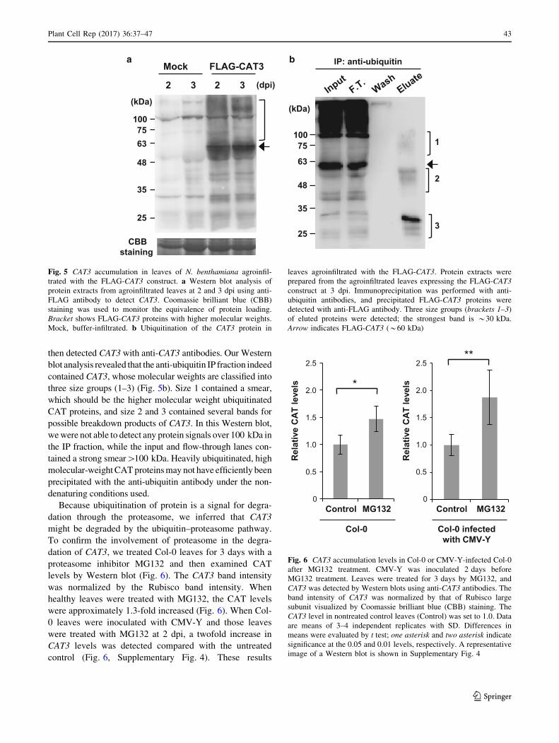
then detected CAT3 with anti-CAT3 antibodies. Our Western
blot analysis revealed that the anti-ubiquitin IP fraction indeed
contained CAT3, whose molecular weights are classified into
three size groups (1–3) (Fig. 5b). Size 1 contained a smear,
which should be the higher molecular weight ubiquitinated
CAT proteins, and size 2 and 3 contained several bands for
possible breakdown products of CAT3. In this Western blot,
wewere not able to detect any protein signals over 100 kDa in
the IP fraction, while the input and flow-through lanes con-
tained a strong smear[100 kDa. Heavily ubiquitinated, high
molecular-weightCATproteinsmay not have efficiently been
precipitated with the anti-ubiquitin antibody under the non-
denaturing conditions used.
Because ubiquitination of protein is a signal for degra-
dation through the proteasome, we inferred that CAT3
might be degraded by the ubiquitin–proteasome pathway.
To confirm the involvement of proteasome in the degra-
dation of CAT3, we treated Col-0 leaves for 3 days with a
proteasome inhibitor MG132 and then examined CAT
levels by Western blot (Fig. 6). The CAT3 band intensity
was normalized by the Rubisco band intensity. When
healthy leaves were treated with MG132, the CAT levels
were approximately 1.3-fold increased (Fig. 6). When Col-
0 leaves were inoculated with CMV-Y and those leaves
were treated with MG132 at 2 dpi, a twofold increase in
CAT3 levels was detected compared with the untreated
control (Fig. 6, Supplementary Fig. 4). These results
a b
2
FLAG-CAT3Mock
3 2 3
CBBstaining
(dpi)
(kDa)
25
35
48
6375
100
IP: anti-ubiquitin
1
2
325
35
48
63
75100
(kDa)
Fig. 5 CAT3 accumulation in leaves of N. benthamiana agroinfil-
trated with the FLAG-CAT3 construct. a Western blot analysis of
protein extracts from agroinfiltrated leaves at 2 and 3 dpi using anti-
FLAG antibody to detect CAT3. Coomassie brilliant blue (CBB)
staining was used to monitor the equivalence of protein loading.
Bracket shows FLAG-CAT3 proteins with higher molecular weights.
Mock, buffer-infiltrated. b Ubiquitination of the CAT3 protein in
leaves agroinfiltrated with the FLAG-CAT3. Protein extracts were
prepared from the agroinfiltrated leaves expressing the FLAG-CAT3
construct at 3 dpi. Immunoprecipitation was performed with anti-
ubiquitin antibodies, and precipitated FLAG-CAT3 proteins were
detected with anti-FLAG antibody. Three size groups (brackets 1–3)
of eluted proteins were detected; the strongest band is *30 kDa.
Arrow indicates FLAG-CAT3 (*60 kDa)
Col-0
MG132
*
Control
Col-0 infected with CMV-Y
Control MG132
Rel
ativ
e C
AT le
vels
2.5
2.0
1.5
1.0
0.5
0
**
Rel
ativ
e C
AT le
vels
2.5
2.0
1.5
1.0
0.5
0
Fig. 6 CAT3 accumulation levels in Col-0 or CMV-Y-infected Col-0
after MG132 treatment. CMV-Y was inoculated 2 days before
MG132 treatment. Leaves were treated for 3 days by MG132, and
CAT3 was detected by Western blots using anti-CAT3 antibodies. The
band intensity of CAT3 was normalized by that of Rubisco large
subunit visualized by Coomassie brilliant blue (CBB) staining. The
CAT3 level in nontreated control leaves (Control) was set to 1.0. Data
are means of 3–4 independent replicates with SD. Differences in
means were evaluated by t test; one asterisk and two asterisk indicate
significance at the 0.05 and 0.01 levels, respectively. A representative
image of a Western blot is shown in Supplementary Fig. 4
Plant Cell Rep (2017) 36:37–47 43
123
suggest that proteasome activity is, at least partially,
responsible for the CAT3 degradation, which is promoted
by CMV infection.
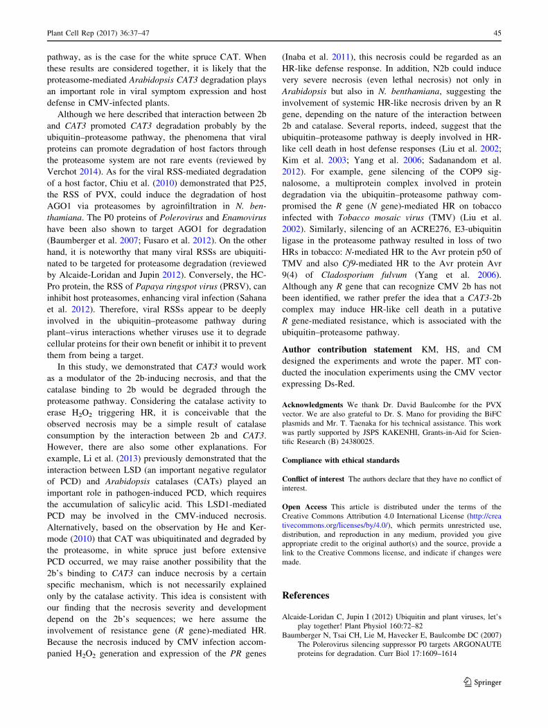
We next investigated whether 2b can affect CAT3
degradation by the Western blot analysis following the
transient overexpression of CAT3 and 2b by agroinfiltra-
tion. The overexpression of CAT3-FLAG itself caused a
slight decrease in the CAT3 level compared with the Mock
control (Fig. 7 lanes 1, 3). In contrast, the coexpression of
CAT3 and Y2b markedly reduced the CAT3 levels (lanes 5,
6). On the other hand, the overexpression of N2b did not
reduce the CAT3 level as efficiently as Y2b did (lanes 7, 8).
In this experiment, we also observed that Y2b was barely
detected compared with the distinct band of N2b in the
Agrobacterium-infiltrated tissues (Fig. 7), suggesting that
Y2b was less stable than N2b. These results suggest that
the degradation of Y2b and CAT3 was simultaneously
taken place when they were co-expressed but that in the
combination of N2b and CAT3, they were more slowly
degraded.
Discussion
To analyze the interaction between 2b and CAT3, we
previously produced transgenic Col-0 plants that overex-
press the CAT3 gene under the 35S promoter (Inaba et al.
2011). With difficulty, we finally obtained several CAT3-
overexpressing transgenic lines, but they produced CAT3 at
most *twofold more than in the wild type (Inaba et al.
2011). We assumed that when the accumulation levels of
CAT3 exceed a certain threshold, the CAT3 protein levels
must be lowered by a well-regulated mechanism, because
the CAT3 levels should be very critical to maintain the
cellular redox balance. Based on the detection of the higher
molecular weight, ubiquitinated smear band (above the
57 kDa) in the agroinfiltration experiment, we considered
that the proteasome pathway regulated CAT3 degradation.
CAT3 expression levels are drastically controlled by a
circadian rhythm, in which the amplitude of the oscillations
in CAT3 mRNA accumulation is *fivefold; the lowest
peak is in the early morning (Zhong and McClung 1996). If
CAT3 functions according to the circadian rhythm and is
regulated at the mRNA level, the synthesized protein
should be quickly degraded. We thus consider that even in
healthy Col-0 plants, CAT3 levels must be reset by protein
degradation along with the circadian rhythm. In fact, a
proteasome inhibitor, MG132, treatment increased 1.3-fold
CAT3 accumulation compared to the untreated control,
while the CAT3 levels were increased ca. twofold by
MG132 when plants were infected with CMV-Y (Fig. 6).
These results provide the evidence that the accumulation
levels of CAT3 in the presence of CMV 2b are significantly
affected by the ubiquitin–proteasome pathway.
As for the involvement of the proteasome in the regu-
lation of cellular CAT3 levels and association of pro-
grammed cell death with the generation of ROS, this study
is not the first one. He and Kermode (2010) have already
demonstrated that white spruce plants actually used the
proteasome to control the levels of CAT (a homolog of
Arabidopsis CAT3) during the seedling development. One
important observation that we share with them is the
finding of several proteins with lower molecular weights as
breakdown products of CAT after MG132 treatment. As
shown in Fig. 5, we also detected several lower molecular
weights bands (20–30 kDa) in our Western blots, sug-
gesting that those breakdown products of CAT3 may have
been generated through the same ubiquitin–proteasome
a Anti-Cat3
(kDa)S PS PS PS P
1006348
252017
Mock CAT3 +Y2bCAT3 CAT3
+N2b
b Anti-2b
1720
(kDa) S PS P
Y2b N2b
ABstaining
1 2 3 4 5 6 7 8
*
Fig. 7 Degradation of CAT3 protein in leaves agroinfiltrated with
CAT3-FLAG and CMV 2b (Y2b or N2b). a Western blot analysis of
the protein extracts from agroinfiltrated leaves at 3 dpi. CAT3-FLAG
was detected using anti-CAT3 antibodies. Protein samples were
prepared as essentially described in Masuta et al. (1995). Briefly, leaf
tissue was homogenized in 1 ml of PBS/Tween and centrifuged at
100009g for 20 min to separate the supernatant (S) and pellet (P).
After Laemmli’s sample buffer was added to each of S and P, the
samples were subjected to SDS-PAGE. Arrow indicates both the
endogenous tobacco catalases and CAT3-FLAG proteins, and asterisk
shows a possible breakdown product derived from the catalases. The
blot was stained with amido black (AB) to confirm the equivalence of
protein loading. b Western blot with anti-2b antibodies. Y2b and N2b
were detected on the same blot. Arrows indicate the 2b protein. The
loading controls of S and P lanes of Y2b and N2b are equal to lanes 5
and 6 (for Y2b) and lanes 7 and 8 (for N2b) of AB staining in a,respectively
44 Plant Cell Rep (2017) 36:37–47
123
pathway, as is the case for the white spruce CAT. When
these results are considered together, it is likely that the
proteasome-mediated Arabidopsis CAT3 degradation plays
an important role in viral symptom expression and host
defense in CMV-infected plants.
Although we here described that interaction between 2b
and CAT3 promoted CAT3 degradation probably by the
ubiquitin–proteasome pathway, the phenomena that viral
proteins can promote degradation of host factors through
the proteasome system are not rare events (reviewed by
Verchot 2014). As for the viral RSS-mediated degradation
of a host factor, Chiu et al. (2010) demonstrated that P25,
the RSS of PVX, could induce the degradation of host
AGO1 via proteasomes by agroinfiltration in N. ben-
thamiana. The P0 proteins of Polerovirus and Enamovirus
have been also shown to target AGO1 for degradation
(Baumberger et al. 2007; Fusaro et al. 2012). On the other
hand, it is noteworthy that many viral RSSs are ubiquiti-
nated to be targeted for proteasome degradation (reviewed
by Alcaide-Loridan and Jupin 2012). Conversely, the HC-
Pro protein, the RSS of Papaya ringspot virus (PRSV), can
inhibit host proteasomes, enhancing viral infection (Sahana
et al. 2012). Therefore, viral RSSs appear to be deeply
involved in the ubiquitin–proteasome pathway during
plant–virus interactions whether viruses use it to degrade
cellular proteins for their own benefit or inhibit it to prevent
them from being a target.
In this study, we demonstrated that CAT3 would work
as a modulator of the 2b-inducing necrosis, and that the
catalase binding to 2b would be degraded through the
proteasome pathway. Considering the catalase activity to
erase H2O2 triggering HR, it is conceivable that the
observed necrosis may be a simple result of catalase
consumption by the interaction between 2b and CAT3.
However, there are also some other explanations. For
example, Li et al. (2013) previously demonstrated that the
interaction between LSD (an important negative regulator
of PCD) and Arabidopsis catalases (CATs) played an
important role in pathogen-induced PCD, which requires
the accumulation of salicylic acid. This LSD1-mediated
PCD may be involved in the CMV-induced necrosis.
Alternatively, based on the observation by He and Ker-
mode (2010) that CAT was ubiquitinated and degraded by
the proteasome, in white spruce just before extensive
PCD occurred, we may raise another possibility that the
2b’s binding to CAT3 can induce necrosis by a certain
specific mechanism, which is not necessarily explained
only by the catalase activity. This idea is consistent with
our finding that the necrosis severity and development
depend on the 2b’s sequences; we here assume the
involvement of resistance gene (R gene)-mediated HR.
Because the necrosis induced by CMV infection accom-
panied H2O2 generation and expression of the PR genes
(Inaba et al. 2011), this necrosis could be regarded as an
HR-like defense response. In addition, N2b could induce
very severe necrosis (even lethal necrosis) not only in
Arabidopsis but also in N. benthamiana, suggesting the
involvement of systemic HR-like necrosis driven by an R
gene, depending on the nature of the interaction between
2b and catalase. Several reports, indeed, suggest that the
ubiquitin–proteasome pathway is deeply involved in HR-
like cell death in host defense responses (Liu et al. 2002;
Kim et al. 2003; Yang et al. 2006; Sadanandom et al.
2012). For example, gene silencing of the COP9 sig-
nalosome, a multiprotein complex involved in protein
degradation via the ubiquitin–proteasome pathway com-
promised the R gene (N gene)-mediated HR on tobacco
infected with Tobacco mosaic virus (TMV) (Liu et al.
2002). Similarly, silencing of an ACRE276, E3-ubiquitin
ligase in the proteasome pathway resulted in loss of two
HRs in tobacco: N-mediated HR to the Avr protein p50 of
TMV and also Cf9-mediated HR to the Avr protein Avr
9(4) of Cladosporium fulvum (Yang et al. 2006).
Although any R gene that can recognize CMV 2b has not
been identified, we rather prefer the idea that a CAT3-2b
complex may induce HR-like cell death in a putative
R gene-mediated resistance, which is associated with the
ubiquitin–proteasome pathway.
Author contribution statement KM, HS, and CM
designed the experiments and wrote the paper. MT con-
ducted the inoculation experiments using the CMV vector
expressing Ds-Red.
Acknowledgments We thank Dr. David Baulcombe for the PVX
vector. We are also grateful to Dr. S. Mano for providing the BiFC
plasmids and Mr. T. Taenaka for his technical assistance. This work
was partly supported by JSPS KAKENHI, Grants-in-Aid for Scien-
tific Research (B) 24380025.
Compliance with ethical standards
Conflict of interest The authors declare that they have no conflict of
interest.
Open Access This article is distributed under the terms of the
Creative Commons Attribution 4.0 International License (http://crea
tivecommons.org/licenses/by/4.0/), which permits unrestricted use,
distribution, and reproduction in any medium, provided you give
appropriate credit to the original author(s) and the source, provide a
link to the Creative Commons license, and indicate if changes were
made.
References
Alcaide-Loridan C, Jupin I (2012) Ubiquitin and plant viruses, let’s
play together! Plant Physiol 160:72–82
Baumberger N, Tsai CH, Lie M, Havecker E, Baulcombe DC (2007)
The Polerovirus silencing suppressor P0 targets ARGONAUTE
proteins for degradation. Curr Biol 17:1609–1614
Plant Cell Rep (2017) 36:37–47 45
123
Chiu MH, Chen IH, Boulcombe DC, Tsai CH (2010) The silencing
suppressor P25 of Potato virus X interacts with Argonaute1 and
mediates its degradation through the proteasome pathway. Mol
Plant Pathol 11:641–649
Ding SW, Anderson BJ, Haase HR, Symons RH (1994) New
overlapping gene encoded by the cucumber mosaic virus
genome. Virology 198:593–601
Ding SW, Li WX, Symons RH (1995) A novel naturally occurring
hybrid gene encoded by a plant RNA virus facilitates long
distance virus movement. EMBO J 14:5762–5772
Frugoli JA, Zhong HH, Nuccio ML, McCourt P, McPeek MA,
Thomas TL, McClung CR (1996) Catalase is encoded by a
multigene family in Arabidopsis thaliana (L.) Heynh. Plant
Physiol 112:327–336
Fukamatsu Y, Yabe N, Hasunuma K (2003) Arabidopsis NDK1 is a
component of ROS signaling by interacting with three catalases.
Plant Cell Physiol 44:982–989
Fusaro AF, Correa RL, Nakasugi K, Jackson C, Kawchuk L, Vaslin
MF, Waterhouse PM (2012) The Enamovirus P0 protein is a
silencing suppressor which inhibits local and systemic RNA
silencing through AGO1 degradation. Virology 426:178–187
Goto K, Kobori T, Kosaka Y, Natsuaki T, Masuta C (2007)
Characterization of silencing suppressor 2b of cucumber mosaic
virus based on examination of its small RNA-binding abilities.
Plant Cell Physiol 48:1050–1060
Havelda Z, Maule AJ (2000) Complex spatial responses to cucumber
mosaic virus infection in susceptible Cucurbita pepo cotyledons.
Plant Cell 12:1975–1985
He X, Kermode AR (2010) Programmed cell death of the megaga-
metophyte during post-germinative growth of white spruce
(Picea glauca) seeds is regulated by reactive oxygen species and
the ubiquitin-mediated proteolytic system. Plant Cell Physiol
51:1707–1720
Hu YQ, Liu S, Yuan HM, Li J, Yan DW, Zhang JF, Lu YT (2010)
Functional comparison of catalase genes in the elimination of
photorespiratory H2O2 using promoter- and 30-untranslatedregion exchange experiments in the Arabidopsis cat2 photores-
piratory mutant. Plant Cell Environ 33:1656–1670
Inaba J, Kim BM, Shimura H, Masuta C (2011) Virus-induced
necrosis is a consequence of direct protein-protein interaction
between a viral RNA-silencing suppressor and a host catalase.
Plant Physiol 156:2026–2036
Ji LH, Ding SW (2001) The suppressor of transgene RNA silencing
encoded by Cucumber mosaic virus interferes with salicylic
acid-mediated virus resistance. Mol Plant-Microbe Interact
14:715–724
Kim M, Ahn JW, Jin UH, Choi D, Paek KH, Pai HS (2003)
Activation of the programmed cell death pathway by inhibition
of proteasome function in plants. J Biol Chem 278:19406–19415
Kim B, Masuta C, Matsuura H, Takahashi H, Inukai T (2008) Veinal
necrosis induced by Turnip mosaic virus infection in Arabidopsis
is a form of defense response accompanying HR-like cell death.
Mol Plant-Microbe Interact 21:260–268
Lewsey M, Surette M, Robertson FC, Ziebell H, Choi SH, Ryu KH,
Canto T, Palukaitis P, Payne T, Walsh JA, Carr JP (2009) The
role of the Cucumber mosaic virus 2b protein in viral movement
and symptom induction. Mol Plant-Microbe Interact 22:
642–654
Li Y, Chen L, Mu J, Zuo J (2013) LESION SIMULATING
DISEASE1 interacts with catalases to regulate hypersensitive
cell death in Arabidopsis. Plant Physiol 163:1059–1070
Liu T, Schiff M, Serino G, Deng XW, Dinesh-Kumar SP (2002) Role
of SCF ubiquitin-ligase and the COP9 signalosome in the N
gene–mediated resistance response to Tobacco mosaic virus.
Plant Cell 14:1483–1496
Lucy AP, Guo HS, Li WX, Ding SW (2000) Suppression of post-
transcriptional gene silencing by a plant viral protein localized in
the nucleus. EMBO J 19:1672–1680
Masuta C, Tanaka H, Uehara K, Kuwata S, Koiwai A, Noma M
(1995) Broad resistance to plant viruses in transgenic plants
conferred by antisense inhibition of a host gene essential in S-
adenosylmethionine-dependent transmethylation reactions. Proc
Natl Acad Sci USA 92:6117–6121
Masuta C, Inaba J, Shimura H (2012) The 2b proteins of Cucumber
mosaic virus generally have the potential to differentially induce
necrosis on Arabidopsis. Plant Signal Behav 7:43–45
Matsuo K, Hong JS, Tabayashi N, Ito A, Masuta C, Matsumura T
(2007) Development of Cucumber mosaic virus as a vector
modifiable for different host species to produce therapeutic
proteins. Planta 225:277–286
Otagaki S, Arai M, Takahashi A, Goto K, Hong JS, Masuta C,
Kanazawa A (2006) Rapid induction of transcriptional and post-
transcriptional gene silencing using a novel Cucumber mosaic
virus vector. Plant Biotechnol 23:259–265
Petrova D, Marinova G, Chaneva G, Kapchina-Toteva V, Stoimenova
E (2009) Local and systemic responses of antioxidants to
Cucumber mosaic virus infection in pepper plants. Biotechnol
Biotechnol Eq 23:516–518
Sadanandom A, Bailey M, Ewan R, Lee J, Nelis S (2012) The
ubiquitin–proteasome system: central modifier of plant signal-
ing. New Phytol 196:13–28
Sahana N, Kaur H, Basavaraj Tena F, Jain RK, Paulkaitis P, Canto T,
Praveen S (2012) Inhibition of the host proteasome facilitates
papaya ringspot virus accumulation and proteosomal catalytic
activity is modulated by viral factor HcPro. PLoS one 7:e52546
Shi BJ, Miller J, Symons RH, Palukaitis P (2003) The 2b protein of
cucumoviruses has a role in promoting the cell-to-cell movement of
pseudorecombinant viruses. Mol Plant-Microbe Interact 16:261–267
Shimura H, Kogure Y, Goto K, Masuta C (2008a) Degree of RNA
silencing and ability of a viral suppressor vary depending on the
cell species in a protoplast system. J Gen Plant Pathol 74:326–330
Shimura H, Fukagawa T, Meguro A, Yamada H, Oh-hira M, Sano S,
Masuta C (2008b) A strategy for screening an inhibitor of viral
silencing suppressors, which attenuates symptom development
of plant viruses. FEBS Lett 582:4047–4052
Singh T, Hayashi M, Mano S, Arai Y, Goto S, Nishimura M (2009)
Molecular components required for the targeting of PEX7 to
peroxisomes in Arabidopsis thaliana. Plant J 60:488–498
Soards AJ, Murphy AM, Palukaitis P, Carr JP (2002) Virulence and
differential local and systemic spread of Cucumber mosaic virus
in tobacco are affected by the CMV 2b protein. Mol Plant-
Microbe Interact 15:647–653
Suzuki M, Kuwata S, Kataoka J, Masuta C, Nitta N, Takanami Y
(1991) Functional analysis of deletion mutants of Cucumber
mosaic virus RNA3 using an in vitro transcription system.
Virology 183:106–113
Takeshita M, Koizumi E, Noguchi M, Sueda K, Shimura H, Ishikawa
N, Matsuura H, Ohshima K, Natsuaki T, Kuwata S, Furuya N,
Tsuchiya K, Masuta C (2012) Infection dynamics in viral spread
and interference under the synergism between Cucumber mosaic
virus and Turnip mosaic virus. Mol Plant-Microbe Interact
25:18–27
Verchot J (2014) The ER quality control and ER associated
degradation machineries are vital for viral pathogenesis. Front
Plant Sci 5:66
Verslues PE, Batelli G, Grillo S, Agius F, Kim YS, Zhu J, Agarwal
M, Katiyar-Agarwal S, Zhu JK (2007) Interaction of SOS2 with
nucleoside diphosphate kinase 2 and catalases reveals a point of
connection between salt stress and H2O2 signaling in Arabidop-
sis thaliana. Mol Cell Biol 27:7771–7780
46 Plant Cell Rep (2017) 36:37–47
123

Yang CW, Gonzalez-Lamothe R, Ewan RA, Rowland O, Yoshioka H,
Shenton M, Ye H, O’Donnell E, Jones JDG, Sadanandom A
(2006) The E3 ubiquitin ligase activity of Arabidopsis PLANT
U-BOX17 and its functional tobacco homolog ACRE276 are
required for cell death and defense. Plant Cell 18:1084–1098
Zhong HH, McClung CR (1996) The circadian clock gates expression
of two Arabidopsis catalase genes to distinct and opposite
circadian phases. Mol Gen Genet 251:196–203
Zimmermann P, Heinlein C, Orendi G, Zentgraf U (2006) Senes-
cence-specific regulation of catalases in Arabidopsis thaliana
(L.) Heynh. Plant Cell Environ 29:1049–1060
Zou JJ, Li XD, Ratnasekera D, Wang C, Liu WX, Song LF, Zhang
WZ, Wu WH (2015) Arabidopsis CALCIUM-DEPENDENT
PROTEIN KINASE8 and CATALASE3 function in abscisic
acid-mediated signaling and H2O2 homeostasis in stomatal guard
cells under drought stress. Plant Cell 27:1445–1460
Plant Cell Rep (2017) 36:37–47 47
123


![Review Article Earthworm Protease · fetida [27]. It has strong antiviral activities against cucumber mosaic virus and tomato mosaic virus. The protease (27,000 Da) is the most active](https://img.pdfslide.us/doc/110x75/5fd08da243d0e50fda5f4e1a/review-article-earthworm-fetida-27-it-has-strong-antiviral-activities-against.jpg)