Embed Size (px)
Citation preview

Institute of Structural Engineering
Division of Computer Aided Design
Laboratory of Biomechanics
2
Department of Structural Mechanics, Biomechanical Laboratory, Poznan University of Technology
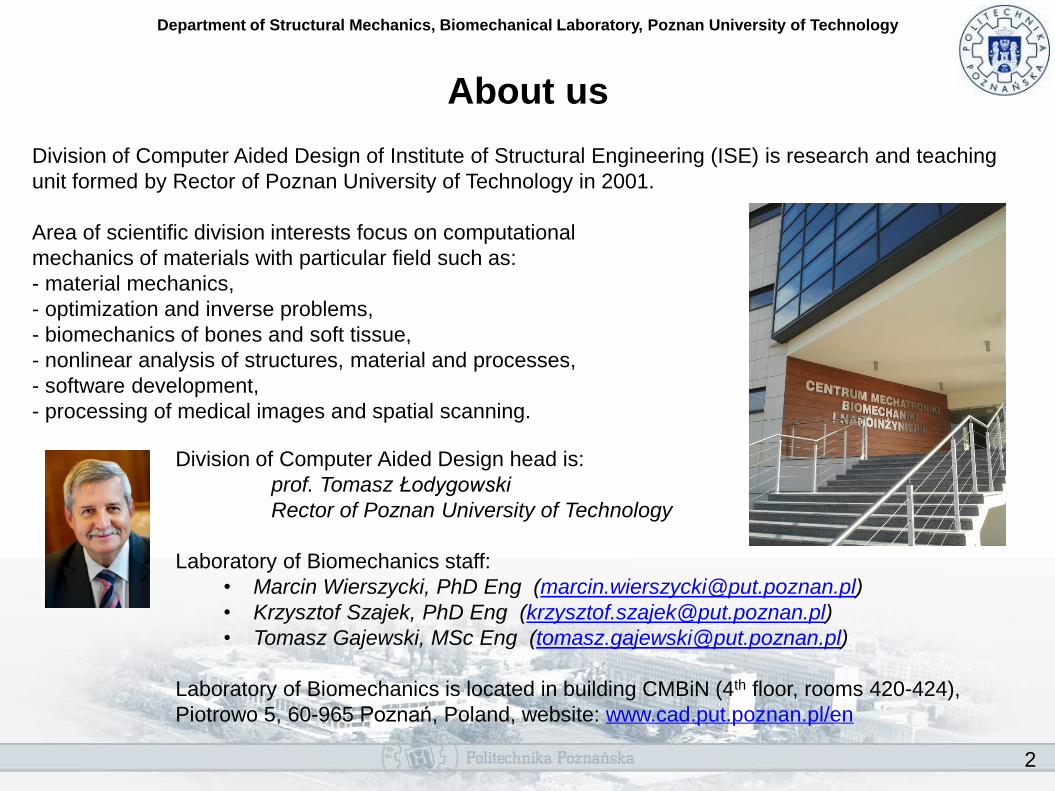
About us
Division of Computer Aided Design of Institute of Structural Engineering (ISE) is research and teaching
unit formed by Rector of Poznan University of Technology in 2001.
Area of scientific division interests focus on computational
mechanics of materials with particular field such as:
- material mechanics,
- optimization and inverse problems,
- biomechanics of bones and soft tissue,
- nonlinear analysis of structures, material and processes,
- software development,
- processing of medical images and spatial scanning.
Division of Computer Aided Design head is:
prof. Tomasz Łodygowski
Rector of Poznan University of Technology
Laboratory of Biomechanics staff:
• Marcin Wierszycki, PhD Eng ([email protected])
• Krzysztof Szajek, PhD Eng ([email protected])
• Tomasz Gajewski, MSc Eng ([email protected])
Laboratory of Biomechanics is located in building CMBiN (4th floor, rooms 420-424),
Piotrowo 5, 60-965 Poznań, Poland, website: www.cad.put.poznan.pl/en

3
Department of Structural Mechanics, Biomechanical Laboratory, Poznan University of Technology
Examples of our activity
• Analyses of biomechanical systems
Numerical modeling of spinal motion segment
Ligamentotaxis procedure simulation
Stress-strain analysis, fatigue life prediction and tightening/loosening simulation of
dental Implant
Calculation of contact pressure in mucosa under complete prothesis
Multiscale modelling of bone tissue
• Optimization
Two-component dental implant
Bone remodelling driven by mechanical stimulus
• Materials modeling
Modeling of the soft and hard tissues of spine
Implementation of hyperelastic model of arteries with damage and distributed fiber
orientations
• Processing of medical images and spatial scanning
Geometry model based on medical imaging (CT, MRI)
Assigning mechanical properties based on medical imaging
Spatial scanning of prothesis and tissues (mandible bone example)
• Software
Optimization module for Abaqus/CAE
Analyses of biomechanical systems

5
Department of Structural Mechanics, Biomechanical Laboratory, Poznan University of Technology
Numerical modeling of spinal motion segment
Purpose: The goal of the study was to provide 3D mechanical model of the human lumbar spine which
can be used in: simulation of surgery and systems of stabilization, analyses of spinal equilibrium and
stability and supporting of medical diagnosis and rehabilitation.
Material and methods: Geometry of the FE model of human lumbar spine motion segment (L4-L5) was created based on CT data. For bone modeling the linear, orthotropic elastic materials was used. For ligaments the hyperelastic material was used. Simulations were conducted in commercial finite element method software Abaqus Unified (Dassault Systemes).
Results: The global stiffness characteristics of spine motion segment has been compared successfully with experimental data. The obtained relative displacements and rotations allow to verify positively the hierarchical concept of intervertebral disc simplifications.
Conclusion: The finite element method enables to obtain useful and helpful results for human lumbar spine motion segment in quasi-static analysis.
Additional info: This research was supported by the grant no. 8 T07A 046 21.

6
Department of Structural Mechanics, Biomechanical Laboratory, Poznan University of Technology
Ligamentotaxis procedure simulation
Purpose: The goal of this study was to determine the forces acting on longitudinal ligaments connected
with damaged vertebral body during relocation procedure. Understanding relocation phenomenon may
also allow to make the surgical technique more objective and in some cases for improving efficiency of
surgical treatment.
Material and methods: The 3D finite element model of fractured human thoracolumbar spine segments was created. Special attention was focused on the modelling of soft tissues: ligaments and intervertebral discs (IVD) in the surgical procedure. The FE model contains beam model of Schanz screw system for spinal fracture reduction (USS Fracture, Synthes). The nonlinear effects e.g. contact constrains or pretension loads have been take into consideration to capture the key mechanisms of ligamentotaxis procedure.
Results: This evaluation of global deformation of fractured human thoracolumbar spine segments was done based on the pre and post procedural X-ray images and experimental results. The obtained values of stresses and strains are compared with the literature based strength of tissues.
Conclusion: The basic assumptions and foundations of ligamentotaxis procedure can be confirmed based on results of carried out FE numerical simulation. The simulation of whole surgical procedure enables us to evaluate stresses and strains in biological structures and mechanical parts as well.
Additional info: This research was done in cooperation with Warsaw University of Technology and Military Medical Institute in Warsaw. It was supported by the grant no. NN518382437.

7
Department of Structural Mechanics, Biomechanical Laboratory, Poznan University of Technology
Stress-strain analysis, fatigue life prediction and
tightening/loosening simulation of dental Implant Purpose: For more than fifty percent of dental treatment patients diverse complications are observed.
In some cases, these problems are caused by mechanical reasons such as loosing of the retaining
screws or fracture and cracking of the dental implant components. FEA simulations are used to
realistically predict dental implant behavior under restoration treatment as well as service loads.
Material and methods: The computer simulations of implant behavior with the use of ABAQUS and fe-safe software were used to consider fatigue changes of dental material and screw loosening phenomenon. For fatigue calculation the strain-life fatigue (Manson-Coffin) algorithm with Principal Strain Criterion is used. For the full simulation of screw loosening, a 3D model, which includes a spiral thread was used. The modeling of tightening is crucial aspect of screw loosing simulation. It was carried out in such a way that it describes a real physical process including rotation of the screw and frictional contact.
Results: The fatigue analysis enable us to predict fatigue life and Factor Of Strength – a scale factor which should be applied to stresses at each node to achieve the assumed design life. In the simulation of screw loosening analysis the friction dissipated energy was used as a measure of the retaining screw loosing resistance.
Conclusion: On the basis of the results of fatigue analysis, it can be claimed that the material fatigue is the basic reason of the observed complications. Three dimensional model of an implant and the use of explicit codes let us effectively carry out the simulation of a screw tightening.
Additional info: Research was done in cooperation with the dental implant producer – OSTEOPLANT. The financial support was provided by the grant no. 3 T11F 026 28.
8
Department of Structural Mechanics, Biomechanical Laboratory, Poznan University of Technology
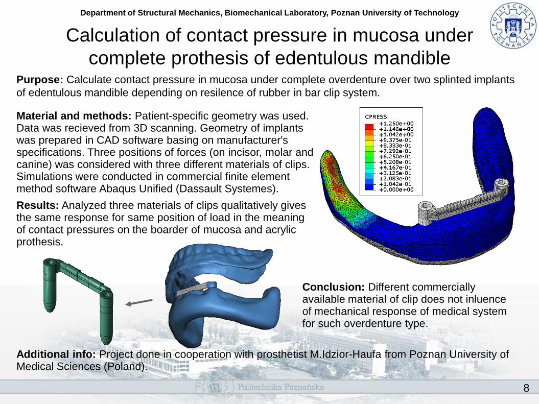
Calculation of contact pressure in mucosa under
complete prothesis of edentulous mandible Purpose: Calculate contact pressure in mucosa under complete overdenture over two splinted implants
of edentulous mandible depending on resilence of rubber in bar clip system.
Material and methods: Patient-specific geometry was used. Data was recieved from 3D scanning. Geometry of implants was prepared in CAD software basing on manufacturer's specifications. Three positions of forces (on incisor, molar and canine) was considered with three different materials of clips. Simulations were conducted in commercial finite element method software Abaqus Unified (Dassault Systemes).
Results: Analyzed three materials of clips qualitatively gives the same response for same position of load in the meaning of contact pressures on the boarder of mucosa and acrylic prothesis.
Conclusion: Different commercially available material of clip does not inluence of mechanical response of medical system for such overdenture type.
Additional info: Project done in cooperation with prosthetist M.Idzior-Haufa from Poznan University of Medical Sciences (Poland).

9
Department of Structural Mechanics, Biomechanical Laboratory, Poznan University of Technology
Two-scale modelling of bone tissue
Purpose: Development and validation of FEA modelling technique of cancellous bone for large models.
Material and methods: Two-scale modelling of bone microstructure is considered. The approach is based on a first-order computational homogenization technique. The coincidence of macro- and micro-model kinematics is done with the use of uniform displacement and traction boundary conditions. The micro and macro FE models are calculated in Abaqus code. The computational homogenization procedure is driven by a self-prepared manager which is coded in Python.
Results: The procedure were tested based on a series of test structures (solid and porous) and load cases. The results confirmed the procedure accuracy and effectiveness (FE bone model consisting more than 40 mln elements was analysed).
Conclusions: The developed procedure allows to analyse a real size bone considering details of its anatomy with acceptable accuracy.
Additional info: Project done in cooperation with prof. Michal Nowak in frame of grant N N518 328835 “Multiscale analysis of adaptive bone remodelling driven by mechanical stimulus”

Material modeling
11
Department of Structural Mechanics, Biomechanical Laboratory, Poznan University of Technology
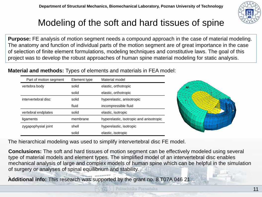
Modeling of the soft and hard tissues of spine
Purpose: FE analysis of motion segment needs a compound approach in the case of material modeling.
The anatomy and function of individual parts of the motion segment are of great importance in the case
of selection of finite element formulations, modeling techniques and constitutive laws. The goal of this
project was to develop the robust approaches of human spine material modeling for static analysis.
Material and methods: Types of elements and materials in FEA model: The hierarchical modeling was used to simplify intervertebral disc FE model. Conclusions: The soft and hard tissues of motion segment can be effectively modeled using several type of material models and element types. The simplified model of an intervertebral disc enables mechanical analysis of large and complex models of human spine which can be helpful in the simulation of surgery or analyses of spinal equilibrium and stability. . Additional info: This research was supported by the grant no. 8 T07A 046 21.
Part of motion segment Element type Material model
vertebra body solid elastic, orthotropic
solid elastic, orthotropic
intervertebral disc solid hyperelastic, anisotropic
fluid incompressible fluid
vertebral endplates solid elastic, isotropic
ligaments membrane hyperelastic, isotropic and anisotropic
zygapophysial joint shell hyperelastic, isotropic
solid elastic, isotropic
AF

12
Department of Structural Mechanics, Biomechanical Laboratory, Poznan University of Technology
Implementation of hyperelastic model of arteries
with damage and fiber orientations Purpose: Implement hyperelastic material model of artery tissues with damage and distributed fiber
orientations. Constitutive law base on model proposed by Holzapfel et. al (2000) and extended by
Weisbecker et. al (2012).
Material and methods: Anisotropic material model represents hiperelastic properties of adventitial and intimal layers of arterial walls, base on fibre-reinforced structural model with incompressibility. Distribution of fiber orientations and mean directions can be identified by laboratory tests. Application in commercial finite element method software Abaqus Unified (Dassault Systemes) as User MATerial subroutine (UMAT).
Results: UMAT subroutine simulation gives satisfactory results in comparision to experimental results. Damaged behavior of collagen fiber fabric is recreated.
Conclusion: Implemented material model can be used in simulations of medical treatments, e.g. balloon angioplasty.
Additional info: Project done in cooperation with prof. Gerhard Holzapfel and MSc. Eng. Hannah Weisbecker from Graz University of Technology (Austria).
Holzapfel et. al (2000)

Optimization

14
Department of Structural Mechanics, Biomechanical Laboratory, Poznan University of Technology
Two-component dental implant optimization
Purpose: Designing of a new implant. Parametric optimization of geometry and assembly screw preload in context of fatigue life, static resistence, screw loosening, tightening inaccuracy, tightness and micromotions and implant diameter reduction. Multiobjective studies were also carried out.
Material and methods: Optimization utilized parametrized FE model build in Abaqus/CAE. The changes were driven by genetic algorithm hybridized with Hooke-Jeeves procedure.
Results: All optimization processes finished with success providing the improved designs which maintain all assumed constraints. The implant prototypes were manufactured and tested. The tests confirmed the obtained results.
Conclusions: The obtained results confirmed the methodology applied as a effective tool for dental implant designing.
Additional info: Project was done in cooperation with the dental implant producer – OSTEOPLANT in frame of grant R13 0020 06 “Development and preparation of dental implant prototypes”

15
Department of Structural Mechanics, Biomechanical Laboratory, Poznan University of Technology
Bone remodelling
Purpose: Development and validation of a two-scale remodelling procedure of bone tissue driven by mechanical stimulus.
Material and methods: In microscale the faithful trabecular bone structure is considered. The whole microstructure is divided into cubical volumes represented by finite elements in macroscale. The remodelling is done in each volume of microstructure separately using optimization procedure. The microstructure changes can occure only on the trabecular bone surface and are driven by strains (as a result of macro model deformation). The procedure was driven by the self-prepared programm.
Results: The verification using data on rat femur bone remodelling confirmed qualitative agreement. The procedure sucessfully remodelled the FE bone model consisting more than 40 mln elements.
Conclusions: The procedure allows to analyse a real size bone considering details of its anatomy. The bone stiffness changes due to trabecular bone remodelling.
Additional info: Project was done in cooperation with prof. Michal Nowak in frame of grant N N518 328835 “Multiscale analysis of adaptive bone remodelling driven by mechanical stimulus”
Processing of medical images and spatial
scanning

17
Department of Structural Mechanics, Biomechanical Laboratory, Poznan University of Technology
Geometry model based of medical imaging (CT, MRI)
Purpose: Obtaining thoracolumbar vertebrae and soft tissue geometry based on medical imaging.
Material and methods: The procedure utilizes the series of plane sections obtained with computer tomography imaging stored in DICOM format. In the first step, for each section the geometry is extracted based on assumed treshold (in Hounsfield’s units) for particular tissues and provide the series of plain contours. Next, the contours
Results: The spatial bone and soft tissue geometry was obtained. The geometry is characterizes by acceptable accuracy and can be directly used for FE models.
Conclusions: Used methodology is effective tool of obtaining of geometry maintaining patient individual variability or pathological changes.
Additional info: Project was done in cooperation with Anna Dąbrowska-Tkaczyk, PhD, in frame of grant N N518 382437 “Analysis of adjustment mechanisms of thoracolumbar vertebral using ligamentotaxis”
are transformed into closed exterior surface according to leading curves. In the last step, after manual correction of contours, the volume model is generated.
18
Department of Structural Mechanics, Biomechanical Laboratory, Poznan University of Technology
Assigning mechanical properties based on
medical imaging
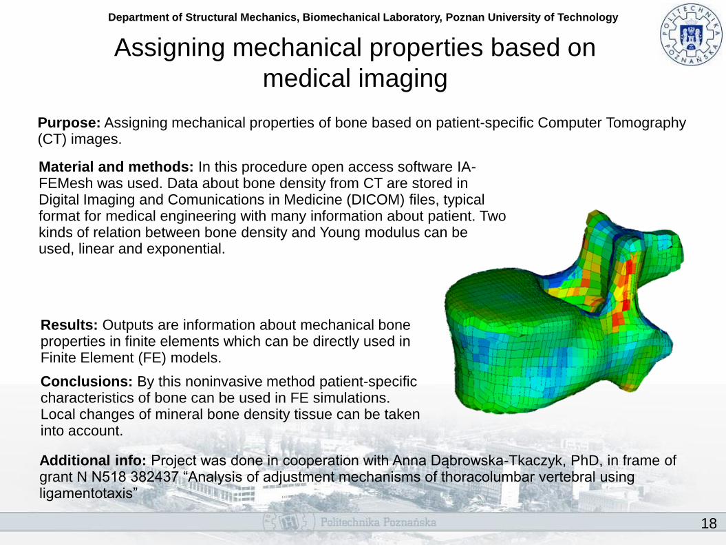
Purpose: Assigning mechanical properties of bone based on patient-specific Computer Tomography (CT) images.
Material and methods: In this procedure open access software IA-FEMesh was used. Data about bone density from CT are stored in Digital Imaging and Comunications in Medicine (DICOM) files, typical format for medical engineering with many information about patient. Two kinds of relation between bone density and Young modulus can be used, linear and exponential.
Results: Outputs are information about mechanical bone properties in finite elements which can be directly used in Finite Element (FE) models.
Conclusions: By this noninvasive method patient-specific characteristics of bone can be used in FE simulations. Local changes of mineral bone density tissue can be taken into account.
Additional info: Project was done in cooperation with Anna Dąbrowska-Tkaczyk, PhD, in frame of grant N N518 382437 “Analysis of adjustment mechanisms of thoracolumbar vertebral using ligamentotaxis”
19
Department of Structural Mechanics, Biomechanical Laboratory, Poznan University of Technology
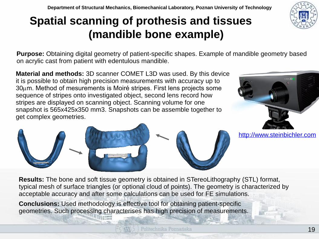
Material and methods: 3D scanner COMET L3D was used. By this device it is possible to obtain high precision measurements with accuracy up to 30mm. Method of mesurements is Moiré stripes. First lens projects some sequence of stripes onto investigated object, second lens record how stripes are displayed on scanning object. Scanning volume for one snapshot is 565x425x350 mm3. Snapshots can be assemble together to get complex geometries.
Spatial scanning of prothesis and tissues
(mandible bone example)
Purpose: Obtaining digital geometry of patient-specific shapes. Example of mandible geometry based on acrylic cast from patient with edentulous mandible.
Results: The bone and soft tissue geometry is obtained in STereoLithography (STL) format, typical mesh of surface triangles (or optional cloud of points). The geometry is characterized by acceptable accuracy and after some calculations can be used for FE simulations.
Conclusions: Used methodology is effective tool for obtaining patient-specific geometries. Such processing characterises has high precision of measurements.
http://www.steinbichler.com

Software
21
Department of Structural Mechanics, Biomechanical Laboratory, Poznan University of Technology
Optimization module for Abaqus/CAE
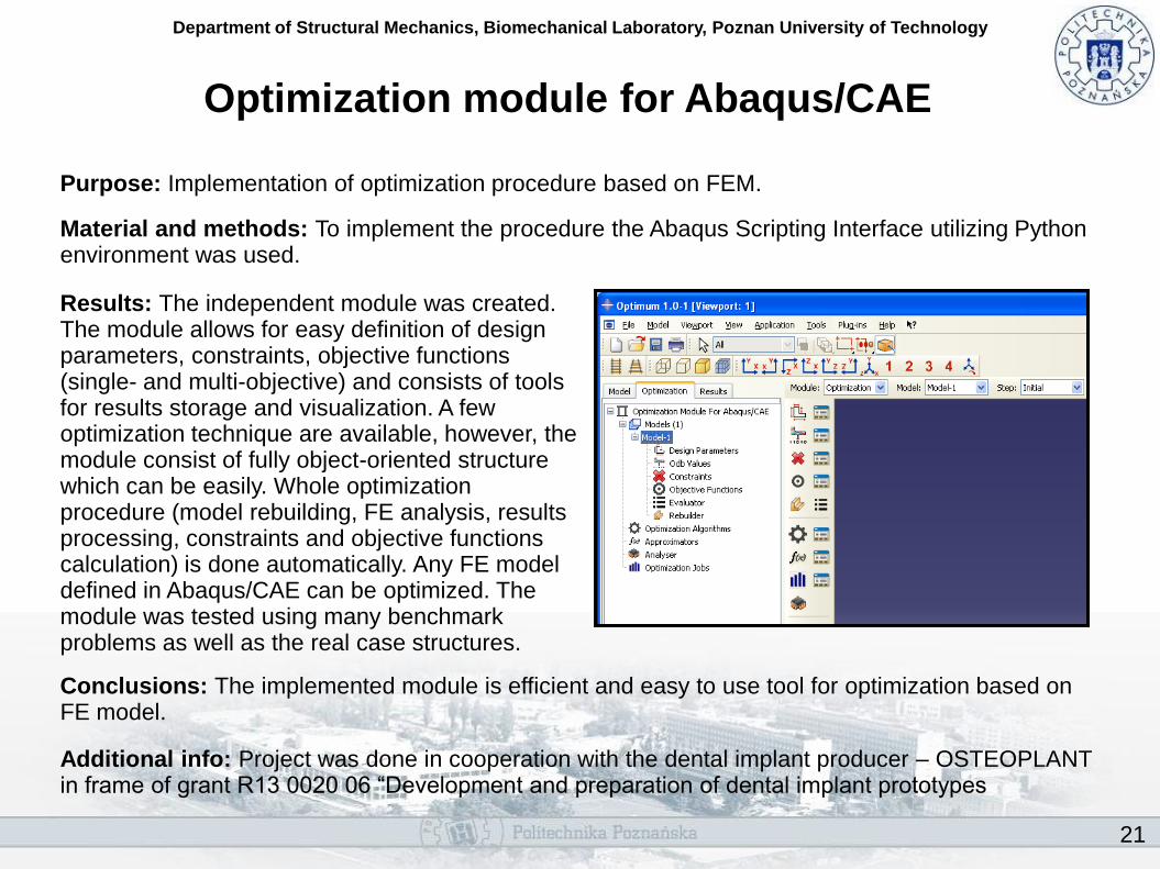
Purpose: Implementation of optimization procedure based on FEM.
Material and methods: To implement the procedure the Abaqus Scripting Interface utilizing Python environment was used.
Results: The independent module was created. The module allows for easy definition of design parameters, constraints, objective functions (single- and multi-objective) and consists of tools for results storage and visualization. A few optimization technique are available, however, the module consist of fully object-oriented structure which can be easily. Whole optimization procedure (model rebuilding, FE analysis, results processing, constraints and objective functions calculation) is done automatically. Any FE model defined in Abaqus/CAE can be optimized. The module was tested using many benchmark problems as well as the real case structures.
Conclusions: The implemented module is efficient and easy to use tool for optimization based on FE model.
Additional info: Project was done in cooperation with the dental implant producer – OSTEOPLANT in frame of grant R13 0020 06 “Development and preparation of dental implant prototypes