Embed Size (px)
Citation preview

at SciVerse ScienceDirect
Insect Biochemistry and Molecular Biology 43 (2013) 292e307
Contents lists available
Insect Biochemistry and Molecular Biology
journal homepage: www.elsevier .com/locate/ ibmb
Venom gland extract is not required for successful parasitism in thepolydnavirus-associated endoparasitoid Hyposoter didymator (Hym.Ichneumonidae) despite the presence of numerous novel andconserved venom proteins
Tristan Dorémus a, Serge Urbach b, Véronique Jouan a, François Cousserans a, Marc Ravallec a,Edith Demettre b, Eric Wajnberg d, Julie Poulain c, Carole Azéma-Dossat c, Isabelle Darboux a,Jean-Michel Escoubas a, Dominique Colinet d, Jean-Luc Gatti d, Marylène Poirié d, Anne-Nathalie Volkoff a,*a INRA (UMR 1333), Université de Montpellier 2, “Insect-Microorganisms Diversity, Genomes and Interactions”, Place Eugène Bataillon, CC101, 34095 Montpellier Cedex, Franceb “Functional Proteomics Platform” BioCampus Montpellier, CNRS UMS3426, INSERM US009, Institut de Génomique Fonctionnelle, CNRS UMR5203, INSERM U661, Université deMontpellier 1 et 2, 34094 Montpellier, FrancecCommissariat à l’Energie Atomique (CEA), Institut de Génomique (IG), “Génoscope”, 2, rue Gaston-Crémieux, CP 5706, 91057 Evry, Franced INRA (UMR 1355), CNRS (UMR 7254), Université Nice Sophia Antipolis, “Institut Sophia Agrobiotech” (ISA), 400 route des Chappes, 06903 Sophia Antipolis, France
a r t i c l e i n f o
Article history:Received 25 October 2012Received in revised form21 December 2012Accepted 21 December 2012
Keywords:VenomHymenopteraParasitoidIchneumonidHyposoter didymatorPolydnavirusSpodoptera frugiperdaHost regulationParasitism successInsect immunityProteomicsTranscriptomeMicroscopy
* Corresponding author.E-mail address: [email protected] (A.-N. Vol
0965-1748/$ e see front matter � 2013 Elsevier Ltd.http://dx.doi.org/10.1016/j.ibmb.2012.12.010
a b s t r a c t
The venom gland is a conserved organ in Hymenoptera that shows adaptations associated with life-stylediversification. Few studies have investigated venom components and function in the highly diverseparasitic wasps and all suggest that the venom regulates host physiology. We explored the venom of theendoparasitoid Hyposoter didymator (Campopleginae), a species with an associated polydnavirus pro-duced in the ovarian tissue. We investigated the effects of the H. didymator venom on two physiologicaltraits of the host Spodoptera frugiperda (Noctuidae): encapsulation response and growth rate. We foundthat H. didymator venom had no significant effect on host cellular immunity or development, suggestingthat it does not contribute to parasitism success. The host physiology seemed to be modified essentiallyby the ovarian fluid containing the symbiotic polydnaviruses. Proteomic analyses indicated that theH. didymator venom gland produces a large variety of proteins, consistent with the classical hymenop-teran venom protein signature, including: reprolysin-like, dipeptidyl peptidase IV, hyaluronidase, argi-nine kinase or allergen proteins. The venom extracts also contained novel proteins, encoded by venomgenes conserved in Campopleginae ichneumonids, and proteins with similarities to active moleculesidentified in other parasitoid species, such as calreticulin, reprolysin, superoxide dismutase and serpin.However, some of these proteins appear to be produced only in small amounts or to not be secreted.Possibly, in Campopleginae carrying polydnaviruses, the host-modifying activities of venom becameredundant following the acquisition of polydnaviruses by the lineage.
� 2013 Elsevier Ltd. All rights reserved.
1. Introduction
The venom gland is a conserved organ in Hymenoptera, butquestions about the evolution and functional diversity of venomproteins remain unanswered. The use of high-throughput tech-nologies has made a large contribution to improving our knowl-edge of hymenoptera venom components in the last 10 years
koff).
All rights reserved.
(Asgari and Rivers, 2011; Formesyn et al., 2012). However, researchhas long focused on Aculeates such as bees, social wasps, ants orsolitary hunting wasps (dos Santos et al., 2011; Piek, 1986; Woodand Hoffman, 1983) whereas Terebrant lineages (parasitic wasps)have been less studied. Parasitic wasps are a group of particularinterest for investigations of the evolution of the nature and func-tion of Hymenoptera venoms due to the extreme diversity of theirlifestyles. Studies of phylogeny suggest that they are descendedfrom an ancestor with a phytophagous lifestyle (Quicke, 1997;Sharkey et al., 2012) and they include ectoparasites and endopar-asites that could be idiobionts (no regulation of the host) or

T. Dorémus et al. / Insect Biochemistry and Molecular Biology 43 (2013) 292e307 293
koinobionts (activemanipulation of the physiology of the host). Thegenerally accepted model is that endoparasites evolved from ec-toparasites (Gauld, 1988; Whitfield, 1998). However, recent studiessuggest a more complex picture with species evolving back andforth between the ectoparasite and endoparasite lifestyles (Sharkeyet al., 2012). In parallel with these transitions in lifestyles, para-sitoid venom may have undergone changes in function. Venom inHymenoptera thus switched from its ancestral function in phy-tophagous species of being a lubricant during egg laying to a role inparasitic wasps of modifying host physiology to facilitate successfulparasitism (Asgari, 2012; Gauld et al., 1988; Piek, 1986).
Host manipulation by parasitoid venoms displays great diversitybetween parasitoid species with different lifestyles (Asgari, 2012).Induction of host paralysis, that may facilitate oviposition and larvalfeeding, is a frequent effect of ectoparasitoid venoms (Piek, 1986;Quistad et al., 1994; Yamamoto et al., 2007). For example, a set ofparalytic peptides has been described in the venom of Braconhebetor, and these peptides also display insecticidal activity(Quistad et al., 1994). Endoparasitoid venoms may also inducetransient paralysis; this is the case for the ichneumonids Pimplahypochondriaca (Parkinson et al., 2002b) and Venturia canescens(Piek, 1986), and the braconids Asobara spp. (Mabiala-Moundoungou et al., 2010) and Chelonus inanitus (Kaeslin et al.,2010). Venoms can modify host development, for example delay-ing in molting, altering pupation or reducing weight gain (Richardsand Edwards, 1999). Such effects have been described for ectopar-asitoid species from the genus Euplectrus (Nakamatsu and Tanaka,2003) and the endoparasitoid Pteromalus puparum, whose ven-omous proteins may disturb the host endocrine system (Zhu et al.,2009). However, only a few venom molecules acting on hostdevelopment have been characterized. They include a reprolysin-type metalloprotease from Eulophus pennicornis venom that in-duces developmental alterations in the host Lacanobia oleracea(Price et al., 2009), and a venom gamma-glutamyl transpeptidase ofthe braconid Aphidius ervi involved in aphid host castration byinducing apoptosis in the host ovarioles (Falabella et al., 2007).Venom components can alter host immunity. Indeed, the presenceof large foreign objects such as parasitoid eggs inside the insectbody triggers an immune response that leads to their encapsulationand death (Strand, 2012). This phenomenon generally involves therecruitment and adhesion of hemocytes that form a cell multilayeraround the foreign invader (Aylin et al., 2011), as well as activationof the phenoloxidase (PO) cascade that leads to melanization of thecapsule and production of toxic radicals (Cerenius and Söderhäll,2004). Diverse venom proteins including the hemocyte anti-aggregation protein Vrp3 from P. hypochondriaca (Richards andDani, 2008), the calreticulin from the braconid Cotesia rubecula(Zhang et al., 2006), and a RhoGAP from the figitid Leptopilinaboulardi (Labrosse et al., 2005) have been shown to inhibit thecellular immune response. Venom proteins that alter the melani-zation response include serine protease homologs and a cysteine-rich/Kunitz-type protease inhibitor from C. rubecula (Asgari et al.,2003a, 2003b), an extracellular superoxide dismutase (SOD) anda serpin (LbSPNy) from L. boulardi (Colinet et al., 2011, 2009).
In numerous species, venom is the main source of factors reg-ulating the host. However, symbiotic viruses i.e. polydnaviruses(PDV), virus like particles (VLP), and teratocytes are also sources ofsuch factors (Beckage and Drezen, 2012). Braconids and ichneu-monids can harbor PDVs or VLPs that are both produced in a spe-cific ovarian tissue, the calyx, whereas figitid VLPs are produced inthe venom apparatus. PDVs and VLPs are injected into the hostduring oviposition whereas teratocytes, large cells from the serosalmembrane found in Braconids and Scelionids, are released into thehost upon parasitoid hatching. These different factors from diversesources may have synergistic effects on host regulation as such
effects would confer a selective advantage on parasitoid species andtherefore favor their emergence and maintenance through evolu-tion. Complementarity between venom and other factors hasindeed been demonstrated in a few models. For instance, venomfrom the braconids of the Cotesia genus (C. glomerata, C. kariyai andC. rubecula) and C. inanitus is required for successful parasitism andsynergizes with the effects of the associated PDV (Asgari, 2012;Kaeslin et al., 2010; Kitano, 1986; Wago and Tanaka, 1989; Zhanget al., 2004). Also, a peptide in C. rubecula venom is involved inregulation of PDV gene expression in the lepidopteran host (Zhanget al., 2004). The venom of braconid wasps carrying PDV has thusretained functions in host regulation, at least as a co-factor for theassociated bracovirus. Ichneumonid species are associated withsymbiotic viruses from another PDV genus, the ichnoviruses (IV),whose ancestor is different to that of the bracoviruses (Volkoffet al., 2010). The venom glands of ichneumonid wasps appear tonot necessarily produce host regulation factors: Campoletis sonor-ensis venom is not needed for parasitism success, whereas the calyxfluid containing the PDVs is essential (Vinson and Stoltz, 1986;Webb and Luckhart, 1994), and Tranosema rostrale venom is notinvolved in modifying host development (Doucet and Cusson,1996). However, except for these few studies, there has been norigorous analysis of the contents and physiological effects of thevenoms of ichneumonid species with associated PDVs.
We report the first exhaustive proteomic analysis of the contentof the venom of an ichneumonid wasp carrying PDV: the Campo-pleginae Hyposoter didymator, a solitary koinobiont endoparasitoidof Noctuid larvae, associated with the Hyposoter didymator Ichno-Virus (HdIV). We also analyzed the host (Spodoptera frugiperda) lifetraits following injection of venom, with or without calyx fluid.H. didymator venom did not affect host cellular immunity ordevelopment, or the number of parasitoids that succeeded indeveloping. This finding was surprising as proteomic analysisrevealed a large set of proteins in H. didymator venom; they includeboth proteins previously described in other parasitic wasp venoms,some involved in parasitism success, and proteins of unknownfunction encoded by genes strongly transcribed in the venomgland. Possibly, in ichneumonids carrying PDVs, the host-modifyingactivities of venom became redundant following the acquisition ofviruses by the lineage.
2. Materials and methods
2.1. Biological materials
2.1.1. Insect rearing and parasitismS. frugiperda was obtained from a laboratory strain and main-
tained at 24 � 2 �C, 65% relative humidity, and 16/8 light/dark ona semi-synthetic diet. H. didymatorwas maintained on S. frugiperdalarvae in the same abiotic conditions, using 2nd instar larvae forparasitism.
For experiments requiring 100% parasitism, S. frugiperda larvae(2nd, 3rd or 4th instar) were individually introduced into a glassvial containing 10 2-day-old H. didymator female wasps. The hostlarvae were removed immediately after being stung and main-tained in an incubator as described above.
2.1.2. Preparation of H. didymator maternal extractsVenom and calyx fluid (containing the PDV particles) were
extracted from H. didymator females anesthetized on ice and dis-sected in phosphate-buffered saline (PBS) under a light microscope.The venom apparatus (gland and reservoir) and the ovaries werecollected separately and pooled in 250 ml PCR tubes. The volumewas adjusted with PBS to reach the desired concentration in waspequivalents (w.e., for example: venom apparatus from 10 wasps in
T. Dorémus et al. / Insect Biochemistry and Molecular Biology 43 (2013) 292e307294
25 ml of PBS for injection of 100 nl containing 0.04 w.e./larva). Thedose injected was chosen according to the egg load of 50 eggs atfemale emergence; each egg was considered to have been injectedwith 1/50 w.e. of maternal fluid (0.02 w.e.), and we choose to injecttwice this quantity, 0.04 w.e.. Venom gland and calyx were dis-rupted by several passages through a 20 ml micropipette cone.Tubes containing the extracts were centrifuged for 5 min at5000 rpm to eliminate tissue. Supernatants containing the venomor calyx extracts were stored on ice and injected within 6 h intoS. frugiperda larvae. For injections with a mixture of venom andcalyx fluid, equal volumes of the two extracts (each at double theroutine concentration) were mixed before injection experiments.
2.1.3. Transmission electron microscopy (TEM)Venom apparatus were dissected from female abdomen in PBS
and fixed with 2% (v/v) glutaraldehyde in 0.1 M cacodylate bufferpH ¼ 7.4 for overnight at þ4 �C then post-fixed with 2% (v/v)osmium tetroxide in the same buffer for 1 h at room temperature.Samples were dehydrated through an ethanol series and embeddedin Epon. Ultrathin sections contrasted with uranyl acetate and leadcitrate were examined under an electron microscope Zeiss EM 10CR at 80 kV.
2.1.4. Injections in S. frugiperda larvaeInjections were given to S. frugiperda larvae anesthetized with
CO2 using the Nanoject II� Auto-Nanoliter Injector (Drummond). Inall experiments, 100 nl of PBS (control), or 0.04 w.e. of venom, calyxfluid or a mixture of venom and calyx fluid in 100 nl were injectedinto 3rd or 4th instar S. frugiperda larvae, less than 1 day old. TheS. frugiperda larvaewere then kept individually in 24-wells plates at25 � 2 �C and fed with a semi-synthetic diet.
2.1.5. Isolation of H. didymator eggs for injectionS. frugiperda larvae (2nd or 3rd instar) were parasitized 2 or 3
times and rapidly dissected in PBS to recover the H. didymator eggs(2e3 eggs per larvae). The eggs were pooled in a Petri dish con-taining 20 ml of PBS, washed twice with 20 ml of PBS, and stored inPBS at room temperature for less than 2 h before being injected intoS. frugiperda larvae. The cleaned eggs were screened by TEM toensure that the egg chorion did not contain HdIV particles (data notshown). Similarly, 3rd instar S. frugiperda larvae were each injectedwith two cleaned eggs. After 24 h, ten S. frugiperda larvae werecollected to test for HdIV gene expression by RT-PCR (using primersspecific to two genes known to be abundantly transcribed, M24 andP30) to further ensure absence of HdIV contamination (data notshown).
2.2. Effect of H. didymator venom and calyx fluid
2.2.1. Analysis of the parasitoid larval survivalTo analyze the effect of H. didymator extracts on parasitoid larval
survival, two 2 h-old H. didymator eggs were injected together with100 nl of PBS or female wasp extracts into 4th instar S. frugiperdalarvae. As a control, single parasitized S. frugiperda larvae wereinjected with 100 nl of PBS containing 1 egg. The number of par-asitoid larvae was verified 10 days after parasitism, i.e. 2 days afterthe normal time at which mature larva egresses from the host. Inthe absence of H. didymator mature larvae, host caterpillars dis-playing symptoms of parasitism (instar delay and lowweight) weredissected to determine the parasitoid status (dead or alive, egg orlarva). In every case, only one alive parasitoid larva was observed ineach dissected S. frugiperda host. Parasitoid larva survival (L_10d)was estimated as the number of parasitoids still alive as a per-centage of the number of S. frugiperda larvae injected. Both thelarvae still inside the host and those that had egressed from the
host were counted. Egressed larvae were kept until day 19 todetermine the proportion that reached adulthood (Ad_19d), alsocalculated as a percentage of the number of host caterpillarsinjected. A total of 116 S. frugiperda larvae were injected with PBS,87 with venom, 135 with calyx fluid, and 116 with both calyx fluidand venom. As controls, 37 larvae were parasitized.
2.2.2. Measurement of bead encapsulation rateTo analyze the effect of venom and calyx fluid on the ability of
S. frugiperda to encapsulate foreign bodies, 3 to 4 Sephadex G-75beads (40e120 mm) were injected together with 100 nl of PBS orH. didymator extracts into S. frugiperda 4th instar larvae. As a con-trol, single parasitized 4th instar S. frugiperda larvae were injectedwith 100 nl of PBS containing 3-4 beads. S. frugiperda larvae weredissected 12 h or 72 h post-injection (p.i.) in PBS to recover thebeads. Recovered beads were photographed under a phase contrastmicroscope. The number of encapsulated beads was recorded foreach condition and time. For each encapsulated bead, the sizes ofthe bead (B) and the hemocyte layer (H) were measured usingImage J software. The bead covering factor (CovF) was calculated as:CovF ¼ H/B. A total of 69, 46, 29, 39 and 46 beads were analyzed12 h p.i. and a total of 122, 85, 69, 31 and 34 beads were observed72 h p.i. for PBS, venom, calyx fluid, combined calyx fluid andvenom injections, and parasitism, respectively.
2.2.3. Measurement of S. frugiperda larvae weightTo analyze the effect of venom and calyx fluid on theweight gain
of S. frugiperda 3rd instar larvae, hosts injectedwith PBS (control) orwith H. didymator maternal extracts were individually weighed 6,24, 48 h, 6 and 7 days p.i. A total of 75,151, 55, 36 and 42 larvaewereweighed for PBS, venom, calyx fluid, combined calyx fluid andvenom injections, and parasitism, respectively.
2.2.4. Statistical analysesBoth S. frugiperda 3rd instar weight gain and bead covering
factors could be considered to be normally distributed. Therefore,ANOVA was used to compare average values obtained with differ-ent H. didymator maternal extracts. For S. frugiperda 3rd instarweight gain, one-way ANOVA was used to analyze venom, calyxfluid, combined calyx fluid and venom, and parasitism against PBScondition for the different post-injection times. For bead coveringfactors, a one-way ANOVA with interaction was used to test theeffect of the conditions. For bead encapsulation rates and parasitoidsurvival, however, logistic regression with a generalized linearmodel specially designed for modeling binomial data using thelogistic link function was used to compare average values(McCullagh and Nelder, 1989). For the effect of the conditions onbead encapsulation rates, differences between 12 h and 72 h p.i.times and the interaction between these two main effects weretested. The effects of the conditions on parasitoid survival rate(L_10d and Ad_19d) were tested. SAS software (SAS Institute Inc.,1999) was used for all computations.
2.3. Venom protein analyses
2.3.1. H. didymator reference transcriptomeTwo H. didymator libraries were used for proteomic analyses.
The first was a venom EST library. Total RNA was extracted fromvenom organs collected from adult wasps using the Qiagen RNeasyMini Kit according to themanufacturer’s protocol. The cDNA librarywas then obtained using the Creator SMART cDNA Library Con-struction Kit (Clontech) according to the manufacturer’s protocol. Atotal of 3367 clones were sequenced by the Genoscope using Big-Dye Termination kits on Applied Biosystems 3730xl DNA Analysers.After cleaning to remove vector stretches and polyA tails, and

T. Dorémus et al. / Insect Biochemistry and Molecular Biology 43 (2013) 292e307 295
elimination of sequences shorter than 100 nt, 2994 EST sequenceswere obtained. These ESTs were then clustered using the TIGRsoftware TGI Clustering tool (TGICL) (Negre et al., 2006); the 330clusters identified constituted the first transcriptome database,DAT_Hd-EST, used for venom protein identification (see below).The second library was generated from total RNA extracted fromeggs, larvae and adults using the Qiagen RNeasy Mini Kit accordingto the manufacturer’s recommendations. This RNA was used toproduce an equilibrated cDNA library, and sequencing was per-formed using GS FLX (Roche/454), Titanium chemistry (performedby GATC Biotech AG, Germany). The 451,267 sequences obtainedwere cleaned and clustered (GATC Biotech AG). The final database,named DAT_Hd-454, contained a total of 130,275 clusters.
All the sequences for both libraries were then submitted toTGICL for assembly using CAP3 Assembly software (Pertea et al.,2003). The resulting database (DAT_Hd-Contig) contained a totalof 19,416 contig sequences. Despite the risk of chimeras, thisapproach was expected to maximize the total length of the avail-able sequences and the probability of obtaining sequences of 50
regions to facilitate peptide matching and similarity searches withother protein-coding sequences.
2.3.2. SDS-PAGE and protein identificationVenom proteins were extracted from the whole venom gland
as described above (section 2.1.2). The proteins were separatedusing Precast 15% READY GEL (BIORAD), under denaturating con-ditions. Samples equivalent to the contents of 10 venom reservoirswere loaded in each lane. Electrophoresis was performed in25 mM TriseHCl pH8.8, 195 mM glycine, and 0.1% (w/v) SDS ata constant current of 35 mA. Gels were stained with colloidal blue(Fermentas), and gel slices were cut out. Enzymatic in-gel diges-tion was performed according to the modified Shevchenko pro-tocol (Shevchenko et al., 1996). Briefly, gel slices were destained bythree washes in 50% acetonitrile, 50 mM NH4HCO3 and incubatedovernight at 25 �C (with shaking) with 15 ng/ml trypsin (Gold,Promega, Charbonnières, France) in 100 mM NH4HCO3. Trypticfragments were extracted with 50% acetonitrile and 5% formicacid, and dehydrated in a vacuum centrifuge. Samples (1 ml) wereanalyzed on-line in a nanoESI LTQ_XL Orbitrap mass spectrometer(Thermo Fisher Scientific, Waltham, MA) coupled to an Ultimate3000 HPLC (Dionex, Amsterdam, Netherlands). Samples weredesalted and pre-concentrated on-line on a Pepmap� precolumn(0.3 mm � 10 mm). They were eluted from the capillary(0.075 mm � 150 mm) reverse-phase column (Pepmap�, Dionex)with a gradient of 0e40% B in 60 min, 80% B for 15 min (A ¼ 0.1%formic acid, 2% acetonitrile in water; B ¼ 0.1% formic acid inacetonitrile) at 300 nl/min. Nano-ESI was performed with a sprayvoltage of 2.4 kV, a heated capillary temperature of 200 �C, anda tube lens voltage of 140 V. A cycle of one full-scan mass spec-trum (400e1600 m/z) at a resolution of 30,000, followed by fivedata-dependent MS/MS spectra was repeated continuouslythroughout the nanoLC separation. All MS/MS spectra wererecorded using normalized collision energy (35%, activation Q 0.25and activation time 30 ms), and an isolation window of 3 m/z. Datawere acquired using Xcalibur software (v 2.0.7, Thermo FisherScientific, Waltham, MA).
Proteins were identified by searching against the entries inthe three databases described above (DAT_Hd-EST, DAT_Hd-454and DAT_Hd-Contig) with the Mascot v2.2 algorithm (MatrixScience Inc., Boston, USA). ProteomeDiscoverer v1.1 (ThermoFisher Scientific, Waltham, MA) was used for data submission.Peptides with scores greater than the identity score (p < 0.05)were considered as significant matches. All spectra were man-ually validated for proteins identified. Considering only se-quences where at least two peptides match, we identified 943
non-redundant sequences (927 assembled sequences fromDAT_Hd-Contig, 15 singletons from DAT_Hd-454 and 1 singletonfrom DAT_Hd-EST) coding for proteins in the H. didymator venomextract.
2.3.3. Sequence analysesAll 943 sequences identified were automatically annotated by
Blast similarity searches using the Blast2GO online tool (http://www.blast2go.com)(Conesa et al., 2005). The Blastx algorithmwas used with a local non-redundant protein database (NCBI,release March 1, 2011) with an E-value > e�05 as the cut-off(Supplementary data 1).
The sequences were then assigned by manual annotation to the22 functional categories usually found in transcriptomic and pro-teomic analyses of venoms (Supplementary data 2): hydrolases(peptidases, esterases, glycosylases and other hydrolases); enzymeinhibitors; oxidoreductases; transferases; isomerases; ligases; ly-ases; chaperones; substrate-specific transport proteins; storageproteins; mitochondrial proteins; nucleotide-binding proteins;proteasome complex proteins; ribosomal proteins; structural pro-teins; proteins involved in cell traffic; proteins involved in regu-lation of transcription/translation; proteins with other functionsand finally, proteins with unknown function.
Sequence alignments were performed using the Multalin onlinetool (Corpet, 1988) and visualized by Jalview (Waterhouse et al.,2009). Peptide signal cleavage sites were predicted using SignalP4software (www.cbs.dtu.dk/services/SignalP).
2.4. Characterization of Hd-VenA and Hd-VenB venom genes
For each gene, available EST sequences were aligned usingCodon code aligner (Codon Code Corporation) to generate a full-length consensus sequence. For amplification of correspondingcDNA or genomic sequences, specific PCR primers were designedusing Primer3 software (http://frodo.wi.mit.edu). The primers werelocated at the extreme ends, andwhen possible in the 50 and 30-UTRsequences, of the available consensus mRNA sequences. Sequencesof the primers are given in Supplementary data 3.
H. didymator genomic DNA was extracted from pooled malesusing Wizard Genomics DNA Purification Kits (Promega) accordingto the manufacturer’s recommendations. Total RNA was extractedfrom ovaries, venom apparatus and head and thorax tissues dis-sected from 15 H. didymator females using RNeasy Mini kits (Qia-gen) according to the manufacturer’s recommendations. DNA waseliminated by treatment of 10 mg RNA with 2 U of TURBO DNase(Ambion) in a total volume of 20 ml. Then, cDNA was synthesizedfrom 5 mg RNA in a total volume of 20 ml using 200 U of SuperScriptIII (Invitrogen), 40 U of RNasin (Promega), 500 mM oligo-dT and10 mM of each dNTP.
PCR was performed in a total volume of 50 mL using 1.25 U ofGoTaq DNA Polymerase (Promega) per reaction in a GeneAmp PCRSystem 2700 (Applied Biosystems). Each reaction contained 50 ngof genomic DNA or cDNA, 200 mM dNTP mix and 0.4 mM of eachprimer. Following an initial step at 95 �C for 2 min, the reactionmixtures were subjected to 30 cycles of denaturation at 95 �C for30 s, primer annealing at 59 �C for 30 s, and DNA extension for3min at 72 �C. The reactionwas completed by a final extension stepat 72 �C for 5 min.
Amplification products were purified using the MiniElute gelextraction kit (Qiagen) according to the manufacturer’s recom-mendations. Purified PCR products were ligated into pGEM-T EasyVector System (Promega) and the resulting constructs were used totransform TOP10 electro-competent cells (Invitrogen). Plasmidswere then purified with QIAprep Spin Miniprep kits (Qiagen) ac-cording to the manufacturer’s instructions.

T. Dorémus et al. / Insect Biochemistry and Molecular Biology 43 (2013) 292e307296
Insert DNA sequencing was carried out by GATC using the PCRprimers. Intron sequences were identified by alignment of genomicsequences with EST sequences using ClustalW2 software (http://www.ebi.ac.uk/Tools/msa/clustalw2)(Larkin et al., 2007).
3. Results and discussion
3.1. Structure of the H. didymator venom apparatus
The venom apparatus of H. didymator (Fig. 1 A) shows the typicaltripartite organization of Hymenoptera (Piek, 1986; Quicke, 1997),with a venom gland connected to a large, transparent and bulbshaped venom reservoir, and a venom duct linking the reservoir tothe ovipositor. The venom gland has a branched tubular structureas in other Campopleginae species such as C. sonorensis (Ferrareseet al., 2009), Venturia canescens and Bathyplectes curculionis (Rav-allec et al., unpublished data).
Transmission electron microscopy (TEM) revealed that thegland is mainly constituted of large cells with few organelles butnumerous secretory vacuoles (Fig. 1B). All cells contain intracellularcanals, into which secretion products are released, probably bya fusion mechanism (Piek, 1986). Intracellular canals have beenobserved in venom glands of many parasitoid species and havebeen called end apparatus (Zhu et al., 2008) and vesicular
Fig. 1. Structure of H. didymator venom apparatus. A. Light microscopy overview of the venoand venom duct (VD) which opens into the ovipositor (OP). B. Venom gland observed by(delineated by dashed lines): nuclei (Nu), secretory vesicles (SV), rough canals bordered byand bordered by cells of the intimal layer (IL). C. Details of the end of a rough canal borderedof the venom reservoir showing the nuclei (Nu) of epithelial squamous cell (Sq), the uneven creservoir lumen (RL). E. Portion of the venom reservoir surrounded by muscle fibers (Mu).
organelles (Wan et al., 2006). As described in Leptopilina spp.(Ferrarese et al., 2009), a portion named the rough canal, borderedwith numerous microvilli (detail in Fig. 1C), extended as far asa second portion, named the smooth canal, lined with cuticle. Thissupra-cellular system of canals in the gland allows secretoryproducts to flow to the cuticle-lined gland lumen bordered bya second type of small and narrow cell forming the intimal layer,and then to the reservoir.
There are secretory cells close to the junction between the glandand the reservoir, but further downstream they are replaced byepithelial non-secretory cells, as described in other parasitoidspecies (Wan et al., 2006). These cells contain very few organellesand resemble squamous cells described in other parasitoid species(Wan et al., 2006) that have a role in the maintenance of the chitinintima of varying thickness that covers the lumen intima (Fig. 1Dand E). Putative aggregates of venom components are found withinthe reservoir lumen or covering the intima (Fig. 1D). The venomapparatus is surrounded by a thin muscular sheath (detail inFig.1E), similar to the “type 2” venom apparatus (Edson and Vinson,1979) described in braconid wasps (Asgari, 2012).
The last part of the venomous organ is the primary duct (Fig. 1A)that allows venom transport to the ovipositor. The duct presentsa spiraled chitin structure, which probably prevents it fromcollapsing.
m apparatus of 2 day-old female H. didymator: venom gland (VG), venom reservoir (VR)transmission electronic microscopy (TEM) showing ultra-structure of secretory cellsmicrovilli (RC), smooth canals lined with cuticle (SC), and lumen lined with cuticle (L)by abundant microvilli (mv), into which secreted products are discharged. D. TEM viewhitinous ridge (CR) lining the reservoir wall, and putative venom aggregates (VA) in the

T. Dorémus et al. / Insect Biochemistry and Molecular Biology 43 (2013) 292e307 297
3.2. Impact of venom extracts on parasitoid survival and hostphysiology
To assess involvement of H. didymator venom in parasitismsuccess, we investigated the effect of venom injections (i) on par-asitoid survival, (ii) on the host cellular immune response and (iii)on S. frugiperda larval development.
3.2.1. Parasitoid survivalParasitoid eggs were injected with 0.04 w.e. of venom, calyx
fluid, or both, into S. frugiperda 4th instar larvae, and parasitoidlarval survival (L_10d) was calculated as the number of larvaerecovered (inside or egressed from the host) as a percentage of thenumber of S. frugiperda injected (Fig. 2). Almost all parasitizedS. frugiperda ensured survival of one parasitoid 10 days p.i.: survivalwas 95%. Survival was significantly lower (31%) in PBS-injectedcaterpillars, but similar (91%) in eggs injected with calyx fluid.Survival in eggs injected with venom (26%) was similar to that ineggs injected with PBS, and co-injection of venom with calyx fluiddid not significantly change parasitoid survival compared to in-jection of calyx fluid only (venom plus calyx fluid: 84% vs. calyxfluid only: 91% survival). Thus venom extract, unlike calyx fluid, hadno noticeable effect on parasitoid egg and larval survival and didnot appear to complement the effects of maternal ovarian factors.However, in about 20% of the hosts, eggs were able to evade theS. frugiperda immune response and to develop in the absence ofcalyx fluid. This suggests either that they were injected in immune-deficient caterpillars or that PDVs are not the unique factorsessential for successful development of H. didymator in this host.The numbers of parasitoid larvae that spun cocoons and of adultswere proportional to the number of larvae that survived: injectionof eggs with PBS or venom alone resulted in very few adults (0.8and 2% of the hosts, respectively) whereas adults were obtainedfrom 30% to 13% of the S. frugiperda hosts into which calyx fluid wasintroduced, whether by parasitism or injection (Fig. 2). However,only 4% of hosts co-injected with calyx fluid and venom lead toadult parasitoids, a value not significantly different from that for thePBS group. Thus the proportion of larvae that reached adulthoodwas much lower for the group with venom and calyx fluid co-injection that for the group with calyx fluid alone; possibly
Fig. 2. Effects of venom and calyx fluid on parasitoid development. Mean percentagesand standard errors of larvae and adults recovered after injection of H. didymatorparasitoid eggs into S. frugiperda 4th instar larvae. The eggs were injected togetherwith phosphate-buffered saline (PBS), or with 0.04 wasp equivalents of venom (V),calyx fluid (CF) or calyx fluid and venom (CF þ V), or injected into previously para-sitized caterpillars (P). Measures were performed 10 days post-injection (p.i.) to assessthe number of larvae (L_10d) still within the host or egressed from the host. Adultemergence was checked until 19 days p.i. to assess the number of parasitoids that hadcompleted normal pupal development (Ad_19d). Different lowercase letters specifythat results are significantly different (p < 0.0001).
venom has a negative effect during H. didymator preimaginaldevelopment. This finding requires further investigation. Interest-ingly, similar observations have been reported for natural para-sitism by C. sonorensis: ablation of the venom apparatus resulted in13% more offspring than from control females (Webb and Luckhart,1994).
H. didymator venom extract clearly did not improve larval sur-vival in S. frugiperda. This does not imply that the host caterpillar isnot affected by the venom. Therefore, we investigated host lifehistory traits classically affected by endoparasitic wasp venoms,such as the immune response and development.
3.2.2. S. frugiperda cellular immune responseEncapsulation by S. frugiperda was evaluated by recovering
Sephadex beads 12 h and 72 h after injection into 4th instar larvaeand measuring both a quantitative (percent of encapsulated beads;Fig. 3A) and a qualitative (bead covering factor, covF; Fig. 3B) in-dicator. As expected, almost all beads recovered from PBS-injectedS. frugiperda larvae were encapsulated (98% and 94% at 12 h and72 h p.i. respectively; Fig. 3A) and covered with a thick layer ofhemocytes (covF ¼ 1.2; Fig. 3B and C). For parasitized larvae, thepercentage of encapsulated beads was significantly lower (62% and75% at 12 h and 72 h p.i., respectively, Fig. 3A) and beads were onlypartially encapsulated or covered with a thin layer of hemocytes(covF ¼ 0.5; Fig. 3B and C). Parasitism thus inhibited theS. frugiperda immune response. To investigate the involvement ofvenom in this phenomenon, beads were co-injected with 0.04 w.e.of venom, calyx fluid, or both. Injections of venom led to resultssimilar to PBS injection, with up to 92% of the beads substantiallyencapsulated (covF ¼ 1) at all times p.i. tested (Fig. 3A and B). Afterinjection of calyx fluid, the number of encapsulated beads wasinitially much smaller (10% at 12 h p.i.); this value was much lowerthan that in parasitized larvae 12 h p.p., probably because the doseof calyx fluid injected may be higher than that females normallyinject during parasitism. The percentage of encapsulated beadsincreased to 69% 72 h p.i., reaching a value and a coverage factor(covF ¼ 0.45) similar to that observed in parasitized larvae (Fig. 3Aand B). Addition of venom to the calyx fluid did not significantlychange the results (Fig. 3B and C). These experiments indicate thatthe alteration of encapsulation observed in parasitized larvae isentirely due to calyx fluid factors with venom components makingno significant contribution.
3.2.3. S. frugiperda larval growthWe investigated the effects of the venom on the development of
S. frugiperda larvae bymeasuring the weight gain of parasitized andof injected caterpillars. Newly molted 3rd instar S. frugiperda larvaewere weighed at various times after parasitism or injections of PBSor 0.04 w.e. of calyx fluid and/or venom extracts. Compared to PBS-treated larvae, parasitism significantly reduced larval weight gainfrom 24 h post treatment until the 7th day, one day before egres-sion of the mature parasitoid larva (Table 1). Injections of venomextract did not significantly affect S. frugiperda weight gain com-pared to PBS injections (with the exception of time points 2 h and 6days; Table 1). Even when higher doses of venom were injected(0.15 and 0.25 w.e.), the weight gain of venom-injected larvae wasnot significantly different from that of PBS-injected larvae (data notshown). Calyx fluid significantly reduced larval weight gain as earlyas 24 h post treatment and until the 7th day (Table 1). This indi-cated that calyx fluid, but not venom, slowed larval weight gain.The weight gain difference between parasitized and calyx fluid-injected S. frugiperda could have been due to the presence of thedeveloping parasitoid larvae (hatching normally 48 h post para-sitism). Finally, co-injections of venom extract with calyx fluid havethe same effect on weight gain as calyx fluid alone, indicating that
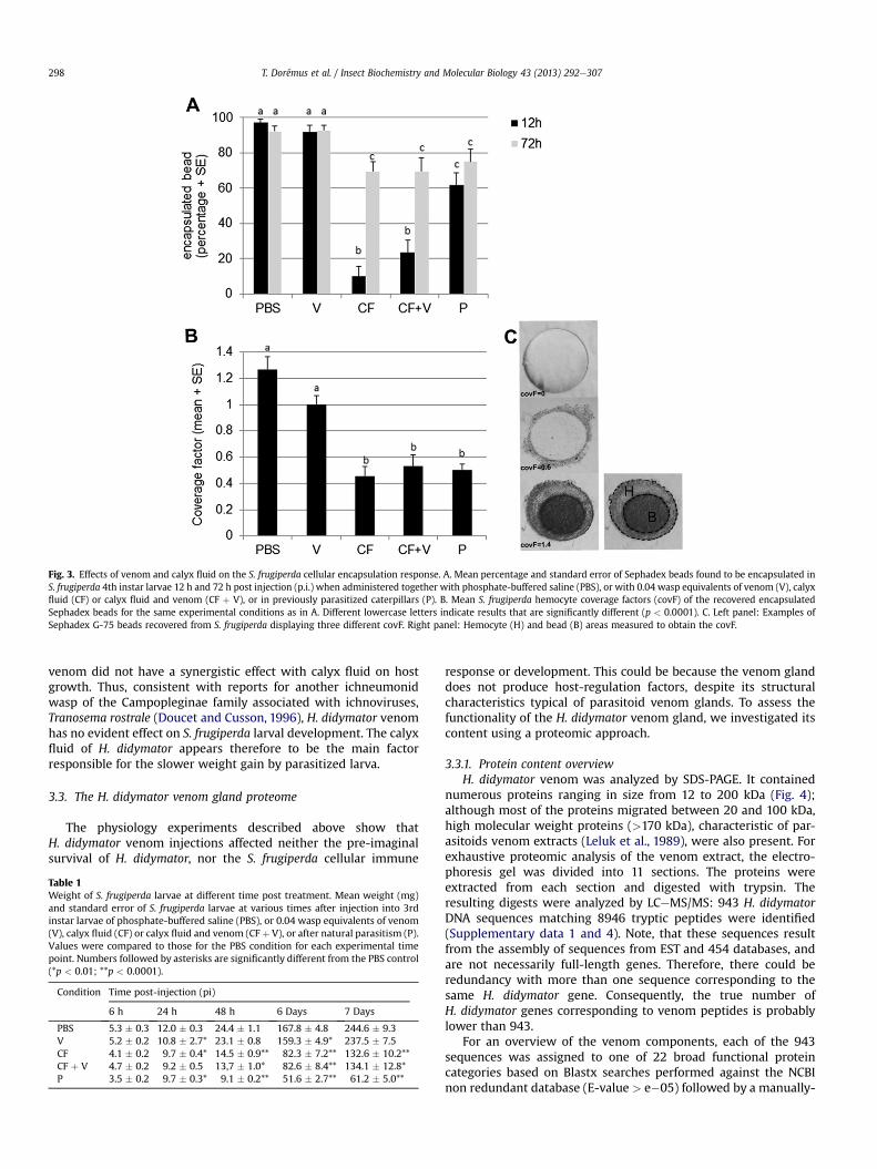
Fig. 3. Effects of venom and calyx fluid on the S. frugiperda cellular encapsulation response. A. Mean percentage and standard error of Sephadex beads found to be encapsulated inS. frugiperda 4th instar larvae 12 h and 72 h post injection (p.i.) when administered together with phosphate-buffered saline (PBS), or with 0.04 wasp equivalents of venom (V), calyxfluid (CF) or calyx fluid and venom (CF þ V), or in previously parasitized caterpillars (P). B. Mean S. frugiperda hemocyte coverage factors (covF) of the recovered encapsulatedSephadex beads for the same experimental conditions as in A. Different lowercase letters indicate results that are significantly different (p < 0.0001). C. Left panel: Examples ofSephadex G-75 beads recovered from S. frugiperda displaying three different covF. Right panel: Hemocyte (H) and bead (B) areas measured to obtain the covF.
T. Dorémus et al. / Insect Biochemistry and Molecular Biology 43 (2013) 292e307298
venom did not have a synergistic effect with calyx fluid on hostgrowth. Thus, consistent with reports for another ichneumonidwasp of the Campopleginae family associated with ichnoviruses,Tranosema rostrale (Doucet and Cusson, 1996), H. didymator venomhas no evident effect on S. frugiperda larval development. The calyxfluid of H. didymator appears therefore to be the main factorresponsible for the slower weight gain by parasitized larva.
3.3. The H. didymator venom gland proteome
The physiology experiments described above show thatH. didymator venom injections affected neither the pre-imaginalsurvival of H. didymator, nor the S. frugiperda cellular immune
Table 1Weight of S. frugiperda larvae at different time post treatment. Mean weight (mg)and standard error of S. frugiperda larvae at various times after injection into 3rdinstar larvae of phosphate-buffered saline (PBS), or 0.04 wasp equivalents of venom(V), calyx fluid (CF) or calyx fluid and venom (CF þ V), or after natural parasitism (P).Values were compared to those for the PBS condition for each experimental timepoint. Numbers followed by asterisks are significantly different from the PBS control(*p < 0.01; **p < 0.0001).
Condition Time post-injection (pi)
6 h 24 h 48 h 6 Days 7 Days
PBS 5.3 � 0.3 12.0 � 0.3 24.4 � 1.1 167.8 � 4.8 244.6 � 9.3V 5.2 � 0.2 10.8 � 2.7* 23.1 � 0.8 159.3 � 4.9* 237.5 � 7.5CF 4.1 � 0.2 9.7 � 0.4* 14.5 � 0.9** 82.3 � 7.2** 132.6 � 10.2**CF þ V 4.7 � 0.2 9.2 � 0.5 13,7 � 1.0* 82.6 � 8.4** 134.1 � 12.8*P 3.5 � 0.2 9.7 � 0.3* 9.1 � 0.2** 51.6 � 2.7** 61.2 � 5.0**
response or development. This could be because the venom glanddoes not produce host-regulation factors, despite its structuralcharacteristics typical of parasitoid venom glands. To assess thefunctionality of the H. didymator venom gland, we investigated itscontent using a proteomic approach.
3.3.1. Protein content overviewH. didymator venom was analyzed by SDS-PAGE. It contained
numerous proteins ranging in size from 12 to 200 kDa (Fig. 4);although most of the proteins migrated between 20 and 100 kDa,high molecular weight proteins (>170 kDa), characteristic of par-asitoids venom extracts (Leluk et al., 1989), were also present. Forexhaustive proteomic analysis of the venom extract, the electro-phoresis gel was divided into 11 sections. The proteins wereextracted from each section and digested with trypsin. Theresulting digests were analyzed by LCeMS/MS: 943 H. didymatorDNA sequences matching 8946 tryptic peptides were identified(Supplementary data 1 and 4). Note, that these sequences resultfrom the assembly of sequences from EST and 454 databases, andare not necessarily full-length genes. Therefore, there could beredundancy with more than one sequence corresponding to thesame H. didymator gene. Consequently, the true number ofH. didymator genes corresponding to venom peptides is probablylower than 943.
For an overview of the venom components, each of the 943sequences was assigned to one of 22 broad functional proteincategories based on Blastx searches performed against the NCBInon redundant database (E-value > e�05) followed by a manually-

Fig. 4. SDS-PAGE profile of H. didymator venom extract. Proteins collected from 10venom reservoirs were separated by SDS-PAGE (lane V) and the gel cut into 11 largesections numbered 1 to 11. Tryptic peptides extracted from each section were sub-jected to nano-LCeMS/MS analysis. Some of the proteins identified are indicated,including the five proteins encoded by the genes of the Hd-VenA family and Hd-VenB.Equivalent lane from the same gel (15% SDS-PAGE) showing the molecular massmarkers is on the left (M), stained with Coomassie Brillant blue R-250.
T. Dorémus et al. / Insect Biochemistry and Molecular Biology 43 (2013) 292e307 299
complemented automated annotation (Fig. 5). Matches were foundfor 925 of the 943 sequences, mainly (93%) with hymenopteranproteins (Supplementary data 1). According to this classification,the H. didymator venom extract contained a large number of en-zymes, particularly hydrolases, oxidoreductases and transferases.
Fig. 5. Distribution into functional classes of the sequences matching with venom extracgenerated by nano-LCeMS/MS to H. didymator transcriptomes - was manually assigned to asequences and the corresponding total number of peptides and unique peptides are indica
Hydrolases are often reported in venoms (examples and referencesin Supplementary data 2), including those of ichneumonid wasps.For instance, hydrolase activity has been detected using enzymaticsemi-quantitative colorimetric analysis in the venom of the pupalendoparasitoid, Pimpla hypochondriaca (Dani et al., 2005).H. didymator venom contained a few protease inhibitors, a class ofproteins also frequent in parasitic and social wasp venoms (Colinetet al., 2009; dos Santos et al., 2010; Yamamoto et al., 2007). TheH. didymator venom extract also contained a large number ofsubstrate-specific transporters and storage proteins (e.g. hexam-erin, apolipophorins found in the high molecular weight section ofthe gel, Fig. 4), proteins that are not commonly found in venoms.However, as for other abundant classes such as mitochondrial, ri-bosomal, chaperone and structural proteins, and proteins involvedin cell trafficking (Fig. 5), it is unclear whether they are indeed truevenom components.
This global analysis of the venom content revealed the presenceof two categories of proteins which we examined in more detail: (i)a set of proteins not previously described in parasitoid venoms thatwere encoded by genes strongly transcribed in H. didymator venomapparatus and (ii) a set of proteins similar to proteins found in thevenom of other parasitoid species that have host regulatoryfunctions.
3.3.2. Newly discovered H. didymator venom proteinsAmong the DNA sequences matching peptides described by the
LCeMS/MS analyses, 17% had no similarity with other proteins ingeneralist databases (Fig. 5). In particular, five sequences corre-sponded to transcripts that were abundant in the venom EST library(2588 sequences of the 2994 of the EST library, 86%).
3.3.2.1. Gene sequence analyses. Sequencing following PCR ampli-fication of genomic DNA identified these ESTs as corresponding tofour distinct genes: three belong to a multigenic family named Hd-VenA; the fourth gene was named Hd-VenB.
The three members of the Hd-VenA multigenic family werenamed Hd-VenA1 (GenBank accession # BankIt1561820), Hd-VenA2
t peptides. Each of the 943 contig sequences - identified by comparing the peptidessingle functional class. For each of the 22 predefined classes, the total number of contigted.
T. Dorémus et al. / Insect Biochemistry and Molecular Biology 43 (2013) 292e307300
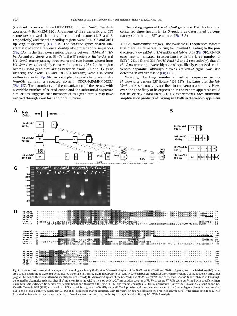
(GenBank accession # BankIt1561824) and Hd-VenA3 (GenBankaccession # BankIt1561826). Alignment of their genomic and ESTsequences showed that they all contained introns (1, 3 and 6,respectively) and that their coding regions were 342, 935 and 2164bp long, respectively (Fig. 6 A). The Hd-VenA genes shared sub-stantial nucleotide sequence identity along their entire sequences(Fig. 6A). In the first exon region, identity between Hd-VenA1, Hd-VenA2 and Hd-VenA3 was 67e73%; the 30-region of Hd-VenA2 andHd-VenA3, encompassing three exons and two introns, absent fromHd-VenA1, was also highly conserved (identity >76% for the regionoverall). Intra-gene similarities between exons 3.3 and 3.7 (94%identity) and exons 3.6 and 3.8 (63% identity) were also foundwithin Hd-VenA3 (Fig. 6A). Accordingly, the predicted protein, Hd-VenA3b, contains a repeated domain “RKGRNAEAMNMDRER”(Fig. 6D). The complexity of the organization of the genes, witha variable number of related exons and the substantial sequencesimilarities, suggests that members of this gene family may haveevolved through exon loss and/or duplication.
Fig. 6. Sequence and transcription analyses of the multigenic family Hd-VenA. A. Schematic dstop codon. Exons are represented by numbered boxes and introns by plain lines. Percent o(regions for which there is less than 5% identity are not labeled). B. Schematic diagram of thegenerated by alternative splicing; sizes (bp) are given from the ATG to the stop codon. C. Trusing total RNA extracted from dissected female heads and thoraxes (HT), ovaries (OV) anVenA3b. Genomic DNA (DNA) was used as a PCR control. D. Alignment of H. didymator Hd-EST1a and b) and Campoletis sonorensis EST (Cs-EST1) sequences sharing similarity with HdRepeated amino acid sequences are underlined. Boxed sequences correspond to the tryptic
The coding region of the Hd-VenB gene was 1194 bp long andcontained three introns in its 50-region, as determined by com-paring genomic and EST sequences (Fig. 7 A).
3.3.2.2. Transcription profiles. The available EST sequences indicatethat there is alternative splicing for Hd-VenA3, leading to the pro-duction of twomRNAs: Hd-VenA3a and Hd-VenA3b (Fig. 6B). RT-PCRexperiments indicated, in accordance with the large number ofESTs (1713, 413 and 331 for Hd-VenA1, 2 and 3 respectively), that allHd-VenA transcripts were highly and specifically expressed in thevenom apparatus, although a weak Hd-VenA2 signal was alsodetected in ovarian tissue (Fig. 6C).
Similarly, the large number of related sequences in theH. didymator venom EST library (131 ESTs) indicates that the Hd-VenB gene is strongly transcribed in the venom apparatus. How-ever, the specificity of its expression in the venom apparatus couldnot be clearly established: RT-PCR experiments gave numerousamplification products of varying size both in the venom apparatus
iagram of the Hd-VenA1, Hd-VenA2 and Hd-VenA3 genes, from the initiation (ATG) to thef identity between paired sequences are given for regions sharing sequence similaritiesHd-VenA1 and Hd-VenA2 mRNAs and of the two Hd-VenA3a and Hd-VenA3b transcripts
anscription patterns of Hd-VenA genes: RT-PCRs were performed with specific primersd venom apparatus (V) for four transcripts: Hd-VenA1, Hd-VenA2, Hd-VenA3a and Hd-VenA proteins and translated sequences of the Campopleginae Venturia canescens (Vc--VenA. An asterisk indicates the predicted cleavage site of the signal peptide sequence.peptides identified by LCeMS/MS analysis.

Fig. 7. Sequence and transcription analyses of the Hd-VenB gene. A. Schematic diagram of Hd-VenB, from the initiation (ATG) to the stop codon. Exons are represented by numberedboxes and introns by plain lines. B. Schematic diagram of the Hd-VenB mRNA; size (bp) is given from the ATG to the stop codon. C. Transcription patterns of Hd-VenB: RT-PCR wasperformed with specific primers as in Fig. 4. Genomic DNA (DNA) was used as a PCR control. The expected amplification products are indicated with arrows. D. Alignment ofH. didymator Hd-VenB proteins and translated sequences of the Campopleginae Venturia canescens venom EST sequence (Vc-EST2) sharing similarity with Hd-VenB. An asteriskindicates the predicted cleavage site of the signal peptide sequence. Repeated amino acid sequences are underlined. Boxed sequences correspond to the tryptic peptides identifiedby LCeMS/MS analysis.
T. Dorémus et al. / Insect Biochemistry and Molecular Biology 43 (2013) 292e307 301
and in head and thorax (Fig. 7C). Further work is required to elu-cidate this presence of diverse amplification products.
3.3.2.3. Characteristics of Hd-VenA and B proteins. In agreementwith the predicted presence of a peptide signal (Figs. 6D and 7D),mass spectrometry analysis detected the corresponding proteins inthe venom extract (Supplementary data 1). Hd-VenA are proteins ofpredicted low molecular weight, 8e18 kDa, consistent with thesections of the SDS-PAGE gel from which they were identified(Fig. 4). The predicted Hd-VenB protein is 310 amino-acids long(34 kDa), in agreement with the detection of most of the peptidescorresponding to the protein in Section 4 of the SDS-PAGE gel(Fig. 4). The protein sequence contains a putative signal peptide,and 10 direct repeats of 20 amino acids, as predicted by RADARsoftware (http://www.ebi.ac.uk/Tools/Radar) (Fig. 7D). None of theproteins displayed a domain conserved in sequences present ingeneralist databases.
Although they encode proteins predicted to be secreted andfound in H. didymator venom extract, only a small number ofpeptides (4e18 peptides) matched with these proteins. In addition,no major bands in the molecular weight range corresponding tothese proteins were observed in the electrophoretic profile of thevenom (Fig. 4). Therefore, Hd-VenA and Hd-VenB may not beabundant in H. didymator venom. On the other hand, they may be
unstable or be processed/degraded very rapidly and thus difficult todetect using the above techniques.
3.3.2.4. Proteins conserved in Campopleginae wasps. Although Hd-VenA and Hd-VenB proteins have not been previously reported toNCBI/EMBL databases, sequences encoding similar proteins werefound in EST libraries from two Campopleginae species, C. sonor-ensis (whole body mixed male and female EST library) and Venturiacanescens (venom gland EST library, private library).
Alignment of H. didymator Hd-VenA protein sequences show30e45% amino acid sequence identity with translated C. sonorensis(CsEST1, LIBEST_026437) and V. canescens (VcEST1a and VcEST1b,GenBank accession # KC012591 and KC012592 respectively) ESTsequences. Two domains were found to be conserved in all Hd-VenA proteins and also CsEST1 and VcEST1a proteins: a “FGIIFxx-xAxVFAxxxxxxxEEAEA” motif in the N-terminal region, anda “RLGRSADPSMKKxxEMIKAR” motif in the C-terminal region(Fig. 6D).
Like Hd-VenA genes, Hd-VenB-related sequence was identifiedin the venom EST library from V. canescens (VcEST2, GenBankaccession # KC012593), suggesting that this gene is also conservedin Campopleginae species and probably transcribed in the venomapparatus (97 of 3085 ESTs in the V. canescens library). TheV. canescens translated sequence showed 53% amino acid identity

T. Dorémus et al. / Insect Biochemistry and Molecular Biology 43 (2013) 292e307302
with the H. didymator protein. However, VcEST2 contained onlyfour of the ten repeats present in Hd-VenB (Fig. 7D).
Thus these genes appear to be conserved among Campopleginaewasps associated either with PDVs or VLPs, and are expressed in thevenom apparatus of these phylogenetically closely related species.The presence of the gene products in C. sonorensis and V. canescensvenom extracts has not yet been demonstrated, but at least forV. canescens, the genes are strongly transcribed in the venom gland(1078 out of 3085 ESTs for VcEST1a and b). The function of theseproteins in the venom apparatus or in the host/parasitoid inter-action is unknown.
3.3.3. Proteins previously described in the venom of otherparasitoid species
Some of the 925 H. didymator sequences identified by the pro-teomic analysis (Table 2) share similarity with proteins identified inthe venom of other parasitoid species, including some implicated inaltering the host phenotype. We examined this group to determinewhether they represent functional venom proteins.
3.3.3.1. Potentially functional hydrolases. Although their function inhost/parasitoid interactions is largely uncharacterized, hydrolasesare common in venoms frommany organisms (Vincent et al., 2010).H. didymator venom extract contained proteins (Hd-Ven1 and Hd-Ven2) similar to disintegrin and metalloproteinase with thrombo-spondin motifs (ADAMTS) from the reprolysin family, and proteins(Hd-Ven21 and Hd-Ven22) similar to dipeptidyl peptidase IV(DDPIV). Both corresponded to a large number of peptides identi-fied in the proteomic analysis (Table 2), suggesting they wereabundant in the H. didymator venom.
Metalloproteases of the reprolysin family have been found inmany parasitoid venoms (Table 2) and one acts as a host develop-ment alteration factor (EpMP3) in the interaction betweenE. pennicornis and its host L. oleracea (Price et al., 2009). VenomousADAMTS proteins are also commonly found in snakes or in socialstinging wasps where they are major active components respon-sible for tissue damage (degradation of the extracellular matrix,bleeding or inflammation reaction) (de Graaf et al., 2010a; dosSantos et al., 2010). H. didymator Hd-Ven1 and Hd-Ven2 proteinsshowed low similarity to Nasonia vitripennis ADAMTS-16 (respec-tively 30% and 23% identity; Supplementary data 5, Fig. S1). It is notknown whether these sequences correspond to a single gene,because they were not complete and correspond to the N-ter (Hd-Ven1) and the C-ter (Hd-Ven2) parts, respectively, of theN. vitripennis sequence (Supplementary data 5, Fig. S1). TheH. didymator reprolysin(s)-like protein migrated in the section 5 ofthe SDS-PAGE (Fig. 4), deduced from the number of correspondingtryptic peptides, and this is coherent with the reported size of theP. hypochondriaca (39.9 kDa) reprolysin (Parkinson et al., 2002a).The available H. didymator sequences lacked the N-terminal regionso we do not know whether it has a signal peptide and cannottherefore predict whether it is secreted. However, Hd-Ven2 con-tains the conserved catalytic domain HEXXHXUGUXH suggestingthat it may be a functional zinc metallopeptidase (Supplementarydata 5, Fig. S1).
DDPIVs are membrane-anchored enzymes, sometimes asso-ciated with exosome membranes, and which cleave N-terminaldipeptides. They are found in the venom of snakes and stingingwasps and were recently reported in N. vitripennis (Table 2). Theirfunction in venom remains unclear andmay be limited to activationor inactivation of biomolecules within the venom reservoir(Mentlein, 1999) (Ogawa et al., 2008). For instance, in Apis mellifera,a membrane-anchored DDPIV activates pro-melittin into highlytoxic melittin in the venom reservoir (Kreil et al., 1980). DDPIVsmay thus be, as suggested by (de Graaf et al., 2010a), “venom trace
elements”. The H. didymator Hd-Ven21 and Hd-Ven22 were similarto DPPIV from the venoms of N. vitripennis (26% and 49% identity,respectively) and Vespa basalis (34% and 60% identity, respectively)(Supplementary data 5, Fig. S2). Hd-Ven21 covered the N-terminalregion of the enzyme and Hd-Ven22 the C-terminal region makingimpossible to determine how many genes code for DDPIV in theH. didymator venom gland. The corresponding proteins wererecovered from section 8 of the SDS-PAGE (around 80 kDa, Fig. 4),consistent with the sizes of other venom DPPIVs (86.8 and 88 kDafor venom DPPIV isoform 1 of N. vitripennis and V. basalisrespectively).
Although less abundant in H. didymator venom, as indicated bythe number of corresponding peptides, one sequence, Hd-Ven43,shared 50% and 45% identity with serine proteases in the venomsof the stinging wasps Apis florea and Bombus ardens, respectively(Supplementary data 5, Fig. S3). The Bombus ardens enzyme isa trypsin-like CLIP-domain serine protease, a family involved bothin the activation of the Toll pathway that regulates dorsoeventralaxis formation and innate immunity, as well as in the PO signal-ing pathway (Jang et al., 2008). The Hd-Ven43 sequence also shared27% identity with Cotesia rubecula Vn-50, a serine protease ho-molog which suppresses the humoral response in the host Pierisrapae by competing with the host serine protease involved in thephenoloxidase (PO) activation cascade (Asgari, 2012). TheH. didymator sequence contains part of the catalytic triad suggest-ing that it may be an active serine protease (Supplementary data 5,Fig. S3).
A number of esterases and glycosylases were also identified inH. didymator venom (Fig. 5). Proteomic analyses identified threesequences, Hd-Ven73, Hd-Ven74 and HdVen75, with similaritieswith the venom carboxylesterase-6 described in N. vitripennis (40,47 and 73% identity, respectively) (Supplementary data 5, Fig. S4).All H. didymator carboxylesterase-like sequences contain the cat-alytic triad (residues SeEeH) characteristic of the esterase-lipasefamily, suggesting that the enzymes are functional. Hd-Ven83shows similarities with a hyaluronidase in the venom of thesolitary vespid wasp Anoplius samariensis (56% similarity)(Supplementary data 5, Fig. S5) and was found in section 5 of theSDS-PAGE (Fig. 4). Hyaluronidases are enzymes that catalyze thehydrolysis of hyaluronic acid and other constituents of theextracellular matrix of vertebrates. They are common in thevenoms of snakes (Kemparaju and Girish, 2006), honeybees,wasps and hornets (Schmidt et al., 1986). In parasitoids, hyal-uronidase activity has only previously been described in thevenom of Eupelmus orientalis, an ectoparasitoid of bruchid insects(Doury et al., 1997). Otherwise, an EST sequence coding for a par-tial hyaluronidase lacking a signal peptide was reported inC. inanitus (Vincent et al., 2010). The H. didymator sequence wereport covers the N-terminal region of the protein and containsa signal peptide (Supplementary data 5, Fig. S5) suggesting thatthis endoparasitoid enzyme is secreted, like classical wasp venomhyaluronidases.
This analysis of the sequences available suggests that thesevarious enzymes found in H. didymator venom extract may befunctional hydrolases that could potentially be injected into thehost at oviposition and act on the physiology of the host caterpillar.
3.3.3.2. Potentially active serine proteases inhibitors. H. didymatorvenom contained serine protease inhibitors (serpins). Their presumedrole in venom is to interact with one or several serine proteases thatactivate the PO cascade; they thereby may inhibit PO activation, asdemonstrated for a serpin (LbSPNy) which is one of the most abun-dant proteins in the venom of a strain of the figitid endoparasitoidLeptopilina boulardi (Colinet et al., 2009). Peptides from H. didymatorvenom matched a number of sequences displaying similarities with

Table 2List of selected proteins identified by nano-LC-MS/MS inH. didymator venom extract that share similarities with proteins reported in venoms of others organisms. Predicted proteins are classified according to the nomenclature defined inFig. 5. For each sequence, the amino acid length, prediction of any signal peptide (using SignalP4 software), the total number of peptides, the number of unique peptides, the peptide redundancy (total/unique), the gel section fromwhichmajority of the peptides were obtained and the number of H. didymator-related venom EST sequences are indicated. Species fromwhich venom components are reported are: Apis florae (Af); Apis mellifera (Am); Anoplus samariensi (As);Asobara japonica (Aj); Bracon hebetor (Bh); Cyphononyx dorsalis (Cd); Chelonus inanitus (Ci); Cotesia rubecula (Cr); Eulophus pennicornis (Ep1); Eumenes pomiformis (Ep2); Eupelmus orentalis (Eo); Leptopilina boulardi (Lb); Microctonusaethiopoides (Ma); Microctonus hyperodae (Mh); Nasonia vitripennis (Nv); Orancistrocerus drewseni (Od); Pimpla hypochondriaca (Ph); Polybia paulista (Pp1); Pteromalus puparum (Pp2); and Vespid basalis (Vb). Cc, Ci and Cr are braconidswhich have associated polydnavirus; Ma and Mh carry virus-like particles. “Lep.” corresponds to venomous proteins identified in urticating hairs and ”cone snail” to proteins identified in the venom bulb of the respective organisms.Asterisks (*) indicate that only the transcript has been identified in a venom gland EST library; double asterisks (**) indicate that only the activity has been detected in crude venom by enzymatic tests or antibody-inhibition experiments;symbols (x) indicate that data originates from automatic genome annotation. (1) Abt and Rivers, 2007. (2) Asgari et al., 2003b. (3) Baek and Lee, 2010. (4) Birrell et al., 2007. (5) Cardoso et al., 2010. (6) Carrijo-Carvalho and Chudzinski-Tavassi, 2007. (7) Colinet et al., 2009. (8) Colinet et al., 2011. (9) Crawford et al., 2008. (10) de Graaf et al., 2010a. (11) de Graaf et al., 2010b. (12) dos Santos et al., 2010. (13) dos Santos et al., 2011. (14) Doury et al., 1997. (15) Kemparaju andGirish, 2006. (16) Lee et al., 2007. (17) Mabiala-Moundoungou, 2009. (18) Mentlein et al., 1999. (19) Padavattan et al., 2008. (20) Parkinson et al., 2001. (21) Parkinson et al., 2002a. (22) Peiren et al., 2005. (23) Peiren et al., 2006. (24) Peirenet al., 2008. (25) Price et al., 2009. (26) Rivers et al., 2009. (27) Safavi-Hemami et al., 2010. (28) Schevchenko et al., 2005. (29) Schmidt et al., 1986. (30) Undheim and King, 2011. (31) Veiga et al., 2005. (32) Vincent et al., 2010. (33)Wright etal., 1973. (34) Yamamoto et al., 2007. (35) Zhang et al., 2006. (36) Zhu et al., 2010.
Protein or family previously identified in
Functional class ID Description aa lengh Sequencealignmentsin suppl.data 5
Peptidesignalprediction
Totalpeptides
Uniquespeptides
Peptideredundancy
Gelsection
EST Ichneu-monids
Braco-nids
Otherparasiticwasps
Paralyticwasps
Otherhymenoptera
Othersorganisms
References
Metalloproteases Hd-Ven1 Disintegrinandmetalloproteinasewith thrombo-spondinmotifs
152 Fig. S1 No N-ter 13 7 1.9 5 5 Ph Ci, Ma,Mh
Ep1, Nv Ep2, Od Am, Pp1 Centipedes,snakes
3, 9, 10, 12,13, 21, 25, 30, 32Hd-ven2 78 No N-ter 44 2 22.0 5 1
Dipeptidylpeptidases
Hd-Ven21 Dipeptidyl-peptidase IV
148 Fig. S2 No N-ter 23 5 4.6 8 e e e Nv e Vb, Am Snakes 5, 10, 16, 18Hd-ven22 424 No N-ter 63 10 6.3 8 e
Serineendopetidases
Hd-Ven43 Venomserineprotease
219 Fig. S3 No N-ter 3 2 1.5 6 e Ph Ci, Cr Nv, Pp2 Ep2, Od Afx, Bax,Am, Pp1,Af
Centipedes,Lep.
2, 3, 10, 12, 13,21, 25, 30, 31,32, 36
Esterases Hd-Ven73 Venomcarboxylesterase-6
542 Fig. S4 Yes 7 4 1.8 8 e e e Nv e e e 10Hd-Ven74 57 No N-ter 3 3 1.0 8 e
Hd-Ven75 318 No N-ter 2 2 1.0 8 e
Glycosylases Hd-Ven83 Hyaluronidase 260 Fig. S5 Yes 14 5 2.8 5 1 e Ci* Eo** Asx, Ep2,Od
Am, Pp1,ants**
Centipedes,snakes,spiders**,Lep.
3, 5, 6, 14, 15,22, 24, 29, 30,32, 33, 36
Proteaseinhibitors
Hd-Ven390 Serine proteaseinhibitor
410 Fig. S6 Yes 58 13 4.5 5 e e e Lb e Pp1 Lep. 7, 12, 31
Oxidases Hd-Ven218 Super-oxidedismutase
219 Fig. S7 No 13 7 1.9 2 e e e Lb e Am (wholegland), Pp1
e 8, 12, 24Hd-Ven219 154 No 9 6 1.5 2 e
Hd-Ven204 Phenoloxidase 685 Fig. S8 No 41 21 2.0 8 e Ph e Nv** e e e 1, 20Transferases Hd-Ven236 Arginine kinase 179 Fig. S9 No 29 3 9.7 4 e e Aj Pp2 Cd, Ep2,
OdAm (wholegland), Pp1
Cone snail 3, 12, 17, 24,27, 34, 36Hd-Ven237 218 No N-ter 185 13 14.2 4 e
Immunerelated
Hd-Ven392 Calreticulin 402 Fig. S10 Yes 13 10 1.3 7 1 Ph** Cr, Aj,Ma, Mh
Nv, Pp2 e e Lep. 9, 10, 17, 26,28, 35, 36
Hd-Ven816 C1q-like venomprotein precursor
138 Fig. S11 No N-ter 4 3 1.3 3 e e e Nv e Am e 11
Otherfunctions
Hd-Ven831 C-type lectin 195 Fig. S12 No N-ter 6 4 1.5 3 e Ci Snakes,Lep.
4, 32, 31, 36
Venomallergens
Hd-Ven850 Venom allergen3-like (Ag5 family)
80 Fig. S13 No N-ter 45 2 22.5 3 4 e Ma, Mh,Ci*
Nv e Pp1, Ant,Vm
e 9, 10, 12, 19,23, 32
Hd-Ven841 Icarapin-like 250 Fig. S14 Yes 5 2 2.5 3 e e Ma, Mh e e Am (wholegland)
e 9, 23
T. Dorémus et al. / Insect Biochemistry and Molecular Biology 43 (2013) 292e307304
serpins, including an Antithrombin-III from the ant Camponotus flor-idanus (Hd-Ven383) and a leukocyte elastase inhibitor-like proteinfrom Bombus impatiens (Hd-Ven384) (Supplementary data 1). How-ever, only one of these serpin-like sequences, Hd-Ven390, containsa predicted putative functional reactive center loop (RCL). This proteinwas well represented in H. didymator venom extract (Table 2) andshares similarities with a neuroserpin-like protein in the bee Micro-ctonus rotundata and with the LbSPNy protein (50% and 30% identity,respectively) (Supplementary data 5, Fig. S6). The deduced size of Hd-Ven390 was 45 kDa (42 kDa after peptide signal cleavage), consistentwith the recovery of most of the tryptic peptides from the section 5 ofthe SDS-PAGE gel (Fig. 4). Preliminary data suggest that H. didymatorwhole venom extract does not prevent PO activation (data notshown), so role, if any, remains unclear. Hd-Ven390may indeed be anactive secreted venom serpin but which acts inside the venomapparatus rather than as a true virulence factor, as suggested fora venom serpin from the social wasp Polybia paulista (dos Santos et al.,2010).
3.3.3.3. Other enzymes. Superoxide dismutase (SOD) is an oxidor-eductase found in many wasp venoms (dos Santos et al., 2010;Peiren et al., 2008). In many cases, an intracellular form of the SODis detected, suggesting that the protein may be a cellular con-taminant or act on the maintenance of the venom apparatus tissue(Peiren et al., 2008). One exception is the extracellular SOD abun-dant in the venom gland of the parasitic wasp L. boulardi that in-hibits PO activity in vitro (Colinet et al., 2011). The two SODsidentified in H. didymator venom (Hd-Ven218 and Hd-Ven219;Table 2) display substantial similarities (more than 80% identity)with intracellular SODs of Asobara tabida and L. boulardi andweaker similarities (33 and 50%, respectively) with L. boulardiextracellular SOD (Supplementary data 5, FigS7). The H. didymatorsequences do not contain predicted signal peptides and thus mostprobably correspond to intracellular SODs.
H. didymator venom also contains a proPO, the inactive zymogenform of PO (Hd-Ven204; Table 2). A PO containing a peptide signalallowing secretion of the enzyme has been described inP. hypochondriaca venom (Parkinson et al., 2001). No peptide signalwas predicted for the H. didymator protein which shares somesimilarity (45% identity) with the secreted P. hypochondriaca PO andmore similarity (71% identity) with A. mellifera proPO(Supplementary data 5, Fig. S8). Hd-Ven204 is therefore probablya classical cellular enzyme released by regulated exocytosis asdescribed for PO found in hemocytes of many arthropods (Lai et al.,2002).
Another abundant enzyme in H. didymator venom, as evaluatedfrom the number of peptides, was an arginine kinase highly similar(>70% identity) to other hymenopteran sequences (Table 2;Supplementary data 5, Fig. S9). Arginine kinase has been reportedin the venom of several hymenopteran insects but there is only onereport of a paralytic effect of this type of enzyme: a truncated(25 kDa) form in the spider-hunting wasp Cyphononyx dorsalis(Yamamoto et al., 2007). The arginine kinase identified inH. didymator was found in section 4 of the SDS-PAGE (around 30e40 kDa, Fig. 4) and was thus most probably a full-length enzymeand likely to be intracellular.
Therefore, according to the analysis of the available sequences,the three enzymes described above appear to be intracellular en-zymes and probably are not injected into the insect host.
3.3.3.4. Non enzymatic proteins. Diverse non enzymatic proteins, asfound in the venom of various parasitic wasps, were also identifiedin H. didymator venom. These proteins include immune-relatedproteins, such as calreticulin and a C1q-like venom protein, a C-type lectin and venom allergen proteins (Table 2).
Calreticulin (CRT) is a multifunctional Ca2þ-binding chaperoneprotein found in the venom fluid of the parasitic wasps C. rubecula,P. puparum and N. vitripennis (Table 2). In C. rubecula, CRT preventsencapsulation in vitro by inhibiting hemocyte spreading behavior,although the mechanism is still unclear (Zhang et al., 2006). Onestudy on ichneumonidae P. hypochondriaca venom demonstrateda hemocyte anti-aggregation effect of the venom CRT (Rivers et al.,2009). A CRT-like protein, Hd-Ven392, highly similar (more than70% identity) to other parasitic wasp CRTs was found inH. didymator venom (Supplementary data 5, Fig. S10). The numberof corresponding peptides was low (Table 2) and although this CRTappears to be secreted and functional, the quantity present inH. didymator venommay be insufficient to cause a significant effectonce injected into the host caterpillar.
The H. didymator venom extract also contained a protein withsimilarities to the Tumor Necrosis Factor superfamily member C1q(Hd-Ven816), a subunit of the C1 enzyme complex that activatesserum complement. A C1q-like venom protein precursor has beenidentified in N. vitripennis and A. mellifera (61 and 57% identity,respectively, with Hd-Ven816; Supplementary data 5, Fig. S11) (deGraaf et al., 2010b). In N. vitripennis, the protein is also present ina number of tissues other than the venom gland and it was sug-gested that it is not a true virulence factor (de Graaf et al., 2010a).CRT blocks the interaction between C1q and immunoglobulins inhumans, so the two proteins may interact in the venom glandwhere they are both found, such as suggested for N. vitripennis(Danneels et al., 2010).
H. didymator venom also included a protein with a C-type lectin(CTL) domain (Hd-Ven831). Venomous CTLs have only beendescribed in braconid wasps carrying PDV as C. inanitus (Vincentet al., 2010) where the protein is believed to play a role inimmune-suppression, in the same way as proteins with a CTLdomain encoded by the PDV associated with the braconid Cotesiaplutellae (Lee et al., 2008). The H. didymator sequence is dissimilarto the C. inanitus venom CTL protein (29% identity) (Supplementarydata 5, Fig. S12), but resembles other non-venom specific secretedhymenopteran CTLs (89% identify with the CTL protein from Acro-myrmex echinatior).
Finally an abundant protein in H. didymator venom, Hd-Ven850,shares similarities with venom allergen-3 (or Antigen 5), a proteinof the Antigen 5 family (Ag5) with a cysteine-rich secretorydomain. This type of protein, commonly found in Hymenopteravenoms, has been described in ants (Hoffman, 2006) and in severalparasitic wasps (Table 2). Antigen 5-like proteins generally havea signal peptide for secretion (Padavattan et al., 2008) but theirbiological function is still not known. The available deducedH. didymator sequence (80 aa) encompasses the C-terminal regionof the protein and shares 25% identity with other allergen-3 se-quences (Supplementary data 5, Fig. S13). Its predicted size of20 kDamost probably corresponds to themajor band in Section 3 ofthe SDS-PAGE (Fig. 4). In the allergen category, we also identified anicarapin-like peptide, Hd-Ven841, a novel IgE-binding bee venomprotein of unknown biological function (Peiren et al., 2006). Theavailable sequence has a signal peptide, typical of secreted proteins(Supplementary data 5, Fig. S14).
4. Conclusion
This first exhaustive analysis of a Campopleginae parasitic waspvenom proteome, that of H. didymator, reveals the presence ofseveral components related to venomous proteins. This is inapparent contrast with the observation that injection of venomextract does not have any detectable effect on development orcellular immunity of the host, S. frugiperda. There are various pos-sible reasons for the venom having no apparent effect on host

T. Dorémus et al. / Insect Biochemistry and Molecular Biology 43 (2013) 292e307 305
physiology. Firstly, many proteins that were identified inH. didymator venom are not commonly major components of activevenoms of koinobiont species, although they are often reported inthese species. The toxicity of many of these molecules, such asmetalloproteases, arginine kinase or hyaluronidase, has only beendemonstrated in stinging wasps, and most of them are allergeninducers in mammals (Crawford et al., 2008; de Graaf et al., 2010a;dos Santos et al., 2011; dos Santos et al., 2010; Vincent et al., 2010).Secondly, most of the POs, serpins, CRT and SODs, molecules pre-viously described in other parasitoid species and shown to bedeleterious for host development or immunity, appear to be eithernot secreted or present at only low concentrations in H. didymatorvenom (as estimated from the number of EST sequences or trypticpeptides). A third possibility is that venom components may havedifferent activities in different lepidopteran host species. Indeed,H. didymator naturally parasitizes several noctuid species and wetested the venom extracts only on S. frugiperda. It may therefore beinformative to assess the effect of this venom on several hostspecies, including for exampleHelicoverpa armigera and Autographagamma, and thereby investigate whether the venom may beinvolved in the determination of H. didymator’s host range. Finally,only two physiological traits were explored in this work. Althoughthey are bothmajor processes affected in insect hosts parasitized bykoinobiont species and are important for successful parasitism,other processes may be affected by factors in the parasite’s venom.Other physiological systems such as metabolism, digestion, thenervous system and behavior have to be investigated before it canbe concluded that H. didymator venom makes no contribution toparasitism.
Our proteomic analysis led to the identification of four previ-ously undescribed secreted proteins, three of them being closelyrelated. The corresponding transcripts were abundant in the venomtissue and most were only found in this tissue, strongly suggestingthat they constitute specific venom proteins. The function of theseproteins remains however unclear. Interestingly, related sequenceswere found in other Campopleginae wasps, including V. canescens,a species devoid of PDVs but associated with VLPs; for this species,there is also no clear evidence of an effect of the venom on hostphysiology. Only a transient paralysis following venom injectionhas been reported (Piek, 1986). Similarly, for C. sonorensis, which isassociated with PDV and also contains homologs, no effect of thevenom on parasitism success was reported (Webb and Luckhart,1994). Further investigation is needed to understand functions ofthese proteins, which may be in the venom gland (e.g. venomstorage or tissue protection) rather than during parasitism.
Our study strongly suggests that there are substantial differ-ences in the evolution of venom components between the twofamilies associated with PDVs, the Braconids and the Ichneumo-nids. In H. didymator, calyx fluid alone was sufficient to reproducethe parasitized host phenotype, which is not the case for braconids,where the venom synergizes with PDV (Asgari, 2012). Preliminaryanalyses also indicated that H. didymator venom had no significanteffect on the transcription levels of four HdIV (P30, M24, Vank1803and VankCR1) genes (data not shown). Indeed, the extensiveanalysis ofH. didymator venom did not identify components relatedto proteins demonstrated to synergize with either the effects orexpression of PDV in Braconidae venom. Most Campopleginaespecies studied to date are endoparasitoids that rely on PDVs orVLPs produced in the ovarian tissues tomanipulate host physiology.In these species, “viral” host regulatory factors may have been ac-quired during evolution before specialization of the venom to havehost regulatory functions or viral acquisition may have been fol-lowed by the loss of any such function of the venom. Braconidsdomesticated a virus independently, and consequently PDVacquisition appears to have led to a different evolution of the
relationship between the PDV and the venom, with venom mole-cules required for the success of parasitism. The reasons why thevenom gland in Campopleginae species retains the capacity toproduce a large range of molecules remains to be elucidated. It ispossible that Campopleginae venom simply corresponds to a phy-logenetic signal and no longer has a role, but it may still have anancestral role in ovipositor lubrication or may have acquired newfunctions that have not been identified yet.
Many aspects of the evolution of the nature and function ofvenoms in parasitic wasps, and the consequences of the acquisitionof symbiotic PDV or VLP in ichneumonid wasps remain to be elu-cidated. Analysis of the venom proteomes in a range of species indifferent branches of the phylogenetic tree of the Ichneumonidaewould be informative. Ichneumonids display a great variety ofparasitoid lifestyles and there may have been multiple shift eventsbetween ecto- and endoparasitism (Gauld, 1988). Investigation ofthe venoms of koinobiont species from the Cremastinae, Anom-alinae and Ophioninae sub-families of Ichneumonids, which maynot carry PDV or VLP, may identify venomous factors required forparasitism success, and contribute to our understanding of Cam-popleginae venom evolution.
The evolution of venoms in other groups, such as reptiles whosevenoms only have a function in predation, is better characterized(Wuster, 2010) than that of venoms in Hymenoptera, and moreparticularly in parasitic wasps, with their enormous diversity of lifecycle strategies. Currently, only a few venomous proteins have beendemonstrated to play a role in the hosteparasitoid interaction.Furthermore, they generally have been described in only a smallnumber of genera, reflecting the limited exploration of parasitoidvenom diversity. Following on from the pioneer work based on thestudy of venom apparatus morphology (Robertson, 1968), andvenom protein and enzymatic profiles (Leluk et al., 1989; Schmidtet al., 1986), recent advances in the characterization of hymenop-tera venoms should, as illustrated by our study, provide a betterinsight into venom evolution in the context of the highly diverselife-styles of hymenoptera.
Acknowledgments
We thank M. Jambart, A. Ausset, C. Gibard and G. Clabots forproviding insects and/or reagents for this study. We also thank B.Duvic and M.A. Mannucci for preliminary PO activation assays andN. Cubedo for his technical advice. English language was revised byAlex Edelman & Associates, France. This work received supportfrom the Genoscope (“Parasitoid venoms” project) and from theFrench National Research Agency (Paratoxose project, #ANR-09-BLAN-0243-01).
Appendix A. Supplementary data
Supplementary data related to this article can be found at http://dx.doi.org/10.1016/j.ibmb.2012.12.010.
References
Abt, M., Rivers, D.B., 2007. Characterization of phenoloxidase activity in venom fromthe ectoparasitoid Nasonia vitripennis (Walker) (Hymenoptera: Pteromalidae).Journal of Invertebrate Pathology 94, 108e118.
Asgari, S., 2012. Venoms from endoparasitoids. In: Beckage, N.E., Drezen, J.M. (Eds.),Parasitoid Viruses Symbionts and Pathogens. Elsevier, pp. 217e230.
Asgari, S., Rivers, D.B., 2011. Venom proteins from endoparasitoid wasps and theirrole in host-parasite interactions. Annual Review of Entomology 56, 313e335.
Asgari, S., Zareie, R., Zhang, G., Schmidt, O., 2003a. Isolation and characterization ofa novel venom protein from an endoparasitoid, Cotesia rubecula (Hym: Braco-nidae). Archives of Insect Biochemistry and Physiology 53, 92e100.
Asgari, S., Zhang, G., Zareie, R., Schmidt, O., 2003b. A serine proteinase homologvenom protein from an endoparasitoid wasp inhibits melanization of the hosthemolymph. Insect Biochemistry and Molecular Biology 33, 1017e1024.

T. Dorémus et al. / Insect Biochemistry and Molecular Biology 43 (2013) 292e307306
Aylin, E., Olga, S., Ekrem, E., Fevzi, U., Rivers, D.B., 2011. Venom-induced Immuno-suppression: An Overview of Hemocyte-mediated Responses.
Baek, J.H., Lee, S.H., 2010. Identification and characterization of venom proteins oftwo solitary wasps, Eumenes pomiformis and Orancistrocerus drewseni. Toxicon56, 554e562.
Beckage, N.E., Drezen, J.-M., 2012. Parasitoid Viruses. Academic Press, San Diego.Birrell, G.W., Earl, S.T.H., Wallis, T.P., Masci, P.P., de Jersey, J., Gorman, J.J., Lavin, M.F.,
2007. The diversity of Bioactive proteins in Australian snake venoms. Molecular& Cellular Proteomics 6, 973e986.
Cardoso, K., Da Silva, M., Costa, G., Torres, T., Del Bem, L., Vidal, R., Menossi, M.,Hyslop, S., 2010. A transcriptomic analysis of gene expression in the venomgland of the snake Bothrops alternatus (urutu). BMC Genomics 11, 605.
Carrijo-Carvalho, L.C., Chudzinski-Tavassi, A.M., 2007. The venom of the Lonomiacaterpillar: an overview. Toxicon 49, 741e757.
Cerenius, L., Söderhäll, K., 2004. The prophenoloxidase-activating system in in-vertebrates. Immunological Reviews 198, 116e126.
Colinet, D., Dubuffet, A., Cazes, D., Moreau, S., Drezen, J.-M., Poirié, M., 2009.A serpin from the parasitoid wasp Leptopilina boulardi targets the Droso-phila phenoloxidase cascade. Developmental & Comparative Immunology33, 681e689.
Colinet, D., Cazes, D., Belghazi, M., Gatti, J.-L., Poirié, M., 2011. Extracellular super-oxide dismutase in insects. Journal of Biological Chemistry 286, 40110e40121.
Conesa, A., Götz, S., García-Gómez, J.M., Terol, J., Talón, M., Robles, M., 2005.Blast2GO: a universal tool for annotation, visualization and analysis in func-tional genomics research. Bioinformatics 21, 3674e3676.
Corpet, F., 1988. Multiple sequence alignment with hierarchical clustering. NucleicAcids Research 16, 10881e10890.
Crawford, A.M., Brauning, R., Smolenski, G., Ferguson, C., Barton, D., Wheeler, T.T.,McCulloch, A., 2008. The constituents of Microctonus sp. parasitoid venoms.Insect Molecular Biology 17, 313e324.
Dani, M.P., Edwards, J.P., Richards, E.H., 2005. Hydrolase activity in the venom of thepupal endoparasitic wasp, Pimpla hypochondriaca. Comparative Biochemistryand Physiology Part B: Biochemistry and Molecular Biology 141, 373e381.
Danneels, E.L., Rivers, D.B., De Graaf, D.C., 2010. Venom proteins of the parasitoidwasp Nasonia vitripennis: recent discovery of an untapped pharmacopee. Toxins2, 494e516.
de Graaf, D.C., Aerts, M., Brunain, M., Desjardins, C.A., Jacobs, F.J., Werren, J.H.,Devreese, B., 2010a. Insights into the venom composition of the ectoparasitoidwasp Nasonia vitripennis from bioinformatic and proteomic studies. InsectMolecular Biology 19, 11e26.
de Graaf, D.C., Brunain, M., Scharlaken, B., Peiren, N., Devreese, B., Ebo, D.G.,Stevens, W.J., Desjardins, C.A., Werren, J.H., Jacobs, F.J., 2010b. Two novel pro-teins expressed by the venom glands of Apis mellifera and Nasonia vitripennisshare an ancient C1q-like domain. Insect Molecular Biology 19, 1e10.
dos Santos, L.D., Santos, K.S., Pinto, J.R.A., Dias, N.B., Souza, B.M.d., dosSantos, M.F., Perales, J., Domont, G.B., Castro, F.M., Kalil, J.E., Palma, M.S., 2010.Profiling the proteome of the venom from the social wasp Polybia paulista:a clue to understand the envenoming mechanism. Journal of ProteomeResearch 9, 3867e3877.
dos Santos, L.D., Pieroni, M., Menegasso, A.R.S., Pinto, J.R.A.S., Palma, M.S., 2011.A new scenario of bioprospecting of Hymenoptera venoms through proteomicapproach. Journal of Venomous Animals and Toxins including Tropical Dis-eases 17.
Doucet, D., Cusson, M., 1996. Alteration of developmental rate and growth of Cho-ristoneura fumiferana parasitized by Tranosema rostrale: role of the calyx fluid.Entomologia Experimentalis et Applicata 81, 21e30.
Doury, G., Bigot, Y., Periquet, G., 1997. Physiological and biochemical analysis offactors in the female venom gland and larval salivary secretions of the ecto-parasitoid wasp Eupelmus orientalis. Journal of Insect Physiology 43, 69e81.
Edson, K.M., Vinson, S.B., 1979. A comparative morphology of the venom apparatusof female braconids (Hymenoptera: Braconidae). The Canadian Entomologist111, 1013e1024.
Falabella, P., Riviello, L., Caccialupi, P., Rossodivita, T., Teresa Valente, M., Luisa DeStradis, M., Tranfaglia, A., Varricchio, P., Gigliotti, S., Graziani, F., Malva, C.,Pennacchio, F., 2007. A [gamma]-glutamyl transpeptidase of Aphidius ervivenom induces apoptosis in the ovaries of host aphids. Insect Biochemistry andMolecular Biology 37, 453e465.
Ferrarese, R., Morales, J., Fimiarz, D., Webb, B.A., Govind, S., 2009. A supracellularsystem of actin-lined canals controls biogenesis and release of virulencefactors in parasitoid venom glands. Journal of Experimental Biology 212,2261e2268.
Formesyn, E.M., Danneels, E.L., De Graaf, D.C., 2012. Proteomics of the venom of theParasitoid Nasonia vitripennis. In: Beckage, N.E., Drezen, J.M. (Eds.), ParasitoidViruses Symbionts and Pathogens. Elsevier, pp. 217e230.
Gauld, I.D., 1988. Evolutionary patterns of host utilization by ichneumonoid para-sitoids (Hymenoptera: Ichneumonidae and Braconidae). Biological Journal ofthe Linnean Society 35, 351e377.
Gauld, I.D., Bolton, B., Morgan, D., 1988. The Hymenoptera. Oxford University Press Ed.Hoffman, D., 2006. Hymenoptera venom allergens. Clinical Reviews in Allergy and
Immunology 30, 109e128.Jang, I.-H., Nam, H.-J., Lee, W.-J., 2008. CLIP-domain serine proteases in Drosophila
innate immunity. BMB Reports 41, 102e107.Kaeslin, M., Reinhard, M., Bühler, D., Roth, T., Pfister-Wilhelm, R., Lanzrein, B., 2010.
Venom of the egg-larval parasitoid Chelonus inanitus is a complex mixture andhas multiple biological effects. Journal of Insect Physiology 56, 686e694.
Kemparaju, K., Girish, K.S., 2006. Snake venom hyaluronidase: a therapeutic target.Cell Biochemistry and Function 24, 7e12.
Kitano, H., 1986. The role of Apanteles glomeratus venom in the defensive responseof its host, Pieris rapae crucivora. Journal of Insect Physiology 32, 369e375.
Kreil, G., Haiml, L., Suchanek, G., 1980. Stepwise cleavage of the pro part ofpromelittin by dipeptidylpeptidase IV. European Journal of Biochemistry 111,49e58.
Labrosse, C., Stasiak, K., Lesobre, J., Grangeia, A., Huguet, E., Drezen, J.M., Poirie, M.,2005. A RhoGAP protein as a main immune suppressive factor in the Leptopilinaboulardi (Hymenoptera, Figitidae)eDrosophila melanogaster interaction. InsectBiochemistry and Molecular Biology 35, 93e103.
Lai, S.-C., Chen, C.-C., Hou, R.F., 2002. Immunolocalization of prophenoloxidase inthe process of wound healing in the mosquito Armigeres subalbatus (Diptera:Culicidae). Journal of Medical Entomology 39, 266e274.
Larkin, M.A., Blackshields, G., Brown, N.P., Chenna, R., McGettigan, P.A.,McWilliam, H., Valentin, F., Wallace, I.M., Wilm, A., Lopez, R., Thompson, J.D.,Gibson, T.J., Higgins, D.G., 2007. Clustal W and clustal X version 2.0. Bioinfor-matics 23, 2947e2948.
Lee, V.S.Y., Tu, W.C., Jinn, T.R., Peng, C.C., Lin, L.J., Tzen, J.T.C., 2007. Molecular cloningof the precursor polypeptide of mastoparan B and its putative processingenzyme, dipeptidyl peptidase IV, from the black-bellied hornet, Vespa basalis.Insect Molecular Biology 16, 231e237.
Lee, S., Nalini, M., Kim, Y., 2008. A viral lectin encoded in Cotesia plutellae bracovirusand its immunosuppressive effect on host hemocytes. Comparative Biochemistryand Physiology - Part A: Molecular & Integrative Physiology 149, 351e361.
Leluk, J., Schmidt, J., Jones, D., 1989. Comparative studies on the protein compositionof hymenopteran venom reservoirs. Toxicon 27, 105e114.
Mabiala-Moundoungou, A.D.N., 2009. Mécanismes et agents de la virulence chez lesparasitoïdes hyménoptères Braconides du genre Asobara le cas particulierd’A. japonica. UFR Sciences. Université de, Picardie Jules Verne, Amiens, p. 166.
Mabiala-Moundoungou, A.D.N., Doury, G., Eslin, P., Cherqui, A., Prévost, G., 2010.Deadly venom of Asobara japonica parasitoid needs ovarian antidote to regulatehost physiology. Journal of Insect Physiology 56, 35e41.
McCullagh, P., Nelder, J., 1989. Generalized Linear Models. Chapman and Hall,London, London.
Mentlein, R., 1999. Dipeptidyl-peptidase IV (CD26)-role in the inactivation of reg-ulatory peptides. Regulatory Peptides 85, 9e24.
Nakamatsu, Y., Tanaka, T., 2003. Venom of ectoparasitoid, Euplectrus sp. near pla-thypenae (Hymenoptera: Eulophidae) regulates the physiological state ofPseudaletia separata (Lepidoptera: Noctuidae) host as a food resource. Journal ofInsect Physiology 49, 149e159.
Negre, V., Hotelier, T., Volkoff, A.-N., Gimenez, S., Cousserans, F., Mita, K., Sabau, X.,Rocher, J., Lopez-Ferber, M., d’Alencon, E., Audant, P., Sabourault, C.,Bidegainberry, V., Hilliou, F., Fournier, P., 2006. SPODOBASE: an EST database forthe lepidopteran crop pest Spodoptera. BMC Bioinformatics 7, 322.
Ogawa, Y., Kanai-Azuma, M., Akimoto, Y., Kawakami, H., Yanoshita, R., 2008.Exosome-like vesicles in Gloydius blomhoffii blomhoffii venom. Toxicon 51,984e993.
Padavattan, S., Schmidt, M., Hoffman, D.R., Markovi�c-Housley, Z., 2008. Crystalstructure of the major allergen from fire ant venom, sol i 3. Journal of MolecularBiology 383, 178e185.
Parkinson, N., Smith, I., Weaver, R., Edwards, J.P., 2001. A new form of arthropodphenoloxidase is abundant in venom of the parasitoid wasp Pimpla hypochon-driaca. Insect Biochemistry and Molecular Biology 31, 57e63.
Parkinson, N., Conyers, C., Smith, I., 2002. A venom protein from the endoparasitoidwasp Pimpla hypochondriaca is similar to snake venom reprolysin-type metal-loproteases. Journal of Invertebrate Pathology 79, 129e131.
Parkinson, N., Smith, I., Audsley, N., Edwards, J.P., 2002. Purification of pimplin,a paralytic heterodimeric polypeptide from venom of the parasitoid waspPimpla hypochondriaca, and cloning of the cDNA encoding one of the subunits.Insect Biochemistry and Molecular Biology 32, 1769e1773.
Peiren, N., Vanrobaeys, F., de Graaf, D.C., Devreese, B., Van Beeumen, J., Jacobs, F.J.,2005. The protein composition of honeybee venom reconsidered by a proteo-mic approach. Biochimica et Biophysica Acta 1752, 1e5.
Peiren, N., de Graaf, D.C., Brunain, M., Bridts, C.H., Ebo, D.G., Stevens, W.J., Jacobs, F.J.,2006. Molecular cloning and expression of icarapin, a novel IgE-binding beevenom protein. FEBS Letters 580, 4895e4899.
Peiren, N., de Graaf, D.C., Vanrobaeys, F., Danneels, E.L., Devreese, B., VanBeeumen, J., Jacobs, F.J., 2008. Proteomic analysis of the honey bee workervenom gland focusing on the mechanisms of protection against tissue damage.Toxicon 52, 72e83.
Pertea, G., Huang, X., Liang, F., Antonescu, V., Sultana, R., Karamycheva, S., Lee, Y.,White, J., Cheung, F., Parvizi, B., Tsai, J., Quackenbush, J., 2003. TIGR Gene Indicesclustering tools (TGICL): a software system for fast clustering of large ESTdatasets. Bioinformatics 19, 651e652.
Piek, T., 1986. Venoms of the Hymenoptera: Biochemical, Pharmacological andBehavioral Aspects. Academic press, London; Orlando; San Diego [etc.].
Price, D.R.G., Bell, H.A., Hinchliffe, G., Fitches, E., Weaver, R., Gatehouse, J.A., 2009.A venom metalloproteinase from the parasitic wasp Eulophus pennicornis istoxic towards its host, tomato moth (Lacanobia oleracae). Insect MolecularBiology 18, 195e202.
Quicke, D.L.J., 1997. Parasitic Wasps, first ed. Chapman & Hall, London.Quistad, G.B., Nguyen, Q., Bernasconi, P., Leisy, D.J., 1994. Purification and charac-
terization of insecticidal toxins from venom glands of the parasitic wasp, Braconhebetor. Insect Biochemistry and Molecular Biology 24, 955e961.

T. Dorémus et al. / Insect Biochemistry and Molecular Biology 43 (2013) 292e307 307
Richards, E.H., Dani, M.P., 2008. Biochemical isolation of an insect haemocyteanti-aggregation protein from the venom of the endoparasitic wasp, Pimplahypochondriaca, and identification of its gene. Journal of Insect Physiology 54,1041e1049.
Richards, E.H., Edwards, J.P., 1999. Parasitization of Lacanobia oleracea (Lepidoptera:Noctuidae) by the ectoparasitc wasp, Eulophus pennicornis: effects of para-sitization, venom and starvation on host haemocytes. Journal of Insect Physi-ology 45, 1073e1083.
Rivers, D.B., Dani, M.P., Richards, E.H., 2009. The mode of action of venom from theendoparasitic wasp Pimpla hypochondriaca (Hymenoptera: Ichneumonidae)involves Caþ2-dependent cell death pathways. Archives of Insect Biochemistryand Physiology 71, 173e190.
Robertson, P.L., 1968. A morphological and functional study of the venom apparatusin representatives of some major groups of Hymenoptera. Australian Journal ofZoology 16, 133e166.
Safavi-Hemami, H., Young, N.D., Williamson, N.A., Purcell, A.W., 2010. Proteomicinterrogation of venom delivery in marine cone snails: novel insights into therole of the venom bulb. Journal of Proteome Research 9, 5610e5619.
SAS Institute Inc., 1999. SAS/STAT User’s Guide, Release 6.07, vol. 1. 6.07 ed.,(Cary, NC).
Schevchenko, A., Leal de Sousa, M.M., Waridel, P., Bittencourt, S.T., Valle deSousa, M., Shevchenko, A., 2005. Sequence similarity-based proteomics in In-sects: characterization of the larvae venom of the Brazilian Moth Cerodirphiaspeciosa. Journal of Proteome Research 4, 862e869.
Schmidt, J.O., Blum, M.S., Overal, W.L., 1986. Comparative enzymology of venomsfrom stinging Hymenoptera. Toxicon 24, 907e921.
Sharkey, M.J., Carpenter, J.M., Vilhelmsen, L., Heraty, J., Liljeblad, J., Dowling, A.P.G.,Schulmeister, S., Murray, D., Deans, A.R., Ronquist, F., Krogmann, L.,Wheeler, W.C., 2012. Phylogenetic relationships among superfamilies of Hy-menoptera. Cladistics 28, 80e112.
Shevchenko, A., Wilm, M., Vorm, O., Mann, M., 1996. Mass spectrometricsequencing of proteins from silver-stained polyacrylamide gels. AnalyticalChemistry 68, 850e858.
Strand, M.R., 2012. Polydnavirus gene products that interact with the host immunesystem. In: Beckage, N.E., Drezen, J.M. (Eds.), Parasitoid Viruses Symbionts andPathogens. Elsevier, pp. 149e161.
Undheim, E.A.B., King, G.F., 2011. On the venom system of centipedes (Chilopoda),a neglected group of venomous animals. Toxicon 57, 512e524.
Veiga, A.B.G., Ribeiro, J.M.C., Guimarães, J.A., Francischetti, I.M.B., 2005. A catalog forthe transcripts from the venomous structures of the caterpillar Lonomia obli-qua: identification of the proteins potentially involved in the coagulation dis-order and hemorrhagic syndrome. Gene 355, 11e27.
Vincent, B., Kaeslin, M., Roth, T., Heller, M., Poulain, J., Cousserans, F., Schaller, J.,Poirie, M., Lanzrein, B., Drezen, J.-M., Moreau, S., 2010. The venom compo-sition of the parasitic wasp Chelonus inanitus resolved by combinedexpressed sequence tags analysis and proteomic approach. BMC Genomics11, 693.
Vinson, S.B., Stoltz, D.B., 1986. Cross-protection experiments with two parasitoid(Hymenoptera: Ichneumonidae) viruses. Annals of the Entomological Society ofAmerica 79, 216e218.
Volkoff, A.-N., Jouan, V., Urbach, S., Samain, S., Bergoin, M., Wincker, P., Demettre, E.,Cousserans, F., Provost, B., Coulibaly, F., Legeai, F., Béliveau, C., Cusson, M.,Gyapay, G., Drezen, J.-M., 2010. Analysis of virion structural components revealsvestiges of the ancestral ichnovirus genome. PLoS Pathog 6, e1000923.
Wago, H., Tanaka, T., 1989. Synergistic effects of calyx fluid and venom of Apanteleskariyai watanabe (Hymenoptera: Braconidae) on the granular cells of Pseuda-letia separata Walker (Lepidoptera: Noctuidae): cell biology. Zoological Science6, 691e696.
Wan, Z.W., Wang, H.Y., Chen, X.X., 2006. Venom apparatus of the endoparasitoidwasp Opius caricivorae Fischer (Hymenoptera: Braconidae): morphology andultrastructure. Microscopy Research and Technique 69, 820e825.
Waterhouse, A.M., Procter, J.B., Martin, D.M.A., Clamp, M., Barton, G.J., 2009. Jalviewversion 2da multiple sequence alignment editor and analysis workbench.Bioinformatics 25, 1189e1191.
Webb, B.A., Luckhart, S., 1994. Evidence for an early immunosuppressive role forrelated Campoletis sonorensis venom and ovarian proteins in Heliothis virescens.Archives of Insect Biochemistry and Physiology 26, 147e163.
Whitfield, J.B., 1998. Phylogeny and evolution of host-parasitoid interactions inhymenoptera. Annual Review of Entomology 43, 129e151.
Wood, C.L., Hoffman, D.R., 1983. Two-dimensional polyacrylamide gel electro-phoresis of hymenoptera venom and venom sac extracts. Toxicon 21, 291e299.
Wright, R.P., Elgert, K.D., Campbell, B.J., Barrett, J.T., 1973. Hyaluronidase andesterase activities of the venom of the poisonous brown recluse spider. Archivesof Biochemistry and Biophysics 159, 415e426.
Wuster, W., 2010. What’s your poison? Heredity 104. 519e519.Yamamoto, T., Arimoto, H., Kinumi, T., Oba, Y., Uemura, D., 2007. Identification of
proteins from venom of the paralytic spider wasp, Cyphononyx dorsalis. InsectBiochemistry and Molecular Biology 37, 278e286.
Zhang, G., Schmidt, O., Asgari, S., 2004. A novel venom peptide from an endopar-asitoid wasp is required for expression of polydnavirus genes in host hemo-cytes. Journal of Biological Chemistry 279, 41580e41585.
Zhang, G., Schmidt, O., Asgari, S., 2006. A calreticulin-like protein from endopar-asitoid venom fluid is involved in host hemocyte inactivation. Developmentaland Comparative Immunology 30, 756e764.
Zhu, J.-Y., Ye, G.-Y., Hu, C., 2008. Morphology and ultrastructure of the venomapparatus in the endoparasitic wasp Pteromalus puparum (Hymenoptera:Pteromalidae). Micron 39, 926e933.
Zhu, J.-Y., Ye, G.-Y., Dong, S.-Z., Fang, Q., Hu, C., 2009. Venom of Pteromalus puparum(Hymenoptera: Pteromalidae) induced endocrine changes in the hemolymph ofits host, Pieris rapae (Lepidoptera: Pieridae). Archives of Insect Biochemistry andPhysiology 71, 45e53.
Zhu, J.-Y., Fang, Q., Wang, L., Hu, C., Ye, G.-Y., 2010. Proteomic analysis of the venomfrom the endoparasitoid wasp Pteromalus puparum (Hymenoptera: Pter-omalidae). Archives of Insect Biochemistry and Physiology 75, 28e44.