Embed Size (px)
Citation preview
SHIKARI® Q-RITUX • 1
innovation for health & wellness
innovation for health & wellnessRituximab (Rituxan®, Mabthera®) ELISA
SHIKARI®Q-RITUXEnzyme immunoassay for the quantitative determination of rituximab in serum, plasma and other biological fluids
Instructions for Use
Matriks Biotek Laboratorieswww.matriksbiotek.com
generated at BeQRious.com
“ t ra c e & c a t c h ”
REF TR-RTXv1 12 x 8 2-80
Ci∑ i∑
2 • SHIKARI® Q-RITUX
Contents Page
Intended Use .................................................................................................................. 2
Summary and Explanation.............................................................................................. 2
Test Principle .................................................................................................................. 6
Warnings and Precautions .............................................................................................. 6
Storage and Stability ....................................................................................................... 7
Specimen Collection and Storage ................................................................................... 8
Materials Supplied .......................................................................................................... 8
Materials Required but not Supplied ............................................................................. 9
Procedure Notes ............................................................................................................. 9
Preparation of Component ........................................................................................... 10
Dilution of Samples ...................................................................................................... 10
Test Procedure .............................................................................................................. 11
Interpretation and Calculation of Results ..................................................................... 11
Assay Characteristics .................................................................................................... 12
Specificity .............................................................................................................. 12
Sensitivity .............................................................................................................. 12
Precision of Kit ....................................................................................................... 13
Recovery ................................................................................................................ 13
Automation .................................................................................................................. 13
References .................................................................................................................... 13
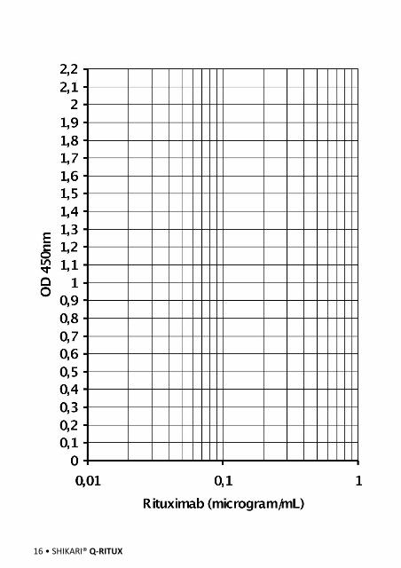
Semi-Log Graph Paper .................................................................................................. 15
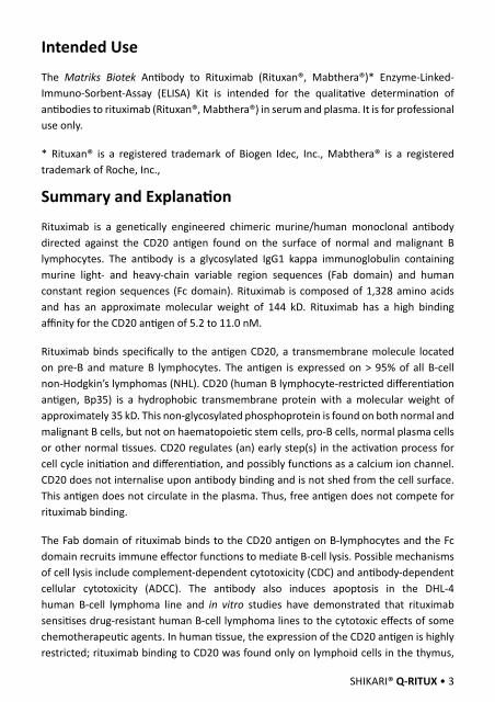
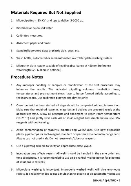
SHIKARI Q-RITUX
Free rituximab (Rituxan®, Mabthera®) quantitative analyses
Required Volume (µl) 10
Total Time (min) 75
Sample Serum, plasma and other biological fluids
Sample Number 96
Dedection Limit (ng/mL) 30
Spike Recovery (%) 98
Shelf Life (year) 1
SHIKARI® Q-RITUX • 3
Intended Use
The Matriks Biotek Antibody to Rituximab (Rituxan®, Mabthera®)* Enzyme-Linked-Immuno-Sorbent-Assay (ELISA) Kit is intended for the qualitative determination of antibodies to rituximab (Rituxan®, Mabthera®) in serum and plasma. It is for professional use only.
* Rituxan® is a registered trademark of Biogen Idec, Inc., Mabthera® is a registered trademark of Roche, Inc.,
Summary and Explanation
Rituximab is a genetically engineered chimeric murine/human monoclonal antibody directed against the CD20 antigen found on the surface of normal and malignant B lymphocytes. The antibody is a glycosylated IgG1 kappa immunoglobulin containing murine light- and heavy-chain variable region sequences (Fab domain) and human constant region sequences (Fc domain). Rituximab is composed of 1,328 amino acids and has an approximate molecular weight of 144 kD. Rituximab has a high binding affinity for the CD20 antigen of 5.2 to 11.0 nM.
Rituximab binds specifically to the antigen CD20, a transmembrane molecule located on pre-B and mature B lymphocytes. The antigen is expressed on > 95% of all B-cell non-Hodgkin’s lymphomas (NHL). CD20 (human B lymphocyte-restricted differentiation antigen, Bp35) is a hydrophobic transmembrane protein with a molecular weight of approximately 35 kD. This non-glycosylated phosphoprotein is found on both normal and malignant B cells, but not on haematopoietic stem cells, pro-B cells, normal plasma cells or other normal tissues. CD20 regulates (an) early step(s) in the activation process for cell cycle initiation and differentiation, and possibly functions as a calcium ion channel. CD20 does not internalise upon antibody binding and is not shed from the cell surface. This antigen does not circulate in the plasma. Thus, free antigen does not compete for rituximab binding.
The Fab domain of rituximab binds to the CD20 antigen on B-lymphocytes and the Fc domain recruits immune effector functions to mediate B-cell lysis. Possible mechanisms of cell lysis include complement-dependent cytotoxicity (CDC) and antibody-dependent cellular cytotoxicity (ADCC). The antibody also induces apoptosis in the DHL-4 human B-cell lymphoma line and in vitro studies have demonstrated that rituximab sensitises drug-resistant human B-cell lymphoma lines to the cytotoxic effects of some chemotherapeutic agents. In human tissue, the expression of the CD20 antigen is highly restricted; rituximab binding to CD20 was found only on lymphoid cells in the thymus,
4 • SHIKARI® Q-RITUX
the white pulp of the spleen and a majority of B lymphocytes in peripheral blood and lymph nodes. Little or no non-specific binding was observed.
Rituximab is a CD20-directed cytolytic antibody indicated for the treatment of patients with: Non-Hodgkin’s Lymphoma (NHL), Chronic Lymphocytic Leukemia, Rheumatoid Arthritis (RA) in combination with methotrexate in adult patients with moderately-to severely-active RA who have inadequate response to one or more TNF antagonist therapies, Wegener’s Granulomatosis (WG) and Microscopic Polyangiitis (MPA) in adult patients in combination with glucocorticoids.
PharmacokineticsNon-Hodgkin’s Lymphoma(NHL): Rituximab at a dose of 375 mg/m2 was administered as an IV infusion at weekly intervals for 4 doses to 203 patients with NHL naive to rituximab. The mean Cmax following the fourth infusion was 486 μg/mL (range 77.5 - 996.6 μg/mL). The peak and trough serum levels of rituximab were inversely correlated with baseline values for the number of circulating CD19-positive B-cells and measures of disease burden. Median steady-state serum levels were higher for responders compared with non-responders. Serum levels were higher in patients with International Working Formulation (IWF) subtypes B, C, and D as compared with those with subtype A. Rituximab was detectable in the serum of patients 3 – 6 months after completion of last treatment. Rituximab at a dose of 375 mg/m2 was administered as an IV infusion at weekly intervals for 8 doses to 37 patients with NHL. The mean Cmax increased with each successive infusion, spanning from a mean of 243 μg/mL (range, 16 – 582 μg/mL) after the first infusion to 550μg/mL (range 171 – 1177 μg/mL) after the eighth infusion. The pharmacokinetic profile of rituximab when administered as 6 infusions of 375 mg/m2 in combination with 6 cycles of CHOP chemotherapy was similar to that seen with rituximab alone.
Chronic Lymphocytic Leukaemia (CLL): Rituximab was administered as an IV infusion at a first-cycle dose of 375 mg/m2 increased to 500 mg/m2 each cycle for a further 5 doses in combination with fludarabine and cyclophosphamide (FC) in CLL patients. The mean Cmax (N=15) was 408 μg/mL (range, 97 – 764 μg/mL) after the fifth 500 mg/m2 infusion.
Rheumatoid Arthritis (RA): The pharmacokinetics of rituximab were assessed following two IV doses of 500 mg and 1000 mg on days 1 and 15 in four studies. In all these studies, rituximab pharmacokinetics were dose proportional over the limited dose range studied. Mean Cmax for serum rituximab following first infusion ranged from 157 to 171 μg/mL for 2 x 500 mg dose and ranged from 298 to 341 μg/mL for 2 x 1000 mg dose. Following second infusion, mean Cmax ranged from 183 to 198 μg/mL for the 2 x 500

SHIKARI® Q-RITUX • 5
mg dose and ranged from 355 to 404 μg/mL for the 2 x 1000 mg dose. Mean terminal elimination half-life ranged from 15 to 16.5 days for the 2 x 500 mg dose group and 17 to 21 days for the 2 x 1000 mg dose group. Mean Cmax was 16 to 19% higher following second infusion compared to the first infusion for both doses. Upon re-treatment with a second course the pharmacokinetics of rituximab were again assessed following two IV doses of 500 mg and 1000 mg. Mean Cmax for serum rituximab following first infusion was 170 to 175 μg/mL for 2 x 500 mg dose and 317 to 370 μg/mL for 2 x 1000 mg dose. Cmax following second infusion, was 207 μg/mL for the 2 x 500 mg dose and ranged from 377 to 386 μg/mL for the 2 x 1000 mg dose. Mean terminal elimination halflife after the second infusion, following the second course, was 19 days for 2 x 500 mg dose and ranged from 21 to 22 days for the 2 x 1000 mg dose. PK parameters for rituximab were comparable over the two treatment courses.
Wegener’s Granulomatosis(WG) and Microscopic Polyangiitis(MPA): Based on the population pharmacokinetic analysis of data in 97 WG and MPA patients who received 375 mg/m2 rituximab once weekly by intravenous infusion for four weeks, the estimated median terminal elimination half-life was 23 days (range, 9 to 49 days). Rituximab mean clearance and volume of distribution were 0. 312 L/day (range, 0.115 to 0.728 L/day) and 4.50 L (range, 2.21 to 7.52 L) respectively. Male patients and patients with higher BSA or positive human anti-chimeric antibody (HACA) levels have higher clearance. However, further dose adjustment based on gender or HACA status is not necessary.
ImmunogenicityAs with all therapeutic proteins, there is a potential for immunogenicity. The observed incidence of antibody (including neutralizing antibody) positivity in an assay is highly dependent on several factors including assay sensitivity and specificity, assay methodology, sample handling, timing of sample collection, concomitant medications, and underlying disease. It was reported that, using an ELISA assay, anti-human anti-chimeric antibody (HACA) was detected in 4 of 356 (1.1%) patients with low-grade or follicular NHL receiving single-agent rituximab. Three of the four patients had an objective clinical response. A total of 273/2578 (11%) patients with RA tested positive for HACA at any time after receiving rituximab. HACA positivity was not associated with increased infusion reactions or other adverse reactions. Upon further treatment, the proportions of patients with infusion reactions were similar between HACA positive and negative patients, and most reactions were reported to be mild to moderate. Four HACA positive patients had serious infusion reactions, and the temporal relationship between HACA positivity and infusion reaction was variable. A total of 23/99 (23%) rituximab-treated patients with WG and MPA tested positive for HACA by 18 months. The clinical relevance of HACA formation in rituximab-treated patients is unclear.
6 • SHIKARI® Q-RITUX
In rituximab-treated cynomolgus monkeys B lymphocyte numbers were reduced by 99% or more in comparison with pre-test values in the peripheral blood of all monkeys, approximately 24 hours after the first dose. Two weeks after the last dose, B lymphocyte numbers were still reduced by more than 99% in 3/6 monkeys dosed for four weeks and in 4/6 monkeys dosed for eight weeks, and B lymphocyte numbers were also depleted in the mandibular lymph nodes and femoral bone marrow. However, a partial recovery of B lymphocyte numbers in the peripheral blood of some monkeys in both dose groups was correlated with the development of antibodies against rituximab.
The use of rituximab (Rituxan®, Mabthera®) was associated to the development of anti-rituximab antibodies, even some might be neutralizing, in various percentages of patients during therapy with the drug. The Matriks Biotek Rituximab-ELISA and Antibody to rituximab ELISA Kits can be efficiently used, for monitoring serum through levels and the presence of anti-rituximab antibodies respectively, during therapy and offers the scientist a tool for decision on possible preventive measures.
Test Principle
The Matriks Biotek rituximab (Rituxan®, Mabthera®) solid phase enzyme-linked immunosorbent assay (ELISA) is based on rituximab-specific 9F9 monoclonal antibody (mAb). Standards and diluted samples are incubated in the microtitre plate coated with 9F9 mAb. After incubation, the wells are washed. Anti-human IgG Fc-specific mAb (clone 1B5) conjugated to horse radish peroxidase (HRP) is added and binds to rituximab specifically captured by the 9F9 on the surface of the wells. Following incubation, wells are washed and the bound enzymatic activity is detected by addition of chromogen-substrate. The colour developed is proportional to the amount of rituximab in the sample or standard. Results of samples can be determined directly using the standard curve.
Warnings and Precautions
1. For professional use only.
2. Before starting the assay, read the instructions completely and carefully. Use the valid version of the package insert provided with the kit. Be sure that everything is understood. For further information (clinical background, test performance, automation protocols, alternative applications, literature, etc.) please refer to the local distributor.
SHIKARI® Q-RITUX • 7
3. In case of severe damage of the kit package please contact Matriksbiotek or your supplier in written form, latest one week after receiving the kit. Do not use damaged components in test runs, but keep safe for complaint related issues.
4. Obey lot number and expiry date. Do not mix reagents of different lots. Do not use expired reagents.
5. Follow good laboratory practice and safety guidelines. Wear lab coats, disposable latex gloves and protective glasses where necessary.
6. Reagents of this kit containing hazardous material may cause eye and skin irritations. See MATERIALS SUPPLIED and labels for details.
7. Chemicals and prepared or used reagents have to be treated as hazardous waste according the national biohazard safety guidelines or regulations.
8. Avoid contact with Stop solution. It may cause skin irritations and burns.
9. Some reagents contain sodium azide (NaN3) as preservatives. In case of contact with eyes or skin, flush immediately with water. NaN3 may react with lead and copper plumbing to form explosive metal azides. When disposing reagents, flush with large volume of water to avoid azide build-up.
10. All reagents of this test kit containing human serum or plasma have been tested and were found negative for HIV I/II, HBsAg and HCV by FDA approved procedures. However, a presence of these or other infectious agents cannot be excluded absolutely and therefore reagents should be treated as potential biohazards in use and for disposal.
Storage and Stability
The kit is shipped at ambient temperature and should be stored at 2-8°C. Keep away from heat or direct sun light. The storage and stability of specimen and prepared reagents is stated in the corresponding chapters.
The strips of microtiter plate is stable up to the expiry date of the kit in the broken, but tightly closed bag when stored at 2–8°C.

8 • SHIKARI® Q-RITUX
Specimen Collection and Storage
Serum, Plasma (EDTA, Heparin)*
The usual precautions for venipuncture should be observed. It is important to preserve the chemical integrity of a blood specimen from the moment it is collected until it is assayed. Do not use grossly hemolytic, icteric or grossly lipemic specimens. Samples appearing turbid should be centrifuged before testing to remove any particulate material.
Storage: 2-8°C -20°C Keep away from heat or direct sun lightAvoid repeated freeze-thaw cyclesStability: 7 d 6 mon
Materials Supplied
1 x 12 x 8 MTP
Microtiter PlateBreak apart strips. Microtiter plate with 12 rows each of 8 wells coated with 9F9 monoclonal antibody specific for rituximab only.
5 x 0.5 mLSTNDA-E
Rituximab Standards A-E1; 0.3; 0.1; 0.03; 0 microgram/mLPurple colored (graded). Ready to use. Used for construction of the standard curve. Contains rituximab, human serum, stabilizer and <0.1% NaN3.
2 x 60 mLASSAYBUF
Assay BufferBlue colored. Ready to use. Contains proteins and <0.1% NaN3.
1 x 12 mLHRPCONJUG
HRP Conjugated Anti-Human IgG mAb (Clone 1B5)Red colored. Ready to use. Contains HRP-Anti-human IgG (Fc-specific) mAb (reactive with human IgG1, IgG2, IgG3 and IgG4 but not with IgA and IgM) and stabilizers.
1 x 12 mLTMBSUBS
TMB Substrate SolutionReady to use. Contains TMB
1 x 12 mLTMBSTOP
TMB Stop Solution Ready to use. 1N HCl.
1 x 50 mLWASHBUFCONC
Wash Buffer, Concentrate (20x)Contains Buffer with Tween 20.
2 x 1 ADH FILMAdhesive FilmFor covering of Microtiter Plate during incubation.
SHIKARI® Q-RITUX • 9
Materials Required But Not Supplied
1. Micropipettes (< 3% CV) and tips to deliver 5-1000 µL.
2. Bidistilled or deionised water
3. Calibrated measures.
4. Absorbent paper and timer.
5. Standard laboratory glass or plastic vials, cups, etc.
6. Wash bottle, automated or semi-automated microtiter plate washing system
7. Microtiter plate reader capable of reading absorbance at 450 nm (reference wavelength 620-690 nm is optional)
Procedure Notes
1. Any improper handling of samples or modification of the test procedure may influence the results. The indicated pipetting volumes, incubation times, temperatures and pretreatment steps have to be performed strictly according to the instructions. Use calibrated pipettes and devices only.
2. Once the test has been started, all steps should be completed without interruption. Make sure that required reagents, materials and devices are prepared ready at the appropriate time. Allow all reagents and specimens to reach room temperature (18-25 °C) and gently swirl each vial of liquid reagent and sample before use. Mix reagents without foaming.
3. Avoid contamination of reagents, pipettes and wells/tubes. Use new disposable plastic pipette tips for each reagent, standard or specimen. Do not interchange caps. Always cap not used vials. Do not reuse wells/tubes or reagents.
4. Use a pipetting scheme to verify an appropriate plate layout.
5. Incubation time affects results. All wells should be handled in the same order and time sequences. It is recommended to use an 8-channel Micropipettor for pipetting of solutions in all wells.
6. Microplate washing is important. Improperly washed wells will give erroneous results. It is recommended to use a multichannel pipette or an automatic microplate
10 • SHIKARI® Q-RITUX
washing system. Do not allow the wells to dry between incubatons. Do not scratch
coated wells during rinsing and aspiraton. Rinse and fll all reagents with care. While
rinsing, check that all wells are flled precisely with Wash Bufer, and that there are
no residues in the wells.
7. Humidity afects the coated wells/tubes. Do not open the pouch untl it reaches
room temperature. Unused wells/tubes should be returned immediately to the
resealed pouch including the desiccant.
Preparaton of Component
Dilute/
disolve
Component with Diluent Relaton Remarks Storage Stability
10 mL Wash
Bufer*
Up to
200
mL
bidist.
Water
1:20 Warm up at 37°C
to dissolve crystals.
Mix vigorously.
2-8 °C 1 w
*Prepare Wash Bufer before startng assay procedure.
*Diluton of Samples
Sample To be diluted Diluent RemarksWith Relaton
Serum/
Plasma
Initally
1:100
Assay bidist.
Bufer Water
1:100
1:1000
For diluton at 1:100;
5 Sample + 495 Assay µL µL
Bufer
For diluton at 1:1000;
5 of 1:100-diluted sample + µL
495 of Assay BuferµL
*. 1:100-diluted samples with a concentraton of rituximab above the measuring range
are to be rated as “> highest standard”. The result must not be extrapolated. The sample
in queston should be further diluted with Assay Bufer and retested.
SHIKARI® Q-RITUX • 11
Test Procedure
1 Pipette 100µL of Assay Buffer non-exceptionally into each of the wells to be used.
2 Pipette 10 µL of each ready-to use Standards, and Diluted Samples into the respective wells of microtiter plate.
WellsA1: Standard AB1: Standard BC1: Standard CD1: Standard DE1: Standard EF1 and on: Sample (Serum/Plasma)
3 Cover the plate with adhesive film. Incubate 30 min at room temperature (18-25°C).
4 Remove adhesive film. Discard incubation solution. Wash plate 3 times each with 300 µL of diluted Wash Buffer. Remove excess solution by tapping the inverted plate on a paper towel.
5 Pipette 100 µL of ready-to use HRP-Anti-human IgG (Fc-specific) mAb Conjugate into each well.
6 Cover the plate with adhesive film. Incubate 30 min at room temperature (18-25°C).
7 Remove adhesive film. Discard incubation solution. Wash plate 3 times each with 300 µL of diluted Wash Buffer. Remove excess solution by tapping the inverted plate on a paper towel.
8 Pipette 100 µL of TMB Substrate Solution into each well.
9 Incubate 15 min (without adhesive film) at room temperature (18-25°C) in the dark.
10 Stop the substrate reaction by adding 100 µL of Stop Solution into each well. Briefly mix contents by gently shaking the plate. Color changes from blue to yellow.
11 Measure optical density (OD) with a photometer at 450 nm within 30 min after pipetting of the Stop Solution.
Interpretation & Calculation of Results
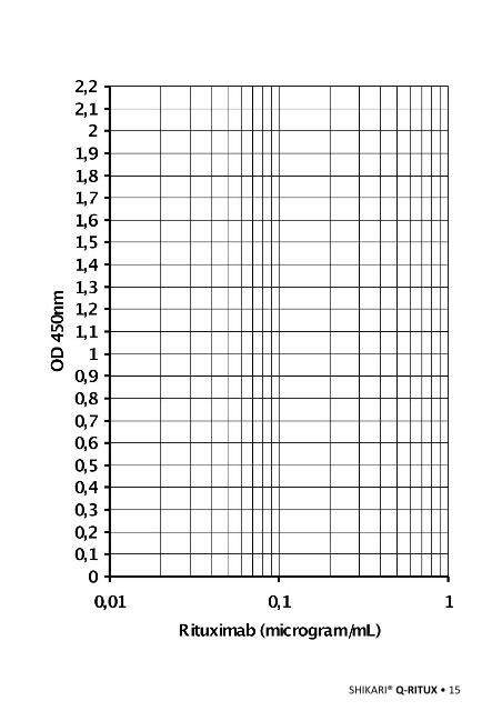
1. Semi-log graph paper should be used for manual construction. Construct a standard curve by plotting the OD450 nm for each of 4 standards (disregarding the zero standard) on the vertical linear y-axis versus the corresponding rituximab concentration on the horizontal logarithmic x-axis, thus create a smooth standard curve with maximum 1 tuning point.
12 • SHIKARI® Q-RITUX
2. The concentration of the samples can be read directly from this standard curve. Using the absorbance value for each sample, determine the corresponding concentration of rituximab from the standard curve. Find the absorbance value on the Y-axis and extend a horizontal line to the standard curve. At the point of intersection, extend a vertical line to the X-axis and read the rituximab concentration for the unknown sample.
3. Any sample diluted at 1:100 and reading an OD450nm lower than that of standard D (30 ng/mL) must not be extrapolated and should be reported as “below detection level.
4. Any sample diluted at 1:100 and still reading greater than the highest standard should be further diluted appropriately with Assay Buffer and retested. Because the samples have been diluted, the concentration determined from the standard-curve must be multiplied by the dilution factor.
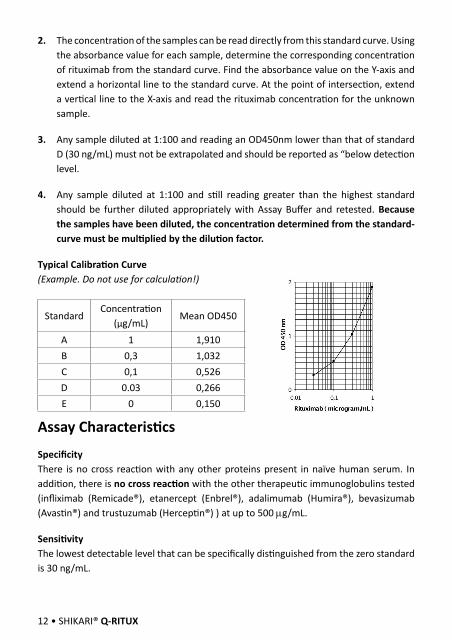
Typical Calibration Curve(Example. Do not use for calculation!)
StandardConcentration
(µg/mL)Mean OD450
A 1 1,910B 0,3 1,032C 0,1 0,526D 0.03 0,266E 0 0,150
Assay Characteristics
SpecificityThere is no cross reaction with any other proteins present in naïve human serum. In addition, there is no cross reaction with the other therapeutic immunoglobulins tested (infliximab (Remicade®), etanercept (Enbrel®), adalimumab (Humira®), bevasizumab (Avastin®) and trustuzumab (Herceptin®) ) at up to 500 mg/mL.
SensitivityThe lowest detectable level that can be specifically distinguished from the zero standard is 30 ng/mL.
SHIKARI® Q-RITUX • 13
Precision of KitIntra-assay CV: <8% at the range of 0.1-1 mg/mL.
Inter-assay CV: <8% at the range of 0.1-1 mg/mL.
RecoveryRecovery rate was found to be equal and higher than 98% with human serum spiked with rituximab at concentrations of 100, 10, and 3mg/mL.
Automation
Experiments have shown that the Matriks Biotek Rituximab ELISA is also suitable to run on an automated ELISA processor.
References1. Datasheet of Rituxan prescribing information.
2. Datasheet of Mabthera prescribing information.
3. Thurlings RM, Teng O, Vos K, Gerlag DM, Aarden L, Stapel SO, JM van Laar, Tak PP, Wolbink GJ, Clinical response, pharmacokinetics, development of human anti-chimaeric antibodies, and synovial tissue response to rituximab treatment in patients with rheumatoid arthritis, Ann Rheum Dis 2010; 69: 409–412.
4. Schmidt E, Hennig K, Mengede C, Zillikens D, Kromminga A, Immunogenicity of rituximab in patients with severe pemphigus, Clinical Immunology, 2009; 132: 334–341.
5. Keystone E, Fleischmann R, Emery P, Furst DE, van Vollenhoven R, Bathon J, Dougados M, Baldassare A, Ferraccioli G, Chubick A, Udell J, Cravets MW, Agarwal S, Cooper S, and Magrini F, Safety and Efficacy of Additional Courses of Rituximab in Patients With Active Rheumatoid Arthritis, ARTHRITIS & RHEUMATISM, 2007; 56: 3896–3908.
6. Shoko Goto, Hiroaki Goto, Reo Tanoshima, Hiromi Kato, Hiroyuki Takahashi, Osamu Sekiguchi, Sumio Kai, Serum sickness with an elevated level of human anti-chimeric antibody following treatment with rituximab in a child with chronic immune thrombocytopenic purpura, Int J Hematol , 2009; 89: 305–309.
7. Tobinai K, Y. Kobayashi, M. Narabayashi, M. Ogura, Y. Kagami, Y. Morishima, T. Ohtsu, T. Igarashi, Y. Sasaki, T. Kinoshita, T. Murate, Feasibility and pharmacokinetic study of a chimeric anti-CD20 monoclonal antibody (IDEC-C2B8, rituximab) in relapsed B-cell lymphoma, Annals of Oncology 1998; 9: 527-534.

14 • SHIKARI® Q-RITUX
8. Magda Nakou, Georgios Katsikas, Prodromos Sidiropoulos, George Bertsias, Eva Papadimitraki, Amalia Raptopoulou, Helen Koutala, Helen A Papadaki, Herakles Kritikos and Dimitrios T Boumpas, rituximab therapy reduces activated B cells in both the peripheral blood and bone marrow of patients with rheumatoid arthritis: depletion of memory B cells correlates with clinical response Arthritis Research & Therapy 2009, 11:R131 (doi:10.1186/ar2798)
9. Pina M. Cardarelli, Maire Quinn, Dana Buckman, Yu Fang, David Colcher, David J. King, Christopher Bebbington, Geoffrey Yarranton, Binding to CD20 by Anti-B1 Antibody or F(ab’)2 is sufficient for induction of apoptosis in B-cell lines, Cancer ImmunolImmunother, 2002; 51: 15–24.
10. Maria Rehnberg, Sylvie Amu, Andrej Tarkowski, Maria I Bokarewa and Mikael Brisslert, Short- and long-term effects of anti-CD20 treatment on B cell ontogeny in bone marrow of patients with rheumatoid arthritis, Arthritis Research & Therapy 2009, 11:R123 (doi:10.1186/ar2789).
11. Ferdinand Breedveld, Sunil Agarwal, Ming Yin, Song Ren, Nicole F. Li, Tim M. Shaw, and Brian E. Davies, Rituximab Pharmacokinetics in Patients With Rheumatoid Arthritis: B-Cell Levels Do Not Correlate With Clinical Response, Journal of Clinical Pharmacology, 2007; 47: 1119-1128.
12. David G. Maloney, Antonio J. Grillo-López, Christine A. White, David Bodkin, Russell J. Schilder, James A. Neidhart, Nalini Janakiraman, Kenneth A. Foon, Tina-Marie Liles, Brian K. Dallaire, Ken Wey, Ivor Royston, Thomas Davis and Ronald Levy, IDEC-C2B8 (Rituximab) Anti-CD20 Monoclonal Antibody Therapy in Patients With Relapsed Low-Grade Non-Hodgkin’s Lymphoma, Blood, 1997; 90: 2188-2195.
13. Dieter Huhn, Christoph von Schilling, Martin Wilhelm, Anthony D. Ho, Michael Hallek, Rolf Kuse, Wolfgang, Knauf, Ute Riedel, Axel Hinke, Stefanie Srock, Stefan Serke, Christian Peschel and Bertold Emmerich, rituximab therapy of patients with B-cell chronic lymphocytic leukemia, Blood, 2001; 98: 1326-1331.
14. Maeda T, Yamada Y, Tawara M, Yamasaki R, Yakata Y, Tsutsumi C, Onimaru Y, Kamihira S, Tomonaga M., Successful treatment with a chimeric anti-CD20 monoclonal antibody (IDEC-C2B8, rituximab) for a patient with relapsed mantle cell lymphoma who developed a human anti-chimeric antibody. Int J Hematol. 2001; 74(1): 70-75.
SHIKARI® Q-RITUX • 15
16 • SHIKARI® Q-RITUX