Embed Size (px)
Citation preview

Initial Management of Suspected Encephalitis
Dr Ruth Palmer
Consultant Microbiologist

CNS infections are urgent and important
• Mortality is significant recovery is slow and and
post infection deficits occur in over 50% of cases • Apart from aciclovir and ART treatment for most
infective causes of encephalitis is non-existent. • Starting aciclovir early is crucial • Negligence settlements for missed HSV can run
into millions of pounds • LP can help in terms of HSV management but
over 62% of patients remain undiagnosed.

Quiz
1. A CT scan should always be performed before a LP
2. You can remove safely 15ml of CSF during an LP
3. A white cell count of 6 in the CSF is considered normal
4. Low CSF glucose indicates bacterial meningitits
5. A negative HSV PCR in CSF excludes HSV encephalitis
6. CSF Neutrophilia excludes encephalitis
7. Parotitis is present in all cases of mumps encephalitis
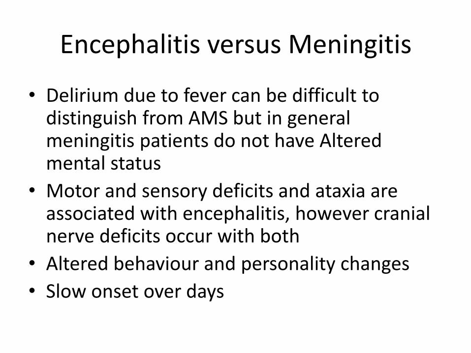
Encephalitis versus Meningitis
• Delirium due to fever can be difficult to distinguish from AMS but in general meningitis patients do not have Altered mental status
• Motor and sensory deficits and ataxia are associated with encephalitis, however cranial nerve deficits occur with both
• Altered behaviour and personality changes
• Slow onset over days
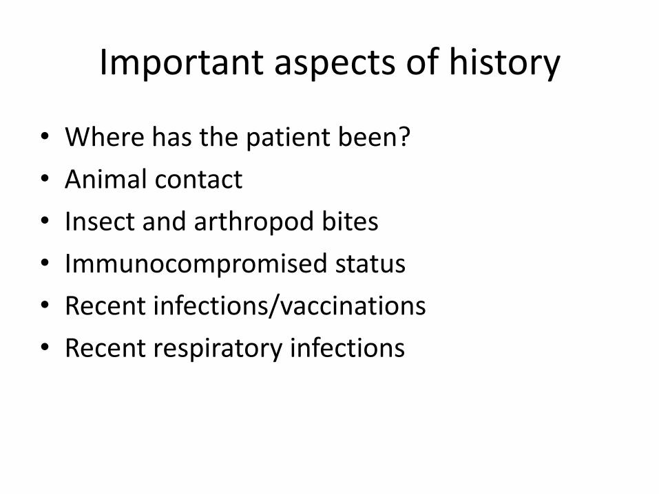
Important aspects of history
• Where has the patient been?
• Animal contact
• Insect and arthropod bites
• Immunocompromised status
• Recent infections/vaccinations
• Recent respiratory infections

Infectious Causes
• HSV/Enterovirus/VZV/HIV/Mumps
• Influenza/Mycoplasma/LCM/Listeria
• EBV/HHV6/CMV/Adenovirus/JC-PMLE
• WNV/Dengue/JE/Lyme
• EE/WEE/St Louis/RMSF
• Rabies
• Nipah/Hendra
• Syphilis
A note on HSV Encephalitis
• Untreated mortality is 70% treated still 19% but 44-62 have significant CNS deficit
• Culture sensitivity is <10% • IgG/IgM sensitivity up to 85% • HSV PCR 98% but please note if CT features and
EEG are suggestive of HSV and CSF is negative then continue treatment.
• HSV PCR remains positive for up to 1 week • The early CT scan can be inconclusive in up to
50% of patients and should be repeated.

Sleepy head!

ead! • 54 yr old taxi driver
• A&E;
– “General slowness” for 1 week
– 7/7 prior home from work with headache & slept for 24hrs
– Then c/o of fever, lethargy & anorexia
– Became unsteady on feet & talking “silly”
– Day 4 GP diagnosed labyrinthitis
– But headaches continued, more unsteady, slurred speech
Sleepy h

Examination
• T 37.6oC, GCS 15/15, HR 58 bpm, BP 132/75 mmHg
• CVS/ RS/GI all normal
• Neuro
– slow but normal gait
– Slurred speech
– Cranial nerves normal
– Tone, power & reflexes normal all 4 limbs
– Coordination deficient upper limbs
– 8/10 mental test score

Differential diagnosis?
• Encephalopathy due to;
– Severe sepsis
– Toxic
– Metabolic
• Ischaemic stroke
• Vasculitis
• Bacterial meningitis
• Encephalitis
Investigations
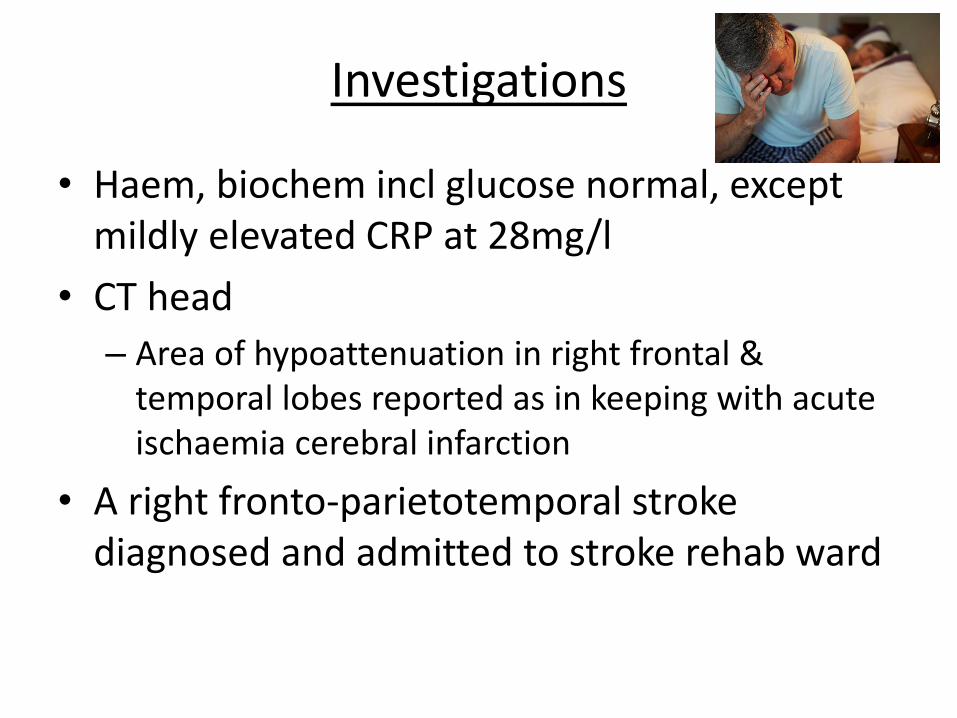
• Haem, biochem incl glucose normal, except mildly elevated CRP at 28mg/l
• CT head
– Area of hypoattenuation in right frontal & temporal lobes reported as in keeping with acute ischaemia cerebral infarction
• A right fronto-parietotemporal stroke diagnosed and admitted to stroke rehab ward

Consultant ward round (Day 3 admission – Mon)
• Symptoms static; Intermittent pyrexia
• Encephalitis considered
• MRI: Diffuse hyperintensities
Right frontal, parietal &
Temporal lobes
Clinical case Normal range (Adult)
Opening pressure 17 cm H2O 9-18cm H2O
Protein 2.90 g/l 0.15-0.45 g/L
CSF glucose Glucose 3.1 (serum 6.6 mmol/l) (47%)
60% of the blood glucose level
Cell counts WCC 5140/mm3 (99% lymphocytes)
WBC 0-5 / mm3
(0 neutrophils, <1 lymphocytes) No RBCs

Lymphocytic CSF
• Viral Meningitis
• Viral Encephalitis
• Mycobacterium tuberculosis
• Listeria monocytogenes
• Fungal – cryptococcal
• Partially treated bacterial meningitis/ early bacterial?
• Parameningeal bacterial infections (cerebral abscesses etc…)
• Mycoplasma
• HIV
• Syphilis
• Drugs e.g. – NSAIDs
– Trimethoprim
• Autoimmune encephalitis
• ADEM
• MS
• Neoplastic/paraneoplastic
• Vasculitis
• Other autoimmune disorders e.g. SLE
• Sarcoid

Progress
• Treatment started on day 3 – IV acyclovir 10mg/kg, amox 3g qds, gent 5mg/kg od
• 3 days into treatment – Less hesitant speech – HSV-1 DNA detected in CSF – Antibacterial drugs stopped – IV aciclovir 2 weeks (then 4 weeks valaciclovir)
WHAT DO YOU THINK OF TREATMENT?
• Despite treatment, patient remained off work and continues to have word-finding difficulties & cognitive slowing

Why encephalitis is missed
• Wrongly attributing a patient’s fever and confusion • Failure to recognise a febrile illness and consider infection • Ignoring a relative says patient behaviour, “not quite right” you
say GCS is 15 • Patient is assumed to be drunk or drugged • Failure to properly investigate a patient with a fever and seizure • Failure to do a lumbar puncture or if delayed LP failure to start
aciclovir.

What are the likely outcomes?
• Death
• Full recovery with no symptoms
• Some disability
– Memory impairment
– Speech impairment
– Unable to walk
– Bed ridden, full care needed
Epidemiology and Incidence
• Viral, bacterial and tick causes
• Total western incidence
• 0.7- 13.8 per 100,000
• Herpes simplex virus encephalitis most common
• Average DGH (300,000) – 15-30 cases per year – 1-2 viral encephalitis per month

Clinical presentation of encephalitis • Classically
– Headache – Altered or reduced consciousness – Personality or behaviour change in a patient with a fever or
history of febrile illness • Subtle presentations
– Low grade fever,
– Behavioural changes
– Speech and language disturbances
• HSV-1 features where temporal or frontal lobes affected may include – Olfactory hallucinations – Simple or complex partial seizures – Bizarre behaviour – Neuropsychiatric features

LP pack - new
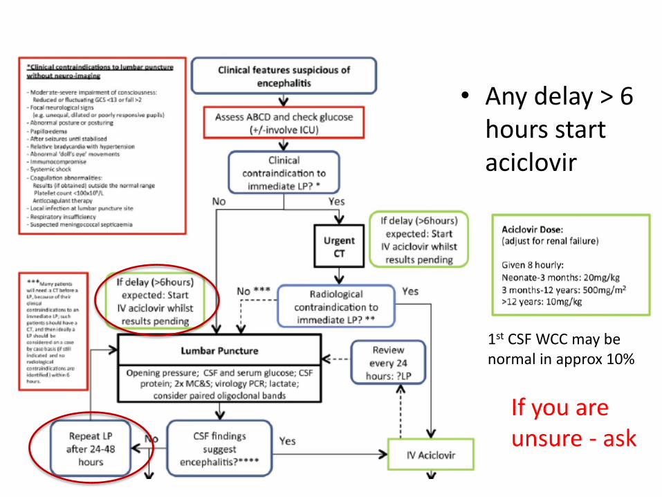
• Any delay > 6 hours start aciclovir
1st CSF WCC may be normal in approx 10%
If you are unsure - ask

Opening
Pressure
High/Very
High Normal/High High 10-20cm High
Colour Clear/Cloudy “Gin” Clear Cloudy Clear Cloudy/Yellow
Cells/mm3 Normal-High
0-1000
Slightly
Increased
5-1000
High/Very
High
100-50000
<5
Slightly
Increased
25-500
Differential Lymphocytes Lymphocytes Neutrophils Lymphocytes Lymphocytes
CSF/Plasma
Glucose Normal-Low Normal Low 66%
Low-Very Low
(<30%)
Protein
(g/L)
Normal-High
0.2-5.0
Normal-High
0.5-1 High >1 <0.45
High-Very High
1.0-5.0
Diagnosis Normal Purulent
Meningitis
Aseptic meningitis or encephalitis
Fungal Tuberculous meningitis
CSF Interpretation is vital
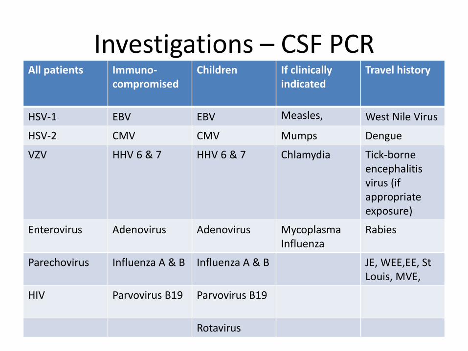
Investigations – CSF PCR All patients Immuno-
compromised Children If clinically
indicated
Travel history
HSV-1 EBV EBV Measles, West Nile Virus
HSV-2 CMV CMV Mumps Dengue
VZV HHV 6 & 7 HHV 6 & 7 Chlamydia Tick-borne encephalitis virus (if appropriate exposure)
Enterovirus Adenovirus Adenovirus Mycoplasma Influenza
Rabies
Parechovirus
Influenza A & B Influenza A & B JE, WEE,EE, St Louis, MVE,
HIV Parvovirus B19
Parvovirus B19
Rotavirus

Investigations
• HIV testing in all cases of encephalitis (BHIVA guidelines)
• CSF PCR (usually tiered set of investigations with HSV/VZ/Enterovirus in first tier second tier suggested by evidence of Mumps/Measles recent vaccination, travel history or if Immunocompromised)
• CSF and serum IgG and IgM as appropriate
• T/S and NPA and faeces if enterovirus or respiratory viral ilness considered
• Vesicle fluid culture and Molecular testing
• If associated with atypical pneumonia, test serum for Mycoplasma and chlamydia
• Autoantibodies: NMDAR antibodies, VGKC antibodies
• Brain biopsy, nucal skin testing

Start aciclovir within 6 hours
• HSV encephalitis
– Aciclovir 10mg/kg IV
• +/- antiepileptic for seizures
• +/- steroids or other immunomodulatory agents
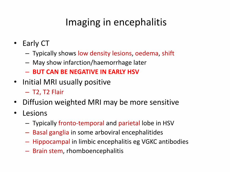
Imaging in encephalitis
• Early CT – Typically shows low density lesions, oedema, shift
– May show infarction/haemorrhage later
– BUT CAN BE NEGATIVE IN EARLY HSV
• Initial MRI usually positive – T2, T2 Flair
• Diffusion weighted MRI may be more sensitive
• Lesions – Typically fronto-temporal and parietal lobe in HSV
– Basal ganglia in some arboviral encephalitides
– Hippocampal in limbic encephalitis eg VGKC antibodies
– Brain stem, rhomboencephalitis
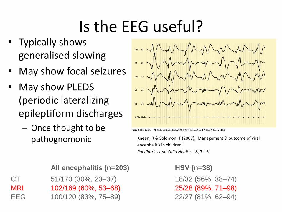
Is the EEG useful? • Typically shows
generalised slowing
• May show focal seizures
• May show PLEDS (periodic lateralizing epileptiform discharges
– Once thought to be pathognomonic
Kneen, R & Solomon, T (2007), 'Management and outcome of viral encephalitis in children', Paediatrics and Child Health, 18, 7-16.
Kneen, R & Solomon, T (2007), 'Management & outcome of viral
encephalitis in children',
Paediatrics and Child Health, 18, 7-16.
All encephalitis (n=203) HSV (n=38)
CT 51/170 (30%, 23–37) 18/32 (56%, 38–74)
MRI 102/169 (60%, 53–68) 25/28 (89%, 71–98)
EEG 100/120 (83%, 75–89) 22/27 (81%, 62–94)
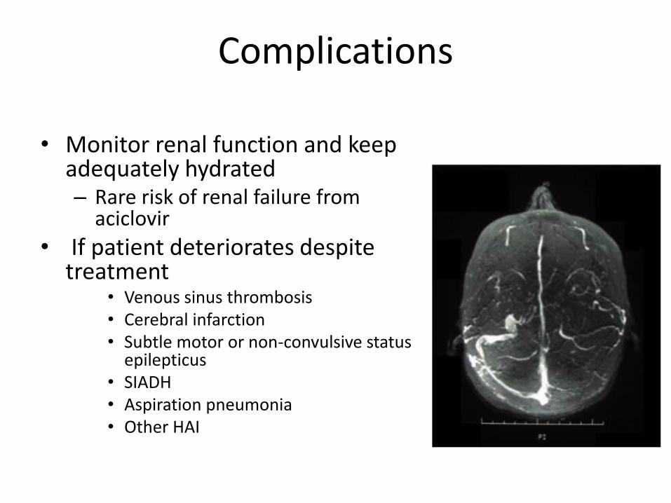
Complications
• Monitor renal function and keep adequately hydrated – Rare risk of renal failure from
aciclovir
• If patient deteriorates despite treatment
• Venous sinus thrombosis • Cerebral infarction • Subtle motor or non-convulsive status
epilepticus • SIADH • Aspiration pneumonia • Other HAI

Patients and their family should be put in contact with patient-orientated support services
www.encephalitis.info

Questions?