Embed Size (px)
Citation preview
Inhibition of Post-Transcriptional RNA Processing by CDKInhibitors and Its Implication in Anti-Viral TherapyJitka Holcakova1, Petr Muller1, Peter Tomasec2, Roman Hrstka1, Marta Nekulova1, Vladimir Krystof3,4,
Miroslav Strnad3,4, Gavin W. G. Wilkinson2, Borivoj Vojtesek1*
1 Regional Centre for Applied Molecular Oncology, Masaryk Memorial Cancer Institute, Brno, Czech Republic, 2 School of Medicine, Cardiff University, Cardiff, United
Kingdom, 3 Laboratory of Growth Regulators, Faculty of Science, Palacky University, Olomouc, Czech Republic, 4 Institute of Experimental Botany AS CR, Olomouc, Czech
Republic
Abstract
Cyclin-dependent kinases (CDKs) are key regulators of the cell cycle and RNA polymerase II mediated transcription. Severalpharmacological CDK inhibitors are currently in clinical trials as potential cancer therapeutics and some of them also exhibitantiviral effects. Olomoucine II and roscovitine, purine-based inhibitors of CDKs, were described as effective antiviral agentsthat inhibit replication of a broad range of wild type human viruses. Olomoucine II and roscovitine show high selectivity forCDK7 and CDK9, with important functions in the regulation of RNA polymerase II transcription. RNA polymerase II isnecessary for viral transcription and following replication in cells. We analyzed the effect of inhibition of CDKs byolomoucine II on gene expression from viral promoters and compared its effect to widely-used roscovitine. We found thatboth roscovitine and olomoucine II blocked the phosphorylation of RNA polymerase II C-terminal domain. However therepression of genes regulated by viral promoters was strongly dependent on gene localization. Both roscovitine andolomoucine II inhibited expression only when the viral promoter was not integrated into chromosomal DNA. In contrast,treatment of cells with genome-integrated viral promoters increased their expression even though there was decreasedphosphorylation of the C-terminal domain of RNA polymerase II. To define the mechanism responsible for decreased geneexpression after pharmacological CDK inhibitor treatment, the level of mRNA transcription from extrachromosomal DNAwas determined. Interestingly, our results showed that inhibition of RNA polymerase II C-terminal domain phosphorylationincreased the number of transcribed mRNAs. However, some of these mRNAs were truncated and lacked polyadenylation,which resulted in decreased translation. These results suggest that phosphorylation of RNA polymerase II C-terminal domainis critical for linking transcription and posttrancriptional processing of mRNA expressed from extrachromosomal DNA.
Citation: Holcakova J, Muller P, Tomasec P, Hrstka R, Nekulova M, et al. (2014) Inhibition of Post-Transcriptional RNA Processing by CDK Inhibitors and ItsImplication in Anti-Viral Therapy. PLoS ONE 9(2): e89228. doi:10.1371/journal.pone.0089228
Editor: Fatah Kashanchi, George Mason University, United States of America
Received October 8, 2013; Accepted January 17, 2014; Published February 21, 2014
Copyright: � 2014 Holcakova et al. This is an open-access article distributed under the terms of the Creative Commons Attribution License, which permitsunrestricted use, distribution, and reproduction in any medium, provided the original author and source are credited.
Funding: The work was supported by the European Regional Development Fund and the State Budget of the Czech Republic (RECAMO, CZ.1.05/2.1.00/03.0101),by Czech Science Foundation (GACR P206/12/G151) and by Ministry of Health, Czech Republic - conceptual development of research organization (MMCI,00209805). The funders had no role in study design, data collection and analysis, decision to publish, or preparation of the manuscript.
Competing Interests: The authors have declared that no competing interests exist.
* E-mail: [email protected]
Introduction
Pharmacological inhibitors of cyclin dependent kinases (PCIs)
represent a heterogeneous group of compounds that are defined
by their capacity to inhibit preferentially cyclin dependent kinases
(CDKs) involved in cell cycle regulation (CDK1, CDK2, CDK4,
CDK6 and CDK7), transcription (CDK7 and CDK9), or
neuronal function (CDK5) [1]. Since many CDKs are critical
regulators of cellular division, the pharmaceutical industry has
been focused on the discovery and development of pharmacolog-
ical CDK inhibitors as potential anticancer drugs [2].
Olomoucine II (OCII; 6-(2-Hydroxybenzylamino)-2(R)-[[1-hy-
droxymethyl)propyl]amino]-9-isopropylpurine) is a 2,6,9-trisubsti-
tuted purine derivative and, like roscovitine (Rosc), is a potent
reversible inhibitor of CDKs. Purine-derived CDK inhibitors
preferentially target CDKs 1, 2, 5, 7 and 9, because of a shared
capacity for competing for the ATP-binding pocket within CDKs
and arresting the cell cycle in G2/M phase. OCII and Rosc are
further known to induce the nuclear accumulation of the tumor
suppressor protein p53 thereby promoting its role as a transcrip-
tion factor [3,4]. Despite the structural similarity between these
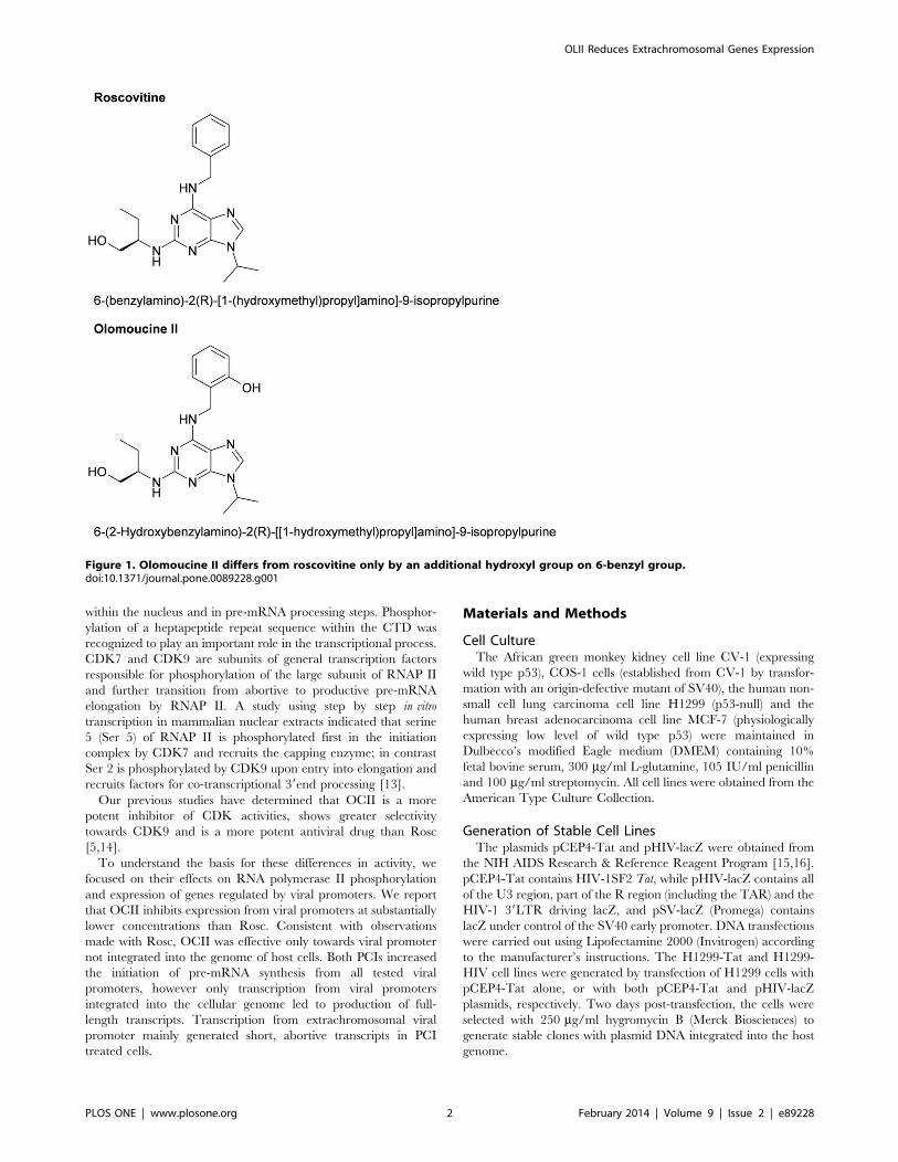
two PCIs (OCII differs from Rosc only by the presence of an
additional ortho-hydroxyl group on benzyl ring, see Figure 1),OCII displays about 10-fold higher inhibitory effect on CDK9,
and preferentially inhibits the S to G2 transition [5].
While CDK inhibitors clearly hold great promise as anticancer
drugs, interest in their capacity to inhibit viral replication keeps
growing [6,7]. Recently, CDKs have been shown to be required
for the efficient replication of a number of clinically-important
viruses, including papillomaviruses, human immunodeficiency
virus type-1 (HIV-1), human cytomegalovirus (HCMV), herpes
simplex virus (HSV) type 1 and HSV-2 [8,9]. Rosc, olomoucine
(the precursor of OCII) and several non-purine derived PCIs (such
as flavopiridol, FVP) inhibit the replication of viruses that are
known to affect cell cycle progression, including HCMV [6], HSV
[10] and HIV-1, -2 [11].
Virus gene transcription is often dependent on host cell factors
such as RNA polymerase II (RNAP II) [12]. C-terminal domain
(CTD), the largest subunit of RNAP II, is an essential transcrip-
tional enhancer that participates in organizing transcription foci
PLOS ONE | www.plosone.org 1 February 2014 | Volume 9 | Issue 2 | e89228
within the nucleus and in pre-mRNA processing steps. Phosphor-
ylation of a heptapeptide repeat sequence within the CTD was
recognized to play an important role in the transcriptional process.
CDK7 and CDK9 are subunits of general transcription factors
responsible for phosphorylation of the large subunit of RNAP II
and further transition from abortive to productive pre-mRNA
elongation by RNAP II. A study using step by step in vitro
transcription in mammalian nuclear extracts indicated that serine
5 (Ser 5) of RNAP II is phosphorylated first in the initiation
complex by CDK7 and recruits the capping enzyme; in contrast
Ser 2 is phosphorylated by CDK9 upon entry into elongation and
recruits factors for co-transcriptional 39end processing [13].
Our previous studies have determined that OCII is a more
potent inhibitor of CDK activities, shows greater selectivity
towards CDK9 and is a more potent antiviral drug than Rosc
[5,14].
To understand the basis for these differences in activity, we
focused on their effects on RNA polymerase II phosphorylation
and expression of genes regulated by viral promoters. We report
that OCII inhibits expression from viral promoters at substantially
lower concentrations than Rosc. Consistent with observations
made with Rosc, OCII was effective only towards viral promoter
not integrated into the genome of host cells. Both PCIs increased
the initiation of pre-mRNA synthesis from all tested viral
promoters, however only transcription from viral promoters
integrated into the cellular genome led to production of full-
length transcripts. Transcription from extrachromosomal viral
promoter mainly generated short, abortive transcripts in PCI
treated cells.
Materials and Methods
Cell CultureThe African green monkey kidney cell line CV-1 (expressing
wild type p53), COS-1 cells (established from CV-1 by transfor-
mation with an origin-defective mutant of SV40), the human non-
small cell lung carcinoma cell line H1299 (p53-null) and the
human breast adenocarcinoma cell line MCF-7 (physiologically
expressing low level of wild type p53) were maintained in
Dulbecco’s modified Eagle medium (DMEM) containing 10%
fetal bovine serum, 300 mg/ml L-glutamine, 105 IU/ml penicillin
and 100 mg/ml streptomycin. All cell lines were obtained from the
American Type Culture Collection.
Generation of Stable Cell LinesThe plasmids pCEP4-Tat and pHIV-lacZ were obtained from
the NIH AIDS Research & Reference Reagent Program [15,16].
pCEP4-Tat contains HIV-1SF2 Tat, while pHIV-lacZ contains all
of the U3 region, part of the R region (including the TAR) and the
HIV-1 39LTR driving lacZ, and pSV-lacZ (Promega) contains
lacZ under control of the SV40 early promoter. DNA transfections
were carried out using Lipofectamine 2000 (Invitrogen) according
to the manufacturer’s instructions. The H1299-Tat and H1299-
HIV cell lines were generated by transfection of H1299 cells with
pCEP4-Tat alone, or with both pCEP4-Tat and pHIV-lacZ
plasmids, respectively. Two days post-transfection, the cells were
selected with 250 mg/ml hygromycin B (Merck Biosciences) to
generate stable clones with plasmid DNA integrated into the host
genome.
Figure 1. Olomoucine II differs from roscovitine only by an additional hydroxyl group on 6-benzyl group.doi:10.1371/journal.pone.0089228.g001
OLII Reduces Extrachromosomal Genes Expression
PLOS ONE | www.plosone.org 2 February 2014 | Volume 9 | Issue 2 | e89228
Production of PCIsBoth roscovitine and olomoucine II have been synthesized in-
house by experienced medicinal chemist. The compounds have
been purified by column chromatography and re-crystallizations.
The purity was confirmed by HPLC/DAD/MS and elemental
analyses to be approximately 99.6%. Structures were confirmed by
1H-NMR and 13C-NMR (Bruker Avance III 400 spectrometer)
and mass spectrometry (Finnigan MAT, LCQ ion trap) [3,4].
b-galactosidase AssayCells were harvested 12 h post-transfection and lysed by
sonication in 0.25 M TrisHCl, pH 7.5. To measure b-galactosi-dase activity, 30 ml of cell lysate (50 mg of total protein) was mixed
with 3 ml of buffer 1 (0.1 M MgCl2, 4.5 M b–mercaptoethanol),
66 ml of ONPG buffer (4 mg/ml o-nitrophenyl-b-D-galactopyr-
anoside in 0.1 M sodium phosphate buffer, pH 7.5) and 201 ml of0.1 M sodium phosphate buffer, pH 7.5. The reaction was
incubated for 30 min at 37uC, stopped with 500 ml of 1 M
Na2CO3 and the absorbance was determined at 420 nm in a
microplate reader. Samples were analyzed in three independent
biological replicates. The error bars illustrate the standard
deviation.
ImmunoblottingCells were detached with cell scrapers, washed three times with
ice-cold PBS and resuspended in lysis buffer (50 mM TrisHCl,
pH 7.4, 250 mM NaCl, 5 mM EDTA, 50 mM NaF, 1 mM
Na3CO4, 1% Nonidet P40) containing protease inhibitor cocktail
and phosphatase inhibitor cocktail 2 (Sigma-Aldrich). 20 mg of
total proteins were separated by SDS-polyacrylamide gel electro-
phoresis (SDS-PAGE) on 8% or 10% gels and transferred onto
nitrocellulose membranes. Membranes were blocked in 5% milk
and 0.1% Tween 20 in PBS and probed overnight with specific
monoclonal antibodies or rabbit polyclonal sera. Primary
antibodies specific for RNA polymerase II CTD (all from Santa
Cruz Biotechnology) included: (i) N-20 used at 1 mg/ml; (ii) H14,
specific for form phosphorylated at Ser 5, used at 6 mg/ml; (iii) H5,
specific for the form phosphorylated at Ser 2 used at 4 mg/ml.
Additional primary monoclonal antibodies used in immunoblots
included (iv) anti-p53 (DO-1, in house [17]), used at 1 mg/ml; (v)
anti-p21WAF21 (118, in house [18]), used at 1 mg/ml; (vi) anti-actin
(A-2066, Sigma-Aldrich) used at 1 mg/ml; (vii) anti-T-antigen
(419, in house [19]), used at 1 mg/ml. All primary antibodies were
diluted in PBS containing 5% milk and 0.1% Tween 20.
Peroxidase conjugated rabbit anti-mouse immunoglobulin or
porcine anti-rabbit immunoglobulin antisera (DAKO Glostrup)
were used as the secondary antibodies and visualized with ECL
reagents (Amersham-Pharmacia). The selected bands from
immunoblots were quantified using TotalLab Quant software.
Band intensities were normalized to actin as loading control,
displayed values represent fold changes relative to untreated
controls.
InfectionThe cell lines MCF-7 and T47D were infected with SV40 virus
(MOI= 10) for 2 h in serum-free DMEM. After infection, DMEM
was supplemented with 10% fetal bovine serum and either 8 mMOCII or 20 mM Rosc added immediately or 16 h post-infection.
Infected cells were harvested at 45 h.
Real-time PCRTotal cellular RNA was extracted using the RNeasy Mini kit
(Qiagen) according to the protocol for cultured cells. cDNA
synthesis was carried out with 500 ng of total RNA using M-MLV
reverse transcriptase (Fermentas Life Sciences) in a total volume of
34 ml according to the manufacturer’s protocol. Samples in
triplicate were subjected to qRT-PCR analysis using SYBR Green
in a 7900 HT Fast Real-Time PCR System (all by Applied
Biosystems,) at conditions: 50uC for 2 min, 95uC, 10 min, and 40
cycles of 95uC for 15 sec, 60uC for 1 min. The primer pair specific
for the N-terminus of b-galactosidase cDNA were B-GAL mRNA
F1 59CTGGCGTAATAGCGAAGAGG39 and B-GAL mRNA
R1 59AGTTTGAGGGGACGACGACAG 39. Primers for the C-
terminus of b-galactosidase cDNA were B-GAL mRNA F2 59
TGGCGATTACCGTTGATGTTGAAG39 and B-GAL mRNA
R2 59 CAGTAAGGCGGTCGGGATAGTTTT39 oligonucleo-
tides. The relative quantification of gene expression was deter-
mined by the comparative CT method using ACTB (human beta-
actin) mRNA as endogenous control, amplified with primer pair
ACTB 1350F 59GGAACGGTGAAGGTGACAGC39 and ACTB
1561R 59ACCTCCCCTGTGTGGACTTG39. Results from
three independent experiments were averaged. Asterisk denotes
significant difference with p,0.05, determined by one-way
ANOVA, The error bars illustrate the standard deviation.
Results
Inhibition of Gene Expression from SV40 Promoter byOCII and RoscSimian virus 40 (SV40) is a DNA virus that potentially causes
tumors. The cellular-encoded RNA polymerase II acts to promote
SV40 early gene expression and the resulting mRNA is translated
into the small and large T antigens. SV40 large T-antigen exerts
pleiotropic effects and was shown to be responsible for multiple
functions involving regulation of virus infection and promoting
tumorigenesis. Among the wide range of characterized interac-
tions, T-antigen forms a stable complex with p53 and by
promoting pRb dephosphorylation it inactivates members of the
pRb family [20–22]. The inactivation of p53 is critical for SV40-
mediated cell transformation, whereas the concomitant destabili-
zation of the inhibitory complex between dephosphorylated pRb
and the E2F family of transcription factors facilitates the
transcription of target genes to promote cell cycle entry [23,24].
We evaluated the effect of PCIs on large T-antigen expression
and its impact on p53 level and activity. The functional activity of
p53 was determined by analysis of its target protein p21WAF21.
MCF-7 (wild type p53) cell line was infected with SV40 and the
effect of OCII and Rosc on the expression of T-antigen, p53 and
p21WAF21 was examined. The expression of T-antigen was
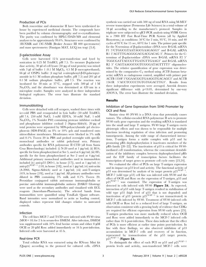
detected in cells infected with SV40 (Figure 2A). As expected,
interaction of p53 with large T-antigen resulted in stabilization of
wild type p53 (high level of p53 that is not degraded) and
inactivation of p53 (protein p21WAF21 is not transactivated) in
MCF-7 cells infected by SV40. Treatment of SV40 infected cells
with OCII or Rosc led to a reduced level of large T-antigen, an
observation consistent with a growing body of evidence that CDKs
are required for efficient expression from SV40 early genes [25].
T-antigen production was more markedly reduced when OCII
and Rosc were added immediately to the MCF-7 infected cells
rather than 16 h post-infection. These data indicate that the effect
of PCIs is more efficient at earlier time points post-infection. In
line with these findings, we also observed inhibition of p53
accumulation in MCF-7 cells and recovery of its function,
represented by transactivation of the p53 target protein,
p21WAF21 (Figure 2A).
To distinguish the effect of each PCI on p53 and p21WAF21
protein levels and activity, non-transfected MCF-7 cells were
OLII Reduces Extrachromosomal Genes Expression
PLOS ONE | www.plosone.org 3 February 2014 | Volume 9 | Issue 2 | e89228
treated with OCII or Rosc. Both agents enhanced the protein
levels of p53 and p21WAF21 (that is tightly controlled by p53) in
MCF-7 cells that normally leads to cell cycle arrest (Figure 2B).
To ascertain whether PCIs inhibited only large T-antigen
expression or whether their effects were connected generally with
SV-40 promoter transcription, we tested their inhibitory effects on
an isolated viral promoter within a plasmid vector. MCF-7 cells
transiently transfected with plasmid pSV-lacZ were treated with
OCII or Rosc and relative b-galactosidase activities determined.
We observed significant (p.0.05) reductions of b-galactosidaseactivity in cells treated with PCIs (Figure 3A) indicating inhibitionof b-galactosidase expression from the viral SV-40 promoter.
These results showed that PCIs affected gene expression from the
SV40 promoter.
The Effect of PCIs on Expression of T-antigen IntegratedInto the Cellular GenomeIt has been described that virus replication or transcription can
be more sensitive to CDK9 or p-TEFb activity than transcription
of cellular genes [26,27]. Moreover, while we observed inhibition
of expression from viral promoter, expression of cellular genes
TP53 and CDKN1A increased (Figure 2B). This observation led
us to test the effect of OCII and Rosc on T-antigen expression in
COS-1 cells, where the sequences encoding large T-antigen are
integrated into the cellular genome. COS-1 is an African green
monkey kidney cell line that was established from CV-1 by
transformation with an origin-defective mutant of SV40. CV-1 cell
line was used as a control. While expression of T-antigen in SV40-
infected cells (SV40 genome was not integrated) was inhibited by
PCIs (Figure 2A), in COS-1 cells OCII and Rosc treatment
stimulated large T-antigen expression, while p53 levels remained
constant and the protein transcriptionally inactive (p21WAF21 was
not activated). Conversely, in the control CV-1 cells, the
transcriptional activity of p53 and in turn p21WAF21 levels
increased after the PCI treatment (Figure 3B). These results
indicate that PCIs may have either an inhibiting or a stimulating
effect on a viral promoter, depending on its location.
Inhibition of Expression from the HIV PromoterTo further analyze the effect that PCIs exert on expression from
integrated and extrachromosomal viral DNA elements, we
decided to test expression from HIV promoter, taking advantage
of the fact that transcription from the HIV genome is sensitive to
CDK9 activity and can be suppressed by PCI treatment at
concentrations that do not impair cellular transcription [26,27].
HIV requires a viral transactivator called Tat (Trans-Activator of
Transcription) to stimulate the processivity of RNAPII and
initiation from the viral LTR promoter. In the absence of Tat,
only short, abortive transcripts are generated.
Transcription elongation factor p-TEFb represents a cellular co-
factor for HIV-1 Tat transcriptional activation and it was found
that CDK9 interacts functionally with the transactivation domain
of Tat, illustrated by the fact that CDK9 depletion blocks Tat-
dependent transactivation and activity [28,29]. Tat is also
phosphorylated by CDK2 and this modification is important both
for HIV-1 transcription and activation of integrated HIV-1
provirus [30]. It was described that Roscovitine as CDK2 and
CDK9 inhibitor effectively suppress wild type and resistant HIV-1
mutants and could selectively sensitize HIV-1-infected cells to
apoptosis at concentrations that did not impede the growth and
proliferation of uninfected cells [31–33].
We constructed two stable cell lines: H1299-Tat (with
incorporated plasmid pCEP4-Tat) and H1299-HIV (with incor-
porated plasmids pCEP4-Tat and pHIV-lacZ). Control cell line
H1299 and H1299-Tat cells were transiently transfected with a
plasmid encoding b-galactosidase under the control of the
promoter in the HIV LTR (pHIV-lacZ) and together with
H1299-HIV cells were treated with OCII or Rosc followed by
b-galactosidase activity measurement. The expression of the
reporter in H1299-Tat stable cell line transiently transfected with
pHIV-lacZ was inhibited by PCI treatment and more efficiently by
OCII than by Rosc (Figure 4). However, an inverse effect of PCI
treatment on the HIV promoter integrated into the cellular
genome (H1299-HIV cells) was observed and the expression of b-galactosidase in H1299-HIV cells increased about 4-fold following
treatment with OCII or 2.5-fold following treatment with Rosc
(p,0.05) (Figure 4). These findings are in agreement with results
obtained with SV40 (Figure 3B) and indicate that effect of PCIs is
not limited to one type of virus promoter, and is dependent on
virus promoter extrachromosomal localization.
Phosphorylation of the C-terminal Domain of RNAPolymerase IIModulation of expression from the HIV promoter by PCI
treatment may be mediated by transcriptional regulation. The C-
terminus of the largest subunit of RNAP II contains a key
regulatory domain referred to as CTD (C-terminal domain).
Therefore, we investigated the effect of PCIs on phosphorylation
of CTD at positions Ser 2 and Ser 5. The levels of both Ser 2 and
Ser 5 phosphorylation in OCII or Rosc treated COS-1, CV-1 and
MCF-7 cells were substantially reduced (Figure 5A). Although the
phosphorylation of RNAP II was suppressed by PCIs, the
expression from viral promoter integrated into the host cell
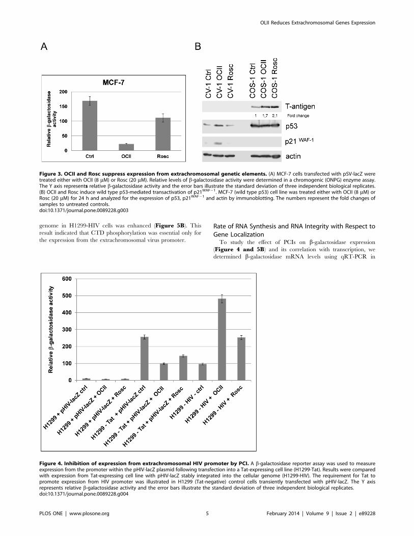
Figure 2. OCII and Rosc suppress T-antigen expression. (A) OCIIand Rosc suppress T-antigen expression and restore p53–mediatedtransactivation of p21WAF21 during SV40 infection. MCF-7 (wild typep53) cell line infected with SV40 was treated either with 8 mM OCII or20 mM Rosc, either immediately or 16 h post-infection. (OCII 16 h, Rosc16 h). Infected cells were harvested at 45 h and then analyzed for theexpression of large T-antigen, p53, p21WAF21 and actin by immuno-blotting. (B) Inhibition of expression from viral promoter integrated intocellular genome. Cell lines CV-1 and COS-1 were treated either with8 mM OCII or 20 mM Rosc and analyzed for the expression of large T-antigen, p53, p21WAF21 and actin by immunoblotting. CV-1 cell line wasused as SV40-negative control cell line (no large T-antigen expression).The numbers represent the fold changes of samples to untreatedcontrols.doi:10.1371/journal.pone.0089228.g002
OLII Reduces Extrachromosomal Genes Expression
PLOS ONE | www.plosone.org 4 February 2014 | Volume 9 | Issue 2 | e89228
genome in H1299-HIV cells was enhanced (Figure 5B). Thisresult indicated that CTD phosphorylation was essential only for
the expression from the extrachromosomal virus promoter.
Rate of RNA Synthesis and RNA Integrity with Respect toGene LocalizationTo study the effect of PCIs on b-galactosidase expression
(Figure 4 and 5B) and its correlation with transcription, we
determined b-galactosidase mRNA levels using qRT-PCR in
Figure 3. OCII and Rosc suppress expression from extrachromosomal genetic elements. (A) MCF-7 cells transfected with pSV-lacZ weretreated either with OCII (8 mM) or Rosc (20 mM). Relative levels of b-galactosidase activity were determined in a chromogenic (ONPG) enzyme assay.The Y axis represents relative b-galactosidase activity and the error bars illustrate the standard deviation of three independent biological replicates.(B) OCII and Rosc induce wild type p53-mediated transactivation of p21WAF21. MCF-7 (wild type p53) cell line was treated either with OCII (8 mM) orRosc (20 mM) for 24 h and analyzed for the expression of p53, p21WAF21 and actin by immunoblotting. The numbers represent the fold changes ofsamples to untreated controls.doi:10.1371/journal.pone.0089228.g003
Figure 4. Inhibition of expression from extrachromosomal HIV promoter by PCI. A b-galactosidase reporter assay was used to measureexpression from the promoter within the pHIV-lacZ plasmid following transfection into a Tat-expressing cell line (H1299-Tat). Results were comparedwith expression from Tat-expressing cell line with pHIV-lacZ stably integrated into the cellular genome (H1299-HIV). The requirement for Tat topromote expression from HIV promoter was illustrated in H1299 (Tat-negative) control cells transiently transfected with pHIV-lacZ. The Y axisrepresents relative b-galactosidase activity and the error bars illustrate the standard deviation of three independent biological replicates.doi:10.1371/journal.pone.0089228.g004
OLII Reduces Extrachromosomal Genes Expression
PLOS ONE | www.plosone.org 5 February 2014 | Volume 9 | Issue 2 | e89228
stable cell lines H1299-HIV and H1299-Tat transiently transfect-
ed by pHIV-lacZ.
Previous work described the importance of CTD phosphoryla-
tion for RNA posttranscriptional processing rather than for RNA
synthesis, where Serine-5 phosphorylation was associated with
recruitment of capping enzymes to 59 ends, whereas Ser 2
phosphorylation was associated with recruitment of cleavage and
polyadenylation factors to the 39 ends [34].
Therefore, we developed real time PCR based assays to analyze
the amount of RNAs and its polyadenylation. Random hexamers
were used to quantify total transcribed b-galactosidase whereas
oligo dT primers were used to analyze only polyadenylated RNA.
The integrity of 59 or 39 end of RNA was assessed using PCR
primer pairs amplifying the corresponding parts of the RNA
termini (Figure 6).
We found that PCIs stimulated the initiation of RNA synthesis
from viral promoters regardless of their localization, as suggested
by the increased levels of PCR-1 transcripts after PCI treatments
compared to control PCR-1 (p,0.001) (Figure 7A, Figure 7Brandom hexamers). However, full length polyadenylated RNA
transcripts were generated to a large extent only from viral
promoters incorporated in the cellular genome (increased levels of
PCR-2 after treatments compared to control, (p,0.001),
Figure 7A, Oligo dT). Conversely, transcripts produced from
extrachromosomal viral promoters after PCI treatments were
probably much shorter and abortive because of reduced amounts
of amplified PCR-2 products from cDNA templates generated
using oligo dT primers in H1299-Tat cells (Figure 7B, Oligo dT).
We also tested the levels of Tat mRNA after OCII and Rosc
treatments and did not find any significant changes (Figure S1).
Inhibition of Expression from the HIV Promoter byFlavopiridolInhibitors of CDK9 have been characterized as inhibitors of
viral transcription and replication [2]. We tested whether the effect
on extrachromosomal and integrated viral promoter is uniform for
these agents. Flavopiridol (FVP) is a highly specific P-TEFb
(CycT1:CDK9) kinase inhibitor that dramatically reduces the
global levels of Ser 2 phosphorylated CTD [27]. We therefore
tested the influence of FVP on RNAP II CTD phosphorylation,
RNA synthesis and protein expression from viral promoters
(Figures S2, S3). H1299-Tat and H1299-HIV cells were treated
with two concentrations of FVP (25 and 100 nM). Both doses
decreased the expression of mRNA from extrachromosomal HIV
promoter in H1299-Tat cells (p,0.05) and in turn led to a
reduction of b-galactosidase protein. The lower concentration of
FVP (25 nM) did not change the expression of b-galactosidasefrom HIV promoter incorporated into the genome of H1299-HIV
cells (p,0.05). In contrast, 100 nM FVP induced expression from
this promoter (p,0.05) and increased the level of b-galactosidaseprotein similarly to OCII and Rosc treatments (Figures S2, S3).FVP in contrast to OCII and Rosc did not increase the rate of
mRNA synthesis from both viral promoters (except higher
concentration of FVP in H1299-HIV cells, p,0.05). However,
FVP impacted on mRNA elongation initiated from these
promoters as indicated by differences in the levels of N- and C-
terminal b-galactosidase PCR products.
Discussion
We have demonstrated that OCII is a potent inhibitor of viral
transcription with an inhibitory effect stronger than Rosc.
Activities of both agents have been variously associated with the
inhibition of CDK1/cyclin B, CDK2/cyclin E, CDK2/cyclin A
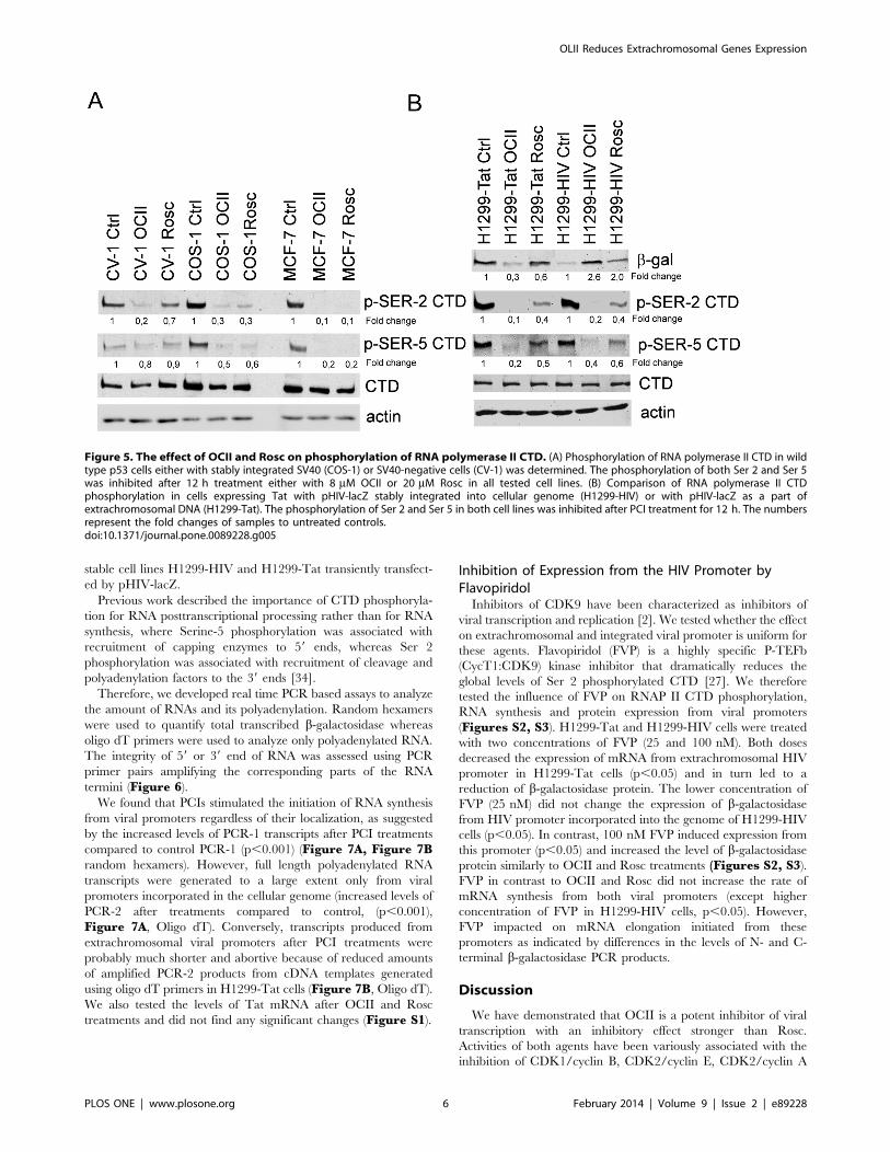
Figure 5. The effect of OCII and Rosc on phosphorylation of RNA polymerase II CTD. (A) Phosphorylation of RNA polymerase II CTD in wildtype p53 cells either with stably integrated SV40 (COS-1) or SV40-negative cells (CV-1) was determined. The phosphorylation of both Ser 2 and Ser 5was inhibited after 12 h treatment either with 8 mM OCII or 20 mM Rosc in all tested cell lines. (B) Comparison of RNA polymerase II CTDphosphorylation in cells expressing Tat with pHIV-lacZ stably integrated into cellular genome (H1299-HIV) or with pHIV-lacZ as a part ofextrachromosomal DNA (H1299-Tat). The phosphorylation of Ser 2 and Ser 5 in both cell lines was inhibited after PCI treatment for 12 h. The numbersrepresent the fold changes of samples to untreated controls.doi:10.1371/journal.pone.0089228.g005
OLII Reduces Extrachromosomal Genes Expression
PLOS ONE | www.plosone.org 6 February 2014 | Volume 9 | Issue 2 | e89228
and to a lesser extent CDK4/cyclin D. In addition to inhibitory
effects on kinases directly involved in the regulation of the cell
cycle, OCII and Rosc also inhibit CDK7/cyclin H and CDK9/
cyclin T, the non-cell cycle kinases [35,36]. OCII is more selective
for CDK9 than Rosc, whereas CDK2 and CDK7 activities are
affected approximately to the same extent by both drugs. On the
other hand Rosc is a stronger inhibitor of CDK1, CDK4 and Erk2
[5].
We observed that OCII and Rosc mediated suppression of large
T-antigen expression during SV40 infection. The reduced level of
T-antigen was connected with releasing p53 from inhibitory
complex with T-antigen and with restoration of p53 transcrip-
tional activity. Interestingly, this suppression was dependent on the
time period of the MCF-7 cell line treatment, as the depletion of
T-antigen was significantly lower when PCIs were added 16 h
post-infection with SV40. Likewise, time-dependent effects have
also been described for Rosc treatment in HCMV-infected cells,
where the level of inhibition was dependent on whether the drug
was added before or after the onset of viral DNA replication [37].
Addition of Rosc at the time of infection altered the accumulation
and processing of selected early genes and inhibited HCMV DNA
replication. Delaying the addition of the drug until 6 h post-
infection abrogated the deleterious effect on early gene expression
and viral DNA synthesis [37,38]. Comparable effects have also
been observed in cells infected with herpes simplex virus [25].
These data indicate that the mechanism of OCII and Rosc
inhibitory effects on SV40 could be the same as for HCMV and
the impact of PCIs is much stronger when they are added before
SV40 early gene expression and DNA replication. We confirm the
influence of PCIs on expression from SV40 promoter using pSV-
lacZ vector, where a stronger effect of OCII was identified.
Transcription of viral genes was described to be more sensitive to
inhibition of CDK9 or p-TEF-b activity than cellular genes. We
tested if the inhibitory effects of OCII and Rosc on expression
from viral promoters changed when the virus is incorporated into
cellular genome. We utilized the COS-1 cell line containing
defective mutant of SV40. We found that PCI treatments
surprisingly increased the transcription of T-antigen in COS-1
cells. Likewise, the complex of p53/T-antigen remained stable and
the transcriptional activity was limited. The same results were
obtained with stable cell lines H1299-Tat and H1299-HIV, where
the treatment with PCIs increased the expression from HIV
promoter and the effect of OCII was markedly stronger.
Casse et al. reported that low concentrations of actinomycin D
and a-amatinin (inhibitor of RNA transcription and inhibitor of
RNAP II) enhance transcriptional elongation from the HIV-1
LTR. This effect may result from increased CDK9 activity
induced by transcriptional arrest. In HeLa cells, ,50% of CDK9/
cyclin T complexes are kinase-inactive and bound to 7SK small
nuclear RNA [39]. The inhibition of transcription with actino-
mycin D or a-amatinin induces rapid release of active CDK9/
cyclin T complexes and thereby promotes an increase in the
proportion of RNAP II phosphorylated on its CTD [40]. CTD
phosphorylation can also be initiated by osmotic shock, UV-
irradiation, okadaic acid or heat shock. All these factors activate
HIV LTR transcription, however this enhancement can be
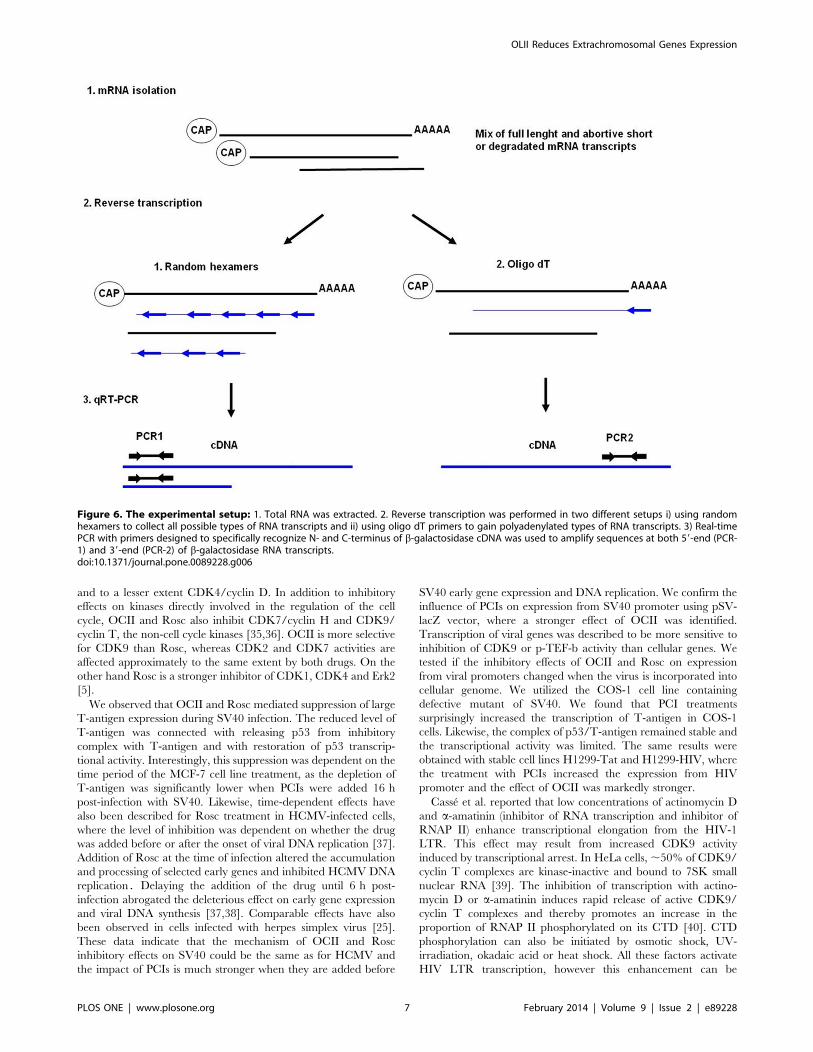
Figure 6. The experimental setup: 1. Total RNA was extracted. 2. Reverse transcription was performed in two different setups i) using randomhexamers to collect all possible types of RNA transcripts and ii) using oligo dT primers to gain polyadenylated types of RNA transcripts. 3) Real-timePCR with primers designed to specifically recognize N- and C-terminus of b-galactosidase cDNA was used to amplify sequences at both 59-end (PCR-1) and 39-end (PCR-2) of b-galactosidase RNA transcripts.doi:10.1371/journal.pone.0089228.g006
OLII Reduces Extrachromosomal Genes Expression
PLOS ONE | www.plosone.org 7 February 2014 | Volume 9 | Issue 2 | e89228
suppressed by CDK9 inhibitors [39]. OCII and Rosc induce
transcription stress [5] and would therefore be expected to release
active CDK9/cyclin complex. Therefore we tested the level of
CTD RNAP II phosphorylation in cells treated by OCII and
Rosc.
The C-terminal domain of largest subunit of RNA PII contains
a series of ‘‘YSPTSPS’’ heptapeptide repeats that are multiply-
phosphorylated during eukaryotic transcription. In the process of
CTD phosphorylation at Ser 2 by P-TEFb, the CDK9/cyclin T1
complex has been implicated in productive elongation and the
39end processing of the primary transcripts such as 39end cleavage
and polyadenylation [41,42]. CTD phosphorylation at Ser 5 by
TFIIH kinase is a critical step in mRNA synthesis, namely RNAP
II promoter clearance (for transition from initiation to early
elongation) and mRNA 59-capping. We found that OCII and also
Rosc dramatically decreased the phosphorylation of CTD,
especially at Ser 2 but also at Ser 5 in all tested cell lines. Hong
et al. [43] demonstrated that inhibition of TFIIH resulted in
defective mRNA capping for the majority of yeast genes. This led
to the degradation of mRNAs by exonucleases, resulting in a
dramatic reduction in steady-state mRNA levels. However,
inhibition of the TFIIH kinase did not significantly affect other
transcriptional processes, such as overall RNAP II density and
CTD phosphorylation at Ser 2. CDK9, the main CTD Ser 2
kinase, is critical for 39end processing of pre-mRNA, as well as for
RNAP II transcription but to a lesser extent [13,44]. OCII and
Rosc as potent inhibitors of CDK9 and CDK7 could reduce the
expression from viral promoters in both ways. We demonstrate
that PCIs increase the initiation of RNA synthesis from viral
promoters, but newly produced transcripts are immediately
degraded due to defects in pre-mRNA elongation, 39end
processing and possibly in 59-capping as well. Importantly, we
observed this effect significantly in case of extrachromosomal
plasmids and viral infections but only if the viral promoter was not
incorporated into the cellular genome. The above mentioned
variable effects of OCII and Rosc on expression from viral
promoters are undoubtedly connected with localization of these
promoters in host chromosome. Pirngruber et al. described that
CDK9 plays an important role in the regulation of transcription
not only by directing RNAP II activity but also through the
modification of chromatin binding and modifying factors. CDK9
guides a complex network of chromatin modifications including
histone H2B monoubiquitination (H2Bub1), H3 lysine 4 trimethy-
lation (H3K4me3) and H3K36me3. CDK9 directs H2Bub1,
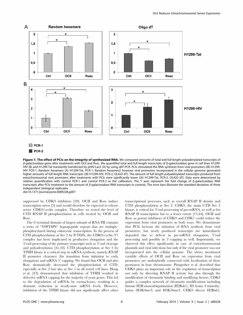
Figure 7. The effect of PCIs on the integrity of synthesized RNA.We compared amounts of total and full length polyadenylated transcripts ofb-galactosidase gene after treatments with OLII and Rosc. We quantified total and full length transcripts of b-galactosidase gene in cell lines H1299-HIV (B) and H1299-Tat transiently transfected by pHIV-LacZ (A) by using qRT-PCR. PCIs stimulated the RNA synthesis from viral promoters ((B) H1299-HIV PCR-1, Random hexamers (A) H1299-Tat, PCR-1, Random hexamers) however viral promoters incorporated in the cellular genome generatedhigher amounts of full length RNA transcripts ((B) H1299-HIV, PCR-2, OLIGO dT). The amount of full length polyadenylated transcripts produced fromextrachomosomal viral promoters after treatments with PCIs were significantly lower ((A) H1299-Tat, PCR-2, OLIGO dT). Data were determined byrelative quantification with control PCR-1 and control PCR-2 as the calibrators. The Y axes represent the fold change of b-galactosidase RNAtranscripts after PCIs treatment to the amount of b-galactosidase RNA transcripts in controls. The error bars illustrate the standard deviation of threeindependent biological replicates.doi:10.1371/journal.pone.0089228.g007
OLII Reduces Extrachromosomal Genes Expression
PLOS ONE | www.plosone.org 8 February 2014 | Volume 9 | Issue 2 | e89228
which influences the accessibility of the newly synthesized mRNA
or the recruitment of 39end processing machinery by modifying
the chromatin structure [45]. CDK9 knockdown increases the
production of polyadenylated transcripts of replication-dependent
histone mRNAs. Thus, CDK9 acts to integrate phosphorylation
during transcription with chromatin modifications to control co-
transcriptional histone pre-mRNA processing [45]. The increase
in the polyadenylation of pre-mRNA is likely to be specific for this
class of mRNAs and not a general defect in polyadenylation site
selection [46]. It is likely that chromatin arrangement of viral
DNA is changed after incorporation in host chromosome, so the
described phenomenon could help to explain the different effects
of PCIs on extra- and intrachromosomal viral promoters. Our
statement is in line with published results, where Zhou et al. [47]
demonstrated that the presence of H3K4me3 and H3K36me3 on
the HIV-1 gene decreased following CDK9 inhibition by
flavopiridol. The dependence of HIV-1 transcription on chroma-
tin modification and histone acetylation was also demonstrated by
van Lint et al. [48]. They demonstrated that global hyperacetyla-
tion of cellular histones in cells latently infected with HIV-1
increases the transcriptional activation of the HIV-1 promoter.
Moreover Keskin et al. [49] described the upregulation of some
Primary Response Genes (PRGs) after treatment with FVP. PRGs
have constitutively open chromatin architecture and show high
level of preexisting H3K4me3 across the promoter region as well
as H3K36me3 in coding region [50].
In summary, we show that PCIs inhibit gene expression form
viral promoters by generating defects in pre-mRNA elongation
and 39end processing. The results indicate that the inhibitory effect
of PCIs on transcription from viral promoters depends on their
localization in cells and the CTD phosphorylation is critical for
linking transcription and posttrancriptional processing of mRNA
expressed particularly from extrachromosomal DNA.
Supporting Information
Figure S1 The expression of Tat gene in cell linesH1299- Tat and H1299-HIV. (A) We evaluated Tat mRNA
basal level in cell lines H1299-HIV and H1299-Tat. Samples in
triplicate were subjected to qRT-PCR analysis using SYBR Green
with the primer pair specific for Tat cDNA: TAT-F 59ATG-
GAGCCAGTAGATCCTAA93 and TAT-R
59GGGTTGCTTTGATAGAGAAGC93. The relative quantifi-
cation of gene expression was determined by the comparative CT
method using ACTB (human beta-actin) mRNA as endogenous
control. The results from three independent experiments were
averaged and the error bars illustrate the standard deviation. We
found that in both cell lines are the comparable amounts of Tat
mRNAs (A) that did not shown any significant changes after OCII
and Rosc treatments ((B) - H1299-Tat, (C) – H1299-HIV). The Y
axes represent the fold change of Tat RNA transcripts in H1299-
Tat and H1299-HIV cell lines compared to negative control
H1299 (without transfected vector pCEP4-Tat) (A) and the fold
change of Tat RNA transcripts after PCI treatment to the amount
of Tat RNA transcripts in controls (B, C). The error bars illustrate
the standard deviation of three independent biological replicates.
(TIF)
Figure S2 Inhibition of expression from the HIVpromoter using Flavopiridol. H1299-Tat and H1299-HIV
cell lines were treated with Flavopiridol (25 nM and 100 nM) for
12 h and the levels of RNA polymerase II CTD phosphorylation
on Ser-2 and Ser-5, b-galactosidase protein and actin were
analyzed by immunoblotting. FVP moderately decreased phos-
phorylation of Ser 2 RNA polymerase II CTD and significantly
decreased the level of b-galactosidase protein in H1299-Tat cells.
The impact of FVP in H1299-HIV cells was dependent on its
concentration. The effect of 25 nM FVP was similar in both cell
lines. In contrast, 100 nM FVP (similar to OCII and Rosc)
increased the level of b-galactosidase protein in H1299-HIV cells.
(TIF)
Figure S3 The effect of Flavopiridol on the integrity ofsynthesized RNA. qRT-PCR was performed in H1299-HIV
and H1299-Tat cell lines treated with 25 nM and 100 nM FVP.
Total RNA was extracted and reverse transcription was performed
in two different setups i) using random hexamers and ii) oligo dT
primers to gain all possible types of RNA transcripts. Real-time
PCR with primers designed to specifically recognize N- and C-
terminus of b-galactosidase cDNA was used to amplify sequences
at both 59- and 39-end of b-galactosidase RNA transcripts. We
compared the amounts of full length and short abortive transcripts
of b-galactosidase gene. The effect of FVP was dependent on the
concentration. 25 nM FVP did not increase expression from either
viral promoter (PCR-1 random hexamers) and decreased the
quantity of b-galactosidase full length mRNA transcripts (PCR-2
oligo dT). Treatment by 100 nM FVP increased the expression
from HIV-promoter (PCR-1 random hexamers) and the number
of b-galactosidase full length mRNA transcripts in H1299-HIV
cells (PCR-2 oligo dT).
(TIF)
Author Contributions
Conceived and designed the experiments: JH PM. Performed the
experiments: JH PM MN. Analyzed the data: RH PT. Contributed
reagents/materials/analysis tools: VK MS. Wrote the paper: JH PM GW
BV.
References
1. Knockaert M, Greengard P, Meijer L (2002) Pharmacological inhibitors of
cyclin-dependent kinases. Trends Pharmacol Sci 23: 417–425.
2. Arris CE, Boyle FT, Calvert AH, Curtin NJ, Endicott JA, et al. (2000)Identification of novel purine and pyrimidine cyclin-dependent kinase inhibitors
with distinct molecular interactions and tumor cell growth inhibition profiles.J Med Chem 43: 2797–2804.
3. Havlicek L, Hanus J, Vesely J, Leclerc S, Meijer L et al. (1997) Cytokinin-
derived cyclin-dependent kinase inhibitors: synthesis and cdc2 inhibitory activityof olomoucine and related compounds. J Med Chem 40: 408–412.
4. Krystof V, Lenobel R, Havlicek L, Kuzma M, Strnad M (2002) Synthesis and
biological activity of olomoucine II. Bioorg Med Chem Lett 12: 3283–3286.
5. Krystof V, McNae IW, Walkinshaw MD, Fischer PM, Muller P, et al. (2005)
Antiproliferative activity of olomoucine II, a novel 2,6,9-trisubstituted purine
cyclin-dependent kinase inhibitor. Cell Mol Life Sci 62: 1763–1771.
6. Bresnahan WA, Boldogh I, Chi P, Thompson EA, Albrecht T (1997) Inhibition
of cellular Cdk2 activity blocks human cytomegalovirus replication. Virology
231: 239–247.
7. Mancebo HS, Lee G, Flygare J, Tomassini J, Luu P, et al. (1997) P-TEFb kinaseis required for HIV Tat transcriptional activation in vivo and in vitro. Genes
Dev 11: 2633–2644.
8. Schang LM, Bantly A, Knockaert M, Shaheen F, Meijer L, et al. (2002)Pharmacological cyclin-dependent kinase inhibitors inhibit replication of wild-
type and drug-resistant strains of herpes simplex virus and human immunode-ficiency virus type 1 by targeting cellular, not viral, proteins. J Virol 76: 7874–
7882.
9. Schang LM, St Vincent MR, Lacasse JJ (2006) Five years of progress on cyclin-dependent kinases and other cellular proteins as potential targets for antiviral
drugs. Antivir Chem Chemother 17: 293–320.
10. Diwan P, Lacasse JJ, Schang LM (2004) Roscovitine inhibits activation ofpromoters in herpes simplex virus type 1 genomes independently of promoter-
specific factors. J Virol 78: 9352–9365.
11. Pisell TL, Ho O, Lee G, Butera ST (2001) Spectrum of cdk-9 inhibitor activity
against HIV-1 replication among various models of chronic and latent infection.
Antivir Chem Chemother 12 Suppl 1: 33–41.
OLII Reduces Extrachromosomal Genes Expression
PLOS ONE | www.plosone.org 9 February 2014 | Volume 9 | Issue 2 | e89228
12. Gale M, Jr., Tan SL, Katze MG (2000) Translational control of viral gene
expression in eukaryotes. Microbiol Mol Biol Rev 64: 239–280.13. Ahn SH, Kim M, Buratowski S (2004) Phosphorylation of serine 2 within the
RNA polymerase II C-terminal domain couples transcription and 39 end
processing. Mol Cell 13: 67–76.14. Holcakova J, Tomasec P, Bugert JJ, Wang EC, Wilkinson GW, et al. (2010) The
inhibitor of cyclin-dependent kinases, olomoucine II, exhibits potent antiviralproperties. Antivir Chem Chemother 20: 133–142.
15. Maio JJ, Brown FL (1988) Regulation of expression driven by human
immunodeficiency virus type 1 and human T-cell leukemia virus type I longterminal repeats in pluripotential human embryonic cells. J Virol 62: 1398–
1407.16. Chang LJ, Urlacher V, Iwakuma T, Cui Y, Zucali J (1999) Efficacy and safety
analyses of a recombinant human immunodeficiency virus type 1 derived vectorsystem. Gene Ther 6: 715–728.
17. Vojtesek B, Bartek J, Midgley CA, Lane DP (1992) An immunochemical analysis
of the human nuclear phosphoprotein p53. New monoclonal antibodies andepitope mapping using recombinant p53. J Immunol Methods 151: 237–244.
18. Fredersdorf S, Milne AW, Hall PA, Lu X (1996) Characterization of a panel ofnovel anti-p21Waf1/Cip1 monoclonal antibodies and immunochemical analysis
of p21Waf1/Cip1 expression in normal human tissues. Am J Pathol 148: 825–
835.19. Harlow E, Crawford LV, Pim DC, Williamson NM (1981) Monoclonal
antibodies specific for simian virus 40 tumor antigens. J Virol 39: 861–869.20. Stubdal H, Zalvide J, DeCaprio JA (1996) Simian virus 40 large T antigen alters
the phosphorylation state of the RB-related proteins p130 and p107. J Virol 70:2781–2788.
21. Sullivan CS, Cantalupo P, Pipas JM (2000) The molecular chaperone activity of
simian virus 40 large T antigen is required to disrupt Rb-E2F family complexesby an ATP-dependent mechanism. Mol Cell Biol 20: 6233–6243.
22. Zalvide J, Stubdal H, DeCaprio JA (1998) The J domain of simian virus 40 largeT antigen is required to functionally inactivate RB family proteins. Mol Cell Biol
18: 1408–1415.
23. Ali SH, Kasper JS, Arai T, DeCaprio JA (2004) Cul7/p185/p193 binding tosimian virus 40 large T antigen has a role in cellular transformation. J Virol 78:
2749–2757.24. Kasper JS, Kuwabara H, Arai T, Ali SH, DeCaprio JA (2005) Simian virus 40
large T antigen’s association with the CUL7 SCF complex contributes to cellulartransformation. J Virol 79: 11685–11692.
25. Schang LM, Rosenberg A, Schaffer PA (1999) Transcription of herpes simplex
virus immediate-early and early genes is inhibited by roscovitine, an inhibitorspecific for cellular cyclin-dependent kinases. J Virol 73: 2161–2172.
26. Chao SH, Fujinaga K, Marion JE, Taube R, Sausville EA, et al. (2000)Flavopiridol inhibits P-TEFb and blocks HIV-1 replication. J Biol Chem 275:
28345–28348.
27. Chao SH, Price DH (2001) Flavopiridol inactivates P-TEFb and blocks mostRNA polymerase II transcription in vivo. J Biol Chem 276: 31793–31799.
28. Yang X, Gold MO, Tang DN, Lewis DE, Aguilar-Cordova E, et al. (1997)TAK, an HIV Tat-associated kinase, is a member of the cyclin-dependent family
of protein kinases and is induced by activation of peripheral blood lymphocytesand differentiation of promonocytic cell lines. Proc Natl Acad Sci U S A 94:
12331–12336.
29. Zhu Y, Pe’ery T, Peng J, Ramanathan Y, Marshall N, et al. (1997) Transcriptionelongation factor P-TEFb is required for HIV-1 tat transactivation in vitro.
Genes Dev 11: 2622–2632.30. Ammosova T, Berro R, Jerebtsova M, Jackson A, Charles S, et al. (2006)
Phosphorylation of HIV-1 Tat by CDK2 in HIV-1 transcription. Retrovirology
3: 78.
31. Guendel I, Agbottah ET, Kehn-Hall K, Kashanchi F (2010) Inhibition ofhuman immunodeficiency virus type-1 by cdk inhibitors. AIDS Res Ther 7: 7.
32. Agbottah E, de La Fuente C, Nekhai S, Barnett A, Gianella-Borradori A, et al.
(2005) Antiviral activity of CYC202 in HIV-1-infected cells. J Biol Chem 280:3029–3042.
33. Wang D, de la Fuente C, Deng L, Wang L, Zilberman I, et al. (2001) Inhibition
of human immunodeficiency virus type 1 transcription by chemical cyclin-dependent kinase inhibitors. J Virol 75: 7266–7279.
34. Garrido-Lecca A, Blumenthal T (2010) RNA polymerase II C-terminal domain
phosphorylation patterns in Caenorhabditis elegans operons, polycistronic geneclusters with only one promoter. Mol Cell Biol 30: 3887–3893.
35. Fischer PM, Endicott J, Meijer L (2003) Cyclin-dependent kinase inhibitors.
Prog Cell Cycle Res 5: 235–248.
36. Morgan DO (1995) Principles of CDK regulation. Nature 374: 131–134.
37. Sanchez V, McElroy AK, Yen J, Tamrakar S, Clark CL, et al. (2004) Cyclin-
dependent kinase activity is required at early times for accurate processing andaccumulation of the human cytomegalovirus UL122–123 and UL37 immediate-
early transcripts and at later times for virus production. J Virol 78: 11219–
11232.
38. Sanchez V, Spector DH (2006) Cyclin-dependent kinase activity is required for
efficient expression and posttranslational modification of human cytomegalovi-
rus proteins and for production of extracellular particles. J Virol 80: 5886–5896.
39. Casse C, Giannoni F, Nguyen VT, Dubois MF, Bensaude O (1999) The
transcriptional inhibitors, actinomycin D and alpha-amanitin, activate the HIV-1 promoter and favor phosphorylation of the RNA polymerase II C-terminal
domain. J Biol Chem 274: 16097–16106.
40. Nguyen VT, Kiss T, Michels AA, Bensaude O (2001) 7SK small nuclear RNAbinds to and inhibits the activity of CDK9/cyclin T complexes. Nature 414:
322–325.
41. Phatnani HP, Greenleaf AL (2006) Phosphorylation and functions of the RNApolymerase II CTD. Genes Dev 20: 2922–2936.
42. Orphanides G, Reinberg D (2002) A unified theory of gene expression. Cell 108:
439–451.
43. Hong SW, Hong SM, Yoo JW, Lee YC, Kim S, et al. (2009) Phosphorylation of
the RNA polymerase II C-terminal domain by TFIIH kinase is not essential for
transcription of Saccharomyces cerevisiae genome. Proc Natl Acad Sci U S A106: 14276–14280.
44. Ni Z, Schwartz BE, Werner J, Suarez JR, Lis JT (2004) Coordination of
transcription, RNA processing, and surveillance by P-TEFb kinase on heat shockgenes. Mol Cell 13: 55–65.
45. Pirngruber J, Shchebet A, Schreiber L, Shema E, Minsky N, et al. (2009) CDK9
directs H2B monoubiquitination and controls replication-dependent histonemRNA 39-end processing. EMBO Rep 10: 894–900.
46. Pirngruber J, Shchebet A, Johnsen SA (2009) Insights into the function of the
human P-TEFb component CDK9 in the regulation of chromatin modificationsand co-transcriptional mRNA processing. Cell Cycle 8: 3636–3642.
47. Zhou M, Deng L, Lacoste V, Park HU, Pumfery A, et al. (2004) Coordination of
transcription factor phosphorylation and histone methylation by the P-TEFbkinase during human immunodeficiency virus type 1 transcription. J Virol 78:
13522–13533.
48. Van Lint C, Emiliani S, Ott M, Verdin E (1996) Transcriptional activation and
chromatin remodeling of the HIV-1 promoter in response to histone acetylation.
Embo J 15: 1112–1120.
49. Keskin H, Garriga J, Georlette D, Grana X (2012) Complex effects of
flavopiridol on the expression of primary response genes. Cell Div 7: 11.
50. Fowler T, Sen R, Roy AL (2011) Regulation of primary response genes. MolCell 44: 348–360.
OLII Reduces Extrachromosomal Genes Expression
PLOS ONE | www.plosone.org 10 February 2014 | Volume 9 | Issue 2 | e89228
![[V]. Process of Transcription and Transcriptional Control of Gene Expression 1 RNA polymerases and Initiation of transcription Transcriptional elongation](https://img.pdfslide.us/doc/110x75/56649e595503460f94b52b31/v-process-of-transcription-and-transcriptional-control-of-gene-expression.jpg)


![RNA interference (RNAi) [aka post-transcriptional gene silencing (PTGS)]](https://img.pdfslide.us/doc/110x75/5681446b550346895db0fc8c/rna-interference-rnai-aka-post-transcriptional-gene-silencing-ptgs-56945a2ebdd61.jpg)
