Embed Size (px)
Citation preview
JOURNAL OF VIROLOGY, Sept. 2004, p. 9030–9040 Vol. 78, No. 170022-538X/04/$08.00�0 DOI: 10.1128/JVI.78.17.9030–9040.2004Copyright © 2004, American Society for Microbiology. All Rights Reserved.
Inhibition of Hepatitis C Virus-Like Particle Binding to Target Cellsby Antiviral Antibodies in Acute and Chronic Hepatitis C
Daniel Steinmann,1 Heidi Barth,1 Bettina Gissler,1 Peter Schurmann,1 Mohammed I. Adah,1J. Tilman Gerlach,2 Gerd R. Pape,2 Erik Depla,3 Dirk Jacobs,3 Geert Maertens,3
Arvind H. Patel,4 Genevieve Inchauspe,5 T. Jake Liang,6Hubert E. Blum,1 and Thomas F. Baumert1*
Department of Medicine II, University of Freiburg, Freiburg,1 and Department of Medicine II, Klinikum Grosshadern,University of Munich, Munich,2 Germany; Innogenetics, Ghent, Belgium3; MRC Virology Unit, Institute of
Virology, University of Glasgow, Glasgow, United Kingdom4; CNRS-bioMerieux, Lyon, France5;and Liver Diseases Section, National Institute of Diabetes and Digestive and
Kidney Diseases, National Institutes of Health, Bethesda, Maryland6
Received 9 December 2003/Accepted 6 April 2004
Hepatitis C virus (HCV) is a leading cause of chronic viral hepatitis worldwide. The study of antibody-mediated virus neutralization has been hampered by the lack of an efficient and high-throughput cell culturesystem for the study of virus neutralization. The HCV structural proteins have been shown to assemble intononinfectious HCV-like particles (HCV-LPs). Similar to serum-derived virions, HCV-LPs bind and enterhuman hepatocytes and hepatoma cell lines. In this study, we developed an HCV-LP-based model system fora systematic functional analysis of antiviral antibodies from patients with acute or chronic hepatitis C. Wedemonstrate that cellular HCV-LP binding was specifically inhibited by antiviral antibodies from patients withacute or chronic hepatitis C in a dose-dependent manner. Using a library of homologous overlapping envelopepeptides covering the entire HCV envelope, we identified an epitope in the N-terminal E2 region (SQKIQLVNTNGSWHI; amino acid positions 408 to 422) as one target of human antiviral antibodies inhibiting cellularparticle binding. Using a large panel of serum samples from patients with acute and chronic hepatitis C, wedemonstrated that the presence of antibodies with inhibition of binding activity was not associated with viralclearance. In conclusion, antibody-mediated inhibition of cellular HCV-LP binding represents a convenientsystem for the functional characterization of human anti-HCV antibodies, allowing the mapping of envelopeneutralization epitopes targeted by naturally occurring antiviral antibodies.
Hepatitis C virus (HCV), a member of the Flaviviridae, is amajor cause of chronic viral hepatitis in the world (28, 31).Progression to chronic disease occurs in the majority of HCV-infected persons, and end-stage liver disease due to chronicHCV infection has become the main indication for liver trans-plantation (28, 31). Resolution of chronic infection is ex-tremely rare; more often, chronic infection results in chronichepatitis, liver cirrhosis, or hepatocellular carcinoma (23).Worldwide, an estimated 170 million people are infected withHCV. Treatment options for chronic HCV infection are lim-ited, and a vaccine to prevent HCV infection is not available(15, 23).
Although the humoral and cellular immune responses in-duced by HCV have been analyzed in great detail, the mech-anisms of viral clearance and persistence are still poorly un-derstood (46). HCV can establish persistent infection despite ahumoral and cellular immune response that is generally tar-geted against all viral proteins (30, 46). A strong cellular im-mune response appears to be important for virus clearance (28,46). Chronic HCV infection results in the induction of a stronghumoral immune response (30), and anti-HCV antibodies canbe detected easily by using synthetic peptides or recombinant
proteins in serologic assays (41). Using the chimpanzee model,antibodies with neutralizing properties have been described(18). These antibodies were directed against epitopes in thehypervariable region of the envelope glycoprotein 2 (E2) andappeared to be isolate specific. Antibody-mediated neutraliza-tion is also suggested by a study of patients undergoing livertransplantation for HCV- and hepatitis B virus-related livercirrhosis. Infusion of anti-HBs hyperimmune globulin contain-ing anti-HCV appeared to reduce HCV infection in the trans-planted liver (19). Due to the pending development of conve-nient and efficient model systems for the analysis of largeserum panels for virus neutralization, a systematic analysis ofantibodies with neutralizing properties obtained from patientswith acute or chronic HCV infection is lacking.
Several groups have recently described the synthesis ofHCV-like particles (HCV-LPs) in insect cells using a recom-binant baculovirus containing the cDNA of the HCV structuralproteins core, E1, and E2 (6, 7, 13, 44, 45, 50). Recently, theformation of HCV-LPs in mammalian cells has also been de-scribed (10, 17). HCV-LPs exhibit morphological, biophysical,and antigenic properties similar to those of putative virionsisolated from HCV-infected patients. In contrast to recombi-nant C-terminally truncated HCV E2 protein, the envelopeproteins of HCV-LPs are presumably presented in a native,virion-like conformation. They may, therefore, interact withanti-HCV antibodies directed against nonlinear or conforma-
* Corresponding author. Mailing address: Dept. of Medicine II,University of Freiburg, Hugstetter Strasse 55, D-79106 Freiburg, Ger-many. Phone: (49-761) 270-3401. Fax: (49-761) 270-3259. E-mail:[email protected].
9030
on May 1, 2018 by guest
http://jvi.asm.org/
Dow
nloaded from
tional epitopes of HCV envelope proteins that may representneutralizing epitopes (8, 13, 45). Indeed, a previous study dem-onstrated that antiviral antibodies in acute and chronic HCVinfections interact with HCV-LPs when used as the captureantigen in an enzyme-linked immunosorbent assay (ELISA).Furthermore, HCV-LPs induce potent humoral and cellularimmune responses in vivo and therefore offer a promisingapproach for vaccine development (7, 29, 33, 37).
Recent studies have demonstrated that HCV-LPs interactwith defined human cell lines and hepatocytes similar to viralparticles isolated from human serum. The interaction of HCV-LPs with target cell lines therefore represents a novel modelsystem for the study of viral binding and entry (4, 44, 48, 50).In this study, we demonstrate that the HCV-LP-based systemcan be conveniently used for the functional characterization ofanti-HCV antibodies in acute and chronic hepatitis C. Weshow that cellular binding of HCV-LPs is inhibited by humananti-HCV antibodies, and we define the envelope epitopestargeted by these antibodies. Furthermore, we use this systemas a surrogate model to study the roles of neutralizing anti-bodies in viral clearance and the clinical outcome of HCVinfection.
MATERIALS AND METHODS
Patients. Serial anti-HCV-positive serum samples (n � 93; sampled at timepoints from 0 to 18 months following the diagnosis of HCV infection) wereobtained from 21 patients with acute symptomatic hepatitis C (8 patients withacute self-limited hepatitis and viral clearance and 13 patients with acute hepa-titis that progressed to chronic infection who were prospectively followed at theDepartment of Medicine II, Klinikum Grosshadern, University of Munich, Mu-nich, Germany, between 1995 and 1999 [22]). The diagnosis of acute hepatitis Cwas based on the following criteria (22): (i) elevated alanine aminotransferaselevels at least 20 times the upper limit of normal; (ii) seroconversion to anti-HCV-positive status by second- or third-generation ELISA or recombinant im-munoblot assay II (EIA II [Abbott Laboratories, Chicago, Ill.] and RIBA II[Ortho Diagnostics, Raritan, N.J.]), respectively; (iii) positive PCR for HCVRNA (Amplicor; Roche Diagnostics, Branchburg, N.J.); and (iv) a history ofsudden onset of liver disease in previously healthy individuals. Potential causes ofacute hepatitis, such as other forms of viral hepatitis, autoimmune hepatitis,alcoholic liver disease, toxins, or metabolic etiologies, were ruled out. In addi-tion, sera (n � 10; sampled �6 months following the diagnosis of HCV infection)from 10 patients with chronic HCV infection were obtained from the De-partment of Medicine II, University of Freiburg, Freiburg, Germany. Allpatients were serologically negative for hepatitis B virus and human immu-nodeficiency virus infection. HCV genotypes were determined by INNO-LiPAHCV II (Innogenetics, Ghent, Belgium) or VERSANT(r) HCV Geno-type Assay (Bayer HealthCare LLC, Morristown, N.J.). Sera from patientswith acute symptomatic hepatitis C were analyzed for anti-core, -NS3, -NS4,and -NS5 antibodies using the INNOTESTHCV Ab III assay (Innogenetics).The sera were also analyzed for anti-envelope antibodies using recombinantC-terminally truncated envelope glycoproteins E1 and E2 or a panel of envelope-specific peptides as described previously (45).
Synthesis and purification of HCV-LPs. Procedures for the expression andpurification of HCV-LPs have been described previously (6, 48). HCV-LPs ofgenotype 1b were derived from the cDNA of the HCV-J strain (26), and HCV-LPs of genotype 1a were obtained from the infectious clone H77C (51). Controlpreparations were derived from insect cells infected with a recombinant bacu-lovirus containing the cDNA for �-glucuronidase (GUS). Insect cell controlpreparations served as negative controls in all binding experiments. The HCV-LP E2 concentration was determined by an E2-specific ELISA, as described re-cently (48).
Cell lines. HuH-7 and HepG2 cells were maintained in Dulbecco’s modifiedEagle’s medium (Gibco Life Technologies, Gaithersburg, Md.) containing 10%fetal calf serum (PAA Laboratories, Linz, Austria). The maintenance of Spodo-ptera frugiperda Sf9 insect cells has been described in detail (6).
Analysis of cellular HCV-LP binding by flow cytometry. Binding of HCV-LPsto cell lines was performed as described recently (38). Cells (1.5 � 105 per assay)
were incubated with HCV-LPs or a control insect cell preparation (GUS) inphosphate-buffered saline (PBS)–2% bovine serum albumin (BSA; pH 5.2) (A3912; Sigma-Aldrich, St. Louis, Mo.) (final volume, 100 �l) at various concen-trations for 1 h at 4°C. After the removal of nonbound HCV-LPs by three washeswith PBS–2% BSA, the cells were incubated for 40 min at 4°C with the mousemonoclonal anti-E2 antibodies 16A6 (48) or AP33 (44) or human polyclonalanti-HCV antibody diluted in 50 �l of PBS–2% BSA (1:100). Human polyclonalanti-HCV antibody was obtained from the serum of a 50-year-old female patientwith chronic hepatitis C (HCV genotype 1a) followed at the Department ofMedicine of the University of Freiburg and is available on request. Titration byan E2-specific ELISA (48) revealed an anti-E2 antibody titer of 1:25,600. Theanti-HCV-LP titer was 1:512,000, as determined by an HCV-LP-specific ELISA(8). The cells were washed three times in PBS–2% BSA at 4°C. The cells wereincubated for 30 min at 4°C with phycoerythrin (PE)-conjugated anti-mouseimmunoglobulin G (IgG) antibody (Jackson ImmunoResearch, West Grove, Pa.)or fluorescein isothiocyanate (FITC)-conjugated anti-human IgG antibody (ICNBiomedicals Inc., Aurora, Ohio) diluted in 50 �l of PBS–2% BSA (1:100). Thecells were washed three times in PBS at 4°C. Cell-bound fluorescence wasanalyzed with a FACScalibur flow cytometer (Becton Dickinson) using Cellquestversion 3.11 and CellquestPro software. These programs produce histograms ofeach sample and calculate the mean fluorescence intensity (MFI) of the cellpopulation, which relates directly to the surface density of PE-labeled E2 boundto hepatocytes (38).
Analysis of HCV-LP binding and internalization using immunofluorescenceand laser scanning confocal microscopy. For the study of cellular HCV-LPbinding, HuH-7 cells (0.5 � 105 per assay) were incubated with HCV-LPs(genotype 1a) at 4°C for 40 min. For the study of temperature-dependentHCV-LP entry (44), cells were incubated for an additional 60 min at 37°C. Afterremoval of nonbound HCV-LPs by washing the cells with ice-cold PBS, the cellswere added to poly-L-lysine-coated cover slides, fixed with PBS containing 3.5%paraformaldehyde, and permeabilized using PBS containing 0.1% Triton X-100.The cells were stained for HCV-LP binding and entry using the mouse anti-E2antibody 16A6 (dilution, 1:100 in PBS) and Cy3-conjugated anti-mouse IgG(dilution, 1:250 in PBS; Jackson ImmunoResearch). For costaining of the cyto-skeleton, cells were coincubated with a polyclonal rabbit anti-actin antibody(dilution, 1:200 in PBS; Sigma-Aldrich) and FITC-conjugated anti-rabbit IgG(dilution, 1:500 in PBS; ICN Biomedicals). Between antibody incubations (1 h atroom temperature [RT]), the cells were washed three times in PBS. For stainingof the nucleus, cells were incubated with DRAQ-5 (Biostatus, Shepshed, Leic-estershire, United Kingdom), a highly permeable DNA-interactive agent, ac-cording to the manufacturer’s protocol. Prior to analysis by confocal laser scan-ning microscopy, the cover slides were mounted in antifade reagent(Fluoroguard; Bio-Rad Laboratories, Hercules, Calif.) to minimize photobleach-ing. The stained cells were analyzed in cross section using an LSM 410 laserscanning confocal microscope (Carl Zeiss Corp., Jena, Germany) with argon(488-nm wavelength) and helium-neon (543- and 633-nm wavelength) lasers.Digitalized images were analyzed using Zeiss-LSM Image Browser version 2.8.
Antibody-mediated inhibition of cellular HCV-LP binding. To study whetherHCV-LP binding can be inhibited by serum-derived antibodies, HCV-LPs (0.1�g of HCV-LP E2/ml) were mixed with human serum in various dilutions andincubated for 1 h at 37°C. HuH-7 cells (1.5 � 105) were added to the mixture andincubated for 1 h at 4°C to allow HCV-LP binding. After the cells were washed,the amount of HCV-LP bound to the cells was assessed as described above. Allsera were screened for inhibition of HCV-LP binding using both mouse mono-clonal anti-E2 and human polyclonal anti-HCV antibodies. Only sera demon-strating a decrease in cellular binding of �50% in both assays were consideredinhibitory. Since the supply of human polyclonal anti-HCV antibody was limited,mouse monoclonal anti-E2 antibody was used for further characterization ofinhibition of HCV-LP binding following the assessment of samples in screeningassays. MFI values from HCV-LPs incubated with control serum (positive con-trol), with anti-HCV-positive serum (experimental values), and control prepa-rations (GUS) with control serum (negative control) were measured, and thespecific neutralization was determined as described previously (38), according tothe following equation: specific neutralization � [(positive control MFI � ex-perimental MFI)/(positive control MFI � negative control MFI)] � 100. Theinhibition of the binding titer was defined as the serum dilution that showed�50% inhibition of cellular HCV-LP binding.
IgG purification from human serum. In order to demonstrate that inhibitionof binding was mediated by anti-HCV antibodies, IgG was purified fromHCV-positive and HCV-negative control sera using a MabTrap kit (Amer-sham Biosciences, Freiburg, Germany) (9). Briefly, a HiTrap protein G col-umn was equilibrated with 3 ml of binding buffer. Serum samples were diluted1:1 in binding buffer and applied to the column (total volume, 1 ml). Follow-
VOL. 78, 2004 ANTIVIRAL ANTIBODIES IN ACUTE AND CHRONIC HEPATITIS C 9031
on May 1, 2018 by guest
http://jvi.asm.org/
Dow
nloaded from
ing washing of the column with 7 ml of binding buffer, the purified IgG waseluted using 5 ml of elution buffer and neutralized using 75 �l of neutralizingbuffer according to the manufacturer’s protocol. Fractions were analyzed forthe presence of anti-HCV antibodies using an HCV-LP-based ELISA (8).Subsequently, IgG-containing fractions were pooled and desalted using aHiTrap desalting column (Amersham Biosciences). The purified IgG wasanalyzed using sodium dodecyl sulfate-polyacrylamide gel electrophoresisand Coomassie blue staining. The concentration of purified IgG was deter-mined by Bradford assay using a Bio-Rad protein assay. The serum IgGconcentration was determined using a turbidimetric immunoassay (RocheDiagnostics, Mannheim, Germany).
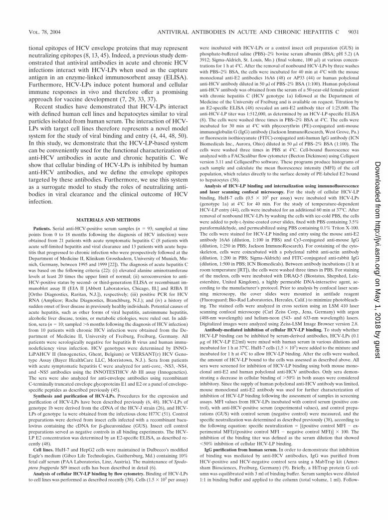
Epitope mapping of antibodies inhibiting cellular binding of HCV-LPs. Toidentify HCV-LP envelope epitopes targeted by human antibodies inhibitingcellular HCV-LP binding, an anti-HCV-positive serum with strong inhibitionof binding activity was incubated with overlapping 15-mer peptides of theHCV envelope glycoproteins (comprising amino acid positions 201 to 758)derived from the HCV-J strain (26) or PBS (as a control) for 1 h at RT(peptide concentration, 100 �g/ml; serum dilution, 1:50). The peptides were
provided through the European Research Network HCVacc (QLK2-1999-00356) and were synthesized by Clonestar Corp. (Brno, Czech Republic).HCV-LPs (derived from the homologous isolate as the peptides) were thenadded to the peptide-antibody complexes and incubated for 1 h at 37°C.Finally the HCV-LPs, antibodies, and peptides were added to HuH-7 cellsand incubated for 1 h at 4°C. Binding of HCV-LPs was detected by flowcytometry using the monoclonal anti-E2 antibody AP33 as described above.For the assessment of concentration-dependent reversion of HCV-LP inhi-bition of binding activity, an anti-HCV-positive serum (dilution, 1:50) wasincubated in the presence of various concentrations of E2 peptide 408 (aminoacid sequence, SQKIQLVNTNGSWHI, corresponding to amino acid posi-tions 408 to 422). Reversion of HCV-LP inhibition of binding activity wascalculated by the following equation: MFI � experimental MFI (anti-HCV-positive serum � HCV envelope peptide) � positive control MFI (anti-HCVpositive serum � PBS).
Statistical analysis. Comparison between subgroups was performed by thechi-square test using the SAS Analyst statistical software package, version 6.12(SAS Institute, Cary, N.C.).
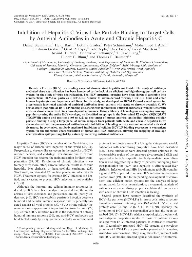
FIG. 1. Concentration-dependent and saturable binding of HCV-LPs to and entry into human hepatoma cells. (A and B) Binding of HCV-LPsto target cells is concentration-dependent and saturable. HuH-7 (A) and HepG2 (B) cells were incubated with increasing concentrations ofHCV-LPs of genotype 1a (triangles), as well as genotype 1b (squares), and particle binding was analyzed by flow cytometry as described inMaterials and Methods. On the y axis, net MFI values for each HCV-LP E2 concentration were calculated by subtracting the MFI of the negativecontrol with anti-E2 and PE-conjugated anti-mouse IgG antibodies from that obtained with the respective HCV-LP concentration (x axis). (C andD) Analysis of HCV-LP entry into target cells using anti-E2-specific immunofluorescence and cross section laser scanning confocal microscopy.(C) Cellular binding of HCV-LP was assessed by incubation of HuH-7 cells with HCV-LPs (genotype 1a) at 4°C for 40 min. (D) Fortemperature-dependent HCV-LP entry, cells were incubated for an additional 60 min at 37°C. After removal of nonbound HCV-LPs by washingthe cells with ice-cold PBS, the cells were added to poly-L-lysine-coated cover slides, fixed, permeabilized, and stained for HCV-LPs using anti-E2and Cy3-conjugated anti-mouse IgG antibodies (arrows). For the costaining of the cytoskeleton, cells were coincubated with a rabbit anti-actin andFITC-conjugated anti-rabbit IgG antibody. For costaining of the nucleus, the cells were incubated with DRAQ-5, a highly permeable DNA-interactive agent.
9032 STEINMANN ET AL. J. VIROL.
on May 1, 2018 by guest
http://jvi.asm.org/
Dow
nloaded from
RESULTS
HCV-LP-based model system for viral envelope binding andentry. To establish a convenient model system for the study ofantibody-mediated inhibition of viral envelope binding to tar-get cells, we first characterized the interaction of HCV-LPswith the hepatoma cell lines HuH-7 and HepG2. These liver-derived cell lines have been used as model systems to study theinteraction of viral envelope glycoproteins with the host cellmembrane (4, 20, 40, 44, 48). Furthermore, defined hepatomacells have been shown to allow the entry of virus, virus-likeparticles, and retroviral HCV pseudoparticles (2, 5, 24, 44),and they may support low-level HCV infection (42). HCV-LPsof genotypes 1a and 1b were synthesized using recombinantbaculoviruses containing the cDNA for the HCV structuralproteins of strains HCV-J and HCV-H77. Using flow cytom-etry and anti-envelope antibodies for the detection of cellular
HCV-LP binding, HCV-LPs demonstrated concentration-de-pendent and saturable binding to HuH-7 and HepG2 cells,respectively (Fig. 1A and B). This side-by-side analysis ofHCV-LPs of genotypes 1a and 1b extends a previous studydescribing the binding of HCV-LPs of the infectious cloneH77C to target cells (48).
The two hepatoma cell lines exhibited similar HCV-LP bind-ing profiles, although saturation of HCV-LP binding to HuH-7cells was reached at a slightly lower HCV-LP E2 concentrationthan for HepG2 cells. Saturation of HCV-LP binding wasreached at an E2 concentration of 0.5 to 1 �g/ml for HuH-7cells (Fig. 1A) and 1 to 2 �g/ml for HepG2 cells (Fig. 1B).These data may indicate a higher number of HCV-LP E2binding sites on HepG2 than on HuH-7 cells. HCV-LPs ofgenotypes 1a and 1b exhibited similar binding profiles (satura-tion of binding at 0.5 and 1 �g of E2/ml for HCV-LPs of
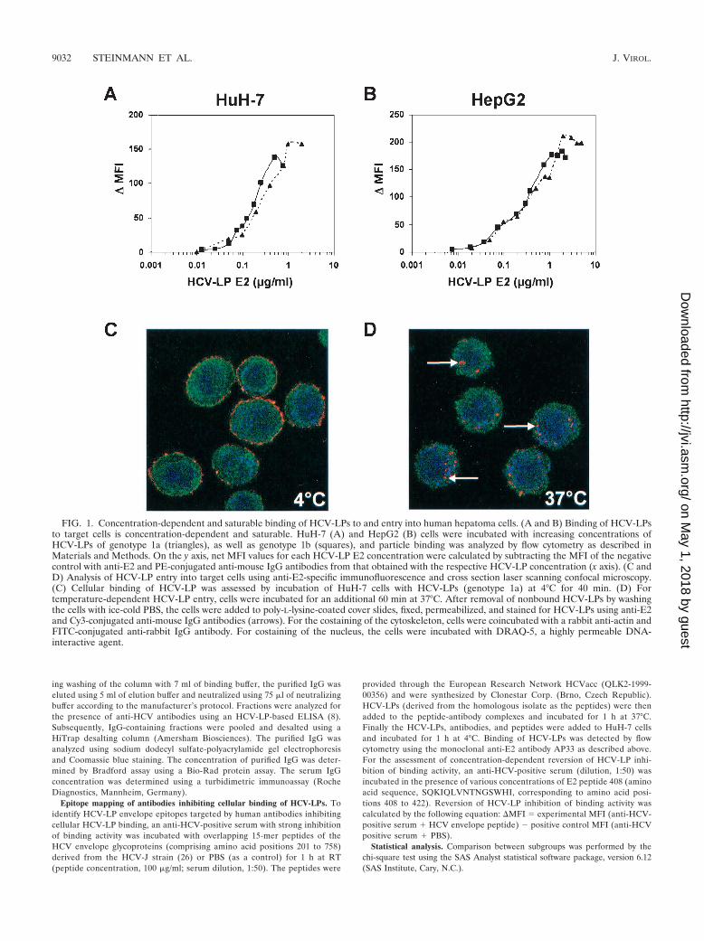
FIG. 2. Antibody-mediated inhibition of cellular HCV-LP binding. HCV-LPs were incubated with anti-HCV-positive serum or a pool ofanti-HCV-negative control sera (dilution, 1:50). HCV-LP–antibody complexes were added to HuH-7 cells for 1 h at 4°C. After removal ofnonbound HCV-LP–antibody complexes by washing the cells in PBS–2% BSA, the binding of HCV-LPs was detected by flow cytometry using themouse monoclonal anti-E2 antibody AP33 and PE-conjugated anti-mouse IgG (A and B) or human polyclonal anti-HCV and FITC-conjugatedanti-human IgG antibody (C). The fluorescence intensity and relative cell numbers (Counts) are shown on the x and y axes, respectively. NC,negative control, corresponding to HuH-7 cells incubated with control insect cell preparations (GUS) and control serum. (D) For the assessmentof concentration-dependent antibody-mediated inhibition of binding, HCV-LPs (genotype 1b) were incubated in subsaturating concentrations withan anti-HCV-positive serum or a pool of anti-HCV-negative control sera (at various dilutions as indicated on the x axis) for 1 h at 37°C.HCV-LP–antibody complexes were added to HuH-7 cells for 1 h at 4°C. HCV-LP binding to the HuH-7 cells was detected by flow cytometry asdescribed above. Inhibition of cellular HCV-LP binding (y axis) was calculated as described in Materials and Methods.
VOL. 78, 2004 ANTIVIRAL ANTIBODIES IN ACUTE AND CHRONIC HEPATITIS C 9033
on May 1, 2018 by guest
http://jvi.asm.org/
Dow
nloaded from
genotype 1a or 1b and HuH-7 cells, respectively). Half-maxi-mal saturation of HCV-LP binding was present at E2 concen-trations between 0.2 and 0.4 �g/ml.
The next step after particle binding is uptake, or entry ofparticles into the cell. To investigate whether HCV-LPs wereinternalized into HuH-7 cells following binding to the cellsurface, we performed anti-E2-specific immunofluorescenceand confocal laser scanning microscopy of HuH-7 cells incu-bated with HCV-LPs. As shown in Fig. 1C and D, incubationof cells with HCV-LPs (genotype 1a) at 4°C resulted in theexclusive detection of HCV-LP E2 at the cell surface, consis-tent with HCV-LP binding to the cell membrane. By contrast,incubation of HuH-7 cells with HCV-LPs at 37°C resulted inthe translocation of E2 immunoreactivity inside the cell, con-sistent with temperature-dependent cellular HCV-LP entry.These data indicate that HCV-LPs are taken up by HuH-7 ina temperature-dependent manner following cell surface bind-ing. These findings corroborate and extend previous experi-ments using dye-labeled HCV-LPs (44).
Inhibition of cellular HCV-LP binding by serum-derivedanti-HCV antibodies. We next studied whether cellular HCV-LP binding is inhibited by sera from HCV-infected individuals.As shown in Fig. 2, anti-HCV-positive sera from patients withhepatitis C inhibited cellular HCV-LP binding (Fig. 2). Toconfirm that the observed inhibition of binding was due toanti-HCV antibodies present in these sera, cellular HCV-LPbinding was studied in the presence of 10 anti-HCV-negativecontrol sera. None of these control sera inhibited cellularHCV-LP binding (Fig. 2A and B). To exclude the possibilitythat the observed inhibition of HCV-LP binding was due tomasking of HCV-LP epitopes by nonneutralizing anti-HCVantibodies present in the sera, inhibition of binding was studiedusing a human serum containing high-titer anti-E2 and anti-HCV-LP antibodies (human polyclonal anti-HCV) (Fig. 2C).Revealing the binding with antibodies from the same species asthe neutralizing serum (human-human) is critical because non-neutralizing anti-envelope antibodies present in human serumcould cover E2 after it is bound to target cells and could,therefore, interfere with the assessment of neutralization if thebinding was revealed with an anti-E2 antibody from a differentspecies (such as a mouse monoclonal antibody) (38). Only serademonstrating a reproducible decrease in cellular binding of�50% were considered inhibitory.
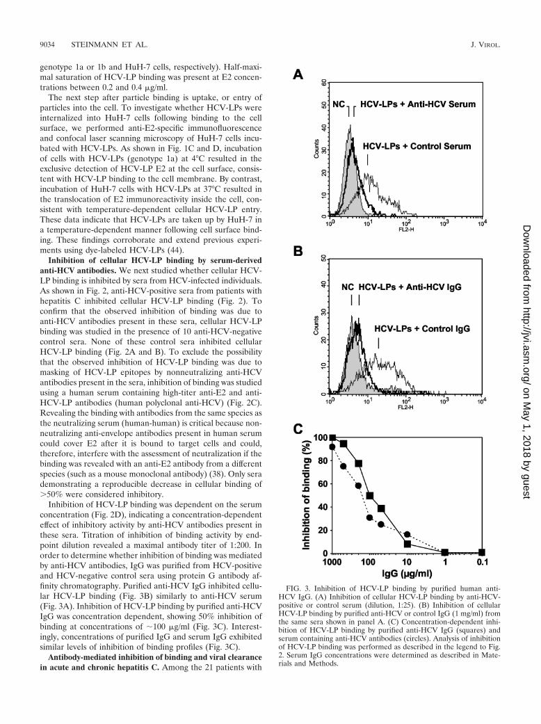
Inhibition of HCV-LP binding was dependent on the serumconcentration (Fig. 2D), indicating a concentration-dependenteffect of inhibitory activity by anti-HCV antibodies present inthese sera. Titration of inhibition of binding activity by end-point dilution revealed a maximal antibody titer of 1:200. Inorder to determine whether inhibition of binding was mediatedby anti-HCV antibodies, IgG was purified from HCV-positiveand HCV-negative control sera using protein G antibody af-finity chromatography. Purified anti-HCV IgG inhibited cellu-lar HCV-LP binding (Fig. 3B) similarly to anti-HCV serum(Fig. 3A). Inhibition of HCV-LP binding by purified anti-HCVIgG was concentration dependent, showing 50% inhibition ofbinding at concentrations of 100 �g/ml (Fig. 3C). Interest-ingly, concentrations of purified IgG and serum IgG exhibitedsimilar levels of inhibition of binding profiles (Fig. 3C).
Antibody-mediated inhibition of binding and viral clearancein acute and chronic hepatitis C. Among the 21 patients with
FIG. 3. Inhibition of HCV-LP binding by purified human anti-HCV IgG. (A) Inhibition of cellular HCV-LP binding by anti-HCV-positive or control serum (dilution, 1:25). (B) Inhibition of cellularHCV-LP binding by purified anti-HCV or control IgG (1 mg/ml) fromthe same sera shown in panel A. (C) Concentration-dependent inhi-bition of HCV-LP binding by purified anti-HCV IgG (squares) andserum containing anti-HCV antibodies (circles). Analysis of inhibitionof HCV-LP binding was performed as described in the legend to Fig.2. Serum IgG concentrations were determined as described in Mate-rials and Methods.
9034 STEINMANN ET AL. J. VIROL.
on May 1, 2018 by guest
http://jvi.asm.org/
Dow
nloaded from
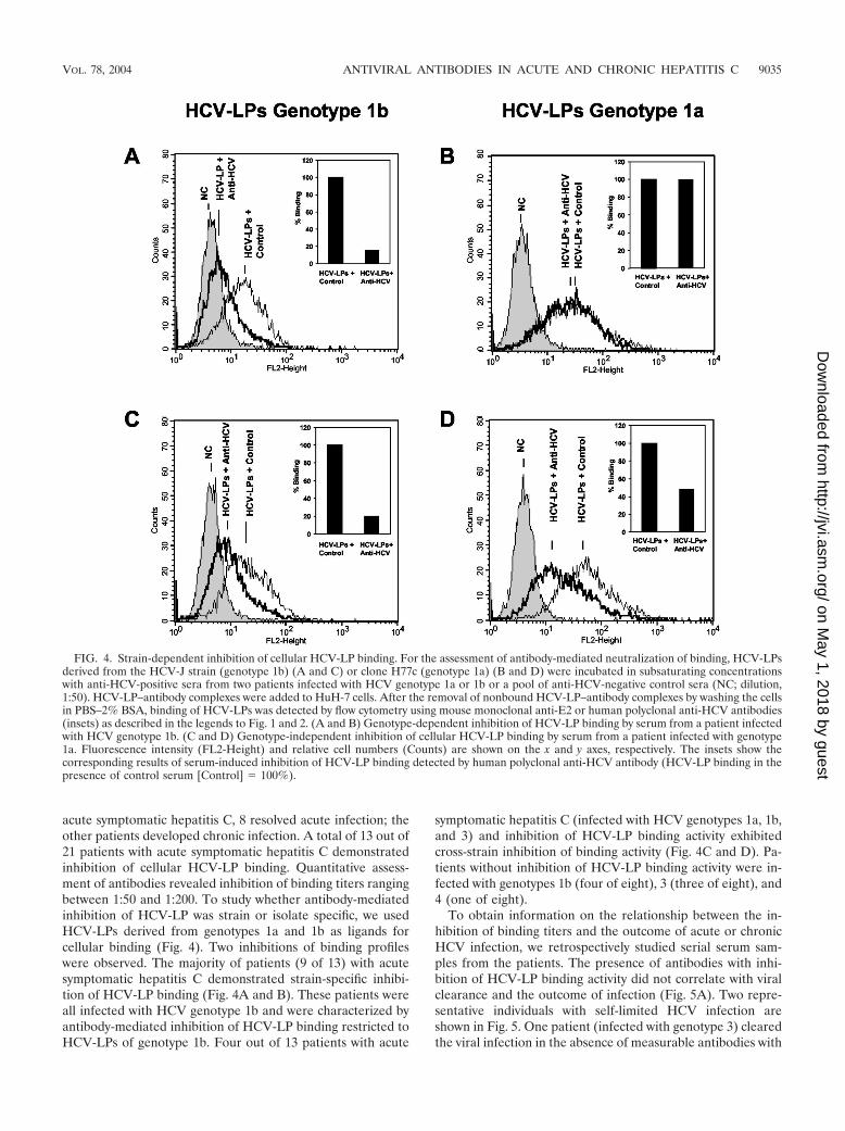
acute symptomatic hepatitis C, 8 resolved acute infection; theother patients developed chronic infection. A total of 13 out of21 patients with acute symptomatic hepatitis C demonstratedinhibition of cellular HCV-LP binding. Quantitative assess-ment of antibodies revealed inhibition of binding titers rangingbetween 1:50 and 1:200. To study whether antibody-mediatedinhibition of HCV-LP was strain or isolate specific, we usedHCV-LPs derived from genotypes 1a and 1b as ligands forcellular binding (Fig. 4). Two inhibitions of binding profileswere observed. The majority of patients (9 of 13) with acutesymptomatic hepatitis C demonstrated strain-specific inhibi-tion of HCV-LP binding (Fig. 4A and B). These patients wereall infected with HCV genotype 1b and were characterized byantibody-mediated inhibition of HCV-LP binding restricted toHCV-LPs of genotype 1b. Four out of 13 patients with acute
symptomatic hepatitis C (infected with HCV genotypes 1a, 1b,and 3) and inhibition of HCV-LP binding activity exhibitedcross-strain inhibition of binding activity (Fig. 4C and D). Pa-tients without inhibition of HCV-LP binding activity were in-fected with genotypes 1b (four of eight), 3 (three of eight), and4 (one of eight).
To obtain information on the relationship between the in-hibition of binding titers and the outcome of acute or chronicHCV infection, we retrospectively studied serial serum sam-ples from the patients. The presence of antibodies with inhi-bition of HCV-LP binding activity did not correlate with viralclearance and the outcome of infection (Fig. 5A). Two repre-sentative individuals with self-limited HCV infection areshown in Fig. 5. One patient (infected with genotype 3) clearedthe viral infection in the absence of measurable antibodies with
FIG. 4. Strain-dependent inhibition of cellular HCV-LP binding. For the assessment of antibody-mediated neutralization of binding, HCV-LPsderived from the HCV-J strain (genotype 1b) (A and C) or clone H77c (genotype 1a) (B and D) were incubated in subsaturating concentrationswith anti-HCV-positive sera from two patients infected with HCV genotype 1a or 1b or a pool of anti-HCV-negative control sera (NC; dilution,1:50). HCV-LP–antibody complexes were added to HuH-7 cells. After the removal of nonbound HCV-LP–antibody complexes by washing the cellsin PBS–2% BSA, binding of HCV-LPs was detected by flow cytometry using mouse monoclonal anti-E2 or human polyclonal anti-HCV antibodies(insets) as described in the legends to Fig. 1 and 2. (A and B) Genotype-dependent inhibition of HCV-LP binding by serum from a patient infectedwith HCV genotype 1b. (C and D) Genotype-independent inhibition of cellular HCV-LP binding by serum from a patient infected with genotype1a. Fluorescence intensity (FL2-Height) and relative cell numbers (Counts) are shown on the x and y axes, respectively. The insets show thecorresponding results of serum-induced inhibition of HCV-LP binding detected by human polyclonal anti-HCV antibody (HCV-LP binding in thepresence of control serum [Control] � 100%).
VOL. 78, 2004 ANTIVIRAL ANTIBODIES IN ACUTE AND CHRONIC HEPATITIS C 9035
on May 1, 2018 by guest
http://jvi.asm.org/
Dow
nloaded from
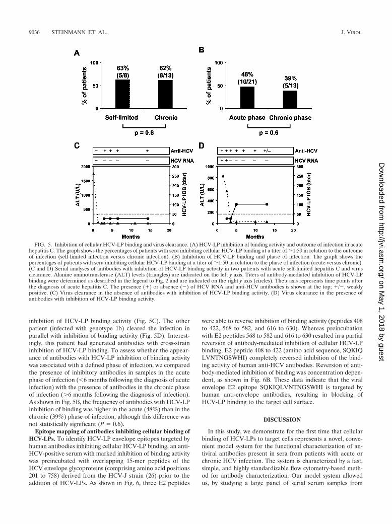
inhibition of HCV-LP binding activity (Fig. 5C). The otherpatient (infected with genotype 1b) cleared the infection inparallel with inhibition of binding activity (Fig. 5D). Interest-ingly, this patient had generated antibodies with cross-straininhibition of HCV-LP binding. To assess whether the appear-ance of antibodies with HCV-LP inhibition of binding activitywas associated with a defined phase of infection, we comparedthe presence of inhibitory antibodies in samples in the acutephase of infection (�6 months following the diagnosis of acuteinfection) with the presence of antibodies in the chronic phaseof infection (�6 months following the diagnosis of infection).As shown in Fig. 5B, the frequency of antibodies with HCV-LPinhibition of binding was higher in the acute (48%) than in thechronic (39%) phase of infection, although this difference wasnot statistically significant (P � 0.6).
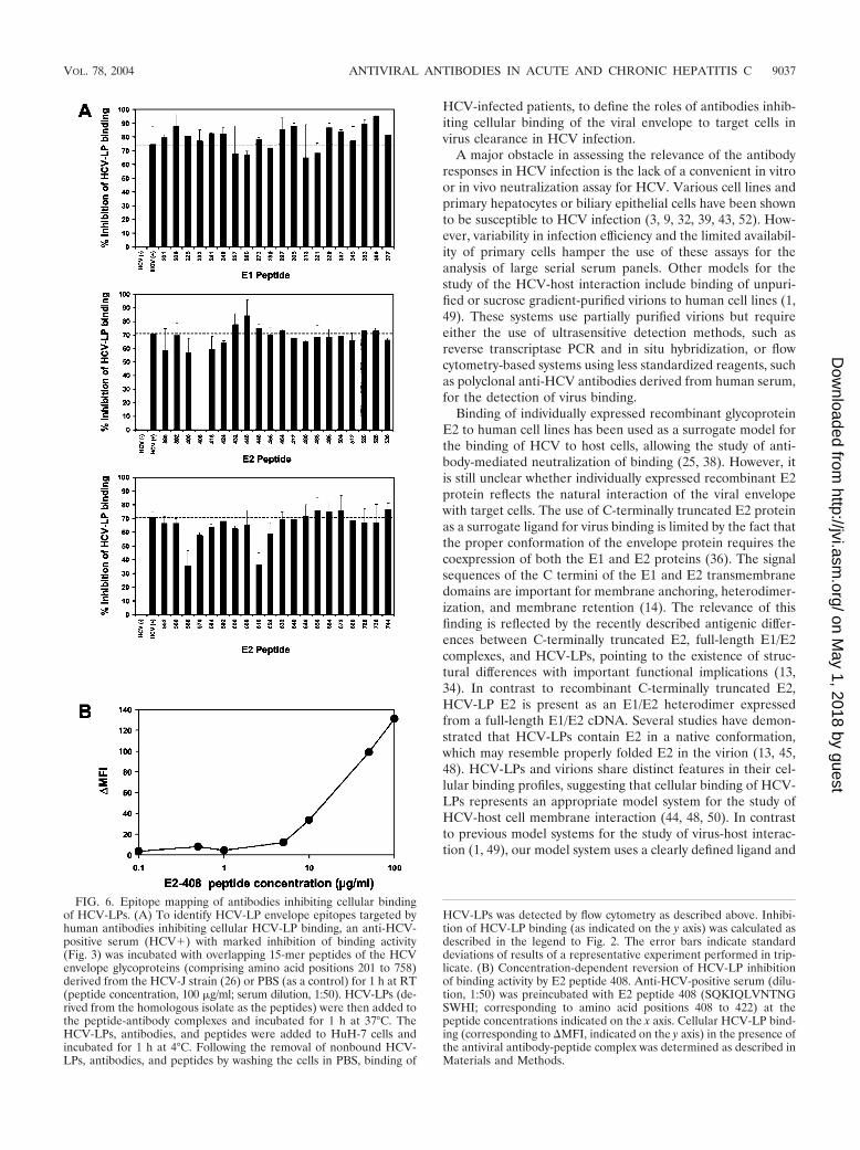
Epitope mapping of antibodies inhibiting cellular binding ofHCV-LPs. To identify HCV-LP envelope epitopes targeted byhuman antibodies inhibiting cellular HCV-LP binding, an anti-HCV-positive serum with marked inhibition of binding activitywas preincubated with overlapping 15-mer peptides of theHCV envelope glycoproteins (comprising amino acid positions201 to 758) derived from the HCV-J strain (26) prior to theaddition of HCV-LPs. As shown in Fig. 6, three E2 peptides
were able to reverse inhibition of binding activity (peptides 408to 422, 568 to 582, and 616 to 630). Whereas preincubationwith E2 peptides 568 to 582 and 616 to 630 resulted in a partialreversion of antibody-mediated inhibition of cellular HCV-LPbinding, E2 peptide 408 to 422 (amino acid sequence, SQKIQLVNTNGSWHI) completely reversed inhibition of the bind-ing activity of human anti-HCV antibodies. Reversion of anti-body-mediated inhibition of binding was concentration depen-dent, as shown in Fig. 6B. These data indicate that the viralenvelope E2 epitope SQKIQLVNTNGSWHI is targeted byhuman anti-envelope antibodies, resulting in blocking ofHCV-LP binding to the target cell surface.
DISCUSSION
In this study, we demonstrate for the first time that cellularbinding of HCV-LPs to target cells represents a novel, conve-nient model system for the functional characterization of an-tiviral antibodies present in sera from patients with acute orchronic HCV infection. The system is characterized by a fast,simple, and highly standardizable flow cytometry-based meth-od for antibody characterization. Our model system allowedus, by studying a large panel of serial serum samples from
FIG. 5. Inhibition of cellular HCV-LP binding and virus clearance. (A) HCV-LP inhibition of binding activity and outcome of infection in acutehepatitis C. The graph shows the percentages of patients with sera inhibiting cellular HCV-LP binding at a titer of �1:50 in relation to the outcomeof infection (self-limited infection versus chronic infection). (B) Inhibition of HCV-LP binding and phase of infection. The graph shows thepercentages of patients with sera inhibiting cellular HCV-LP binding at a titer of �1:50 in relation to the phase of infection (acute versus chronic).(C and D) Serial analyses of antibodies with inhibition of HCV-LP binding activity in two patients with acute self-limited hepatitis C and virusclearance. Alanine aminotransferase (ALT) levels (triangles) are indicated on the left y axis. Titers of antibody-mediated inhibition of HCV-LPbinding were determined as described in the legend to Fig. 2 and are indicated on the right y axis (circles). The x axis represents time points afterthe diagnosis of acute hepatitis C. The presence (�) or absence (�) of HCV RNA and anti-HCV antibodies is shown at the top; �/�, weaklypositive. (C) Virus clearance in the absence of antibodies with inhibition of HCV-LP binding activity. (D) Virus clearance in the presence ofantibodies with inhibition of HCV-LP binding activity.
9036 STEINMANN ET AL. J. VIROL.
on May 1, 2018 by guest
http://jvi.asm.org/
Dow
nloaded from
HCV-infected patients, to define the roles of antibodies inhib-iting cellular binding of the viral envelope to target cells invirus clearance in HCV infection.
A major obstacle in assessing the relevance of the antibodyresponses in HCV infection is the lack of a convenient in vitroor in vivo neutralization assay for HCV. Various cell lines andprimary hepatocytes or biliary epithelial cells have been shownto be susceptible to HCV infection (3, 9, 32, 39, 43, 52). How-ever, variability in infection efficiency and the limited availabil-ity of primary cells hamper the use of these assays for theanalysis of large serial serum panels. Other models for thestudy of the HCV-host interaction include binding of unpuri-fied or sucrose gradient-purified virions to human cell lines (1,49). These systems use partially purified virions but requireeither the use of ultrasensitive detection methods, such asreverse transcriptase PCR and in situ hybridization, or flowcytometry-based systems using less standardized reagents, suchas polyclonal anti-HCV antibodies derived from human serum,for the detection of virus binding.
Binding of individually expressed recombinant glycoproteinE2 to human cell lines has been used as a surrogate model forthe binding of HCV to host cells, allowing the study of anti-body-mediated neutralization of binding (25, 38). However, itis still unclear whether individually expressed recombinant E2protein reflects the natural interaction of the viral envelopewith target cells. The use of C-terminally truncated E2 proteinas a surrogate ligand for virus binding is limited by the fact thatthe proper conformation of the envelope protein requires thecoexpression of both the E1 and E2 proteins (36). The signalsequences of the C termini of the E1 and E2 transmembranedomains are important for membrane anchoring, heterodimer-ization, and membrane retention (14). The relevance of thisfinding is reflected by the recently described antigenic differ-ences between C-terminally truncated E2, full-length E1/E2complexes, and HCV-LPs, pointing to the existence of struc-tural differences with important functional implications (13,34). In contrast to recombinant C-terminally truncated E2,HCV-LP E2 is present as an E1/E2 heterodimer expressedfrom a full-length E1/E2 cDNA. Several studies have demon-strated that HCV-LPs contain E2 in a native conformation,which may resemble properly folded E2 in the virion (13, 45,48). HCV-LPs and virions share distinct features in their cel-lular binding profiles, suggesting that cellular binding of HCV-LPs represents an appropriate model system for the study ofHCV-host cell membrane interaction (44, 48, 50). In contrastto previous model systems for the study of virus-host interac-tion (1, 49), our model system uses a clearly defined ligand and
FIG. 6. Epitope mapping of antibodies inhibiting cellular bindingof HCV-LPs. (A) To identify HCV-LP envelope epitopes targeted byhuman antibodies inhibiting cellular HCV-LP binding, an anti-HCV-positive serum (HCV�) with marked inhibition of binding activity(Fig. 3) was incubated with overlapping 15-mer peptides of the HCVenvelope glycoproteins (comprising amino acid positions 201 to 758)derived from the HCV-J strain (26) or PBS (as a control) for 1 h at RT(peptide concentration, 100 �g/ml; serum dilution, 1:50). HCV-LPs (de-rived from the homologous isolate as the peptides) were then added tothe peptide-antibody complexes and incubated for 1 h at 37°C. TheHCV-LPs, antibodies, and peptides were added to HuH-7 cells andincubated for 1 h at 4°C. Following the removal of nonbound HCV-LPs, antibodies, and peptides by washing the cells in PBS, binding of
HCV-LPs was detected by flow cytometry as described above. Inhibi-tion of HCV-LP binding (as indicated on the y axis) was calculated asdescribed in the legend to Fig. 2. The error bars indicate standarddeviations of results of a representative experiment performed in trip-licate. (B) Concentration-dependent reversion of HCV-LP inhibitionof binding activity by E2 peptide 408. Anti-HCV-positive serum (dilu-tion, 1:50) was preincubated with E2 peptide 408 (SQKIQLVNTNGSWHI; corresponding to amino acid positions 408 to 422) at thepeptide concentrations indicated on the x axis. Cellular HCV-LP bind-ing (corresponding to MFI, indicated on the y axis) in the presence ofthe antiviral antibody-peptide complex was determined as described inMaterials and Methods.
VOL. 78, 2004 ANTIVIRAL ANTIBODIES IN ACUTE AND CHRONIC HEPATITIS C 9037
on May 1, 2018 by guest
http://jvi.asm.org/
Dow
nloaded from
highly standardized conditions to quantify cellular binding(monoclonal antibodies and flow cytometry). Binding couldeasily be detected and quantified by mouse monoclonal anti-E2 antibodies, whereas human polyclonal anti-HCV antibodyserved as a reagent to confirm the specificity of the inhibitionof binding.
In this study, we demonstrate for the first time that cellularHCV-LP binding is inhibited by anti-HCV-positive sera fromHCV-infected patients. Specific inhibition of HCV-LP bindingby antiviral antibodies is demonstrated by the following exper-imental findings: (i) only sera from HCV-infected patients, butnot from uninfected control subjects, inhibited cellular HCV-LP binding in a concentration-dependent manner (inhibitor-specific inhibition of binding [Fig. 2 and 3]); (ii) only purifiedIgG from HCV-infected patients, but not from uninfectedcontrol subjects, inhibited cellular HCV-LP binding in a con-centration-dependent manner (antibody-specific inhibition ofbinding [Fig. 3]); (iv) antibody-mediated inhibition of bindingis reverted by preincubation with a defined viral envelope pep-tide (Fig. 6).
Using antibody-mediated inhibition of HCV-LP binding as amarker for virus neutralization at the level of envelope bindingto the host cell membrane, our study provides for the first timea detailed functional characterization of anti-HCV antibodiesin acute and chronic HCV infections. The key findings of ourstudy are the following: (i) antibodies inhibiting HCV-LP bind-ing are present in acute or chronic HCV infection; (ii) anti-bodies that inhibit HCV-LP binding are of low titer (�1:200);(iii) in the majority of studied individuals, antibody-mediatedinhibition of cellular HCV-LP binding is subtype or isolatespecific; (iv) the presence of antibodies with inhibition ofHCV-LP binding activity may not predict virus clearance.
Using antibody-mediated inhibition of HCV-LP binding as asurrogate marker for virus neutralization, we conclude thatvirus-neutralizing antibodies are present in acute and chronicHCV infections. Since the presence of such antibodies did notcorrelate with the outcome of infection, we conclude that neu-tralizing antibodies presumably do not play a major role in vi-rus clearance. Our data indicate that limited induction of high-titer antibodies with neutralization capacity, as well as isolate-specific restriction of neutralization, most likely contributes tothe failure of antibody-mediated virus clearance in HCV in-fection.
The absence of a high-titer humoral immune response withneutralizing properties in patients with self-limited acute hep-atitis C is in line with the concept that a robust and multispe-cific antiviral cellular immune response is central for sustainedvirus clearance in acutely infected individuals (22, 23, 30, 46).Several studies with humans have shown that virus clearance isassociated with a strong HCV-specific CD4� T helper andcytotoxic-T-cell response (for a review, see references 23, 30,and 46).
Recently, pseudotyped retroviral particles containing HCVglycoproteins have been suggested as a model system for HCVentry (5, 5a, 24). Whether the entry of retroviral HCV pseu-doparticles containing heterologous retroviral proteins repre-sents HCV entry leading to viral infection is not yet known. Ina previously published pilot study, Bartosch et al. analyzed serafrom four HCV-infected chimpanzees and one HCV-infectedhuman for the presence of antibodies neutralizing pseudopar-
ticle entry (5a). Interestingly, the authors could detect antibod-ies (titer, �1:320) neutralizing pseudoparticle entry in the sin-gle human individual and in two out of four chimpanzees thatprogressed to chronic HCV infection. Cross-neutralization ofpseudoparticle entry was observed in one of the two animalsand the single human (5a). In contrast, no antibodies withneutralizing properties were observed in two chimpanzees withself-limited infections (5a). Since only one human individualand four experimentally infected chimpanzees were assessed inthis study (5a), it is difficult to compare the results obtained inthe HCV pseudoparticle and HCV-LP models at the presenttime. Interestingly, both systems indicate the presence of cross-neutralizing antibodies in chronic HCV infection. A prospec-tive study assessing serum samples from a large number ofHCV-infected humans side by side is needed to compare find-ings obtained using the two systems. Studies are under way toaddress this important question.
We cannot exclude the possibility that anti-envelope anti-bodies with binding-inhibitory properties are present in anti-gen-antibody immune complexes. In this case, these antibodiesare not detected by this assay or by ELISAs based on HCV-LPs (8) or individual envelope proteins (12, 21) or by otherneutralization-of-binding assays (25, 38). Virus neutralizationcan also occur at stages of the viral life cycle following virusbinding to the cell membrane (16). These stages include fusionof the viral envelope with the cell membrane, blocking of viraluncoating, and replication and would not be detected by ourassay.
Interestingly, antibody-mediated inhibition of HCV-LPbinding did not correlate with the immunoreactivity of anti-HCV antibodies against HCV-LPs or recombinant E1/E2 pro-teins as capture antigens in an ELISA (8). Several sera frompatients with chronic HCV infection containing high-titer an-tibodies against HCV-LPs (�1:128,000) (8) or recombinantE1/E2 proteins (�1:64,000) did not result in inhibition ofHCV-LP binding (data not shown). In contrast, several serafrom patients with acute self-limited hepatitis C and low-titeranti-HCV-LP or anti-E1/E2 antibodies exhibited marked inhi-bition of HCV-LP binding (data not shown). The differencebetween the presence (as measured by HCV-LP [8] or E1/E2[48, 29] ELISAs) and function (as measured by inhibition ofHCV-LP binding [this study]) of anti-envelope antibodies isalso reflected by the interaction of HCV-LPs with anti-HCVantibodies from patients infected with different HCV geno-types. Using HCV-LPs as capture antigens in an ELISA for-mat, HCV-LPs of genotype 1b cross-interacted with antibodiesof patients infected with HCV genotypes 1a, 2, 3, 4, 5, and 6(8). In contrast, cross-reactivity between the same HCV sub-types (1a or 1b) was a less frequent event (4 of 13 patients) inantibody-mediated inhibition of HCV-LP binding. Althoughwe cannot exclude the possibility that cross-reactivity observedin studies using the HCV-LP ELISA may be partly due to theinteraction of antibodies with partly deenveloped nucleocap-sids present in the HCV-LP preparation, our data indicate thatHCV elicits abundant antibodies directed against the HCVstructural proteins (as demonstrated in HCV-LP ELISA) (8)that have little or no ability to block envelope docking to thetarget cell (this study).
Several mechanisms may explain this finding. First, the ma-jority of anti-HCV antibodies are targeted against epitopes of
9038 STEINMANN ET AL. J. VIROL.
on May 1, 2018 by guest
http://jvi.asm.org/
Dow
nloaded from
the HCV structural proteins that are not required for virusneutralization. Envelope epitopes presented on the surfaces ofHCV-LPs have been mapped recently (13, 45). Our data areconsistent with the hypothesis that many epitopes exposed onthe HCV-LP surface and recognized by human antiviral anti-bodies are most likely not target epitopes for virus neutraliza-tion. Epitopes mediating viral envelope binding and targetedby virus-neutralizing antibodies are still poorly defined. So far,two epitopes in envelope glycoproteins E1 and E2 (amino acids197 to 207 and 640 to 653) have been proposed to play apotential role in mediating cellular HCV-LP binding (44). Inchimpanzees, the E2 HVR-1 has been inferred to represent apotential B-cell epitope associated with virus neutralization(18). In contrast, antibodies directed against several epitopesof the HCV core protein were not able to block HCV-LPbinding to target cells, suggesting that the nucleocapsid is notinvolved in mediating HCV-LP cellular binding (M. Triyatniand T. J. Liang, unpublished observations). Using a panel ofoverlapping envelope peptides covering the entire HCV enve-lope, we identified an epitope of the N-terminal E2 region(SQKIQLVNTNGSWHI; amino acid positions 408 to 422) asthe target of human antiviral antibodies inhibiting cellular par-ticle binding. This epitope overlaps with the E2 hypervariableregion and corroborates the importance of this region for thebinding of the viral envelope to the cell surface. Interestingly,the same region has been shown to be involved in E2-CD81interaction (34, 35) and in mediating cellular binding and entryof HCV pseudoparticles (5, 24).
Second, the presence of HCV as a highly variable pool ofrapidly mutating viral quasispecies in an individual patient maycontribute to virus escape from antibody-mediated virus neu-tralization. A previous study elegantly demonstrated that anantiserum against the E2 hypervariable region from HCV re-covered from a single patient induced protection against ho-mologous HCV infection in chimpanzees but not against theemergence of neutralization escape mutants that were found tobe already present in the complex viral quasispecies of theinoculum (18). This study, performed with serum samples froma single patient, suggested that neutralizing antibodies can beinduced but isolate-specific virus neutralization and the rapidemergence of viral escape mutants may represent a majorlimitation for antibody-mediated virus neutralization in HCVinfection. Our data identifying antibodies with strain-specificneutralization of viral envelope binding in a large serum panelof 21 well-characterized patients indicates that the proposedmechanism may indeed contribute to the failure of antibody-mediated virus neutralization in patients with HCV infection.
Third, the virus has developed highly specific escape strate-gies to evade antibody-mediated neutralization. These strate-gies may include conformational masking of receptor bindingsites following envelope-antibody interaction (27) or alterationin envelope N-glycosylation motifs (11, 47), as described re-cently for human immunodeficiency virus. The HCV-LP-basedmodel system described in this study may be helpful in definingsimilar mechanisms for HCV.
In summary, this model system may be useful in providing adetailed map of epitopes targeted by human antiviral antibod-ies on the level of envelope-cell surface receptor interaction.Furthermore, the HCV-LP-based model system may allow theelucidation of viral escape mechanisms for the evasion of an-
tibody-mediated neutralization. Understanding these mecha-nisms may ultimately provide important clues for the develop-ment of new antivirals inhibiting virus-cell surface interaction,as well as define strategies for the efficient induction of viruscross-neutralizing antibodies for vaccine development.
ACKNOWLEDGMENTS
We thank F. von Weizsacker and P. Hafkemeyer (Department ofMedicine II, University of Freiburg) for providing HuH-7 and HepG2cell lines, C. Grullich and J. Finke (Department of Medicine I, Uni-versity of Freiburg) for support in flow cytometry analysis, M. Follo(Core Facility, Department of Medicine I, University of Freiburg) forperforming confocal laser scanning microscopy, M. Nauck (Division ofClinical Chemistry, Department of Medicine, University of Freiburg)for determination of serum IgG concentrations, and J. Baumert (Na-tional Research Centre for Environment and Health [GSF], Instituteof Epidemiology, Neuherberg, Germany) for support in statisticalanalysis.
This work was supported by grants from the European Union, Brus-sels, Belgium (QLK-2-1999-00356 and QLK-2-2002-01329); the Wil-helm Sander Foundation, Munich, Germany (99041.1); the DeutscheForschungsgemeinschaft (Ba1417/6-1 and 7-1); the Alexander vonHumboldt Foundation; and the Flemish Government, Brussels, Bel-gium (IWT grant 000405).
REFERENCES
1. Agnello, V., G. Abel, M. Elfahal, G. B. Knight, and Q. X. Zhang. 1999.Hepatitis C virus and other flaviviridae viruses enter cells via low densitylipoprotein receptor. Proc. Natl. Acad. Sci. USA 96:12766–12771.
2. Andre, P., F. Komurian-Pradel, S. Deforges, M. Perret, J. L. Berland, M.Sodoyer, S. Pol, C. Brechot, G. Paranhos-Baccala, and V. Lotteau. 2002.Characterization of low- and very-low-density hepatitis C virus RNA-con-taining particles. J. Virol. 76:6919–6928.
3. Bartenschlager, R., and V. Lohmann. 2000. Replication of hepatitis C virus.J. Gen. Virol. 81:1631–1648.
4. Barth, H., C. Schafer, M. I. Adah, F. Zhang, R. J. Linhardt, H. Toyoda, A.Kinoshita-Toyoda, T. Toida, T. H. Van Kuppevelt, E. Depla, F. Von Weiz-sacker, H. E. Blum, and T. F. Baumert. 2003. Cellular binding of hepatitis Cvirus envelope glycoprotein E2 requires cell surface heparan sulfate. J. Biol.Chem. 278:41003–41012.
5. Bartosch, B., J. Dubuisson, and F. L. Cosset. 2003. Infectious hepatitis Cvirus pseudo-particles containing functional E1-E2 envelope protein com-plexes. J. Exp Med. 197:633–642.
5a.Bartosch, B., J. Bukh, J. C. Meunier, C. Granier, R. E. Engle, W. C. Black-welder, S. U. Emerson, F. L. Cosset, and R. H. Purcell. 2003. In vitro assayfor neutralizing antibody to hepatitis C virus: evidence for broadly conservedneutralization epitopes. Proc. Natl. Acad. Sci. USA 100:14199–14204.
6. Baumert, T. F., S. Ito, D. T. Wong, and T. J. Liang. 1998. Hepatitis C virusstructural proteins assemble into viruslike particles in insect cells. J. Virol.72:3827–3836.
7. Baumert, T. F., J. Vergalla, J. Satoi, M. Thomson, M. Lechmann, D. Herion,H. B. Greenberg, S. Ito, and T. J. Liang. 1999. Hepatitis C virus-like particlessynthesized in insect cells as a potential vaccine candidate. Gastroenterology117:1397–1407.
8. Baumert, T. F., S. Wellnitz, S. Aono, J. Satoi, D. Herion, J. Tilman Gerlach,G. R. Pape, J. Y. Lau, J. H. Hoofnagle, H. E. Blum, and T. J. Liang. 2000.Antibodies against hepatitis C virus-like particles and viral clearance in acuteand chronic hepatitis C. Hepatology 32:610–617.
9. Bichr, S., R. Rende-Fournier, G. Vona, A. M. Yamamoto, E. Depla, G.Maertens, and C. Brechot. 2002. Detection of neutralizing antibodies tohepatitis C virus using a biliary cell infection model. J. Gen. Virol. 83:1673–1678.
10. Blanchard, E., D. Brand, S. Trassard, A. Goudeau, and P. Roingeard. 2002.Hepatitis C virus-like particle morphogenesis. J. Virol. 76:4073–4079.
11. Calarese, D. A., C. N. Scanlan, M. B. Zwick, S. Deechongkit, Y. Mimura, R.Kunert, P. Zhu, M. R. Wormald, R. L. Stanfield, K. H. Roux, J. W. Kelly,P. M. Rudd, R. A. Dwek, H. Katinger, D. R. Burton, I. A. Wilson, R.Pantophlet, X. Wei, J. M. Decker, S. Wang, H. Hui, J. C. Kappes, X. Wu, J. F.Salazar-Gonzalez, M. G. Salazar, J. M. Kilby, M. S. Saag, N. L. Komarova,M. A. Nowak, B. H. Hahn, P. D. Kwong, and G. M. Shaw. 2003. Antibodydomain exchange is an immunological solution to carbohydrate cluster rec-ognition. Science 300:2065–2071.
12. Chen, M., M. Sallberg, A. Sonnerborg, O. Weiland, L. Mattsson, L. Jin, A.Birkett, D. Peterson, and D. R. Milich. 1999. Limited humoral immunity inhepatitis C virus infection. Gastroenterology 116:135–143.
13. Clayton, R. F., A. Owsianka, J. Aitken, S. Graham, D. Bhella, and A. H.
VOL. 78, 2004 ANTIVIRAL ANTIBODIES IN ACUTE AND CHRONIC HEPATITIS C 9039
on May 1, 2018 by guest
http://jvi.asm.org/
Dow
nloaded from
Patel. 2002. Analysis of antigenicity and topology of E2 glycoprotein presenton recombinant hepatitis C virus-like particles. J. Virol. 76:7672–7682.
14. Cocquerel, L., A. Op de Beeck, M. Lambot, J. Roussel, D. Delgrange, A.Pillez, C. Wychowski, F. Penin, and J. Dubuisson. 2002. Topological changesin the transmembrane domains of hepatitis C virus envelope glycoproteins.EMBO J. 21:2893–2902.
15. Di Bisceglie, A. M., and J. H. Hoofnagle. 2002. Optimal therapy of hepatitisC. Hepatology 36:S121–S127.
16. Dimmock, N. J. 1993. Neutralization of animal viruses. Curr. Top. Microbiol.Immunol. 183:1–149.
17. Ezelle, H. J., D. Markovic, and G. N. Barber. 2002. Generation of hepatitisC virus-like particles by use of a recombinant vesicular stomatitis virusvector. J. Virol. 76:12325–12334.
18. Farci, P., A. Shimoda, D. Wong, T. Cabezon, D. De Gioannis, A. Strazzera,Y. Shimizu, M. Shapiro, H. J. Alter, and R. H. Purcell. 1996. Prevention ofhepatitis C virus infection in chimpanzees by hyperimmune serum againstthe hypervariable region 1 of the envelope 2 protein. Proc. Natl. Acad. Sci.USA 93:15394–15399.
19. Feray, C., M. Gigou, D. Samuel, B. Ducot, P. Maisonneuve, M. Reynes, A.Bismuth, and H. Bismuth. 1998. Incidence of hepatitis C in patients receiv-ing different preparations of hepatitis B immunoglobulins after liver trans-plantation. Ann. Intern. Med. 128:810–816.
20. Flint, M., J. Dubuisson, C. Maidens, R. Harrop, G. R. Guile, P. Borrow, andJ. A. McKeating. 2000. Functional characterization of intracellular and se-creted forms of a truncated hepatitis C virus E2 glycoprotein. J. Virol.74:702–709.
21. Fournillier-Jacob, A., F. Lunel, A. Cahour, P. Cresta, L. Frangeul, M.Perrin, M. Girard, and C. Wychowski. 1996. Antibody responses to hepatitisC envelope proteins in patients with acute or chronic hepatitis C. J. Med.Virol. 50:159–167.
22. Gerlach, J. T., H. M. Diepolder, M. C. Jung, N. H. Gruener, W. W. Schraut,R. Zachoval, R. Hoffmann, C. A. Schirren, T. Santantonio, and G. R. Pape.1999. Recurrence of hepatitis C virus after loss of virus-specific CD4� T-cellresponse in acute hepatitis C. Gastroenterology 117:933–941.
23. Hoofnagle, J. H. 2002. Course and outcome of hepatitis C. Hepatology36:S21–S29.
24. Hsu, M., J. Zhang, M. Flint, C. Logvinoff, C. Cheng-Mayer, C. M. Rice, andJ. A. McKeating. 2003. Hepatitis C virus glycoproteins mediate pH-depen-dent cell entry of pseudotyped retroviral particles. Proc. Natl. Acad. Sci.USA 100:7271–7276.
25. Ishii, K., D. Rosa, Y. Watanabe, T. Katayama, H. Harada, C. Wyatt, K.Kiyosawa, H. Aizaki, Y. Matsuura, M. Houghton, S. Abrignani, and T.Miyamura. 1998. High titers of antibodies inhibiting the binding of envelopeto human cells correlate with natural resolution of chronic hepatitis C.Hepatology 28:1117–1120.
26. Kato, N., M. Hijikata, Y. Ootsuyama, M. Nakagawa, S. Ohkoshi, T. Sug-imura, and K. Shimotohno. 1990. Molecular cloning of the human hepatitisC virus genome from Japanese patients with non-A, non-B hepatitis. Proc.Natl. Acad. Sci. USA 87:9524–9528.
27. Kwong, P. D., M. L. Doyle, D. J. Casper, C. Cicala, S. A. Leavitt, S. Majeed,T. D. Steenbeke, M. Venturi, I. Chaiken, M. Fung, H. Katinger, P. W.Parren, J. Robinson, D. Van Ryk, L. Wang, D. R. Burton, E. Freire, R. Wyatt,J. Sodroski, W. A. Hendrickson, and J. Arthos. 2002. HIV-1 evades anti-body-mediated neutralization through conformational masking of receptor-binding sites. Nature 420:678–682.
28. Lauer, G. M., and B. D. Walker. 2001. Hepatitis C virus infection. N. Engl.J. Med. 345:41–52.
29. Lechmann, M., K. Murata, J. Satoi, J. Vergalla, T. F. Baumert, and T. J.Liang. 2001. Hepatitis C virus-like particles induce virus-specific humoraland cellular immune responses in mice. Hepatology 34:417–423.
30. Liang, T. J., B. Rehermann, L. B. Seeff, and J. H. Hoofnagle. 2000. Patho-genesis, natural history, treatment, and prevention of hepatitis C. Ann.Intern. Med. 132:296–305.
31. Major, M. E., B. Rehermann, and S. M. Feinstone. 2001. Hepatitis C viruses,p. 1127–1161. In D. M. Knipe, P. M. Howley, D. E. Griffin, R. A. Lamb,M. A. Martin, B. Roizman, and S. E. Straus (ed.), Fields virology, 4th ed.,vol. 1. Lippincott Williams & Wilkins, Baltimore, Md.
32. Mizutani, T., N. Kato, S. Saito, M. Ikeda, K. Sugiyama, and K. Shimotohno.1996. Characterization of hepatitis C virus replication in cloned cells from ahuman T-cell leukemia virus type 1-infected cell line, MT-2. J. Virol. 70:7219–7223.
33. Murata, K., M. Lechmann, M. Qiao, T. Gunji, H. J. Alter, and T. J. Liang.2003. Immunization with hepatitis C virus-like particles protects mice from
recombinant hepatitis C virus-vaccinia infection. Proc. Natl. Acad. Sci. USA100:6753–6758.
34. Owsianka, A., R. F. Clayton, L. D. Loomis-Price, J. A. McKeating, and A. H.Patel. 2001. Functional analysis of hepatitis C virus E2 glycoproteins andvirus-like particles reveals structural dissimilarities between different formsof E2. J. Gen. Virol. 82:1877–1883.
35. Patel, A. H., J. Wood, F. Penin, J. Dubuisson, and J. A. McKeating. 2000.Construction and characterization of chimeric hepatitis C virus E2 glycopro-teins: analysis of regions critical for glycoprotein aggregation and CD81binding. J. Gen. Virol. 81:2873–2883.
36. Patel, J., A. H. Patel, and J. McLauchlan. 2001. The transmembrane domainof the hepatitis C virus E2 glycoprotein is required for correct folding of theE1 glycoprotein and native complex formation. Virology 279:58–68.
37. Qiao, M., K. Murata, A. R. Davis, S. H. Jeong, and T. J. Liang. 2003.Hepatitis C virus-like particles combined with novel adjuvant systems en-hance virus-specific immune responses. Hepatology 37:52–59.
38. Rosa, D., S. Campagnoli, C. Moretto, E. Guenzi, L. Cousens, M. Chin, C.Dong, A. Weiner, J. Y. N. Lau, Q.-L. Choo, D. Chien, P. Pileri, M. Houghton,and S. Abrignani. 1996. A quantitative test to estimate neutralizing antibod-ies to the hepatitis C virus: cytofluorimetric assessment of envelope glyco-protein 2 binding to target cells. Proc. Natl. Acad. Sci. USA 93:1759–1763.
39. Rumin, S., P. Berthillon, E. Tanaka, K. Kiyosawa, M. A. Trabaud, T. Bizol-lon, C. Gouillat, P. Gripon, C. Guguen-Guillouzo, G. Inchauspe, and C.Trepo. 1999. Dynamic analysis of hepatitis C virus replication and quasispe-cies selection in long-term cultures of adult human hepatocytes infected invitro. J. Gen. Virol. 80:3007–3018.
40. Scarselli, E., H. Ansuini, R. Cerino, R. M. Roccasecca, S. Acali, G. Filocamo,C. Traboni, A. Nicosia, R. Cortese, and A. Vitelli. 2002. The human scaven-ger receptor class B type I is a novel candidate receptor for the hepatitis Cvirus. EMBO J. 21:5017–5025.
41. Schiff, E. R., M. de Medina, and R. S. Kahn. 1999. New perspectives in thediagnosis of hepatitis C. Semin. Liver Dis. 19:3–15.
42. Seipp, S., H. M. Mueller, E. Pfaff, W. Stremmel, L. Theilmann, and T.Goeser. 1997. Establishment of persistent hepatitis C virus infection andreplication in vitro. J. Gen. Virol. 78:2467–2476.
43. Shimizu, Y., A. Iwamoto, M. Hijikata, R. H. Purcell, and H. Yoshikura. 1992.Evidence for in vitro replication of hepatitis C virus genome in a humanT-cell line. Proc. Natl. Acad. Sci. USA 89:5477–5481.
44. Triyatni, M., B. Saunier, P. Maruvada, A. R. Davis, L. Ulianich, T. Heller, A.Patel, L. D. Kohn, and T. J. Liang. 2002. Interaction of hepatitis C virus-likeparticles and cells: a model system for studying viral binding and entry.J. Virol. 76:9335–9344.
45. Triyatni, M., J. Vergalla, A. R. Davis, K. G. Hadlock, S. K. Foung, and T. J.Liang. 2002. Structural features of envelope proteins on hepatitis C virus-likeparticles as determined by anti-envelope monoclonal antibodies and CD81binding. Virology 298:124–132.
46. Ward, S., G. Lauer, R. Isba, B. Walker, and P. Klenerman. 2002. Cellularimmune responses against hepatitis C virus: the evidence base 2002. Clin.Exp. Immunol. 128:195–203.
47. Wei, X., J. M. Decker, S. Wang, H. Hui, J. C. Kappes, X. Wu, J. F. Salazar-Gonzalez, M. G. Salazar, J. M. Kilby, M. S. Saag, N. L. Komarova, M. A.Nowak, B. H. Hahn, P. D. Kwong, and G. M. Shaw. 2003. Antibody neutral-ization and escape by HIV-1. Nature 422:307–312.
48. Wellnitz, S., B. Klumpp, H. Barth, S. Ito, E. Depla, J. Dubuisson, H. E.Blum, and T. F. Baumert. 2002. Binding of hepatitis C virus-like particlesderived from infectious clone H77C to defined human cell lines. J. Virol.76:1181–1193.
49. Wunschmann, S., J. D. Medh, D. Klinzmann, W. N. Schmidt, and J. T.Stapleton. 2000. Characterization of hepatitis C virus (HCV) and HCV E2interactions with CD81 and the low-density lipoprotein receptor. J. Virol.74:10055–10062.
50. Xiang, J., S. Wunschmann, S. L. George, D. Klinzman, W. N. Schmidt, D. R.LaBrecque, and J. T. Stapleton. 2002. Recombinant hepatitis C virus-likeparticles expressed by baculovirus: utility in cell-binding and antibody detec-tion assays. J. Med. Virol. 68:537–543.
51. Yanagi, M., R. H. Purcell, S. U. Emerson, and J. Bukh. 1997. Transcriptsfrom a single full-length cDNA clone of hepatitis C virus are infectious whendirectly transfected into the liver of a chimpanzee. Proc. Natl. Acad. Sci.USA 94:8738–8743.
52. Zhao, X., Z. Y. Tang, B. Klumpp, G. Wolff-Vorbeck, H. Barth, S. Levy, F. vonWeizsacker, H. E. Blum, and T. F. Baumert. 2002. Primary hepatocytes ofTupaia belangeri as a potential model for hepatitis C virus infection. J. Clin.Investig. 109:221–232.
9040 STEINMANN ET AL. J. VIROL.
on May 1, 2018 by guest
http://jvi.asm.org/
Dow
nloaded from