Embed Size (px)
Citation preview
JOURNAL OF VIROLOGY, June 2002, p. 6356–6363 Vol. 76, No. 120022-538X/02/$04.00�0 DOI: 10.1128/JVI.76.12.6356–6363.2002Copyright © 2002, American Society for Microbiology. All Rights Reserved.
NOTES
Half-Life of the Duck Hepatitis B Virus Covalently Closed CircularDNA Pool In Vivo following Inhibition of Viral ReplicationWilliam R. Addison,1* Kathie-Anne Walters,1 Winnie W. S. Wong,2 John S. Wilson,1
Danuta Madej,1 Lawrence D. Jewell,3 and D. Lorne J. Tyrrell1,2
Department of Medical Microbiology and Immunology and Glaxo Wellcome Research Centre, University of Alberta,Edmonton, Alberta, Canada T6G 2S2,1 and Department of Medicine2 and Department of Laboratory
Medicine and Pathology,3 University of Alberta, Edmonton, Alberta, Canada T6G 2R7
Received 10 January 2002/Accepted 16 March 2002
Covalently closed circular DNA (cccDNA) is a crucial intermediate in the replication of hepadnaviruses. Weinhibited the replication of duck hepatitis B virus in congenitally infected ducks with a combination oflamivudine and a dideoxyguanosine prodrug. Inhibition of viral replication should prevent renewal of thecccDNA pool, and its decay was measured in liver biopsy samples collected over a 5-month period. In threeducks, the cccDNA pools declined exponentially, with half-lives ranging from 35 to 57 days. In two others, thepools declined exponentially for about 70 days but then stabilized at about 6 copies/diploid genome. Theselection of drug-resistant virus mutants is an unlikely explanation for this unexpected stabilization of cccDNAlevels. Liver sections stained for the cell division marker PCNA showed that animals in which cccDNA loss wascontinuous had significantly greater numbers of PCNA-positive nuclei than did those animals in which cccDNAlevels had plateaued.
The hepadnaviruses include the human pathogen Hepatitis Bvirus (HBV) and animal viruses such as Duck hepatitis B virus(DHBV) and Woodchuck hepatitis B virus (WHV), which areimportant models for studying hepadnavirus biology. Theseviruses are characterized by small, circular, partially double-stranded DNA genomes. The minus strand of the viral DNA iscovalently linked to protein at its 5� end and contains a nick atits 3� end. The plus strand is only partially complete (for re-views, see references 10 and 21). A crucial event in the repli-cation of these viruses is the conversion of this form of the viralgenome into a covalently closed circular DNA (cccDNA) formwithin the nucleus of a newly infected hepatocyte (15, 17, 26).cccDNA is the template for viral transcripts encoding struc-tural proteins and the viral polymerase. It is also the templatefor the pregenomic RNA which is the precursor of the viralgenome. Early in infection, the pool of nuclear cccDNA isamplified to about 20 molecules/cell (13, 14, 24). This is ac-complished when an intracellular circuit of pregenomic RNAis encapsidated and converted to viral DNA in the cytoplasmand subsequently enters the nucleus and converts to cccDNA(29). Once a pool of cccDNA is established, capsids containingnewly replicated viral DNA are enveloped and exported fromthe cell rather than entering the nucleus. This change in thefate of nucleocapsids is thought to be caused by rising levels ofthe pre-S envelope protein resulting from an increasing pool ofcccDNA template (22, 23).
Attempts to cure hepadnavirus infections by treatment with
antiviral drugs suggest that the cccDNA pool is very stable. IfcccDNA turnover were rapid, then inhibitors of the viral re-verse transcriptase would prevent replenishment of thecccDNA pool, leading to its elimination after a short course oftreatment. However, human HBV carriers treated with lami-vudine for 3 months usually relapse quickly when treatment isdiscontinued, indicating that cccDNA persists (5). If treatmentis continued for 1 to 2 years, drug-resistant mutants often arise,indicating that cccDNA must have persisted for at least as longas the treatment period (2, 11). Similar results are seen inanimal model systems. In cultured woodchuck hepatocytes,WHV cccDNA is maintained over several weeks of antiviraltherapy (4, 18). DHBV-infected ducks treated with lamivudinefor many months nevertheless show a rebound in viremia assoon as drug treatment ceases (unpublished observations).Measurement of the half-life of the cccDNA pool would aid inthe design of more effective antiviral treatment regimens.
The first attempt to measure the stability of the cccDNApool of a hepadnavirus was described by Civitico and Locarnini(3). They measured the turnover rate of the endogenouscccDNA pool when primary duck hepatocytes were transferredto a medium containing bromodeoxyuridine. This nucleosidewas incorporated into newly synthesized cccDNA and in-creased its buoyant density. In this pulse-chase experiment,unlabeled cccDNA of normal buoyant density turned over veryrapidly, with a half-life of only 3 to 5 days. This result is difficultto reconcile with the evident stability of the cccDNA pool invivo. The cccDNA pool is known to rise rapidly as culturedhepatocytes dedifferentiate in vitro to levels that are 50 to 100times higher than that found in an intact liver. In addition, thebromodeoxyuridine density label used in this experiment in-
* Corresponding author. Mailing address: Department of MedicalMicrobiology and Immunology, 622 HMRC, University of Alberta,Edmonton, Alberta, Canada T6G 2S2. Phone: (780) 492-7227. Fax:(780) 492-9828. E-mail: [email protected].
6356
on July 12, 2018 by guesthttp://jvi.asm
.org/D
ownloaded from
hibits DHBV transcription and replication (3, 26). A combi-nation of these effects may have led to an underestimate of thecccDNA half-life in an intact liver.
Zhu et al. used a different strategy to estimate the half-livesof the cccDNA pools in the livers of woodchucks infected withWHV (31). Synthesis of new cccDNA was suppressed withL-FMAU (L-2�-deoxy-2�fluoro-5-methyl-1-�-D-arabinosylura-cil), a chain-terminating nucleoside analog which specificallyinhibits the viral polymerase. The sizes of the cccDNA poolswere measured in liver tissues retrieved by biopsy at varioustimes after drug treatment was initiated. The cccDNA poolsdecayed, with half-lives of 33 days in two animals and 50 daysin a third animal. These results are consistent with the reporteddifficulty in eliminating cccDNA by long-term treatments ofHBV, DHBV, and WHV infections with viral polymerase in-hibitors. Using a similar strategy, we sought to measure thehalf-lives of the cccDNA pools in the livers of ducks congeni-tally infected with DHBV.
Six-week-old Pekin ducks were used in our study and weremaintained according to the regulations of the CanadianCouncil on Animal Care. At 6 weeks of age, a duck’s liver massand cccDNA pool size are stable (30). All of the ducks wereinfected with the Alberta strain of DHBV type 16. Some werecongenitally infected by their mothers; others were infected inovo (25) with high-titer infectious duck serum. Initial viruslevels in serum, ascertained by dot blot analysis, ranged from2 � 108 to 5 � 109 viral genome equivalents (vge)/ml (Tables1 and 2). DHBV replication was strongly inhibited by a com-bination of 40 mg of lamivudine/kg of body weight (a gift ofGlaxo Wellcome, Research Triangle Park, N.C.) and 20 mgof 2-amino-6-methoxypurine-2�,3�-dideoxyriboside/kg. Bothdrugs were administered twice per day by intramuscular injec-tion. The latter is a dideoxyguanosine (ddG) prodrug and wassynthesized according to the method of Robbins et al. (20).The ducks were treated with two drugs, because ddG caninhibit both the protein priming of DHBV replication (12) andchain elongation, the target of lamivudine action. We assumedthat the combined drug treatment would more profoundlyinhibit DHBV replication than would treatment with eitherdrug alone. In addition, preliminary studies have shown thatpolymerase mutations that are resistant to lamivudine remainsensitive to ddG (D. L. J. Tyrrell, M. Robbins, L. Condrey, andK. P. Fischer, Abstr. 48th Meet. Am. Soc. Liver Dis., abstr.1207, 1997). Combined drug therapy should therefore reducethe selection of drug-resistant viral mutants over the course ofthe study.
Our strategy for measuring the half-life of the cccDNA poolwas predicated on a rapid and thorough inhibition of newcccDNA synthesis. In preliminary experiments, the effective-ness of the two drugs at inhibiting DHBV replication was
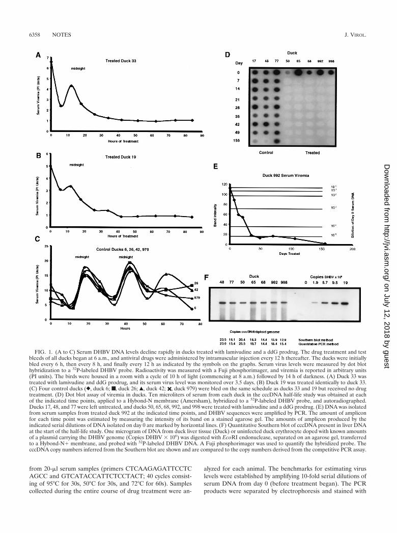
determined by measuring the kinetics of the loss of DHBVDNA from the sera of treated animals. Two congenitally in-fected ducks (ducks 33 and 19) were treated with the drugcombination and were bled at frequent intervals over a 4-dayperiod. Virus levels in serum were measured by dot blot hy-bridization and phosphorimager analysis. The decline in thevirus levels followed a complex course, as shown in Fig. 1A andB. The curves in Fig. 1A and B suggest that a loss in viral DNA,effected by the drugs, was superimposed on pulses of virusreleased into the serum. To test this possibility, four untreated,congenitally infected ducks (ducks 6, 26, 42, and 979) were bledon the same schedule as were the ducks described above. Asshown in Fig. 1C, the virus levels in their sera oscillated, de-clining to a minimum over the first 12 h and subsequentlyfluctuating, with peaks at 18 and 42 h after the experimentbegan. The peaks occurred at midnight, in the middle of theanimals’ “dark period,” and were remarkably coherent for allfour animals. Subsequently, the fluctuations in the animals’virus levels began to lose their periodicity and coherence. Toour knowledge, this is the first report of such oscillations inDHBV levels in serum. We suggest that this response wasinduced by the stress experienced by the animals when beinghandled and bled and that its period was tied to the diurnalrhythm of the animals. The periodicity in serum virus levelswas lost as the animals became habituated to handling. Thisconjecture is reasonable, since young ducks show a markeddiurnal rhythm in the release of the stress hormone cortico-sterone (28). In pigeons, frequent handling increases the cor-ticosterone level but not the rhythm of its release (27). Finally,corticosteroids are known to stimulate DHBV replication (9).We think that in our experiment, inhibition of viral replicationby the drug treatment was partially overcome by a stimulationof replication induced by the stress experienced by the animalsduring handling; the combination of these antagonistic effectsresulted in the curves seen in Fig. 1A and B. Despite thesecomplications, it was evident that the antiviral drug combina-tion resulted in the immediate and rapid inhibition of DHBVreplication that is required for a cccDNA half-life study.
The ducks were bled weekly over the course of the study.The viremia of the treated ducks became undetectable by dotblot assay within the first week (Fig. 1D). The detection limit ofthis assay was only about 0.5% of the initial serum virus levels.A more sensitive PCR assay was used to estimate lower viruslevels. Amplification of the region between positions 1039 and1945 of the DHBV sequence was performed on DNA isolated
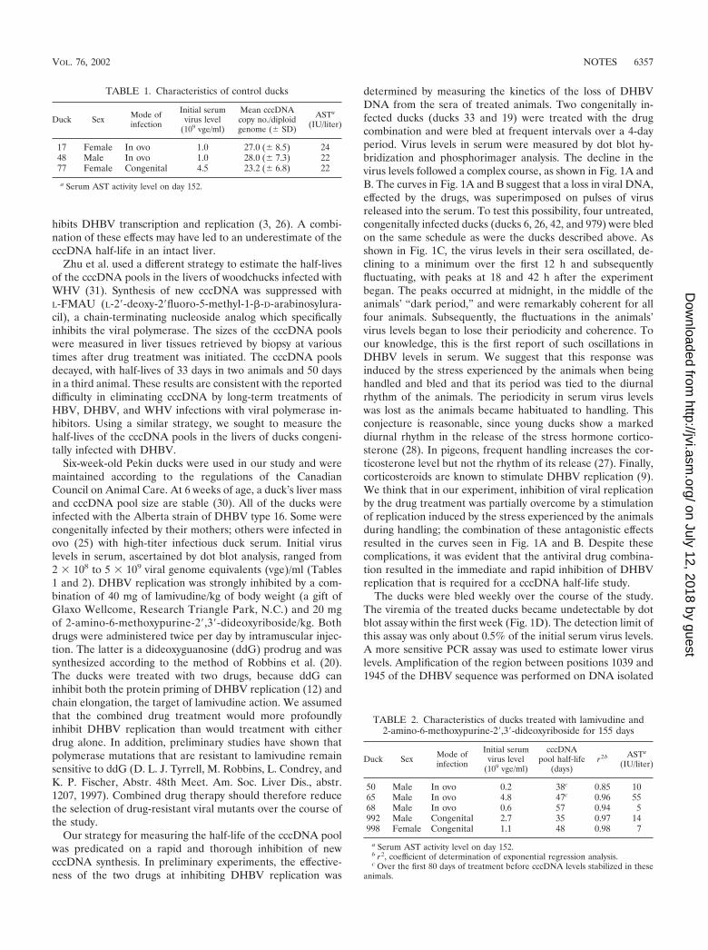
TABLE 1. Characteristics of control ducks
Duck Sex Mode ofinfection
Initial serumvirus level
(109 vge/ml)
Mean cccDNAcopy no./diploidgenome (� SD)
ASTa
(IU/liter)
17 Female In ovo 1.0 27.0 (� 8.5) 2448 Male In ovo 1.0 28.0 (� 7.3) 2277 Female Congenital 4.5 23.2 (� 6.8) 22
a Serum AST activity level on day 152.
TABLE 2. Characteristics of ducks treated with lamivudine and2-amino-6-methoxypurine-2�,3�-dideoxyriboside for 155 days
Duck Sex Mode ofinfection
Initial serumvirus level
(109 vge/ml)
cccDNApool half-life
(days)r2 b ASTa
(IU/liter)
50 Male In ovo 0.2 38c 0.85 1065 Male In ovo 4.8 47c 0.96 5568 Male In ovo 0.6 57 0.94 5992 Male Congenital 2.7 35 0.97 14998 Female Congenital 1.1 48 0.98 7
a Serum AST activity level on day 152.b r2, coefficient of determination of exponential regression analysis.c Over the first 80 days of treatment before cccDNA levels stabilized in these
animals.
VOL. 76, 2002 NOTES 6357
on July 12, 2018 by guesthttp://jvi.asm
.org/D
ownloaded from
from 20-�l serum samples (primers CTCAAGAGATTCCTCAGCC and GTCATACCATTCTCCTACT; 40 cycles consist-ing of 95°C for 30s, 50°C for 30s, and 72°C for 60s). Samplescollected during the entire course of drug treatment were an-
alyzed for each animal. The benchmarks for estimating viruslevels were established by amplifying 10-fold serial dilutions ofserum DNA from day 0 (before treatment began). The PCRproducts were separated by electrophoresis and stained with
FIG. 1. (A to C) Serum DHBV DNA levels decline rapidly in ducks treated with lamivudine and a ddG prodrug. The drug treatment and testbleeds of all ducks began at 6 a.m., and antiviral drugs were administered by intramuscular injection every 12 h thereafter. The ducks were initiallybled every 6 h, then every 8 h, and finally every 12 h as indicated by the symbols on the graphs. Serum virus levels were measured by dot blothybridization to a 32P-labeled DHBV probe. Radioactivity was measured with a Fuji phosphorimager, and viremia is reported in arbitrary units(PI units). The birds were housed in a room with a cycle of 10 h of light (commencing at 8 a.m.) followed by 14 h of darkness. (A) Duck 33 wastreated with lamivudine and ddG prodrug, and its serum virus level was monitored over 3.5 days. (B) Duck 19 was treated identically to duck 33.(C) Four control ducks (}, duck 6; ■, duck 26; Œ, duck 42; ✖, duck 979) were bled on the same schedule as ducks 33 and 19 but received no drugtreatment. (D) Dot blot assay of viremia in ducks. Ten microliters of serum from each duck in the cccDNA half-life study was obtained at eachof the indicated time points, applied to a Hybond-N membrane (Amersham), hybridized to a 32P-labeled DHBV probe, and autoradiographed.Ducks 17, 48, and 77 were left untreated, and ducks 50, 65, 68, 992, and 998 were treated with lamivudine and a ddG prodrug. (E) DNA was isolatedfrom serum samples from treated duck 992 at the indicated time points, and DHBV sequences were amplified by PCR. The amount of ampliconfor each time point was estimated by measuring the intensity of its band on a stained agarose gel. The amounts of amplicon produced by theindicated serial dilutions of DNA isolated on day 0 are marked by horizontal lines. (F) Quantitative Southern blot of cccDNA present in liver DNAat the start of the half-life study. One microgram of DNA from duck liver tissue (Duck) or uninfected duck erythrocyte doped with known amountsof a plasmid carrying the DHBV genome (Copies DHBV � 106) was digested with EcoRI endonuclease, separated on an agarose gel, transferredto a Hybond-N� membrane, and probed with 32P-labeled DHBV DNA. A Fuji phosphorimager was used to quantify the hybridized probe. ThecccDNA copy numbers inferred from the Southern blot are shown and are compared to the copy numbers derived from the competitive PCR assay.
6358 NOTES J. VIROL.
on July 12, 2018 by guesthttp://jvi.asm
.org/D
ownloaded from
ethidium bromide, and the amount of 906-bp amplicon wasmeasured with a video image capture system and NIH ImageGauge software. Figure 1E shows the results obtained for duck992; similar results were obtained for the other animals. Overthe first week of treatment, the virus level was reduced to 10�2
to 10�3 of its initial level; the virus level continued to fall overthe next several weeks and plateaued at or below 10�5 of theinitial level after 5 to 8 weeks of treatment. The inhibition ofreplication we observed was fourfold more rapid than thatreported by Zhu et al. in their study of the half-life of WHVcccDNA (31). The rapid and potent inhibition of viral replica-tion produced by our drug treatment regimen was thought tobe sufficient to prevent renewal of the cccDNA pool and per-mit analysis of its decay over time.
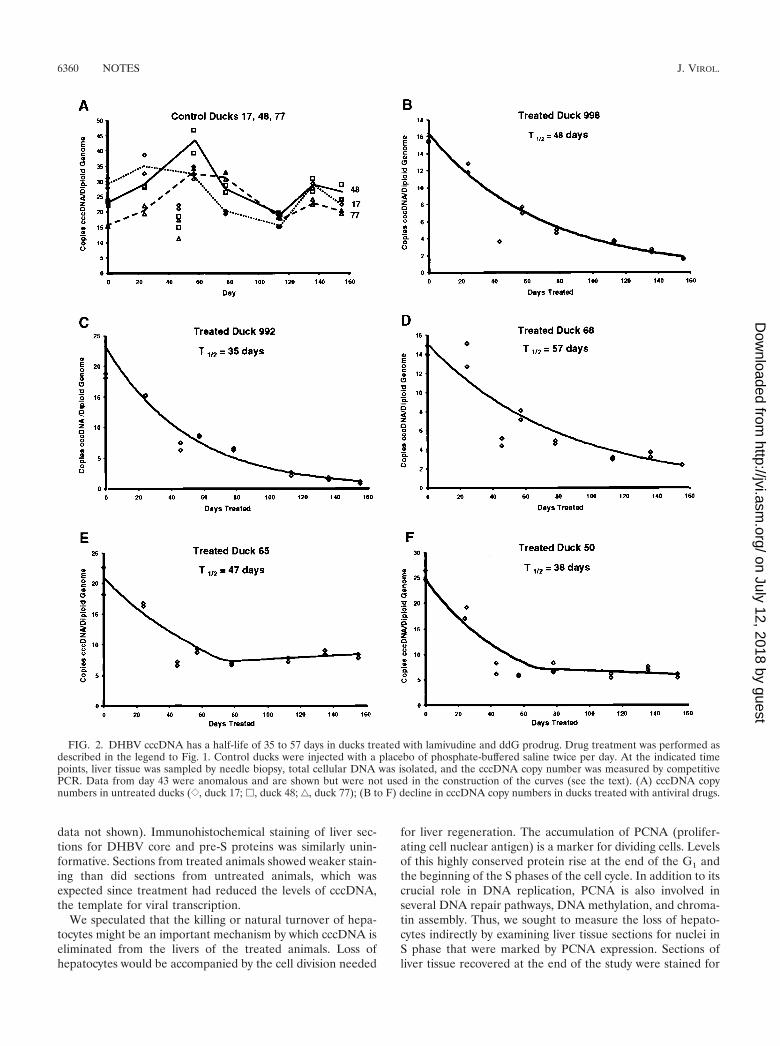
Eight ducks were used in the cccDNA half-life study. Threecontrol ducks were injected with 0.5 ml of phosphate-bufferedsaline, and five ducks were treated with lamivudine and theddG prodrug as described above. At eight points over a 155-day period, liver tissue from each animal was obtained byneedle biopsy, total DNA was isolated without proteinasetreatment, and duplicate competitive PCR assays selective forDHBV cccDNA were performed as previously described (1).The accuracy of the PCR assay at the start of the study, beforedrug treatment had eliminated other viral replicative interme-diates, was confirmed by quantitative Southern blotting. Anautoradiogram of the Southern blot along with a comparisonof the cccDNA copy numbers inferred from the two methodsis shown in Fig. 1F. The two assays yielded very similar esti-mates of the initial cccDNA copy numbers. At the end of thestudy, the cccDNA levels of each animal, expressed as thecopies of cccDNA per diploid genome, were plotted againsttime. Where appropriate, the data were fitted to an exponen-tial decay curve by regression analysis (Microsoft Excel). Thesecurves are shown in Fig. 2. The cccDNA copy numbers insamples taken on day 43 of the experiment were much lowerthan expected for both the control and treated animals. Wethink that these results are anomalous and possibly due to theselective loss of cccDNA from the DNA samples isolated onthat day. While the results of the day 43 assays are shown onthe graphs, they were not included in the regression analysesused to calculate the half-lives of cccDNA pools.
The sizes of the cccDNA pools in untreated control ducksare shown in Fig. 2A. The cccDNA levels at the beginning andthe end of the experiment were similar, but the most notablefeatures of these data are the fluctuations in the cccDNA levelsover time, which are reflected in the large standard deviationsshown in Table 1. We think that these fluctuations are real andnot due to experimental error, since the data for the treatedducks show much less random variation than the data for thecontrols. The patterns of fluctuation were similar for all threeanimals, suggesting that the variation is driven by some envi-ronmental factor. Virus levels in serum also fluctuated over thecourse of the study, but there was no correlation, either posi-tive or negative, between cccDNA levels and serum virus levels(data not shown).
The cccDNA pool sizes in treated ducks 998, 992, and 68decayed in a simple pattern, as shown in Fig. 2B to D. Thesedata fit exponential decay curves very well, as reflected by thecoefficient of determination values that are close to 1 (Table 2).The half-lives of the cccDNA pools in the three animals were
quite different: 48 days for duck 998, 35 days for duck 992, and57 days for duck 68. Both the magnitude of and the animal-to-animal variation in the cccDNA half-lives in these duckswere similar to those reported for WHV cccDNA in wood-chuck livers (half-lives of 33 and 50 days) by Zhu et al. (31).
The cccDNA pools in the two remaining treated animals,ducks 65 and 50, showed an unexpected pattern of decay. Asshown in Fig. 2E and F, the cccDNA pools decayed exponen-tially, with half-lives of 47 and 38 days, respectively, for aboutthe first 70 days of treatment. Then the cccDNA levels abruptlyplateaued at about 8 and 6 copies of cccDNA/diploid genome,respectively. The reason for the stabilization of the cccDNApools in these animals is not clear. All the animals in the studyhad normal serum aspartic acid transaminase (AST) levels onday 152 (Tables 1 and 2), suggesting that impaired liver func-tion could not provide an explanation for the unexpected pat-tern of cccDNA loss in ducks 50 and 65. The emergence of adrug-resistant mutant virus would explain these results. Theselection of such mutants occurs frequently upon treatment ofinfected woodchucks and humans (7, 16, 31). However, wethink this explanation is unlikely. Viral DNA could not bedetected by dot blotting in the sera of these ducks at any pointduring drug treatment (Fig. 1D). The more sensitive PCRassay did not detect any increase in viral DNA in serum fromthe very low levels typical of all treated animals late in thestudy. Had a drug-resistant mutant arisen, a resurgent viremiashould have resulted. PCR amplification of the low levels ofvirus (under the conditions described above) in serum col-lected on day 155 permitted the sequencing of viral DNAencoding the YMDD and upstream FLL motifs that can mu-tate to confer lamivudine resistance (8). The amplicons fromducks 50 and 65 were sequenced, and the sequences of thepolymerase reading frame encoding amino acids 484 to 594, aregion containing both the FLL and YMDD motifs, werefound to be wild type. Thus, the viral polymerase likely re-mained sensitive to inhibition by lamivudine. It is unlikely thatthe emergence of a drug-resistant virus prevented ducks 50 and65 from continuing to lose cccDNA.
The difference in the kinetics of cccDNA loss between thetwo groups of treated ducks prompted us to examine sectionsof liver tissue recovered at the end of the study for histopatho-logical differences. The liver tissue was fixed in 4% bufferedparaformaldehyde, embedded in paraffin, sectioned, andstained with eosin and hematoxylin. The three untreated ani-mals showed mild to moderate portal inflammation and mildpiecemeal and focal necrosis. Ducks 17 and 77 showed fattychanges in their livers, which were severe in the case of duck 17and mild in the case of duck 77. The ducks treated with anti-viral drugs had milder hepatitis than did the control animals.Duck 65 showed no evidence of hepatitis. The four othertreated ducks showed mild portal inflammation. Necrosis wasabsent in all but duck 68, which showed mild focal necrosis. Nohistological difference was seen between animals in whichcccDNA levels stabilized at a low level and those in which thecccDNA pools continued to decline over the course of thestudy. These histological findings were corroborated by testsfor liver enzymes in serum. The serum AST levels, shown inTables 1 and 2, were in the normal range at the end of thestudy, as were those of the other liver enzymes tested (alanineaminotransferase, bilirubin, and �-glutamyl aminotransferase;
VOL. 76, 2002 NOTES 6359
on July 12, 2018 by guesthttp://jvi.asm
.org/D
ownloaded from
data not shown). Immunohistochemical staining of liver sec-tions for DHBV core and pre-S proteins was similarly unin-formative. Sections from treated animals showed weaker stain-ing than did sections from untreated animals, which wasexpected since treatment had reduced the levels of cccDNA,the template for viral transcription.
We speculated that the killing or natural turnover of hepa-tocytes might be an important mechanism by which cccDNA iseliminated from the livers of the treated animals. Loss ofhepatocytes would be accompanied by the cell division needed
for liver regeneration. The accumulation of PCNA (prolifer-ating cell nuclear antigen) is a marker for dividing cells. Levelsof this highly conserved protein rise at the end of the G1 andthe beginning of the S phases of the cell cycle. In addition to itscrucial role in DNA replication, PCNA is also involved inseveral DNA repair pathways, DNA methylation, and chroma-tin assembly. Thus, we sought to measure the loss of hepato-cytes indirectly by examining liver tissue sections for nuclei inS phase that were marked by PCNA expression. Sections ofliver tissue recovered at the end of the study were stained for
FIG. 2. DHBV cccDNA has a half-life of 35 to 57 days in ducks treated with lamivudine and ddG prodrug. Drug treatment was performed asdescribed in the legend to Fig. 1. Control ducks were injected with a placebo of phosphate-buffered saline twice per day. At the indicated timepoints, liver tissue was sampled by needle biopsy, total cellular DNA was isolated, and the cccDNA copy number was measured by competitivePCR. Data from day 43 were anomalous and are shown but were not used in the construction of the curves (see the text). (A) cccDNA copynumbers in untreated ducks ({, duck 17; �, duck 48; ‚, duck 77); (B to F) decline in cccDNA copy numbers in ducks treated with antiviral drugs.
6360 NOTES J. VIROL.
on July 12, 2018 by guesthttp://jvi.asm
.org/D
ownloaded from
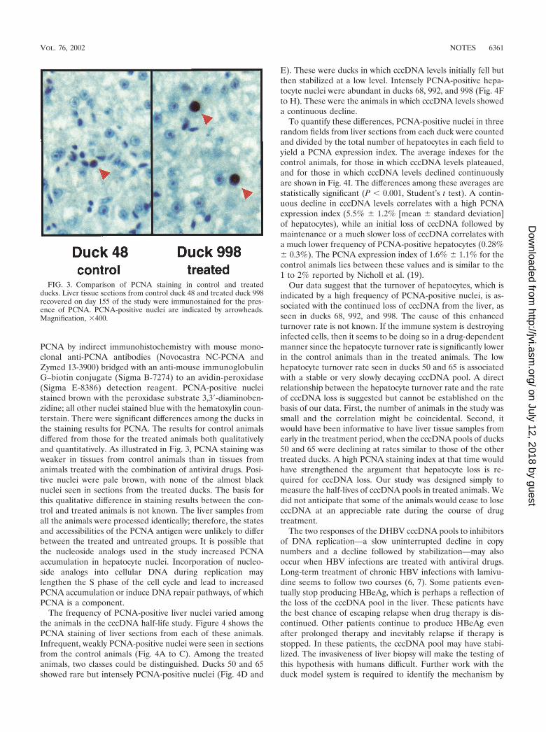
PCNA by indirect immunohistochemistry with mouse mono-clonal anti-PCNA antibodies (Novocastra NC-PCNA andZymed 13-3900) bridged with an anti-mouse immunoglobulinG–biotin conjugate (Sigma B-7274) to an avidin-peroxidase(Sigma E-8386) detection reagent. PCNA-positive nucleistained brown with the peroxidase substrate 3,3�-diaminoben-zidine; all other nuclei stained blue with the hematoxylin coun-terstain. There were significant differences among the ducks inthe staining results for PCNA. The results for control animalsdiffered from those for the treated animals both qualitativelyand quantitatively. As illustrated in Fig. 3, PCNA staining wasweaker in tissues from control animals than in tissues fromanimals treated with the combination of antiviral drugs. Posi-tive nuclei were pale brown, with none of the almost blacknuclei seen in sections from the treated ducks. The basis forthis qualitative difference in staining results between the con-trol and treated animals is not known. The liver samples fromall the animals were processed identically; therefore, the statesand accessibilities of the PCNA antigen were unlikely to differbetween the treated and untreated groups. It is possible thatthe nucleoside analogs used in the study increased PCNAaccumulation in hepatocyte nuclei. Incorporation of nucleo-side analogs into cellular DNA during replication maylengthen the S phase of the cell cycle and lead to increasedPCNA accumulation or induce DNA repair pathways, of whichPCNA is a component.
The frequency of PCNA-positive liver nuclei varied amongthe animals in the cccDNA half-life study. Figure 4 shows thePCNA staining of liver sections from each of these animals.Infrequent, weakly PCNA-positive nuclei were seen in sectionsfrom the control animals (Fig. 4A to C). Among the treatedanimals, two classes could be distinguished. Ducks 50 and 65showed rare but intensely PCNA-positive nuclei (Fig. 4D and
E). These were ducks in which cccDNA levels initially fell butthen stabilized at a low level. Intensely PCNA-positive hepa-tocyte nuclei were abundant in ducks 68, 992, and 998 (Fig. 4Fto H). These were the animals in which cccDNA levels showeda continuous decline.
To quantify these differences, PCNA-positive nuclei in threerandom fields from liver sections from each duck were countedand divided by the total number of hepatocytes in each field toyield a PCNA expression index. The average indexes for thecontrol animals, for those in which cccDNA levels plateaued,and for those in which cccDNA levels declined continuouslyare shown in Fig. 4I. The differences among these averages arestatistically significant (P 0.001, Student’s t test). A contin-uous decline in cccDNA levels correlates with a high PCNAexpression index (5.5% � 1.2% [mean � standard deviation]of hepatocytes), while an initial loss of cccDNA followed bymaintenance or a much slower loss of cccDNA correlates witha much lower frequency of PCNA-positive hepatocytes (0.28%� 0.3%). The PCNA expression index of 1.6% � 1.1% for thecontrol animals lies between these values and is similar to the1 to 2% reported by Nicholl et al. (19).
Our data suggest that the turnover of hepatocytes, which isindicated by a high frequency of PCNA-positive nuclei, is as-sociated with the continued loss of cccDNA from the liver, asseen in ducks 68, 992, and 998. The cause of this enhancedturnover rate is not known. If the immune system is destroyinginfected cells, then it seems to be doing so in a drug-dependentmanner since the hepatocyte turnover rate is significantly lowerin the control animals than in the treated animals. The lowhepatocyte turnover rate seen in ducks 50 and 65 is associatedwith a stable or very slowly decaying cccDNA pool. A directrelationship between the hepatocyte turnover rate and the rateof cccDNA loss is suggested but cannot be established on thebasis of our data. First, the number of animals in the study wassmall and the correlation might be coincidental. Second, itwould have been informative to have liver tissue samples fromearly in the treatment period, when the cccDNA pools of ducks50 and 65 were declining at rates similar to those of the othertreated ducks. A high PCNA staining index at that time wouldhave strengthened the argument that hepatocyte loss is re-quired for cccDNA loss. Our study was designed simply tomeasure the half-lives of cccDNA pools in treated animals. Wedid not anticipate that some of the animals would cease to losecccDNA at an appreciable rate during the course of drugtreatment.
The two responses of the DHBV cccDNA pools to inhibitorsof DNA replication—a slow uninterrupted decline in copynumbers and a decline followed by stabilization—may alsooccur when HBV infections are treated with antiviral drugs.Long-term treatment of chronic HBV infections with lamivu-dine seems to follow two courses (6, 7). Some patients even-tually stop producing HBeAg, which is perhaps a reflection ofthe loss of the cccDNA pool in the liver. These patients havethe best chance of escaping relapse when drug therapy is dis-continued. Other patients continue to produce HBeAg evenafter prolonged therapy and inevitably relapse if therapy isstopped. In these patients, the cccDNA pool may have stabi-lized. The invasiveness of liver biopsy will make the testing ofthis hypothesis with humans difficult. Further work with theduck model system is required to identify the mechanism by
FIG. 3. Comparison of PCNA staining in control and treatedducks. Liver tissue sections from control duck 48 and treated duck 998recovered on day 155 of the study were immunostained for the pres-ence of PCNA. PCNA-positive nuclei are indicated by arrowheads.Magnification, �400.
VOL. 76, 2002 NOTES 6361
on July 12, 2018 by guesthttp://jvi.asm
.org/D
ownloaded from
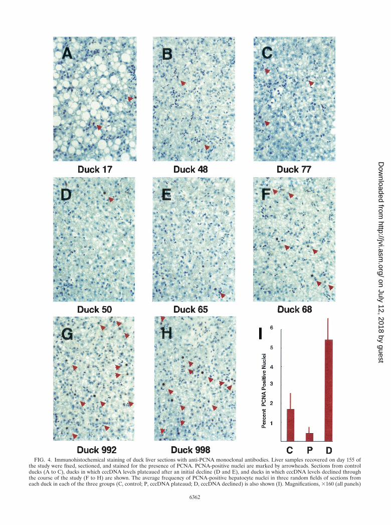
FIG. 4. Immunohistochemical staining of duck liver sections with anti-PCNA monoclonal antibodies. Liver samples recovered on day 155 ofthe study were fixed, sectioned, and stained for the presence of PCNA. PCNA-positive nuclei are marked by arrowheads. Sections from controlducks (A to C), ducks in which cccDNA levels plateaued after an initial decline (D and E), and ducks in which cccDNA levels declined throughthe course of the study (F to H) are shown. The average frequency of PCNA-positive hepatocyte nuclei in three random fields of sections fromeach duck in each of the three groups (C, control; P, cccDNA plateaud; D, cccDNA declined) is also shown (I). Magnifications, �160 (all panels)
6362
on July 12, 2018 by guesthttp://jvi.asm
.org/D
ownloaded from
which cccDNA is lost during prolonged antiviral therapy and tounderstand how that mechanism can fail to eliminate thecccDNA pool in some cases.
Nucleotide sequence accession number. The sequence of theAlberta strain of DHBV type 16 has been deposited in Gen-Bank under accession no. AF047045.
We acknowledge Gerald Lachance for expert care of the animalsand thank Karl Fischer for useful comments on the manuscript.
This research was supported by Glaxo Wellcome Canada.
REFERENCES
1. Addison, W. R., W. W. S. Wong, K. P. Fischer, and D. L. J. Tyrrell. 2000. Aquantitative competitive PCR assay for the covalently closed circular form ofthe duck hepatitis B virus. Antivir. Res. 48:27–37.
2. Chyama, K., Y. Suzuki, M. Kobayashi, M. Kobayashi, A. Tsubota, M. Hashi-moto, Y. Miyano, H. Koike, M. Kobayashi, I. Koida, Y. Arase, S. Saitoh, N.Murashima, K. Ikeda, and H. Kumada. 1998. Emergence and takeover ofYMDD motif mutant hepatitis B virus during long-term lamivudine therapyand re-takeover by wild type after cessation of therapy. Hepatology 27:1711–1716.
3. Civitico, G. M., and S. A. Locarnini. 1994. The half-life of duck hepatitis Bvirus supercoiled DNA in congenitally infected primary hepatocyte cultures.Virology 203:81–89.
4. Dandri, M., M. R. Burda, H. Will, and J. Petersen. 2000. Increased hepa-tocyte turnover and inhibition of woodchuck hepatitis B virus replication byadefovir in vitro do not lead to reduction of the closed circular DNA.Hepatology 32:139–146.
5. Dienstag, J. L., R. P. Perrillo, E. R. Schiff, M. Bartholomew, C. Vicary, andM. Rubin. 1995. A preliminary trial of lamivudine for chronic hepatitis Binfection. N. Engl. J. Med. 333:1657–1661.
6. Dienstag, J. L., E. R. Schiff, T. L. Wright, R. P. Perillo, H.-W. L. Hann, Z.Goodman, L. Crowther, L. Condreay, M. Woessner, M. Rubin, and N. A.Brown. 1999. Lamivudine as initial treatment for chronic hepatitis B in theUnited States. N. Engl. J. Med. 341:1266–1273.
7. Fischer, K. P., K. S. Gutfreund, and D. L. J. Tyrrell. 2001. Lamivudine:resistance mechanisms and their clinical implications. Drug Resist. Updates4:118–128.
8. Fischer, K. P., and D. L. J. Tyrrell. 1996. Generation of duck hepatitis Bvirus polymerase mutants through site-directed mutagenesis which demon-strate resistance to lamivudine [(�)-�-L-2�,3�-dideoxy-3�-thiacytidine] invitro. Antimicrob. Agents Chemother. 40:1957–1960.
9. Fukuda, R., S. Okinaga, S. Akagi, M. Hidaka, N. Ono, S. Fukumoto, and Y.Shimada. 1988. Alteration of infection pattern of duck hepatitis B virus byimmunomodulatory drugs. J. Med. Virol. 27:387–396.
10. Ganem, D., and H. E. Varmus. 1987. The molecular biology of hepatitis Bviruses. Annu. Rev. Biochem. 56:651–693.
11. Gutfreund, K. S., M. Williams, R. George, V. Bain, M. M. Ma, E. M.Yoshida, J.-P. Villeneuve, K. P. Fischer, and D. L. J. Tyrrell. 2000. Genotypicsuccession of mutations of the hepatitis B virus polymerase associated withlamivudine resistance. J. Hepatol. 33:469–475.
12. Howe, A. Y. M., M. J. Robbins, J. S. Wilson, and D. L. J. Tyrrell. 1996.Selective inhibition of the reverse transcription of duck hepatitis B virus bybinding of 2�,3�-dideoxyguanosine-5�-triphosphate to the viral polymerase.Hepatology 23:87–96.
13. Jilbert, A. R., T.-T. Wu, J. M. England, P. M. Hall, N. Z. Carp, A. P.O’Connell, and W. S. Mason. 1992. Rapid resolution of duck hepatitis Bvirus infections occurs after massive hepatocellular involvement. J. Virol.66:1377–1388.
14. Kajino, K., A. R. Jilbert, J. Saputelli, C. E. Aldrich, J. Cullen, and W. S.Mason. 1994. Woodchuck hepatitis virus infections: very rapid recovery aftera prolonged viremia and infection of virtually every hepatocyte. J. Virol.68:5792–5803.
15. Köck, J., and H.-J. Schlicht. 1993. Analysis of the earliest steps of hepad-navirus replication: genome repair after infectious entry into hepatocytesdoes not depend on viral polymerase activity. J. Virol. 67:4867–4874.
16. Mason, W. S., J. Cullen, G. Moraleda, J. Saputelli, C. E. Aldrich, D. S.Miller, B. Tennant, L. Frick, D. Averett, L. D. Condreay, and A. R. Jilbert.1998. Lamivudine therapy of WHV-infected woodchucks. Virology 245:18–32.
17. Miller, R. H., and W. S. Robinson. 1984. Hepatitis B virus DNA forms innuclear and cytoplasmic fractions of infected human liver. Virology 137:390–399.
18. Moraleda, G., J. Saputelli, C. E. Aldrich, D. Averett, L. Condreay, and W. S.Mason. 1997. Lack of effect of antiviral therapy in nondividing hepatocytecultures on the closed circular DNA of woodchuck hepatitis virus. J. Virol.71:9392–9399.
19. Nicholl, A. J., P. W. Angus, S. T. Chou, C. A. Luscombe, R. A. Smallwood,and S. A. Locarnini. 1997. Demonstration of duck hepatitis B virus in bileduct epithelial cells: implications for pathogenesis and persistent infection.Hepatology 26:463–469.
20. Robins, M. J., J. S. Wilson, D. Madej, D. L. J. Tyrrell, W. P. Gati, R. J.Lindmark, and S. F. Wnuk. 2001. Nucleic acid related compounds. 114.Synthesis of 2,6-(disubstituted)purine-2�,3�-dideoxynucleosides and selectedcytotoxic, anti-hepatitis B, and adenosine deaminase substrate activities.J. Heterocycl. Chem. 38:1297–1306.
21. Seeger, C., J. Summers, and W. Mason. 1991. Viral DNA synthesis. Curr.Top. Microbiol. Immunol. 168:41–59.
22. Summers, J., P. M. Smith, and A. L. Horwich. 1990. Hepadnavirus envelopeproteins regulate covalently closed circular DNA amplification. J. Virol.64:2819–2824.
23. Summers, J., P. M. Smith, M. Huang, and M. Yu. 1991. Morphogenetic andregulatory effects of mutations in the envelope proteins of an avian hepad-navirus. J. Virol. 65:1310–1317.
24. Tencza, M. G., and J. E. Newbold. 1997. Heterogeneous response for amammalian hepadnavirus infection to acyclovir: drug-arrested intermediatesof minus-strand viral DNA synthesis are enveloped and secreted from in-fected cells as virion-like particles. J. Med. Virol. 51:6–16.
25. Tsiquaye, K. N., M. Rapicetta, T. F. McCaul, and A. J. Zuckerman. 1985.Experimental in ovo transmission of duck hepatitis B virus. J. Virol. Methods11:49–57.
26. Tuttleman, J. S., C. Pourcel, and J. Summers. 1986. Formation of the poolof covalently closed circular viral DNA in hepadnavirus-infected cells. Cell47:451–460.
27. Westerhof, I., J. A. Mol, W. E. Van den Brom, J. T. Lumeij, and A. Rijnberk.1994. Diurnal rhythms of plasma corticosterone concentrations in racingpigeons (Columba livia domestica) exposed to different light regimens, andthe influence of frequent blood sampling. Avian Dis. 38:428–434.
28. Wilson, S. C., F. J. Cunningham, and T. R. Morris. 1982. Diurnal changes inthe plasma concentrations of corticosterone, luteinizing hormone and pro-gesterone during sexual development and the ovulatory cycle of KhakiCampbell ducks. J. Endocrinol. 93:267–277.
29. Wu, T.-T., L. Coates, C. E. Aldrich, J. Summers, and W. S. Mason. 1990. Inhepatocytes infected with duck hepatitis B virus, the template for viral RNAsynthesis is amplified by an intracellular pathway. Virology 175:255–261.
30. Zhang, Y.-Y., and J. Summers. 2000. Low dynamic state of viral competitionin a chronic avian hepadnavirus infection. J. Virol. 74:5257–5275.
31. Zhu, Y., T. Yamamoto, J. Cullen, J. Saputelli, C. E. Aldrich, D. S. Miller, S.Litwin, P. A. Furman, A. R. Jilbert, and W. S. Mason. 2001. Kinetics ofhepadnavirus loss from the liver during inhibition of viral DNA synthesis.J. Virol. 75:311–322.
VOL. 76, 2002 NOTES 6363
on July 12, 2018 by guesthttp://jvi.asm
.org/D
ownloaded from