Embed Size (px)
Citation preview
JOURNAL OF VIROLOGY, May 1988, p. 1836-18390022-538X/88/051836-04$02.00/0Copyright © 1988, American Society for Microbiology
Vol. 62, No. 5
Presentation and Immunogenicity of the Hepatitis B SurfaceAntigen and a Poliovirus Neutralization Antigen on Mixed Empty
Envelope ParticlesF. DELPEYROUX, N. PEILLON, B. BLONDEL, R. CRAINIC, AND R. E. STREECKt*
Unite de Virologie Medicale, Institut Pasteur, 75724 Paris Cedex 15, France
Received 14 September 1987/Accepted 8 February 1988
Insertion of a synthetic DNA fragment encoding a poliovirus neutralization epitope into the S gene encodingthe major envelope protein of hepatitis B virus has yielded hybrid (HBsPolioAg) particles closely resemblingauthentic 22-nm antigen (HBsAg) particles by expression of the modified gene in mammalian cells. In mice,these hybrid particles induce neutralizing antibodies against poliovirus but only a very weak immune responseto HBsAg (F. Delpeyroux, N. Chenciner, A. Lim, Y. Malpiece, B. Blondel, R. Crainic, S. Van der Werf, andR. E. Streeck, Science 233:472-474, 1986). By cotransfection with different plasmids carrying either modifiedor unmodified S genes, we have now obtained mixed particles presenting both HBsAg and HBsPolioAg. Whensuch particles were inoculated into rabbits, antibodies to both poliovirus and to HBsAg were induced.Moreover, the titers of neutralizing antibodies to poliovirus induced by HBsPolioAg were much higher thanthose previously obtained in mice. The design of multivalent particles carrying various peptide sequences orpresenting several heterologous epitopes may therefore be possible.
The hepatitis B surface antigen (HBsAg) appearing in thesera of patients infected by the hepatitis B virus (HBV) iscarried by three morphologically distinct particles, 42-nmspherical particles representing infectious virions (Dane par-ticles), filaments of about 22 nm in diameter, and 22-nmspherical particles corresponding to empty viral envelopes(18). The 22-nm particles consist of cellular lipids, the majorenvelope protein, and one or two minor envelope proteins.The proteins are present in both the glycosylated andnonglycosylated forms (17) and are linked by disulfidebridges into dimers and higher oligomers. Reduction of theinter- and intramolecular disulfide bridges and dissociationof the particles lead to a drastic decrease of the HBsAgantigenicity (12, 16). Expression of the S gene of the HBVgenome encoding the 226-amino-acid sequence of the majorenvelope protein in mammalian cell lines yields definedlipoprotein particles similar to those from human serumwhich are secreted into the cellular medium (5, 13).We have previously analyzed the formation and secretion
of modified HBsAg particles by creating in-phase insertionsof different lengths and sequences in various regions of the Sgene. Expression of the modified genes was studied in mouseL cells (3). A hybrid HBsAg carrying a poliovirus neutrali-zation epitope inserted next to the major antigenic regionwas shown to be organized into 22-nm particles whichreacted with a poliovirus-specific monoclonal antibody.Mice immunized with these HBsPolioAg particles showed apoor response to HBsAg and a low but significant titer ofpoliovirus-neutralizing antibodies (4). The weak immuneresponse to HBsAg suggested that an important conforma-tional change of the hepatitis B surface antigen might haveoccurred as a result of the insertion of the poliovirus epitope.To further analyze the potential of the HBV envelopeparticles as an immunogenic carrier of heterologous anti-gens, we have carried out this study with the following
* Corresponding author.t Present address: Institute of Medical Microbiology, University
of Mainz, D-6500 Mainz, Federal Republic of Germany.
objectives: to design mixed particles in which both HBsAgand HBsPolioAg could be presented in biologically activeform, to analyze the immunogenicity of the mixed viralantigens, and to extend the system to other species knowingthat mice are poor responders to poliovirus-neutralizingantigens.To obtain expression of HBsAg and HBsPolioAg, we
transfected two plasmids, pLAS and pPAP, into mouse Lcells. pLAS and pPAP both carry the S gene of HBV,including the viral polyadenylation site, and the simian virus40 early promoter. pPAP differs from pLAS by a 39-base-pair insertion of a synthetic DNA fragment in the uniqueBamHI site within the S gene, encoding amino acid residues93 through 103 of the VP1 capsid protein of poliovirus type1 (Mahoney strain) and an additional Gly and Ser residueencoded by the BamHI linker used for insertion. As previ-ously shown by transfection of mouse L cells with pLAS orpPAP, stable clones secreting either HBsAg or hybrid HBsPolioAg particles could be obtained (4).For the expression of HBsAg and HBsPolioAg from the
same cells, pLAS and pPAP were mixed with plasmid pW (2)encoding neomycin resistance and were cotransfected intomouse L cells. For 106 cells, 5 jig each of pLAS and pPAPand 2 pLg of pW were used. Of 12 G418-resistant clonesisolated, 10 were HBsAg positive. To characterize theHBsAg products, proteins of seven clones were labeled invivo with [35S]methionine. Cellular supernatants were clar-ified at 12,000 x g for 5 min, and rabbit antiserum to humanHBsAg particles (Behring) was added at a dilution of 1:100.Protein A-Sepharose (Pharmacia) was added as describedpreviously (3), except that the Sepharose bed was washedthree times with a solution consisting of 10 mM Tris hydro-chloride (pH 7.2), 150 mM NaCl, 1% Triton X-100, 0.1%sodium dodecyl sulfate, and 1% sodium deoxycholate andwas washed twice with 125 mM Tris hydrochloride (pH 6.8).Proteins were eluted by boiling for 5 min in 40 ,ul of samplebuffer and were submitted to polyacrylamide gel electropho-resis in the presence of sodium dodecyl sulfate. One of theseclones (MAP) produced HBsAg and HBsPolioAg in both the
1836
on June 4, 2018 by guesthttp://jvi.asm
.org/D
ownloaded from
NOTES 1837
a b c d e f g h
28.527
s-2*I3
M NI L P L+P L P IL-Pas C3 S 03
FIG. 1. Polyacrylamide gel electrophoresis of HBsAg and HBsPolioAg in the presence of sodium dodecyl sulfate. [35S]methionine-labeled proteins from cellular supernatants were immunoprecipi-tated with antiserum to HBsAg (aS) or the antipoliovirus monoclo-nal antibody C3. The autoradiograms of two different (12.5%) gelsare shown. HBsAg (5), HBsPolioAg (SP), and their glycosylatedforms (*) are indicated. Their molecular sizes deduced from stan-dards (Pharmacia) are indicated in kilodaltons (kDa) between lanesd and e. Lanes: a and c, two independent clones producing onlyHBsPolioAg; b and d, MAP clone producing both HBsAg andHBsPolioAg (M); e and h, LAS clone producing HBsAg (L); f andi, PAP clone producing HBsPolioAg (P); g and j, mixture of LASand PAP supernatants (L+P).
glycosylated and nonglycosylated forms (Fig. 1, lane b).Moreover, all four polypeptides were immunoprecipitatedfrom the supernatant of the MAP clone by the anti-poliovirusC3 monoclonal antibody reacting specifically with HBsPolioAg (Fig. 1, lane d).To check that the immunoprecipitation of HBsAg (lacking
the polio insertion) by the polio-specific monoclonal anti-body was not due to aggregation or to protein exchangebetween different particles, the supernatants of two cellularclones producing either HBsAg or HBsPolioAg alone weremixed and kept at 370C for 36 h. Only HBsPolioAg wasimmunoprecipitated from the mixed supernatants by themonoclonal antibody C3, whereas both proteins reacted withthe antiserum to HBsAg (Fig. 1, lanes e toj). This shows thatimmunoprecipitation of HBsAg by C3 from the'supernatantof the MAP clone was due to the presence of HBsAg andHBsPolioAg in the same particle, probably linked by disul-fide bridges (mixed particles). As expected, mixed HBsAg-HBsPolioAg particles had the same density (about 1.2 g/CM3in CsCI) as HBsAg particles and as HBsPolioAg particles.For the purification of antigens, HBsAg particles from thesupematants of cellular clones grown to confluence wereprecipitated by ammonium sulfate and were submitted totwo successive centrifugations in CsCl using a Beckman VTi50 rotor, followed by a 10 to 30% (wt/wt) linear sucrosegradient in a solution consisting of 10 mM Tris hydrochloride(pH 7.5), 150 mM NaCl, 1 mM EDTA (TNE) with a 0.5-mlcushion of 66% sucrose. Alternatively, HBsAg-containingfractions obtained after the first CsCl Fradient were pooledand recentrifuged in a four-step CsCl gradient from 1.3 to 1.1g/cmowith a Beckman SW41 rotor. Finally, HBsAg-con-taing fractions were pooled and dialyzed against TNE. Ananalysis of the proteins of partially purified mixed particles isshown in Fig. 2. In this preparation, HBsAg representedonly 20% of the proteins of the mixed particles (Fig. 2e),although immunoprecipitation had previously suggested that
roughly equal amounts of HBsAg and HBsPolioAg werepresent (Fig. 1). This discrepancy may be due to a certaininstability of the particular clone used for the two experi-ments.
Antisera were raised in rabbits to HBsAg particles fromhuman serum, HBsPolioAg particles, and mixed HBsAg-HBsPolioAg particles. The analysis of the sera harvestedbefore and 2 weeks after the first and the second injections isshown in Table 1. Whereas HBsAg particles induced highanti-HBsAg titers of antibodies (approximately 15,000 to20,000 radioimmunoassay units/,ug of HBsAg inoculated,detected 2 weeks after the second injection in rabbits LF1and LF2), the HBsAg response induced by HBsPolioAg wasvery low, as previously observed in mice (4). With the sameamount of protein, only about 0.3 to 1% of the antibody titerelicited by HBsAg was observed with HBsPolioAg. How-ever, in mixed HBsAg-HBsPolioAg particles, the immuno-genicity of HBsAg was restored. Assuming that the mixedparticles carried about 20% HBsAg and 80% HBsPolioAg, assuggested by the protein analysis shown in Fig. 2, theantibody titer per microgram of HBsAg obtained was of thesame order as that obtained with unmodified HBsAg parti-cles (10,000 to 15,000 radioimmunoassay units per ,ug ofHBsAg inoculated in rabbits LF7 and LF8).
Neutralizing antibodies against Mahoney poliovirus wereinduced after the first injection in all but one (LF5) of therabbits immunized with particles carrying HBsPolioAg.Moreover, three animals showed an increase of poliovirus-neutralizing antibody titers after the booster injection. Oneof the animals (LF6), immunized with a higher dose ofHBsPolioAg, showed a particularly high titer of neutralizingantibodies (Fig. 3). The antiserum to poliovirus obtained wasspecific for the Mahoney strain. The Sabin type 1 strain, theMahoney-derived attenuated strain, was not neutralized bythe rabbit antisera (Table 1). This was rather surprising,since the C3 monoclonal antibody which was isolated from amouse immunized with heated Mahoney virions and which
a b c_ ;S t_lf""lpi
tI
_v
S!- SMSP_S_. -
d
i m -.1_.4.> =s_ S.1 =*e'~~~~~-8--
e fkkDafr:
I, -1sI&w
_30
- 20.1
FIG. 2. Polyacrylamide gel electrophoresis of proteins from HBsAg and HBsPolioAg particles revealed by silver staining. Twodifferent 12.5% gels are shown. HBsAg (S) and HBsPolioAg (SP)and their glycosylated forms (*) are indicated. Lanes: a and b, 0.75and 1 ,ug of human HBsAg particles from the Hevac B vaccine(Pasteur Vaccins); c and d, 0.25 and 0.5 jig of HBsPolioAg particlesfrom a PAP clone; e, 1 ,ug of mixed particles from a MAP clone; f,HBsAg particles from a Vero cell clone (N. Chenciner, unpublishedresults). Molecular size standards (kilodaltons [kDa]) (Pharmacia)are indicated at the right of the gel.
VOL. 62, 1988
on June 4, 2018 by guesthttp://jvi.asm
.org/D
ownloaded from
1838 NOTES
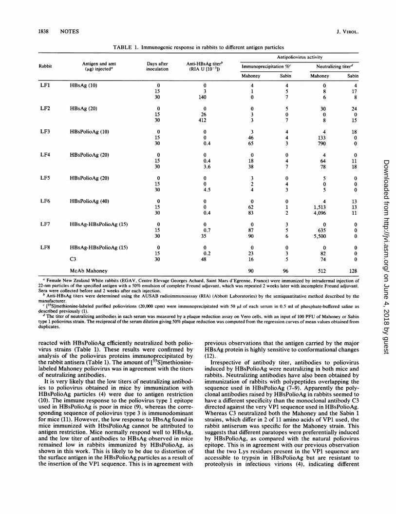
TABLE 1. Immunogenic response in rabbits to different antigen particles
Antipoliovirus activityAntigen and amt Days after Anti-HBsAg titerb..Rabbit Anie n m asatr At-BA ieb Immunoprecipitation %c Neutrailizing titer'(,ug) injected' inoculation (RIA U [10-3J) ___Neutralzing
Mahoney Sabin Mahoney Sabin
LF1 HBsAg (10) 0 0 4 4 0 415 3 1 5 8 1730 140 0 7 6 8
LF2 HBsAg (20) 0 0 0 5 30 2415 26 3 0 0 030 412 3 7 8 15
LF3 HBsPolioAg (10) 0 0 3 4 4 1815 0 46 4 133 030 0.4 65 3 790 0
LF4 HBsPolioAg (20) Q 0 0 0 4 015 0.4 18 4 64 1130 3.6 38 7 78 18
LF5 HBsPolioAg (20) 0 0 3 0 5 015 0 2 4 0 030 4.5 4 3 5 0
LF6 HBsPolioAg (40) 0 0 0 0 4 1315 0 62 1 1,513 1330 0.4 83 2 4,096 11
LF7 HBsAg-HBsPolioAg (15) 0 0 0 3 0 015 0.7 87 5 635 030 35 90 6 5,500 0
LF8 HBsAg-HBsPolioAg (15) 0 0 0 0 0 015 0.2 23 3 82 0
C3 30 48 16 5 74 0
McAb Mahoney 90 96 512 128
a Female New Zealand White rabbits (EGAV, Centre Elevage Georges Achard, Saint Mars d'Egrenne, France) were immunized by intradermal injection of22-nm particles of the specified antigen with a 50% emulsion of complete Freund adjuvant, which was repeated 2 weeks later with incomplete Freund adjuvant.Sera were collected before and 2 weeks after each injection.
b Anti-HBsAg titers were determined using the AUSAB radioimmunoassay (RIA) (Abbott Laboratories) by the semiquantitative method described by themanufacturer.
c [35Slmethionine-labeled purified poliovirions (20,000 cpm) were immunoprecipitated with 50 ,ul of each serum in 0.5 ml of phosphate-buffered saline asdescribed previously (1).
d The titer of neutralizing antibodies in each serum was measured by a plaque reduction assay on Vero cells, with an input of 100 PFU of Mahoney or Sabintype 1 poliovirus strain. The reciprocal of the serum dilution giving 50% plaque reduction was computed from the regression curves of mean values obtained fromduplicates.
reacted with HBsPolioAg efficiently neutralized both polio-virus strains (Table 1). These results were confirmed byanalysis of the poliovirus proteins immunoprecipitated bythe rabbit antisera (Table 1). The amount of [35S]methionine-labeled Mahoney poliovirus was in agreement with the titersof neutralizing antibodies.
It is very likely that the low titers of neutralizing antibod-ies to poliovirus obtained in mice by immunization withHBsPolioAg particles (4) were due to antigen restriction(10). The immune response to the poliovirus type 1 epitopeused in HBsPolioAg is poor in mice (9), wher-eas the corre-sponding sequence of poliovirus type 3 is immunodominantfor mice (11). However, the low response to HbsAg found inmice immunized with HbsPolioAg cannot be attributed toantigen restriction. Mice normally respond well to HBsAg,and the low titer of antibodies to HBsAg observed in miceremained low in rabbits immunized by HBsPolioAg, asshown in this work. This is likely to be due to distortion ofthe surface antigen in the HBsPolioAg particles as a result ofthe insertion of the VP1 sequence. This is in agreement with
previous observations that the antigen carried by the majorHBsAg protein is highly sensitive to conformational changes(12).
Irrespective of antibody titer, antibodies to poliovirusinduced by HBsPolioAg were neutralizing in both mice andrabbits. Neutralizing antibodies have also been obtained byimmunization of rabbits with polypeptides overlapping thesequence used in HBsPolioAg (7-9). Apparently the poly-clonal antibodies raised by HBsPolioAg in rabbits seemed tohave a different specificity than the monoclonal antibody C3directed against the very VP1 sequence used in HBsPolioAg.Whereas C3 neutralized both the Mahoney and the Sabin 1strains, which differ in 2 of 11 amino acids of VP1 used, therabbit antiserum was specific for the Mahoney strain. Thissuggests that different paratopes were preferentially inducedby HBsPolioAg, as compared with the natural poliovirusepitope. This is in agreement with our previous observationthat the two Lys residues present in the VP1 sequence areaccessible to trypsin in HBsPolioAg but are resistant toproteolysis in infectious virions (4), indicating different
J. VIROL.
on June 4, 2018 by guesthttp://jvi.asm
.org/D
ownloaded from
NOTES 1839
4-
- VPi
ZVP2-_ VP3
o I, o | t ° ---- WX s°-0 0 0
LF1 LF2 LF3 LF4 LF5 LF& C3FIG. 3. Polyacrylamide gel electrophoresis of polypeptides of
infectious poliovirus. [35S]methionine-labeled virions were immuno-precipitated with antisera from rabbits LF1 to LF6 (Table 1) or withthe antipoliovirus monoclonal antibody (C3). o, Pre-immune sera; i,sera taken 2 weeks after the second immunization. VP1 throughVP4, the proteins of the poliovirus capsid. For immunoprecipita-tion, [35S]methionine-labeled Mahoney virus (13,500 cpm) (1) wasdiluted in 500 ,ul of phosphate-buffered saline containing 2% bovineserum albumin. Rabbit sera or ascites fluid containing the C3monoclonal antibody was added at a dilution of 1:50 or 1:100,respectively. Incubation was performed overnight at 4°C. Additionof protein A-Sepharose and the subsequent steps were carried out asdescribed previously (3).
structural environments of these residues in the two macro-
molecular complexes.Our work is the first description of 22-nm particles com-
posed of heterologous proteins which differ in internalsequences. The obvious capacity of these proteins to mix inthe membrane of the endoplasmic reticulum and to formmixed aggregates by invagination of this membrane (6, 14)lends credence to the notion that the internal heterologousregion does not play a critical role in the formation and thesecretion of these particles. Moreover, the capacity of themixed particles to induce antibodies to both HBsAg andpoliovirus shows that the two antigens do not structurallyinterfere with each other. By variation of the ratio of the twoplasmids used for transfection, it may be possible to vary therelative concentrations of the two antigens on the 22-nmparticles. Similarly, particles carrying a larger number ofantigens may be produced. These various possibilities mayallow the design of highly sophisticated vaccinating struc-tures.
Paul et al. (15) have described HBsAg particles fromhuman serum which carried normally mutually exclusivesubtype determinants. This finding was explained by pheno-typic mixing resulting from a two-hit infection of the samecell with viruses of different genotype. Our results confirmthat the same HBsAg particle may carry HBsAg encoded bydifferent S genes.
We thank P. Adamowicz for human HBsAg particles and N.Chenciner for encouragement and support. The expert technicalassistance of M. Lambert and A. Candrea is gratefully acknowl-edged. We thank A. Galliot and N. Perrin for typing the manuscript.
This work was partly supported by grants 83.L.0936 and 84.V.816from the Ministere de l'Industrie et de la Recherche and grants83.3008 and 86.3003 from the Institut National de la Sante et de laRecherche Medicale.
LITERATURE CITED1. Blondel, B., R. Crainic, 0. Fichot, ,G. Dufraisse, A. Candrea, D.
Diamond, M. Girard, and F. Horaud. 1986. Mutations confer-
ring resistance to neutralization with monoclonal antibodies intype 1 poliovirus can be located outside or inside the antibody-binding site. J. Virol. 57:81-90.
2. Colbere-Garapin, F., F. Horodniceanu, P. Kourisky, and A. C.Garapin. 1981. A new dominant hybrid selective marker forhigher eukaryotic cells. J. Mol. Biol. 150:1-14.
3. Delpeyroux, F., N. Chenciner, A. Lim, M. Lambert, and R. E.Streeck. 1987. Insertions in the hepatitis B surface antigen:effect on assembly and secretion of 22-nm particles from mam-malian cells. J. Mol. Biol. 195:343-350.
4. Delpeyroux, F., N. Chenciner, A. Lim, Y. Malpiece, B. Blondel,R. Crainic, S. Van der Werf, and R. E. Streeck. 1986. Apoliovirus neutralization epitope expressed on hybrid hepatitisB surface antigen particles. Science 233:472-474.
5. Dubois, M. F., C. Pourcel, S. Rousset, C. Chany, and P. Tiollais.1980. Excretion of hepatitis B surface antigen particles frommouse cells transformed with cloned viral DNA. Proc. Natl.Acad. Sci. USA 77:4549-4553.
6. Eble, B. E., V. R. Lingappa, and D. Ganem. 1986. Hepatitis Bsurface antigen: an unusual secreted protein initially synthe-sized as a transmembrane polypeptide. Mol. Cell. Biol. 6:1454-1463.
7. Emini, E. A., A. J. Bradford, and E. Wimmer. 1983. Priming forand induction of anti-poliovirus neutralizing antibodies by syn-thetic peptides. Nature (London) 304:699-703.
8. Ferguson, M., D. M. A. Evans, D. I. Magrath, P. D. Minor,J. W. Almond, and G. C. Schild. 1985. Induction by syntheticpeptides of broadly reactive, type specific neutralizing antibodyto poliovirus type 3. Virology 143:505-515.
9. Horaud, F., R. Crainic, B. Blondel, 0. Akacem, P. Couillin, S.Van der Werf, C. Wyckowski, P. Bruneau, 0. Siffert, and M.Girard. 1987. Identification and characterization of a cpntinuousneutralization epitope (C3) present on type 1 poliovirus. Prog.Med. Virol. 34:129-155.
10. Icenogle, J. P., P. D. Minor, M. Ferguson, and J. M. Hogle.1986. Modulation of humoral response to a 12-amino-acid siteon the poliovirus virion. J. Virol. 60:297-301.
11. Minor, P. D., M. Ferguson, D. M. A. Evans, J. W. Almond, andJ. P. Icenogle. 1986. Antigenic structure of polioviruses ofserotypes 1, 2 and 3. J. Gen. Virol. 67:1283-1291.
12. Mishiro, S., M. Imai, K. Takabashi, A. Machida, T. Gotanda, Y.Miyakawa, and M. Mayumi. 1980. A 49000 dalton polypeptidebearing all antigenic determinants and full immunogenicity of22nm hepatitis B surface antigen particles. J. Immunol. 124:1584-1592.
13. Moriarty, A. M., B. H. Hayer, J. W.-K. Shih, J. L. Gerin, andD. H. Hamer. 1981. Expression of the hepatitis B virus surfaceantigen gene in cell culture by using a simian virus 40 vector.Proc. Natl. Acad. Sci. USA 78:2606-2610.
14. Patzer, E. J., G. R. Nakamura, C. C. Simonsen, A. Levinson,and R. Brands. 1986. Intracellular assembly and packaging ofhepatitis B surface antigen particles occur in the endoplasmicreticulum. J. Virol. 58:884-892.
15. Paul, D. A., R. H. Purcell, and D. L. Peterson. 1986. Use ofmonoclonal antibodies to determine if HBsAg of mixed subtypeis one particle or two. J. Virol. Methods 13:43-53.
16. Skelly, J., C. R. Howard, and A. J. Zuckerman. 1981. HepatitisB polypeptide vaccine preparation in micelle form. Nature(London) 290:51-54.
17. Stibbe, W., and W. H. Gerlich. 1983. Structural relationshipsbetween minor and major proteins of hepatitis B surface anti-gen. J. Virol. 46:626-628.
18. Tiollais, P., C. Pourcel, and A. Dejean. 1985. The hepatitis Bvirus. Nature (London) 317:489-495.
VOL. 62, 1988
on June 4, 2018 by guesthttp://jvi.asm
.org/D
ownloaded from