Embed Size (px)
Citation preview
A Novel Anti-fibrogenic Factor
Inhibition of CTGF Over-expression in Diabetic Retinopathy by SERPINA3K
via Blocking the WNT/Beta-catenin Pathway
Running Title: A Novel Anti-fibrogenic Factor
Bin Zhang, Kevin K. Zhou and Jian-xing Ma*
Department of Cell Biology, Department of Medicine, The University of Oklahoma Health
Sciences Center, Oklahoma City, OK 73104
*Corresponding Author:
Jian-xing Ma, M.D., Ph.D.
E-mail: [email protected]
Submitted 17 July 2009 and accepted 6 March 2010.
Additional information for this article can be found in an online appendix at
http://diabetes.diabetesjournals.org
This is an uncopyedited electronic version of an article accepted for publication in Diabetes. The American Diabetes Association, publisher of Diabetes, is not responsible for any errors or omissions in this version of the manuscript or any version derived from it by third parties. The definitive publisher-authenticated
version will be available in a future issue of Diabetes in print and online at http://diabetes.diabetesjournals.org.
Diabetes Publish Ahead of Print, published online March 18, 2010
Copyright American Diabetes Association, Inc., 2010
A Novel Anti-fibrogenic Factor
2
Objective: Connective tissue growth factor (CTGF) is a major fibrogenic factor. Increased retinal
CTGF levels have been implicated to play a role in diabetic retinopathy (DR). SERPINA3K is a
serine proteinase inhibitor, and its levels were decreased in the retinas with DR. The purpose of
this study was to investigate the role of SERPINA3K in the regulation of CTGF and fibrogenesis,
and its mechanism of action.
Research Design and Methods: Adenovirus expressing SERPINA3K was injected intravitreally
into streptozotocin-induced diabetic rats. CTGF expression was measured using Western blot
analysis and real-time RT-PCR. Fibrosis was evaluated by quantifying retinal fibronectin using
ELISA. Wnt pathway activation was determined by phosphorylation of low-density lipoprotein
receptor-related protein 6 (LRP6), a co-receptor of Wnt ligands, and stabilization of beta-catenin,
an essential effector of the canonical Wnt pathway.
Results: Ad-SERPINA3K attenuated the CTGF and fibronectin over-expression in the retinas of
diabetic rats. In cultured retinal cells, SERPINA3K blocked the over-production of CTGF
induced by high glucose. DKK1, a specific Wnt antagonist, also attenuated the high glucose-
induced CTGF over-expression, indicating a role of Wnt signaling in CTGF over-expression in
diabetes. Similarly, increased SERPINA3K blocked Wnt pathway activation in the diabetic
retinas and in cells treated with high glucose. Further, SERPINA3K also attenuated the Wnt3a-
induced activation of the canonical Wnt pathway and the over-expression of CTGF.
Conclusion: SERPINA3K is an anti-fibrogenic factor and its anti-fibrogenic activity is through
blocking the Wnt pathway. Decreased SERPINA3K levels may contribute to the fibrosis in DR.
A Novel Anti-fibrogenic Factor
3
ERPINA3K, a serine proteinase
inhibitor (serpin), is expressed
in the liver, kidney, pancreas
and retina (1-3). SERPINA3K specifically
binds to tissue kallikrein to form a covalent
complex and inhibits proteolytic activities of
tissue kallikrein (3), and is believed to
participate in the regulation of vasodilation
and local blood flow via interactions with the
kallikrein-kinin system (4). Later studies
suggest that SERPINA3K has other functions
independent of inhibition of tissue kallikrein.
For example, SERPINA3K has been found to
inhibit retinal neovascularization (NV) in the
ischemia-induced retinopathy, which is not
dependent on its interactions with the
kallikrein-kinin system (5). Further, in a
diabetic rat model, SERPINA3K levels have
been shown to decrease in the retinas,
suggesting that decreased SERPINA3K levels
may contribute to diabetic retinopathy (DR)
(6).
DR is one of the leading causes of
blindness (7). In advanced stages of DR,
retinal fibrosis occurs and fibrovascular
contraction can cause haemorrhages and
retinal detachment (7; 8). Connective tissue
growth factor (CTGF) is a pro-fibrogenic
factor which stimulates fibroblast
proliferation, cell adhesion, and extracellular
matrix production (9; 10). The potential role
of CTGF in pathological fibrosis has been
established (11), and CTGF has been
suggested to be an attractive therapeutic target
in some fibrotic diseases (12). The protein
and mRNA levels of CTGF were found to be
elevated in the retinas with DR (13), and the
roles of CTGF in fibrovascular proliferation
and thickening of capillary basement
membrane were also demonstrated in
proliferative DR (13-16). All of these
previous findings suggest a therapeutic
potential for anti-CTGF therapy in DR.
Wnts are a group of secreted, cysteine-
rich glycoproteins (17). As shown in Figure
S1 (available in the online appendix at
http://diabetes.diabetesjournals.org), in the
absence of Wnt ligands, transcription factor β-
catenin, a down-stream effector of the
canonical Wnt pathway, is phosphorylated by
a protein complex containing GSK-3 in the
cytosol and constantly degraded to prevent its
accumulation (18; 19). Upon binding of
certain Wnt ligands, the Frizzled (Fz) receptor
dimerizes with the co-receptor, low-density
lipoprotein receptor-related protein 5 or 6
(LRP5/6), forming a receptor/co-receptor
complex (17). As a result, the downstream
signaling is stimulated, including
phosphorylation of LRP5/6 and stabilization
of β-catenin (20; 21). β-catenin is
subsequently translocated into the nucleus,
associates with T cell factor (TCF) for DNA
binding and regulates expression of target
genes including CTGF (17).
The Wnt signaling pathway is
involved in multiple physiological and
pathological processes. It has been well
studied in embryogenesis and carcinogenesis
(22). Recent evidence suggests that the Wnt
pathway is also important in ocular diseases,
for example, mutations in the Fz receptor and
LRP co-receptor have been shown to
associate with the vascular developmental
defects (23). Furthermore, it has been
revealed that Wnt signaling is responsible for
pathological fibrosis in the lung, suggesting
that inhibition of Wnt signaling, such as Wnt
antagonists, may represent a therapeutic
option (24-27). As a pro-fibrogenic factor,
CTGF was also found to be regulated by Wnt
signaling in osteoblast differentiation (28; 29).
However, there is little previous evidence to
implicate Wnt signaling in fibrosis in the
retina with DR.
In the present study, we have
investigated the inhibitory effect of
SERPINA3K on the hyperglycemia-induced
CTGF over-expression and Wnt pathway
activation, and further determined if the
S

A Novel Anti-fibrogenic Factor
4
beneficial effects of SERPINA3K in DR are
through the Wnt antagonistic activity.
RESEARCH DESIGN AND METHODS
Cell Culture: rMC-1, a cell line
derived from rat retinal Müller cells, is a kind
gift from Dr. Vijay Sarthy at Northwestern
University, and cultured
in Dulbecco's
Modified Eagle's Medium (DMEM, Cellgro,
Manassas, VA) containing 10% fetal bovine
serum (FBS, Invitrogen, Carlsbad, CA) (30).
HTERT-RPE, a cell line derived from human
RPE cells, was purchased from ATCC
(Manassas, VA) and cultured in DMEM
containing 10% FBS following the ATCC
recommendation. L cells and L cells stably
expressing Wnt3A (L-Wnt3a) were purchased
from ATCC and cultured in DMEM
containing 10% FBS and 0.4 mg/ml G-418
(Invitrogen, Carlsbad, CA). The cells and
conditioned media (1 g/L glucose, 1% FBS)
were harvested following the procedure
recommended by ATCC. The cultured cells
were starved in 1 g/L glucose (5 mM) DMEM
containing 1% FBS overnight before
treatment. For high glucose (HG) treatment,
the cells were exposed to 30 mM D-glucose
(Sigma, St. Louis, MO), and the low glucose
control (LG) included 5 mM D-glucose and
25 mM L-glucose (Sigma, St. Louis, MO) in
the culture medium.
Experimental Animals: Brown
Norway (BN) rats were purchased from
Charles River Laboratories (Wilmington,
MA). Care, use, and treatment of all animals
in this study were in strict agreement with the
ARVO Statement for the Use of Animals in
Ophthalmic and Vision Research and the
guidelines in the Care and Use of Laboratory
Animals set forth by the University of
Oklahoma.
Induction of Experimental Diabetes:
The experimental diabetes was induced as
described previously (31). Briefly, BN rats (8
week of age) were given a single
intraperitoneal injection of streptozotocin
(STZ, 50 mg/kg in 10 mmol/L of citrate
buffer, pH 4.5) after overnight fasting. Serum
glucose levels were monitored 48 hr after the
STZ injection and every two weeks thereafter,
and only the animals with blood glucose
levels higher than 350 mg/dl were used as
diabetic rats.
Recombinant Proteins, Adenovirus,
Plasmids, Transfection and Reporter Assay;
The SERPINA3K cDNA was cloned into the
pET28 vector (Novagen, Madison, WIS) and
the construct was transformed into E. coli
strain BL-21/DE3 (Novagen, Madison, WIS).
The expression and purification followed the
protocol described previously (5). Endotoxin
levels were measured using a limulus
amebocyte kit (Biowhittaker, Walkersville,
MD). Bovine serum albumin (BSA) (Sigma
St. Louis, MO) was used as protein control
for SERPINA3K. Recombinant Dickkopf-1
(DKK1) protein was purchased from R&D
Systems (Minneapolis, MN). To clone the
SERPINA3K cDNA, total RNA was
extracted from the liver and was reverse
transcribed to cDNA. The full-length
sequence of SERPINA3K containing the
signal peptide was cloned into the shuttle
vector. The adenoviral vector used in the
study is human adenovirus serotype 5 (Ad5). Adenoviruses expressing SERPINA3K and
LacZ were generated using AdEasy systems
from Qbiogene (Irvine, CA) following
manufacturer’s protocol. These adenoviruses
were purified using Adeno-X Virus
Purification Kits from BD (San Jose, CA).
TOPFLASH vector was constructed as
described (32). Fugene 6 (Roche Applied
Science, Indianapolis, IN) was used for
transfection following manufacturer’s
protocol. Luciferase reporter assays were
performed in 12-well plates. The TOPFLASH
construct and renilla luciferase pRL-TK
vector were co-transfected into the cells.
TOPFLASH activity was measured using the
dual luciferase reporter system (Promega,
A Novel Anti-fibrogenic Factor
5
Madison, WI) and normalized by renilla
luciferase activity.
Real-time RT-PCR: Total RNA was
isolated using RNeasy Mini Kit (Qiagen
Sciences, Germantown, MD). mRNA was
reverse transcribed to cDNA using TaqMan
kit from Roche (Indianapolis, IN). This
cDNA was then used for specific real-time
PCR. The specific primers for CTGF (5'-
AAGACCTGTGGGATGGGC -3’ and 5'-
TGGTGCAGCCAGAAAGCTC-3’) were
synthesized from IDT (San Diego, CA). To
normalize the variation of the amount of
mRNA in each reaction, 18S rRNA (primers:
5'-TTTGTTGGTTTTCGGAACTGA-3’ and
5'-CGTTTATGGTCGG AACTACGA-3’)
was simultaneously processed in the same
sample as an internal control. iQ SYBR
Green Supermix from BIORAD (Hercules,
CA) was used for real-time PCR reaction
following the manufacturer’s procedure.
Standard curves for CTGF primers
and 18S primers were constructed using serial
1 to 10 dilutions of the cDNA product from
RT reaction (Fig. S6). The efficiency of
CTGF primers was 99.46% and the efficiency
of 18S primers was 97.43%. Dilutions of
1:1000, 1:100 and 1:10 were used in the assay
and all of the samples were diluted by 1:100
for the real-time PCR reaction. To calculate
relative changes in gene expression, we used
the delta-delta method following the
BIORAD’s introduction.
Western Blot Analysis: The same
amounts of proteins from the cytosolic
fraction, total cell lysates and retinal
homogenates were resolved by SDS-PAGE
(8% or 10%) and analyzed by Western
blotting using specific antibodies. For
cytosolic β-catenin measurement, cells were
lysed by three frozen-thaw cycles in PBS with
a protease inhibitor cocktail (Roche Applied
Science, Indianapolis, IN) followed by
centrifugation, and the supernatants were
isolated for Western blot analysis. For total
cell lysates, harvested cells were sonicated in
RIPA (Cell Signaling Technology, Danvers,
MA) buffer containing 1% SDS. For retinal
homogenate preparation, the retinas were
homogenized in PBS with a protease inhibitor
cocktail using a soft tissue pestle (Fisher
Scientific Inc, Pittsburgh, PA). Antibodies for
CTGF, LRP6 and β-catenin were purchased
from Santa Cruz Biotechnology, Inc. (Santa
Cruz, CA), and used at 1:400, 1:500 and
1:2500 dilutions. Antibody for β-actin
(1:3000) was purchased from Invitrogen
(Carlsbad, CA). Antibodies for
phosphorylated LRP6 (S1490),
phosphorylated β-catenin (S33/37/T41),
phosphorylated GSK3β (S9), GSK3β and
Histone H3 were purchased from Cell
Signaling Technology (Danvers, MA), and
used at 1:500 dilution for the first two
antibodies and 1:2500 dilution for the last
three antibodies. The monoclonal antibody for
SERPINA3K (1:1000) was generated using
the recombinant SERPINA3K through
contracted service with Proteintech Group
(Chicago, IL).
Because of the post-translational
modifications, CTGF can display multiple
bands with different molecular weights,
which has been reported in the literature (33).
Typically, a 36 or 38-KD band, or double
bands at these molecular weights can be
detected, dependent on the tissue, cell type
and treatment. In the present study, CTGF
showed double bands (36/38 KD) in the
Western blotting data using the HTERT RPE-
1 cell lysate.
ELISA for Fibronectin: Fibronectin
concentrations in the retina homogenates were
measured using an ELISA kit purchased from
Assaypro (Winfield, MO) according to
manufacturer’s instruction. The working
range of the fibronectin ELISA kit used here
is 4 ng/ml to 1000 ng/ml. All of the samples
were measured in the linear part of the
working range. The assay CVs are less than
2%. The total protein concentration was
measured by Bradford protein assay.
A Novel Anti-fibrogenic Factor
6
Statistical Analysis: Student’s t test
(two tailed) was used for comparisons
between two groups. ANOVA was used for
comparisons between groups in Table 1.
Statistical significance was accepted when the
P value was less than 0.05.
RESULTS
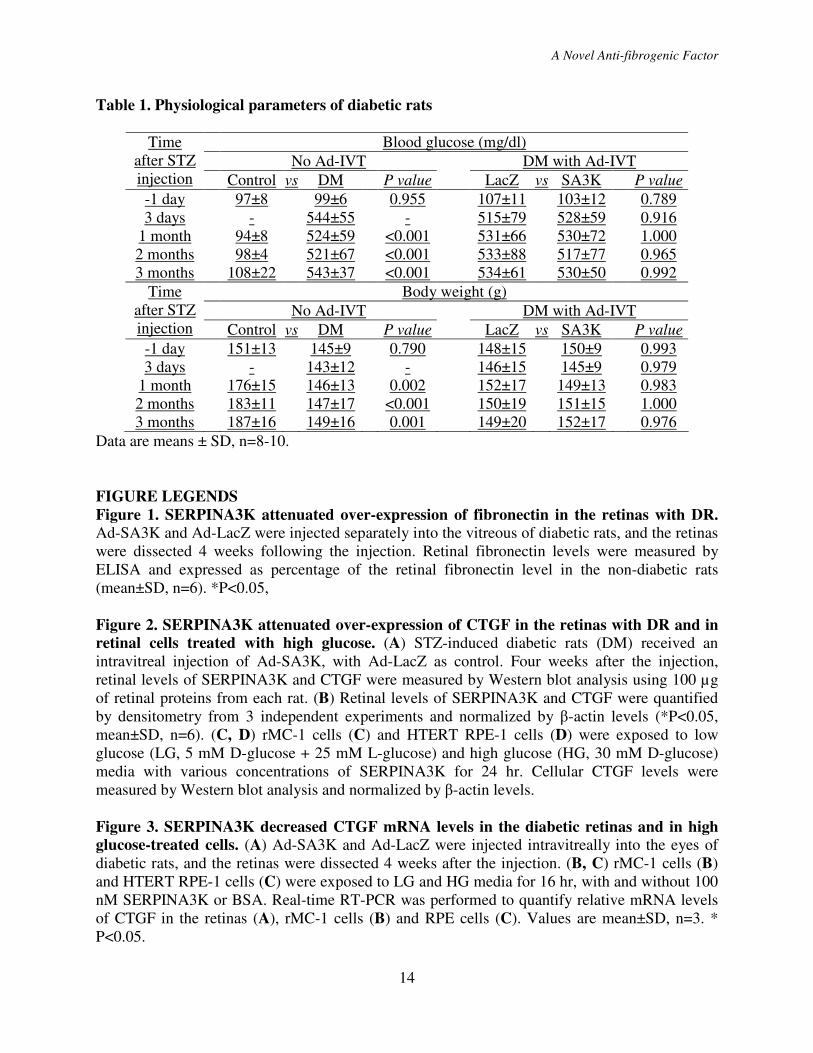
Clinical characteristics of diabetic
animals: Two months after the induction of
diabetes, the diabetic rats received an
intravitreal (IVT) injection of adenovirus
expressing SERPINA3K (Ad-SA3K, 5×10⁷
pfu/eye), and the same titer of adenovirus
expressing LacZ (Ad-LacZ) was injected for
control. The rats were separated into four
groups: normal rats without diabetes (control);
rats with diabetes mellitus (DM); DM rats
with injection of Ad-SA3K (DM-SA3K) and
DM rats with injection of Ad-LacZ (DM-
LacZ). There were 8 to 10 rats in each group.
Before the STZ injection, all of these rats had
similar blood glucose levels (~100 mg/dl) and
similar body weights (~150 g). Three months
after STZ injection, average body weight of
the DM group (149±16g) was significantly
lower than in non-diabetic control group
(187±16g) (P<0.05). At each time point, there
was no significant blood glucose or body
weight difference between the DM group,
DM-LacZ group and DM-SA3K group (Table
1), suggesting that the ocular adenovirus
injection had no systemic effect in the
diabetic animals.
A novel anti-fibrogenic activity of
SERPINA3K in the retina with DR: To
investigate the effect of SERPINA3K on
fibrosis in the retina with DR, we measured
the retinal levels of fibronectin in the STZ-
induced diabetic rats. Fibronectin is an
extracellular matrix protein, and over-
production of fibronectin has been shown to
contribute to capillary basement membrane
thickening in DR (34). Consistent with
previous studies, our ELISA results showed
that fibronectin levels were significantly
higher in the retinas of the diabetic rats,
compared to that in non-diabetic controls (Fig.
1). Ad-SA3K blocked retinal fibronectin
over-expression in diabetic rats (Fig. 1),
suggesting an anti-fibrogenic activity of
SERPINA3K.
Retinal endothelial cells and pericytes
are two major vascular cell types which are
profoundly affected in DR, and their
dysfunctions contribute to the blood retinal
barrier (BRB) breakdown in DR (35-37).
Since these cells are also the sources of
fibronectin production responsible for
thickening of the basement membrane, we
measured the concentration of fibronectin in
primary retinal pericytes and endothelial cells
by ELISA. The high glucose-induced over-
production of fibronectin was attenuated by
SERPINA3K in both of the cell types (Fig.
S2).
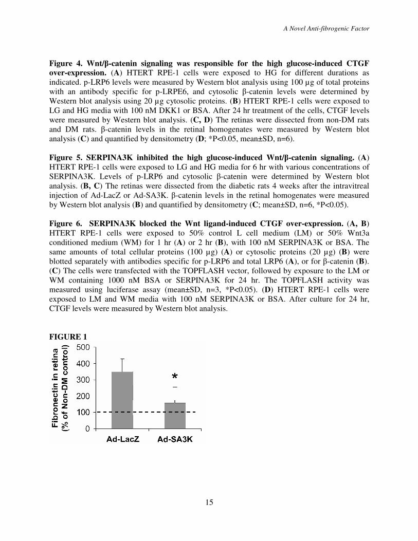
Inhibitory effects of SERPINA3K
on CTGF over-expression: The inhibitory
effect of SERPINA3K on CTGF was
evaluated in the STZ-induced diabetic rats.
Four weeks after the injection of Ad-SA3K or
Ad-LacZ, the retinas were dissected following
a thorough perfusion to remove the blood in
the retinal vasculature. As measured by
Western blot analysis, retinal levels of
SERPINA3K were decreased in the untreated
diabetic rats, compared to that in non-diabetic
rats (Fig. 2A, B). The Ad-SA3K injection
resulted in an increase of SERPINA3K levels
in the retinas of the diabetic rats (Fig. 2A, B).
Retinal levels of CTGF were significantly
increased in the retinas of un-treated diabetic
rats, compared with non-diabetic rats at the
same age. The injection of Ad-SA3K
mitigated the over-expression of CTGF in the
retinas compared to the control Ad-LacZ
virus (Fig. 2A, B).
It has been shown that retinal pigment
epithelium (RPE) cells and retinal Müller
cells are two major cell types expressing
CTGF in the proliferative vitreoretinopathy
(38). The diabetes-induced BRB breakdown
A Novel Anti-fibrogenic Factor
7
occurs predominantly at the level of the
retinal blood vessels (39). However the failure
of the RPE barrier also occurs at an lower
level, suggesting that the pathological change
of the RPE is involved in diabetes (39). To
evaluate the direct effect of SERPINA3K on
the hyperglycemia-mediated CTGF over-
expression in vitro, HTERT RPE-1 cells
(human RPE cell line) and rMC-1 cells (rat
retinal Müller cell line) were exposed to
media containing 30 mM D-glucose (HG).
CTGF expression was significantly elevated
by high glucose exposure, when compared to
low glucose control (5 mM D-glucose and 25
mM L-glucose, LG). SERPINA3K blocked
the high glucose-induced CTGF over-
expression in a dose-dependent manner in
both of the cell lines (Fig. 2C, D).
To further study the mechanism for
the regulation of CTGF by SERPINA3K,
real-time RT-PCR was performed to measure
mRNA levels of CTGF in the retinas and
cultured retinal cells. The mRNA levels of
CTGF were increased in the retinas with DR
and decreased by Ad-SA3K (Fig. 3A).
Similarly, exposure to high glucose media for
16 hr significantly elevated CTGF mRNA
levels in both HTERT RPE-1 cells and rMC-1
cells. In a time course experiment, the cells
were exposed to high glucose medium for 0-
24 hr. The high glucose treatment
continuously increased the CTGF mRNA
level after 4 hr of exposure (Fig. S3). The
high glucose-induced CTGF mRNA over-
expression was attenuated by 100 nM
SERPINA3K (Fig. 3B, C). Taken together,
these results indicate that SERPINA3K blocks
the hyperglycemia-induced CTGF expression
at the transcription level.
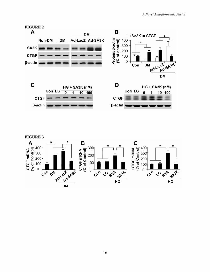
The role of the high glucose-induced
Wnt/β-catenin signaling in CTGF over-
expression: To explore the signaling pathway
mediating the inhibitory effect of
SERPINA3K on CTGF transcription, we first
investigated cell signaling responsible for
CTGF over-expression induced by high
glucose. As the β-catenin-TCF/LEF-binding
(TBE) site was identified in the promoter
region of the CTGF gene, and CTGF has been
shown to be a target gene of β-catenin-
TCF/LEF transcription factor (33), a
downstream effector of the canonical Wnt
pathway, we investigated the role of the Wnt
pathway in the CTGF over-expression in
diabetes. A number of Wnt ligands and Fz
receptors and both LRP5 and 6 were found to
express in the cultured HTERT RPE-1 cells
by RT-PCR, suggesting that this cell line is a
suitable model for Wnt signaling studies (Fig.
S4). As phosphorylation of LRP6 is a critical
step in the canonical Wnt pathway activation
(40), we measured phosphorylated LRP6 (p-
LRP6) levels. As shown by Western blot
analysis using an antibody specific for p-
LRP6, phosphorylation of the endogenous
LRP6 was increased in the RPE cells exposed
to 30 mM glucose, compared to that in control
cells exposed to the low glucose medium (Fig.
4A). In the same cell line, cytosolic β-catenin
levels were also elevated by the high glucose
medium in an exposure time-dependent
manner (Fig. 4A). Further, under high glucose
conditions, phosphorylated β-catenin levels
were decreased, compared to that in low
glucose control (Fig. S5 A), while nuclear β-
catenin levels were elevated (Fig. S5 B).
Consistently, GSK3β phosphorylation
(inactive form) was increased (Fig. S5 A). In
vivo, cytosolic β-catenin levels in the retina
homogenates were also significantly elevated
in the diabetic rats, indicating an activation of
the canonical Wnt pathway in the retinas with
DR (Fig 4C, D). Further, when the Wnt
pathway was blocked by Dickkopf1 (DKK1),
a specific inhibitor of the canonical Wnt
pathway, the high glucose-induced CTGF
over-expression was attenuated (Fig. 4B),
suggesting that the Wnt pathway activation
induced by high glucose and diabetes is
responsible for the CTGF over-expression.
Inhibitory effects of SERPINA3K
on Wnt/β-catenin signaling in DR and high
A Novel Anti-fibrogenic Factor
8
glucose-treated retinal cells: To further
investigate whether SERPINA3K inhibits the
Wnt pathway in high glucose-treated cells and
in diabetic retinas, SERPINA3K was
delivered into the RPE cell culture medium
containing high glucose and into the vitreous
humor of diabetic rats. After 6 hr exposure,
SERPINA3K decreased the high glucose-
induced phosphorylation of LRP6 and
accumulation of β-catenin in a concentration-
dependent manner (Fig. 5A). SERPINA3K at
100 nM decreased p-LRP6 and cytosolic β-
catenin to levels similar to that in the low
glucose control (Fig. 5A). Similarly, 100 nM
SERPINA3K completely reversed the high
glucose-induced changes of phosphorylated
β-catenin levels, nuclear β-catenin levels and
phosphorylated GSK3β levels (Fig. S5 A, B).
In diabetic rats, the injection of Ad-SA3K
also blocked the accumulation of cytosolic β-
catenin in the retina, compared to the control
virus Ad-LacZ, suggesting an inhibitory
effect of SERPINA3K on the canonical Wnt
pathway in diabetes (Fig 5B, C).
The Wnt inhibitory effect of
SERPINA3K was responsible for CTGF
regulation: To determine whether
SERPINA3K also blocks CTGF expression
induced by Wnt signaling, the RPE cells were
exposed to a 50% Wnt3a conditioned medium,
with the 50% L cell medium as control. The
Wnt3a conditioned medium elevated p-LRP6
and cytosolic β-catenin levels in the RPE cells
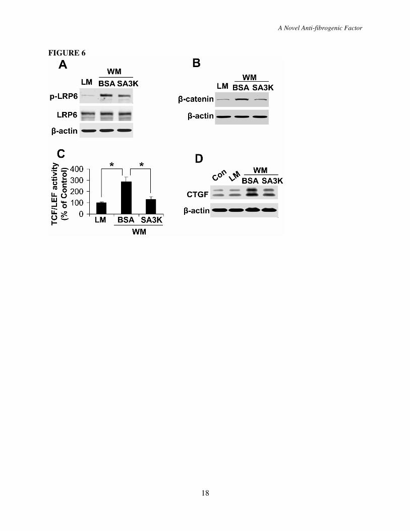
(Fig. 6A, B). SERPINA3K at 100 nM blocked
the increase of p-LRP6 and β-catenin levels
induced by Wnt3a (Fig. 6A, B). TOPFLASH
is a luciferase reporter construct under the
control of a promoter containing the
TCF/LEF-binding sites. To measure β-
catenin-dependent reporter gene transcription,
TOPFLASH activity assay was performed to
further confirm the inhibitory effect of
SERPINA3K on Wnt signaling. The RPE
cells were transfected with the TOPFLASH
construct and then incubated with the 50%
Wnt3a conditioned medium. TOPFLASH
assay showed that Wnt3a induced an
approximate 3-fold increase in TOPFLASH
reporter (TCF/LEF) activity, which was
attenuated by SERPINA3K (Fig. 6C). These
results indicated that SERPINA3K is a Wnt
inhibitor.
To determine the effect of
SERPINA3K on the Wnt-induced CTGF
expression, the 50% Wnt3a conditioned
medium was used to treat the cultured RPE
cells. Wnt3a, as well as high glucose, induced
CTGF over-expression in the RPE cells (Fig.
6D). Correlating with its inhibitory effect on
Wnt signaling, SERPINA3K also inhibited
the Wnt3a-induced CTGF up-regulation,
similar to that in the high glucose model (Fig.
6D).
DISCUSSION
SERPINA3K is an extracellular serpin
and has been found to function as an anti-
angiogenic factor (5) and an anti-
inflammatory factor (41). Our previous
studies showed that SERPINA3K levels are
decreased in the retina of diabetic rats (6).
The present study revealed a novel anti-
fibrogenic activity of this serpin, as it inhibits
CTGF over-expression and reduces the
production of extracellular matrix in the retina
with DR. These findings suggest that
decreased SERPINA3K levels in the diabetic
retina may contribute to pathological fibrosis
in DR. Further, our results demonstrate that
the inhibitory effect of SERPINA3K on
CTGF over-expression is through mitigating
the Wnt signaling activation induced by
diabetes.
Fibrosis is an important pathological
feature of DR (7; 8). At early stages of DR,
increased production of extracellular matrix
has been shown to contribute to the capillary
basement membrane thickening (42). At
advanced stages of DR, fibrosis can result in
contraction and retinal detachment, a major
cause of blindness in DR (7; 8). The
molecular mechanism for retinal fibrosis in
A Novel Anti-fibrogenic Factor
9
DR is not clear. CTGF is a major fibrogenic
factor, and its over-expression has been found
to play a key role in the basement membrane
thickening in DR models (14; 16). CTGF up-
regulates production of extracellular matrix
proteins such as fibronectin (43; 44).
Therefore, CTGF is considered a promising
target for treating retinal fibrosis in DR. The
present study identified SERPINA3K as an
endogenous inhibitor of CTGF and thus, an
anti-fibrogenic factor in the retina. On the
other hand, CTGF has been reported to bind
to Wnt receptor/co-receptor (45). However,
the function of CTGF in canonical Wnt
signaling is still controversial (33; 45).
Despite the possible feedback regulation by
CTGF, blockade of Wnt signaling by
SERPINA3K should result in a decrease of
CTGF levels and inhibition of fibrogenesis,
which supports our conclusion, i.e. the anti-
fibrogenic effect of SERPINA3K is mediated,
at least in part, through the Wnt pathway.
Our previous studies have shown that
retinal levels of SERPINA3K are decreased in
STZ-induced diabetic rats after 1, 2 and 4
months of diabetes (6). In the same animal
model, CTGF over-expression was found in
the diabetic rat retinas 3 months after STZ
injection (13), correlating with the decrease of
SERPINA3K. The disturbed balance between
pro-fibrogenic factor CTGF and anti-
fibrogenic factor SERPINA3K may represent
a new pathogenic mechanism for the
basement membrane thickening in DR.
Wnt signaling is involved in ocular
diseases, such as vasculature disorders in the
retina (23). Our recent study showed that
activation of the Wnt pathway also plays a
pathogenic role in subretinal
neovascularization in an animal model of wet
AMD (46). The role of Wnt signaling in
pathological fibrosis has been revealed in
some tissues, e.g. the lung (24). However, in
most ocular diseases such as DR, the role of
Wnt signaling, especially the association
between Wnt signaling and retinal fibrosis
remains obscure. Here, our results showed
that cytosolic β-catenin, an essential effector
of the canonical Wnt pathway, is accumulated
in both diabetic retinas and in the high
glucose-treated retinal cells. Phosphorylation
of LRP6 is an early, yet essential step in
activation of the canonical Wnt pathway, as
the phosphorylation sites in the LRP6
intracellular domain are known to create
inducible docking sites for Axin, leading to
stabilization of β-catenin in the cytosol and
transduction of the extracellular Wnt signal
into intracellular compartments (40; 47). Our
results showed that phosphorylated LRP6
levels were increased under high glucose
conditions. In the hyperglycemia models, the
induction of Wnt signaling activity was
accompanied by CTGF over-expression.
Same as the high glucose exposure, Wnt3a
ligand also induced Wnt signaling activation
and up-regulated CTGF expression, while
DKK1, a specific inhibitor of the Wnt
pathway, blocked the Wnt pathway activation.
Therefore, our results revealed that Wnt
signaling is activated in DR, which may be
responsible for CTGF over-expression and
retinal fibrosis.
Since DKK1 is a commonly accepted,
specific inhibitor of the canonical Wnt
pathway, DKK1 was used to specifically
attenuate the Wnt signaling activation and
further to reveal the role of Wnt signaling in
the high glucose-induced CTGF over-
expression. DKK1 has been reported to
induce the internalization of LRP6 (48). To
investigate the mechanism for the
SERPINA3K effect on the Wnt pathway, we
have measured several components of the
Wnt pathway at different levels of the cascade.
Further, we measured the cell surface level of
LRP6 to determine the internalization of
LRP6 using extracellular biotin labeling.
Similar to DKK1, pre-incubation of the cells
with SERPINA3K decreased the cell surface
level of LRP6 (Fig. S5 C). This result
suggests that induction of LRP6
A Novel Anti-fibrogenic Factor
10
internalization may represent a mechanism
responsible, at least partially, for the
inhibition of LRP6 phosphorylation by
SERPINA3K.
There are 19 Wnt ligands, many of
which function as agonists of Fz/LRP5/6
receptor complex and thus, activate Wnt/β-
catenin signaling (49). On the other hand,
some natural inhibitors of the Wnt pathway
such as DKK family members and IGFBP-4
have been identified (50-52). Here we showed
that SERPINA3K blocks the Wnt pathway
activation induced by high glucose and by
Wnt ligands, suggesting that SERPINA3K is
a novel endogenous inhibitor of the Wnt
pathway. SERPINA3K is known to
specifically bind to tissue kallikrein, forming
a covalent complex and inhibiting proteolytic
activities of tissue kallikrein (3). Through
interactions with the kallikrein-kinin system,
SERPINA3K participates in the regulation of
blood pressure and local blood flow (4).
However, the kallikrein-binding activity
cannot explain the broad functions of
SERPINA3K, such as anti-angiogenic and
anti-inflammatory activities (5; 41). In
addition, some of these functions have proven
to be independent of interactions with tissue
kallikrein (5). The present study demonstrates
that SERPINA3K has a potent Wnt antagonist
activity. Since the Wnt pathway regulates
multiple pathological processes, including
angiogenesis, inflammation and fibrosis, the
Wnt antagonizing activity of SERPINA3K
may represent a unifying mechanism
responsible for the broad beneficial effects of
this serpin. As an endogenous inhibitor of the
Wnt pathway, this serpin molecule may have
therapeutic potential.
ACKNOWLEDGMENTS
This study was supported by NIH
grants EY012231, EY018659, EY019390,
and P20RR024215 from the National Center
for Research Resources, and grants from
OCAST and ADA.
A Novel Anti-fibrogenic Factor
11
REFERENCES
1. Gettins PG: Serpin structure, mechanism, and function. Chem Rev 102:4751-4804, 2002
2. Chao J, Tillman DM, Wang MY, Margolius HS, Chao L: Identification of a new tissue-
kallikrein-binding protein. Biochem. J. 239:325-331, 1986
3. Chao J, Chai KX, Chen LM, Xiong W, Chao S, Woodley-Miller C, Wang LX, Lu HS, Chao L:
Tissue kallikrein-binding protein is a serpin. I. Purification, characterization, and distribution in
normotensive and spontaneously hypertensive rats. J. Biol. Chem. 265:16394-16401, 1990
4. Ma JX, Yang Z, Chao J, Chao L: Intramuscular delivery of rat kallikrein-binding protein gene
reverses hypotension in transgenic mice expressing human tissue kallikrein. J Biol Chem
270:451-455, 1995
5. Gao G, Shao C, Zhang SX, Dudley A, Fant J, Ma JX: Kallikrein-binding protein inhibits
retinal neovascularization and decreases vascular leakage. Diabetologia 46:689-698, 2003
6. Hatcher HC, Ma JX, Chao J, Chao L, Ottlecz A: Kallikrein-binding protein levels are reduced
in the retinas of streptozotocin-induced diabetic rats. Invest. Ophthalmol. Vis. Sci. 38:658-664,
1997
7. Aiello LP, Gardner TW, King GL, Blankenship G, Cavallerano JD, Ferris FL, 3rd, Klein R:
Diabetic retinopathy. Diabetes Care 21:143-156, 1998
8. Friedlander M: Fibrosis and diseases of the eye. J Clin Invest 117:576-586, 2007
9. Frazier K, Williams S, Kothapalli D, Klapper H, Grotendorst GR: Stimulation of fibroblast
cell growth, matrix production, and granulation tissue formation by connective tissue growth
factor. J Invest Dermatol 107:404-411, 1996
10. Kireeva ML, Latinkic BV, Kolesnikova TV, Chen CC, Yang GP, Abler AS, Lau LF: Cyr61
and Fisp12 are both ECM-associated signaling molecules: activities, metabolism, and
localization during development. Exp Cell Res 233:63-77, 1997
11. Leask A: Transcriptional profiling of the scleroderma fibroblast reveals a potential role for
connective tissue growth factor (CTGF) in pathological fibrosis. Keio J Med 53:74-77, 2004
12. Crean JK, Lappin D, Godson C, Brady HR: Connective tissue growth factor: an attractive
therapeutic target in fibrotic renal disease. Expert Opin Ther Targets 5:519-530, 2001
13. Tikellis C, Cooper ME, Twigg SM, Burns WC, Tolcos M: Connective tissue growth factor is
up-regulated in the diabetic retina: amelioration by angiotensin-converting enzyme inhibition.
Endocrinology 145:860-866, 2004
14. Kuiper EJ, de Smet MD, van Meurs JC, Tan HS, Tanck MW, Oliver N, van Nieuwenhoven
FA, Goldschmeding R, Schlingemann RO: Association of connective tissue growth factor with
fibrosis in vitreoretinal disorders in the human eye. Arch Ophthalmol 124:1457-1462, 2006
15. Hughes JM, Kuiper EJ, Klaassen I, Canning P, Stitt AW, Van Bezu J, Schalkwijk CG, Van
Noorden CJ, Schlingemann RO: Advanced glycation end products cause increased CCN family
and extracellular matrix gene expression in the diabetic rodent retina. Diabetologia 50:1089-
1098, 2007
16. Kuiper EJ, van Zijderveld R, Roestenberg P, Lyons KM, Goldschmeding R, Klaassen I, Van
Noorden CJ, Schlingemann RO: Connective tissue growth factor is necessary for retinal capillary
basal lamina thickening in diabetic mice. J Histochem Cytochem 56:785-792, 2008
17. He X, Semenov M, Tamai K, Zeng X: LDL receptor-related proteins 5 and 6 in Wnt/beta-
catenin signaling: arrows point the way. Development 131:1663-1677, 2004
18. Liu C, Li Y, Semenov M, Han C, Baeg GH, Tan Y, Zhang Z, Lin X, He X: Control of beta-
catenin phosphorylation/degradation by a dual-kinase mechanism. Cell 108:837-847, 2002
19. Polakis P: Casein kinase 1: a Wnt'er of disconnect. Curr Biol 12:R499-R501, 2002
A Novel Anti-fibrogenic Factor
12
20. Orsulic S, Peifer M: Cell-cell signalling: Wingless lands at last. Curr Biol 6:1363-1367, 1996
21. Dale TC: Signal transduction by the Wnt family of ligands. Biochem J 329 ( Pt 2):209-223,
1998
22. Clevers H: Wnt/beta-catenin signaling in development and disease. Cell 127:469-480, 2006
23. Masckauchan TN, Kitajewski J: Wnt/Frizzled signaling in the vasculature: new angiogenic
factors in sight. Physiology (Bethesda) 21:181-188, 2006
24. Morrisey EE: Wnt signaling and pulmonary fibrosis. Am J Pathol 162:1393-1397, 2003
25. Konigshoff M, Balsara N, Pfaff EM, Kramer M, Chrobak I, Seeger W, Eickelberg O:
Functional Wnt signaling is increased in idiopathic pulmonary fibrosis. PLoS ONE 3:e2142,
2008
26. He W, Dai C, Li Y, Zeng G, Monga SP, Liu Y: Wnt/beta-catenin signaling promotes renal
interstitial fibrosis. J Am Soc Nephrol 20:765-776, 2009
27. Cheng JH, She H, Han YP, Wang J, Xiong S, Asahina K, Tsukamoto H: Wnt antagonism
inhibits hepatic stellate cell activation and liver fibrosis. Am J Physiol Gastrointest Liver Physiol
294:G39-49, 2008
28. Si W, Kang Q, Luu HH, Park JK, Luo Q, Song WX, Jiang W, Luo X, Li X, Yin H, Montag
AG, Haydon RC, He TC: CCN1/Cyr61 is regulated by the canonical Wnt signal and plays an
important role in Wnt3A-induced osteoblast differentiation of mesenchymal stem cells. Mol Cell
Biol 26:2955-2964, 2006
29. Luo Q, Kang Q, Si W, Jiang W, Park JK, Peng Y, Li X, Luu HH, Luo J, Montag AG,
Haydon RC, He TC: Connective tissue growth factor (CTGF) is regulated by Wnt and bone
morphogenetic proteins signaling in osteoblast differentiation of mesenchymal stem cells. J Biol
Chem 279:55958-55968, 2004
30. Sarthy VP, Brodjian SJ, Dutt K, Kennedy BN, French RP, Crabb JW: Establishment and
characterization of a retinal Muller cell line. Invest. Ophthalmol. Vis. Sci. 39:212-216, 1998
31. Zhang SX, Ma JX, Sima J, Chen Y, Hu MS, Ottlecz A, Lambrou GN: Genetic difference in
susceptibility to the blood-retina barrier breakdown in diabetes and oxygen-induced retinopathy.
Am J Pathol 166:313-321, 2005
32. Semenov M, Tamai K, He X: SOST is a ligand for LRP5/LRP6 and a Wnt signaling inhibitor.
J Biol Chem 280:26770-26775, 2005
33. Deng YZ, Chen PP, Wang Y, Yin D, Koeffler HP, Li B, Tong XJ, Xie D: Connective tissue
growth factor is overexpressed in esophageal squamous cell carcinoma and promotes
tumorigenicity through beta-catenin-T-cell factor/Lef signaling. J Biol Chem 282:36571-36581,
2007
34. Ljubimov AV, Burgeson RE, Butkowski RJ, Couchman JR, Zardi L, Ninomiya Y, Sado Y,
Huang ZS, Nesburn AB, Kenney MC: Basement membrane abnormalities in human eyes with
diabetic retinopathy. J Histochem Cytochem 44:1469-1479, 1996
35. Cunha-Vaz J, Bernardes R: Nonproliferative retinopathy in diabetes type 2. Initial stages and
characterization of phenotypes. Prog Retin Eye Res 24:355-377, 2005
36. Cunha-Vaz J, Faria de Abreu JR, Campos AJ: Early breakdown of the blood-retinal barrier in
diabetes. Br J Ophthalmol 59:649-656, 1975
37. Cunha-Vaz JG: Pathophysiology of diabetic retinopathy. Br J Ophthalmol 62:351-355, 1978
38. Cui JZ, Chiu A, Maberley D, Ma P, Samad A, Matsubara JA: Stage specificity of novel
growth factor expression during development of proliferative vitreoretinopathy. Eye 21:200-208,
2007
A Novel Anti-fibrogenic Factor
13
39. Do carmo A, Ramos P, Reis A, Proenca R, Cunha-vaz JG: Breakdown of the inner and outer
blood retinal barrier in streptozotocin-induced diabetes. Exp Eye Res 67:569-575, 1998
40. Zeng X, Huang H, Tamai K, Zhang X, Harada Y, Yokota C, Almeida K, Wang J, Doble B,
Woodgett J, Wynshaw-Boris A, Hsieh JC, He X: Initiation of Wnt signaling: control of Wnt
coreceptor Lrp6 phosphorylation/activation via frizzled, dishevelled and axin functions.
Development 135:367-375, 2008
41. Zhang B, Hu Y, Ma JX: SERPINA3K is an Endogenous Anti-inflammatory and Anti-oxidant
Factor in the Retina. Invest Ophthalmol Vis Sci, 2009
42. Evans T, Deng DX, Chen S, Chakrabarti S: Endothelin receptor blockade prevents
augmented extracellular matrix component mRNA expression and capillary basement membrane
thickening in the retina of diabetic and galactose-fed rats. Diabetes 49:662-666, 2000
43. Arnott JA, Nuglozeh E, Rico MC, Arango-Hisijara I, Odgren PR, Safadi FF, Popoff SN:
Connective tissue growth factor (CTGF/CCN2) is a downstream mediator for TGF-beta1-
induced extracellular matrix production in osteoblasts. J Cell Physiol 210:843-852, 2007
44. Crean JK, Finlay D, Murphy M, Moss C, Godson C, Martin F, Brady HR: The role of p42/44
MAPK and protein kinase B in connective tissue growth factor induced extracellular matrix
protein production, cell migration, and actin cytoskeletal rearrangement in human mesangial
cells. J Biol Chem 277:44187-44194, 2002
45. Mercurio S, Latinkic B, Itasaki N, Krumlauf R, Smith JC: Connective-tissue growth factor
modulates WNT signalling and interacts with the WNT receptor complex. Development
131:2137-2147, 2004
46. Chen Y, Hu Y, Lu K, Flannery JG, Ma JX: Very low density lipoprotein receptor, a negative
regulator of the wnt signaling pathway and choroidal neovascularization. J Biol Chem
282:34420-34428, 2007
47. Tamai K, Zeng X, Liu C, Zhang X, Harada Y, Chang Z, He X: A mechanism for Wnt
coreceptor activation. Mol Cell 13:149-156, 2004
48. Yamamoto H, Sakane H, Yamamoto H, Michiue T, Kikuchi A: Wnt3a and Dkk1 regulate
distinct internalization pathways of LRP6 to tune the activation of beta-catenin signaling. Dev
Cell 15:37-48, 2008
49. Nusse R: Wnt signaling in disease and in development. Cell Res 15:28-32, 2005
50. Mao B, Wu W, Li Y, Hoppe D, Stannek P, Glinka A, Niehrs C: LDL-receptor-related protein
6 is a receptor for Dickkopf proteins. Nature 411:321-325, 2001
51. Semenov MV, Tamai K, Brott BK, Kuhl M, Sokol S, He X: Head inducer Dickkopf-1 is a
ligand for Wnt coreceptor LRP6. Curr Biol 11:951-961, 2001
52. Zhu W, Shiojima I, Ito Y, Li Z, Ikeda H, Yoshida M, Naito AT, Nishi J, Ueno H, Umezawa
A, Minamino T, Nagai T, Kikuchi A, Asashima M, Komuro I: IGFBP-4 is an inhibitor of
canonical Wnt signalling required for cardiogenesis. Nature 454:345-349, 2008
A Novel Anti-fibrogenic Factor
14
Table 1. Physiological parameters of diabetic rats
Blood glucose (mg/dl)
No Ad-IVT DM with Ad-IVT
Time
after STZ
injection Control vs DM P value LacZ vs SA3K P value
-1 day 97±8 99±6 0.955 107±11 103±12 0.789
3 days - 544±55 - 515±79 528±59 0.916
1 month 94±8 524±59 <0.001 531±66 530±72 1.000
2 months 98±4 521±67 <0.001 533±88 517±77 0.965
3 months 108±22 543±37 <0.001 534±61 530±50 0.992
Body weight (g)
No Ad-IVT DM with Ad-IVT
Time
after STZ
injection Control vs DM P value LacZ vs SA3K P value
-1 day
151±13 145±9 0.790 148±15 150±9 0.993
3 days - 143±12 - 146±15 145±9 0.979
1 month 176±15 146±13 0.002 152±17 149±13 0.983
2 months 183±11 147±17 <0.001 150±19 151±15 1.000
3 months 187±16 149±16 0.001 149±20 152±17 0.976
Data are means ± SD, n=8-10.
FIGURE LEGENDS
Figure 1. SERPINA3K attenuated over-expression of fibronectin in the retinas with DR.
Ad-SA3K and Ad-LacZ were injected separately into the vitreous of diabetic rats, and the retinas
were dissected 4 weeks following the injection. Retinal fibronectin levels were measured by
ELISA and expressed as percentage of the retinal fibronectin level in the non-diabetic rats
(mean±SD, n=6). *P<0.05,
Figure 2. SERPINA3K attenuated over-expression of CTGF in the retinas with DR and in
retinal cells treated with high glucose. (A) STZ-induced diabetic rats (DM) received an
intravitreal injection of Ad-SA3K, with Ad-LacZ as control. Four weeks after the injection,
retinal levels of SERPINA3K and CTGF were measured by Western blot analysis using 100 µg
of retinal proteins from each rat. (B) Retinal levels of SERPINA3K and CTGF were quantified
by densitometry from 3 independent experiments and normalized by β-actin levels (*P<0.05,
mean±SD, n=6). (C, D) rMC-1 cells (C) and HTERT RPE-1 cells (D) were exposed to low
glucose (LG, 5 mM D-glucose + 25 mM L-glucose) and high glucose (HG, 30 mM D-glucose)
media with various concentrations of SERPINA3K for 24 hr. Cellular CTGF levels were
measured by Western blot analysis and normalized by β-actin levels.
Figure 3. SERPINA3K decreased CTGF mRNA levels in the diabetic retinas and in high
glucose-treated cells. (A) Ad-SA3K and Ad-LacZ were injected intravitreally into the eyes of
diabetic rats, and the retinas were dissected 4 weeks after the injection. (B, C) rMC-1 cells (B)
and HTERT RPE-1 cells (C) were exposed to LG and HG media for 16 hr, with and without 100
nM SERPINA3K or BSA. Real-time RT-PCR was performed to quantify relative mRNA levels
of CTGF in the retinas (A), rMC-1 cells (B) and RPE cells (C). Values are mean±SD, n=3. *
P<0.05.
A Novel Anti-fibrogenic Factor
15
Figure 4. Wnt/β-catenin signaling was responsible for the high glucose-induced CTGF
over-expression. (A) HTERT RPE-1 cells were exposed to HG for different durations as
indicated. p-LRP6 levels were measured by Western blot analysis using 100 µg of total proteins
with an antibody specific for p-LRPE6, and cytosolic β-catenin levels were determined by
Western blot analysis using 20 µg cytosolic proteins. (B) HTERT RPE-1 cells were exposed to
LG and HG media with 100 nM DKK1 or BSA. After 24 hr treatment of the cells, CTGF levels
were measured by Western blot analysis. (C, D) The retinas were dissected from non-DM rats
and DM rats. β-catenin levels in the retinal homogenates were measured by Western blot
analysis (C) and quantified by densitometry (D; *P<0.05, mean±SD, n=6).
Figure 5. SERPINA3K inhibited the high glucose-induced Wnt/β-catenin signaling. (A)
HTERT RPE-1 cells were exposed to LG and HG media for 6 hr with various concentrations of
SERPINA3K. Levels of p-LRP6 and cytosolic β-catenin were determined by Western blot
analysis. (B, C) The retinas were dissected from the diabetic rats 4 weeks after the intravitreal
injection of Ad-LacZ or Ad-SA3K. β-catenin levels in the retinal homogenates were measured
by Western blot analysis (B) and quantified by densitometry (C; mean±SD, n=6, *P<0.05).
Figure 6. SERPINA3K blocked the Wnt ligand-induced CTGF over-expression. (A, B)
HTERT RPE-1 cells were exposed to 50% control L cell medium (LM) or 50% Wnt3a
conditioned medium (WM) for 1 hr (A) or 2 hr (B), with 100 nM SERPINA3K or BSA. The
same amounts of total cellular proteins (100 µg) (A) or cytosolic proteins (20 µg) (B) were
blotted separately with antibodies specific for p-LRP6 and total LRP6 (A), or for β-catenin (B).
(C) The cells were transfected with the TOPFLASH vector, followed by exposure to the LM or
WM containing 1000 nM BSA or SERPINA3K for 24 hr. The TOPFLASH activity was
measured using luciferase assay (mean±SD, n=3, *P<0.05). (D) HTERT RPE-1 cells were
exposed to LM and WM media with 100 nM SERPINA3K or BSA. After culture for 24 hr,
CTGF levels were measured by Western blot analysis.
FIGURE 1
A Novel Anti-fibrogenic Factor
16
FIGURE 2
FIGURE 3
A Novel Anti-fibrogenic Factor
17
FIGURE 4
FIGURE 5
A Novel Anti-fibrogenic Factor
18
FIGURE 6

![The Guide - Diabetic Retinopathy - Vision Lossvisionloss.org.au/wp-content/uploads/2016/05/The... · the guide [diabetic retinopathy] What is Diabetic Retinopathy? Diabetic Retinopathy](https://img.pdfslide.us/doc/110x75/5e3ed00bf9c32e41ea6578a8/the-guide-diabetic-retinopathy-vision-the-guide-diabetic-retinopathy-what.jpg)