Embed Size (px)
Citation preview
RESEARCH ARTICLE Open Access
Increase of reactive oxygen speciescontributes to growth inhibition byfluconazole in Cryptococcus neoformansNadir Hani Dbouk, Madison Bailey Covington, Kenny Nguyen and Srikripa Chandrasekaran*
Abstract
Background: Cryptococcus neoformans, a basidiomycetous yeast, is a fungal pathogen that can colonize the lungsof humans causing pneumonia and fungal meningitis in severely immunocompromised individuals. Recent studieshave implied that the antifungal drug fluconazole (FLC) can induce oxidative stress in C. neoformans by increasingthe production of reactive oxygen species (ROS), as presence of the antioxidant ascorbic acid (AA) could reverse theinhibitory effects of FLC on C. neoformans. However, in Candida albicans, AA has been shown to stimulate theexpression of genes essential for ergosterol biosynthesis. Hence, the contribution of ROS in FLC-mediated growthinhibition remains unclear.
Results: In order to determine whether counteracting ROS generated by FLC in C. neoformans can contribute todiminishing inhibitory effects of FLC, we tested three other antioxidants in addition to AA, namely, pyrrolidinedithiocarbamate (PDTC), retinoic acid (RA), and glutathione (GSH). Our data confirm that there is an increase in ROSin the presence of FLC in C. neoformans. Importantly, all four antioxidants reversed FLC-mediated growth inhibitionof C. neoformans to various extents. We further verified the involvement of increased ROS in FLC-mediated growthinhibition by determining that ROS-scavenging proteins, metallothioneins (CMT1 and CMT2), contribute to growthrecovery by PDTC and AA during treatment with FLC.
Conclusion: Our study suggests that ROS contributes to FLC-mediated growth inhibition and points to a complexnature of antioxidant-mediated growth rescue in the presence of FLC.
Keywords: Antifungal treatment, Antioxidants, ROS, Metallothionein
BackgroundEukaryotic pathogens, including pathogenic fungi are animportant cause of death in immunocompromisedpatients and can colonize immunocompetent individuals[1]. Cryptococcal meningitis caused by Cryptococcusneoformans is the leading cause of fungal central nervoussystem infection in the world, especially among personssuffering from HIV/AIDS [2, 3]. According to CDCreports, annually one million global cases of cryptococcalinfections occur, accounting for up to 600,000 mortal-ities and about one-third of all AIDS-associated deaths.Despite the severity of cryptococcosis, unfortunatelycurrent treatments for cryptococcal infections are inad-equate. A main barrier to establishment of an effective
antifungal drug therapy is increased drug resistance infungi [4–6].Compared to other anti-cryptococcal drugs, flucona-
zole (FLC) is the more affordable and less toxic alterna-tive, which is most commonly prescribed in geographiclocations where cryptococcosis is most prevalent [7, 8].FLC is the drug of choice for moderate pulmonary infec-tions. For central nervous system infections, a combin-ation of more expensive fungicidal drugs amphotericin Band flucytosine are administered [9, 10]; however, thecombination of these two drugs produces more toxicside effects for the host.A well-established mechanism of action of FLC is the
inhibition of Erg11, which is one of the key enzymesparticipating in the synthesis of ergosterol, an importantcomponent of the plasma membrane [11]. One factorthat contributes to failure of FLC-based therapy is the
© The Author(s). 2019 Open Access This article is distributed under the terms of the Creative Commons Attribution 4.0International License (http://creativecommons.org/licenses/by/4.0/), which permits unrestricted use, distribution, andreproduction in any medium, provided you give appropriate credit to the original author(s) and the source, provide a link tothe Creative Commons license, and indicate if changes were made. The Creative Commons Public Domain Dedication waiver(http://creativecommons.org/publicdomain/zero/1.0/) applies to the data made available in this article, unless otherwise stated.
* Correspondence: [email protected] of Biology, Furman University, Greenville, SC, USA
Dbouk et al. BMC Microbiology (2019) 19:243 https://doi.org/10.1186/s12866-019-1606-4
development of drug resistance. FLC resistance in C.neoformans occurs primarily via development of aneuploidcells with elevated levels of Erg11, which prevents dimin-ishment of ergosterol [12]. Other causes for FLC resistancein pathogenic fungi include accumulation of mutations inERG11 [13] and via drug efflux pumps [14, 15]. Import-antly, the mechanisms through which FLC leads to forma-tion of aneuploid and FLC resistant cells remain largelyuncharacterized.While diminishment of ergosterol is a well-documented
cause of FLC-mediated growth inhibition of C. neoformans,additional possible effects of FLC on C. neoformans cellshave been proposed. FLC treatment has been shown tocause an increase in reactive oxygen species (ROS) in Can-dida albicans [16–18] and most recently in C. neoformans[19]. ROS are molecules with unpaired, highly reactive elec-trons called free radicals, generated during basic cellularprocesses, or due to external stress-inducing conditions,including environmental pollutants, foreign compoundssuch as drugs or chemicals, and exposure to X-rays [20].Free radicals are highly reactive and unstable and excessiveamounts of ROS are known to cause cell damage and trig-ger apoptosis. Generation of high amounts of free radicalscan be harmful to biological macromolecules, as it cancause modification of DNA bases [21], lipid peroxidation,and protein carbonylation [22] leading to damage due tooxidative stress. Some examples of ROS include hydroxylradicals, hydroxide anion radicals, singlet oxygen, hydrogenperoxide, hypochlorite, nitric oxide radicals, and peroxyni-trite radicals. FLC-mediated increase in ROS could contrib-ute towards oxidative stress in C. neoformans. Consistentwith FLC-triggered ROS contributing to growth inhibition,co-treatment of C. neoformans cells with FLC and the anti-oxidant ascorbic acid (AA) was shown to partially rescue C.neoformans cells from FLC-mediated growth inhibition[19]. Similarly, co-treatment of C. albicans cells with theanti-fungal drug miconazole and a synthetic antioxidant,pyrrolidine dithiocarbamate (PDTC), has been shown toincrease the Minimal Inhibitory Concentration (MIC) ofmiconazole [18]. These studies suggest an additional effectof anti-fungal azole drugs on pathogenic fungi, which isinducing oxidative stress via an increase in ROS content.Interestingly, treatment of C. albicans with AA has
been shown to increase expression of the gene UPC2,which is involved in regulating ergosterol biosynthesis[23, 24]. This finding suggests that AA might be func-tioning indirectly to regulate ergosterol levels, which isby counteracting FLC-mediated inhibition of ergosterolbiosynthesis. Hence, whether ROS increase triggered byFLC contributes to growth inhibition elicited by FLC re-mains unclear.The metal copper has been shown to be essential for
virulence of C. neoformans [25]. Lack of a copper trans-porter, CTR4, led to reduced virulence in cryptococcosis
models in mice [26]. During infection by C. neoformans,copper acquisition and increased copper levels is essen-tial for melanin formation, which confers virulence to C.neoformans [27]. While elevated copper is essentialduring infection, increased copper can be toxic as itcontributes to increased production of ROS, due to itsparticipation in oxidation and reduction reactions [28].To counteract harmful effects of copper, C. neoformansincreases expression of metallothionein genes, CMT1and CMT2, which bind to and sequester copper [29].Previous studies have shown that C. neoformans mutantslacking metallothionein genes exhibit attenuated viru-lence [30] and show an increased sensitivity to FLC [19].These findings suggest that Cmt1 and Cmt2 proteinsallow for reversal of some of the harmful effects of ROSgenerated in the presence of FLC.The purpose of this study was to perform a more
rigorous test to determine if ROS plays a role in influen-cing sensitivity to FLC in C. neoformans. In order to de-termine whether it is the antioxidant properties of AAthat caused rescue of C. neoformans growth inhibition,we tested three alternative known antioxidants for theirability to reverse the effects of FLC on the wild type aswell as on metallothionein deficient mutants. Our datasuggest that treatment with FLC leads to increase ofROS and this oxidative stress may further contribute toFLC-mediated growth inhibition. Furthermore, this studysuggests that lowering ROS is not the only contributingfactor to the antioxidant-mediated growth rescue andpoints to the complex nature of the physiological effectsof FLC.
ResultsWe wished to determine whether antioxidants with diversechemical structures and modes of action could alleviateFLC-mediated growth inhibition of C. neoformans. Inaddition to AA that has been previously shown to reducegrowth inhibition in the presence of FLC in C. neoformans[19], we tested three chemically unrelated antioxidants: pyr-rolidinedithiocarbamate (PDTC), retinoic acid (RA), and areduced form of glutathione (GSH). The concentrations ofAA, PDTC, and GSH were established based on previousstudies [18, 19, 31]. The concentration of RA was estab-lished as the smallest concentration that rescued growth ofC. neoformans cells in the presence of hydrogen peroxide(as later indicated in Fig. 3a). Growth of cells on plates withmedia supplemented with the respective amounts of theantioxidants and lacking FLC was not inhibited as com-pared to the control YPD media (as indicated in Fig. 2b).As shown in Fig. 1a, in the presence of 32 μg/ml FLC, cellgrowth was significantly inhibited, although single coloniesof cells that were likely resistant to FLC were observed. Co-treatment of cells with both FLC and any of the four anti-oxidants led to rescue of growth. While RA, AA and PDTC
Dbouk et al. BMC Microbiology (2019) 19:243 Page 2 of 10
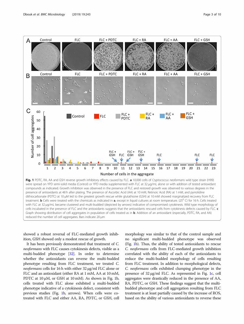
showed a robust reversal of FLC-mediated growth inhib-ition, GSH showed only a modest rescue of growth.It has been previously demonstrated that treatment of C.
neoformans with FLC causes cytokinesis defects, visible as amulti-budded phenotype [32]. In order to determinewhether the antioxidants can reverse the multi-buddedphenotype resulting from FLC treatment, we treated C.neoformans cells for 16 h with either 32 μg/ml FLC alone orFLC and an antioxidant (either RA at 1mM, AA at 10mM,PDTC at 10 μM, or GSH at 10mM). As shown in Fig. 1b,cells treated with FLC alone exhibited a multi-buddedphenotype indicative of a cytokinesis defect, consistent withprevious studies (Fig. 1b, arrows). When cells were co-treated with FLC and either AA, RA, PDTC, or GSH, cell
morphology was similar to that of the control sample andno significant multi-budded phenotype was observed(Fig. 1b). Thus, the ability of tested antioxidants to rescueC. neoformans cells from FLC-mediated growth inhibitioncorrelated with the ability of each of the antioxidants toreduce the multi-budded morphology of cells resultingfrom FLC treatment. In addition to morphological defects,C. neoformans cells exhibited clumping phenotype in thepresence of 32 μg/ml FLC. As represented in Fig. 1c, cellaggregates were drastically reduced in the presence of AA,RA, PDTC, or GSH. These findings suggest that the multi-budded phenotype and cell aggregation resulting from FLCtreatment is at least partially caused by the increase of ROS,based on the ability of various antioxidants to reverse these
Fig. 1 PDTC, RA, AA and GSH reverse growth inhibitory effects caused by FLC. a 10,000 cells of Cryptococcus neoformans wild type strain (H99)were spread on YPD semi-solid media (Control) or YPD media supplemented with FLC at 32 μg/mL alone or with addition of tested antioxidantcompounds as indicated. Growth inhibition was observed in the presence of FLC and restored growth was observed to various degrees in thepresence of antioxidants at 48 h after plating. The presence of Ascorbic Acid (AA) at 10 mM, Retinoic Acid (RA) at 1 mM, and pyrrolidinedithiocarbonate (PDTC) at 10 μM led to the greatest growth rescue while glutathione (GSH) at 10 mM showed marginalized recovery from FLCtreatment. b Cells were treated with the chemicals as indicated in a, except in liquid cultures at room temperature. (250 C) for 16 h. Cells treatedwith FLC at 32 μg/mL became clustered and multi-budded (depicted by arrows) indicative of compromised cytokinesis. Wild type morphology ofcells incubated in the presence of FLC and the antioxidants suggests that the antioxidants rescued cells from cytokinesis defects caused by FLC. cGraph showing distribution of cell aggregates in population of cells treated as in b. Addition of an antioxidant (especially, PDTC, RA, and AA)reduced the number of cell aggregates. Bars indicate 20 μm
Dbouk et al. BMC Microbiology (2019) 19:243 Page 3 of 10
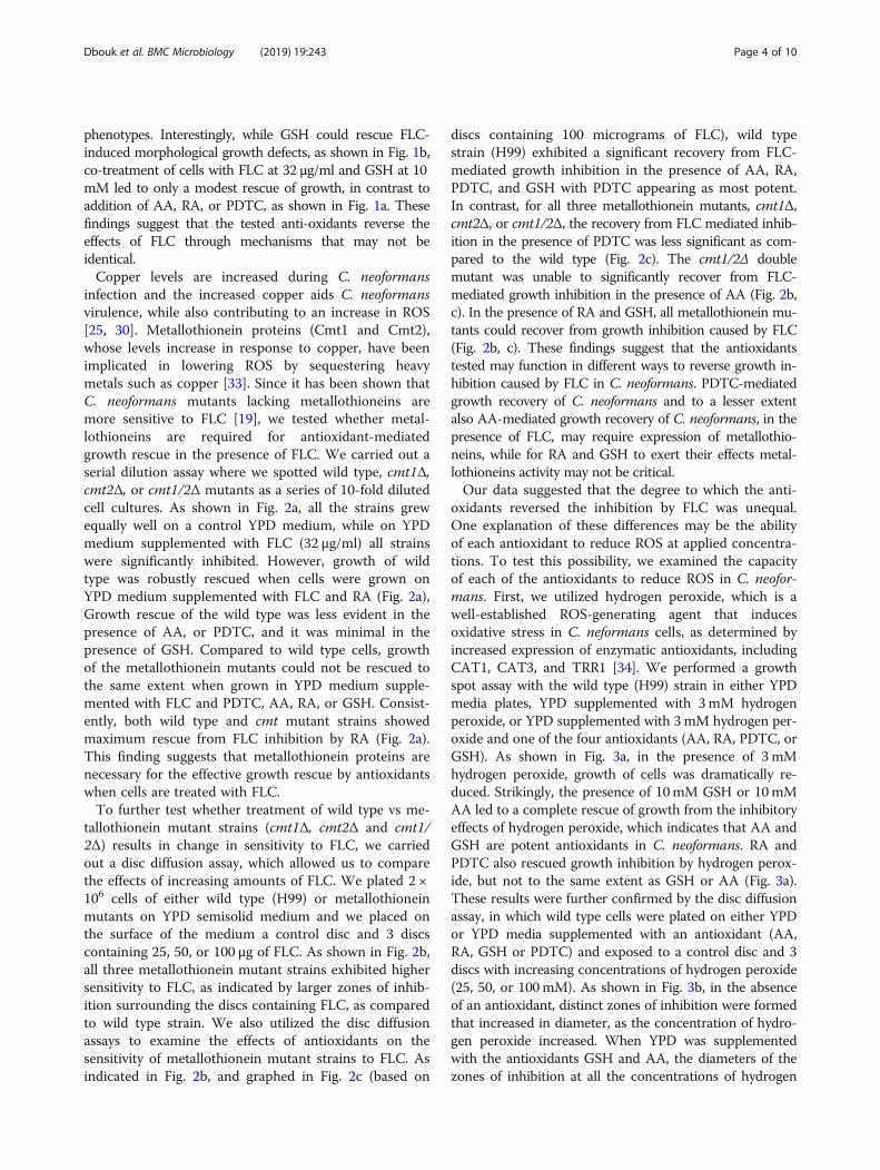
phenotypes. Interestingly, while GSH could rescue FLC-induced morphological growth defects, as shown in Fig. 1b,co-treatment of cells with FLC at 32 μg/ml and GSH at 10mM led to only a modest rescue of growth, in contrast toaddition of AA, RA, or PDTC, as shown in Fig. 1a. Thesefindings suggest that the tested anti-oxidants reverse theeffects of FLC through mechanisms that may not beidentical.Copper levels are increased during C. neoformans
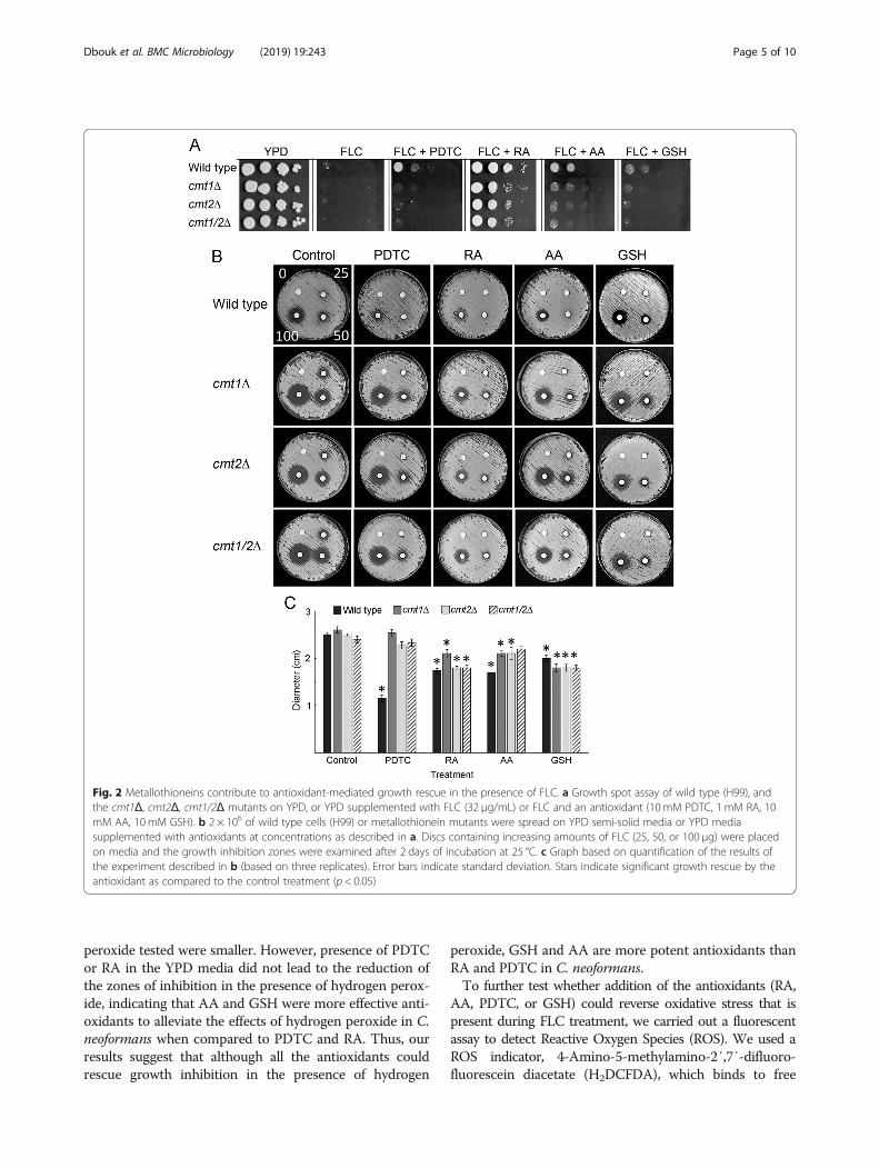
infection and the increased copper aids C. neoformansvirulence, while also contributing to an increase in ROS[25, 30]. Metallothionein proteins (Cmt1 and Cmt2),whose levels increase in response to copper, have beenimplicated in lowering ROS by sequestering heavymetals such as copper [33]. Since it has been shown thatC. neoformans mutants lacking metallothioneins aremore sensitive to FLC [19], we tested whether metal-lothioneins are required for antioxidant-mediatedgrowth rescue in the presence of FLC. We carried out aserial dilution assay where we spotted wild type, cmt1Δ,cmt2Δ, or cmt1/2Δ mutants as a series of 10-fold dilutedcell cultures. As shown in Fig. 2a, all the strains grewequally well on a control YPD medium, while on YPDmedium supplemented with FLC (32 μg/ml) all strainswere significantly inhibited. However, growth of wildtype was robustly rescued when cells were grown onYPD medium supplemented with FLC and RA (Fig. 2a),Growth rescue of the wild type was less evident in thepresence of AA, or PDTC, and it was minimal in thepresence of GSH. Compared to wild type cells, growthof the metallothionein mutants could not be rescued tothe same extent when grown in YPD medium supple-mented with FLC and PDTC, AA, RA, or GSH. Consist-ently, both wild type and cmt mutant strains showedmaximum rescue from FLC inhibition by RA (Fig. 2a).This finding suggests that metallothionein proteins arenecessary for the effective growth rescue by antioxidantswhen cells are treated with FLC.To further test whether treatment of wild type vs me-
tallothionein mutant strains (cmt1Δ, cmt2Δ and cmt1/2Δ) results in change in sensitivity to FLC, we carriedout a disc diffusion assay, which allowed us to comparethe effects of increasing amounts of FLC. We plated 2 ×10⁶ cells of either wild type (H99) or metallothioneinmutants on YPD semisolid medium and we placed onthe surface of the medium a control disc and 3 discscontaining 25, 50, or 100 μg of FLC. As shown in Fig. 2b,all three metallothionein mutant strains exhibited highersensitivity to FLC, as indicated by larger zones of inhib-ition surrounding the discs containing FLC, as comparedto wild type strain. We also utilized the disc diffusionassays to examine the effects of antioxidants on thesensitivity of metallothionein mutant strains to FLC. Asindicated in Fig. 2b, and graphed in Fig. 2c (based on
discs containing 100 micrograms of FLC), wild typestrain (H99) exhibited a significant recovery from FLC-mediated growth inhibition in the presence of AA, RA,PDTC, and GSH with PDTC appearing as most potent.In contrast, for all three metallothionein mutants, cmt1Δ,cmt2Δ, or cmt1/2Δ, the recovery from FLC mediated inhib-ition in the presence of PDTC was less significant as com-pared to the wild type (Fig. 2c). The cmt1/2Δ doublemutant was unable to significantly recover from FLC-mediated growth inhibition in the presence of AA (Fig. 2b,c). In the presence of RA and GSH, all metallothionein mu-tants could recover from growth inhibition caused by FLC(Fig. 2b, c). These findings suggest that the antioxidantstested may function in different ways to reverse growth in-hibition caused by FLC in C. neoformans. PDTC-mediatedgrowth recovery of C. neoformans and to a lesser extentalso AA-mediated growth recovery of C. neoformans, in thepresence of FLC, may require expression of metallothio-neins, while for RA and GSH to exert their effects metal-lothioneins activity may not be critical.Our data suggested that the degree to which the anti-
oxidants reversed the inhibition by FLC was unequal.One explanation of these differences may be the abilityof each antioxidant to reduce ROS at applied concentra-tions. To test this possibility, we examined the capacityof each of the antioxidants to reduce ROS in C. neofor-mans. First, we utilized hydrogen peroxide, which is awell-established ROS-generating agent that inducesoxidative stress in C. neformans cells, as determined byincreased expression of enzymatic antioxidants, includingCAT1, CAT3, and TRR1 [34]. We performed a growthspot assay with the wild type (H99) strain in either YPDmedia plates, YPD supplemented with 3mM hydrogenperoxide, or YPD supplemented with 3mM hydrogen per-oxide and one of the four antioxidants (AA, RA, PDTC, orGSH). As shown in Fig. 3a, in the presence of 3 mMhydrogen peroxide, growth of cells was dramatically re-duced. Strikingly, the presence of 10mM GSH or 10mMAA led to a complete rescue of growth from the inhibitoryeffects of hydrogen peroxide, which indicates that AA andGSH are potent antioxidants in C. neoformans. RA andPDTC also rescued growth inhibition by hydrogen perox-ide, but not to the same extent as GSH or AA (Fig. 3a).These results were further confirmed by the disc diffusionassay, in which wild type cells were plated on either YPDor YPD media supplemented with an antioxidant (AA,RA, GSH or PDTC) and exposed to a control disc and 3discs with increasing concentrations of hydrogen peroxide(25, 50, or 100mM). As shown in Fig. 3b, in the absenceof an antioxidant, distinct zones of inhibition were formedthat increased in diameter, as the concentration of hydro-gen peroxide increased. When YPD was supplementedwith the antioxidants GSH and AA, the diameters of thezones of inhibition at all the concentrations of hydrogen
Dbouk et al. BMC Microbiology (2019) 19:243 Page 4 of 10
peroxide tested were smaller. However, presence of PDTCor RA in the YPD media did not lead to the reduction ofthe zones of inhibition in the presence of hydrogen perox-ide, indicating that AA and GSH were more effective anti-oxidants to alleviate the effects of hydrogen peroxide in C.neoformans when compared to PDTC and RA. Thus, ourresults suggest that although all the antioxidants couldrescue growth inhibition in the presence of hydrogen
peroxide, GSH and AA are more potent antioxidants thanRA and PDTC in C. neoformans.To further test whether addition of the antioxidants (RA,
AA, PDTC, or GSH) could reverse oxidative stress that ispresent during FLC treatment, we carried out a fluorescentassay to detect Reactive Oxygen Species (ROS). We used aROS indicator, 4-Amino-5-methylamino-2′,7′-difluoro-fluorescein diacetate (H2DCFDA), which binds to free
Fig. 2 Metallothioneins contribute to antioxidant-mediated growth rescue in the presence of FLC. a Growth spot assay of wild type (H99), andthe cmt1Δ, cmt2Δ, cmt1/2Δ mutants on YPD, or YPD supplemented with FLC (32 μg/mL) or FLC and an antioxidant (10 mM PDTC, 1 mM RA, 10mM AA, 10mM GSH). b 2 × 10⁶ of wild type cells (H99) or metallothionein mutants were spread on YPD semi-solid media or YPD mediasupplemented with antioxidants at concentrations as described in a. Discs containing increasing amounts of FLC (25, 50, or 100 μg) were placedon media and the growth inhibition zones were examined after 2 days of incubation at 25 °C. c Graph based on quantification of the results ofthe experiment described in b (based on three replicates). Error bars indicate standard deviation. Stars indicate significant growth rescue by theantioxidant as compared to the control treatment (p < 0.05)
Dbouk et al. BMC Microbiology (2019) 19:243 Page 5 of 10
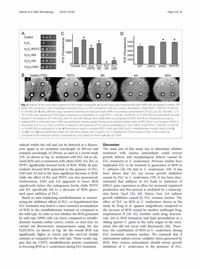
radicals within the cell and can be detected as a fluores-cent signal at an excitation wavelength of 495 nm andemission wavelength of 529 nm, as used in a recent study[19]. As shown in Fig. 3c, treatment with FLC led to ele-vated ROS and co-treatment with either GSH, AA, RA, orPDTC significantly lowered levels of ROS. While all anti-oxidants lowered ROS generated in the presence of FLC,GSH and AA led to the most significant decrease in ROS,while the effect of RA and PDTC was less pronounced.Furthermore, GSH and AA appeared to lower ROSsignificantly below the endogenous levels, while PDTCand RA specifically led to a decrease of ROS gener-ated upon addition of FLC.Based on data implicating metallothioneins in counter-
acting the inhibitory effects of FLC, we hypothesized thatFLC treatment may lead to a more extensive accumulationof ROS in the metallothionein mutants as compared tothe wild type. In order to test whether the ROS generatedby wild type (H99) cells was lower compared to metallo-thionein mutants (either cmt1Δ, cmt2Δ, or cmt1/2Δ), wecarried out fluorescence measurements using the dyeH2DCFDA. As shown in Fig. 3d, the overall ROS wassignificantly higher in cmt2Δ and the cmt1/2Δ doublemutants as compared to the wild type. These results sug-gest that the CMT2 metallothionein protein contributesto lowering ROS in C. neoformans during FLC treatment.
DiscussionThe main aim of this study was to determine whethertreatment with various antioxidants could reversegrowth defects and morphological defects caused byFLC treatment in C. neoformans. Previous studies haveimplicated FLC to be involved in generation of ROS inC. albicans [16–18] and in C. neoformans [19]. It hasbeen shown that AA can rescue growth inhibitioncaused by FLC in C. neoformans [19]. It has been dem-onstrated that addition of AA leads to induction ofERG11 gene expression to allow for increased ergosterolproduction and this process is mediated by a transcrip-tion factor Upc2 [23, 24]. Hence, how AA reducesgrowth inhibition caused by FLC remains unclear. Theeffect of FLC on ROS in C. neoformans shown in thestudy by Peng et al. appears insignificant compared tothe increase of ROS caused by another antifungal drug,amphotericin B [19, 35]. Another azole drug, itracona-zole, led to ROS formation and lipid peroxidation in asibling species C. gattii in the early stages of the treat-ment; this did not occur with fluconazole [36]. There-fore, the contribution of ROS in C. neoformans duringFLC treatment remains unclear. We reasoned that ifantioxidants indeed reverse oxidative stress generated byROS, then various antioxidants should rescue growthinhibition of C. neoformans in the presence of FLC.
Fig. 3 Analysis of the antioxidant potential of the tested compounds. a Growth spot assay showing wild type (H99) cells incubated on either YPDalone, YPD containing 3 mM of hydrogen peroxide (H2O2), or YPD containing 3 mM H2O2 and an antioxidant (10 μM PDTC, 1 mM RA, 10 mM AA,or 10 mM GSH). b Disc diffusion assay represents resistance of wild type strain (H99) to increasing concentrations of H2O2 (25, 50, 100 mM). ~ 2 ×10⁶ of cells were spread over YPD plates containing no antioxidant, or 10 μM PDTC, 1 mM RA, 10 mM AA, or 10 mM GSH. All antioxidants rescuedgrowth in the presence of 3 mM H2O2 with AA and GSH having more visible effect as compared to PDTC and RA. c A fluorescence assay tomeasure ROS in wild type strain (H99) was performed, wherein greater fluorescence indicates higher levels of ROS. There is an increase of ROS inthe presence of FLC (p < 0.01), and ROS is reduced in the presence of FLC and an antioxidant (p < 0.01, either 10 μM PDTC, 1 mM, RA, 10 mM AA,or 10 mM GSH) as indicated by a star. d Fluorescence assay to measure ROS in wild type (H99) and in metallothionein mutant strains (cmt1Δ,cmt2Δ, cmt1/2Δ) was performed where the cells were treated with 32 μg/mL FLC. A significance of the increase in ROS in the mutant ascompared to the wild type control is indicated by a star, based on three replicates (p < 0.05)
Dbouk et al. BMC Microbiology (2019) 19:243 Page 6 of 10

While we found that all tested antioxidants (AA, RA,PDTC, and GSH) could rescue growth inhibited by FLC(using growth assays, spot assays and disc diffusionassays to assess sensitivity to FLC), the rescue was notuniform. If we consider a measure of growth rescue inthe presence of hydrogen peroxide as an indicator of theantioxidant potential, AA and GSH were the mostefficient antioxidants in C. neoformans, while PDTC andRA were less effective as antioxidants compared to AAand GSH. Interestingly, while GSH was one of the morepotent antioxidants, based on the ability of GSH tolower ROS caused by hydrogen peroxide and FLC, GSHonly moderately rescued growth inhibition by FLC. Thissuggests that the antioxidants may be counteracting spe-cific species of ROS not always matching ROS type thatis generated during oxidative stress in the presence ofFLC. A non-exclusive possibility is that the effects of theantioxidants also involve changes in the expression of er-gosterol pathway genes in addition to lowering ROS andcollectively contribute to the survival in the presence ofFLC. Future studies that would examine ergosterol levelsand measure global gene expression in the presence ofFLC and specific antioxidants will help to resolve theseuncertainties.Interestingly, each of the antioxidants tested could
rescue morphological growth defects caused by FLC. C.neoformans wild type (H99) strain treated with (FLC) at32 μg/mL displayed multi-budded phenotype most likelydue to inability to perform cytokinesis. Our study re-vealed that all the antioxidants tested (AA, RA, PDTC,and GSH) can rescue cells from cytokinesis defectscaused by FLC, but not all antioxidants could rescuegrowth inhibition due to FLC to the same extent. Thisresult suggests that while cytokinesis defect may contrib-ute to growth defect in FLC-treated cells, eliminatingthis aberration is not sufficient to restore growth in thepresence of FLC.Another evidence that FLC is contributing to an in-
crease in ROS in C. neoformans is the involvement ofthe metallothionein genes CMT1 and CMT2 in resistingthe inhibitory effects of FLC. Metallothionein proteinsare essential for sequestering copper levels, which areupregulated during infection by C. neoformans [29]. In-creased copper levels can induce ROS, hence during infec-tion by C. neoformans, expression of CMT genes is crucial.Previous studies have shown that mutants of C. neofor-mans, lacking cmt genes are more sensitive to FLC treat-ment [19]. Our results suggest that cells lacking Cmt2 orboth Cmt1 and Cmt2 proteins are more sensitive to FLC.We also find that Cmt mutants are compromised in theirability to recover C. neoformans cells from FLC treatmentwhen antioxidants are added. Interestingly, the extent towhich the mutants could be rescued when co-treated withFLC and an antioxidant varied depending on the type of
antioxidant. We find that Cmt proteins play an importantrole in PDTC-based growth rescue in FLC-treated cells.This further suggests that these antioxidants act throughvarious molecular mechanisms to facilitate rescue fromFLC-mediated cell growth inhibition. Future studies shoulddetermine the effects of the antioxidants on gene expres-sion in cells treated with FLC. Including Cmt mutants intranscriptional profiling of C. neoformans during varioustreatments would shed light on molecular mechanisms re-sponsible for FLC resistance in C. neoformans.The antioxidants we have tested in this study have been
shown to reverse damage caused by many types of freeradicals. PDTC has been shown to reverse oxidative dam-age and carbonylation of proteins by reversing HOCl-mediated oxidative stress [37]. RA has been implicated inhydroxyl radical and lipid peroxide scavenging [38]. AAhas been shown to reverse oxidative stress mainly causedby oxygen free radicals [39, 40]. GSH has been implied inreversing oxidative stress generated by hydrogen peroxide[41] and lipid peroxides [42]. Previous studies using C.glabrata as a model have suggested that FLC causes anincrease in singlet oxygen and peroxide radicals and cancause DNA damage and treatment of Candida with FLCincreased activity of enzymatic antioxidants, namelysuperoxide dismutase (SOD) and glutathione peroxidase(GPx) [16]. It is possible that reversal of ROS and growthdefects in the presence of AA and GSH in C. neoformansis due to the quenching of singlet oxygen species andhydrogen peroxide damage caused by FLC. In addition toDNA damage, it is possible that protein oxidation andcarbonylation could be increased in the presence of FLC,which would explain the role played by PDTC in reversingFLC damage in C. neoformans. FLC has been shown to bemore potent in Candida species strains defective in super-oxide dismutase and catalase activity [43]. Hence RAcould have restored FLC-mediated growth inhibition byregulated SOD levels in the presence of FLC. Furtherinvestigations should determine what specific forms offree radicals are upregulated in the presence of FLC andthe extent of DNA and protein damage that could becaused in the presence of FLC.
ConclusionsIn summary, we conclude that one of the effects of FLCtreatment in C. neoformans is an increase in ROS. Fur-thermore, addition of antioxidants can partially rescuegrowth of C. neoformans in the presence of FLC. How-ever, our results point to a complex nature of the effectsof the antioxidants and suggest that various mechanismscontribute to the antioxidant-mediated growth rescue.The significance of this study is in understanding envir-onmental conditions that can cause rescue of growth ofC. neoformans in the presence of FLC and potentiallydevelopment of resistance to FLC. While formation of
Dbouk et al. BMC Microbiology (2019) 19:243 Page 7 of 10
aneuploid cells is associated with FLC resistance, recentstudies are revealing that counteracting ROS caused byFLC in fungi could also contribute to resisting FLCmode of action. Understanding how individual antioxi-dants could reverse ROS generated by FLC and tyingtheir effects to transcriptional profiling of genes thatget altered during co-treatment with FLC and antioxi-dants would uncover molecular mechanisms that poten-tially lead to FLC resistance in C. neoformans and otherpathogenic fungi.
MethodsReagents usedAscorbic Acid or AA (Fisher Scientific, Cat No A61-25,CAS 5081-7) was prepared from a stock of 1M, and usedat 10mM. A reduced form of glutathione or GSH (AlfaAesar, Cat No AAJ6216606, CAS 70-18-8) was preparedfrom a stock of 0.5M, and used at 10mM. Pyrrolidine-dithiocarbamate or PDTC (Cayman Chemicals, Cat No20713, CAS 5108-96-3) was prepared from a stock of 10mM, and used at 10 μM. Retinoic Acid or RA (CaymanChemical, Cat No 11017, CAS 302-79-4) was preparedfrom a stock of 100mM (dissolved in dimethyl sulfoxide(DMSO)), and used at 1 mM. The fluorescent dye forROS assays, 4-Amino-5-methylamino-2′,7′-difluorofluor-escein diacetate (H2DCFDA) (Sigma, Cat No D6883, CAS4091-99-0), was dissolved in DMSO at a stock concentra-tion of 100mM and used at 10 μM. Fluconazole (CaymanChemical, Cat No 11594, CAS 86386-73-4) was dissolvedin DMSO as a 50mg/ml stock and used at 32 μg/ml.Hydrogen peroxide (Cat No H325-100) was obtained fromFisher Scientific.
Strains and mediaCryptococcus neoformans var. grubii wild type (strainH99 Stud) is the derivative of the original strain isolatedin 1978 by John Perfect at Duke University (ATCC 208821)that has been passaged through a rabbit at that time. Thecmt1Δ, cmt2Δ, cmt1/2Δ deletion mutants isogenic to H99(CMT1, CNAG_05549; CMT2, CNAG_00306) were kindlyprovided by the laboratory of Dr. Lukasz Kozubowski,Clemson University (the metallothionein mutants were ori-ginally obtained from Dr. Dennis J. Thiele, Duke University).Cells were grown on YPD media: (1% yeast extract, 2%
peptone, 2% dextrose, 2% agar), supplemented with che-micals as indicated in the text.
Fluconazole sensitivity plate and spot growth assaysEither wild type, cmt1Δ, cmt2Δ or cmt1/2Δ were grown inliquid YPD broth overnight for 16 h. All strains were di-luted to an Optical Density of OD600 = 0.1 and refreshedin YPD liquid media for 4 h and then counted using aNeubauer Hemocytometer. For growth assays, ~ 10,000cells in exponential growth phase were spread onto plates
containing either YPD media alone, YPD plus 32 μg/μLFLC, and YPD plus 32 μg/μL FLC and an antioxidant,namely, 10 μM PDTC, 1mM RA, 10mM AA, or 10mMGSH. Spot growth assays were performed with a 10-foldserial dilution of cells such that 2 μL contained either 104,103, 102, or 10 cells and were carefully spotted onto YPDplates alone, YPD plus 32 μg/μL FLC, or YPD plus FLCand individual antioxidants, as described above. For bothgrowth assays and spot assays, cells grew for 48 h at 25 °Cbefore recording the data.
Fluorescence assay to detect ROSCells were grown overnight at room temperature in 2mlliquid YPD medium with constant agitation, diluted toan Optical Density OD600 = 0.1, and grown for an add-itional 4 h. Subsequently, the culture was diluted to 10,000 cells/ml and the cultures were either grown as notreatment control, treated with either 32 μg/ml FLC, or32 μg/ml FLC and an antioxidant (either 10 μM PDTC,1 mM, RA, 10 mM AA, or 10 mM GSH) for 12 h. To de-tect ROS, 10 μM of a fluorescent dye, H2DCFDA, wasadded to each of the samples and incubated for 1 h inthe dark at 25 °C. A control set of each of the sampleswere incubated without the fluorescent dye. 250 μL ofthe sample were added to each well of a 96-well micro-plate. ROS was measured as fluorescence emitted by thefluorescent dye, H2DCFDA, at an excitation wavelengthof 485 nm and an emission wavelength of 535 nm. Thefluorescence reading was measured and recorded asRelative Fluorescence Units (RFU). From each reading ofthe sample treated with H2DCFDA the reading obtainedfrom the sample without addition of H2DCFDA wassubtracted. Each treatment was made in triplicate. Alldata points were computed using multifactorial ANOVAand Tukey’s HSD post hoc test.
Disk diffusion assayC. neoformans strains (wild type H99, or mutants,cmt1Δ, cmt2Δ, or cmt1/2Δ) were grown in 2 ml of YPDliquid broth overnight, for 16 h, diluted to an OD600 =0.1 and refreshed for 4 h. Each strain was counted usinga hemocytometer and ~ 2 × 10⁶ cells were plated ontoYPD semi-solid media plates containing either no antioxi-dant (control), AA (10mM), RA (1mM), PDTC (10 μM)or GSH (10mM), and spread with sterile Dynarex cottontipped applicators at opposing 90° angles. The plates wereleft to dry before application of cotton disks. After 10minof drying, 6.6 mm cotton disks were lightly placed perpen-dicular on top of the YPD medium as to not break thesurface of the gel. Depending on the experiment, either in-creasing amounts of 25, 50 and 100 micrograms of FLC,or increasing concentrations of 25, 50, and 100mMhydrogen peroxide were added to the top end of the diskin order for the FLC or hydrogen peroxide to diffuse
Dbouk et al. BMC Microbiology (2019) 19:243 Page 8 of 10
throughout the area surrounding the disc. Finally, thediscs were laid flush onto the medium equidistant fromone another. The cells grew for 48 h at 25 °C and all treat-ments were done in triplicate. Each zone of inhibition wasmeasured and the results from each of the three replicateexperiments were averaged. A multifactorial ANOVAalong with a Tukey’s HSD post hoc test was used to indi-cate significance.
MicroscopyDifferential interference contrast (DIC) microscopy wasused to study C. neoformans cell morphology under vari-ous conditions. C. neoformans cells were grown for 16 h at25 °C in YPD liquid media, diluted down to an OD600 =0.1, and refreshed for 4 h. Cells were then grown with ei-ther no treatment (control cells), treatment with FLC aloneat 32 μg/ml, or FLC at 32 μg/ml and an antioxidant (10 μMPDTC, 1mM RA, 10mM AA, or 10mM GSH) for 16 h.Cells were centrifuged at 3000 x g for 2min and washedwith ice cold PBS (137mM NaCl, 2.7mM KCl, 10mMNa2HPO4, 1.8mM KH2PO4). An agar trap was made tocapture yeast cells, by melting 0.8% agarose on a slide as athin section. Cells were placed in an agar trap, coveredwith a coverslip and visualized by Zeiss Axiovert 200inverted microscope (Carl zeiss, Inc., Thornwood, NY).
Statistical analysesFor all statistical analyses, the Shapiro Wilk Test wasused to test for normality, and afterward, the BartlettTest was used to test for equality of variance. Since bothconditions were met, a multifactorial ANOVA was per-formed. The Tukey HSD test was used to determine ifthe relationship between the control group and variablegroups were statistically significant.
Supplementary informationSupplementary information accompanies this paper at https://doi.org/10.1186/s12866-019-1606-4.
Additional file 1. Raw data corresponding to Figures 1C, 2C, 3C, and 3Dthat are included in the article.
AbbreviationsAA: Ascorbic acid; FLC: Fluconazole; GSH: Glutathione; H2DCFDA: 4-Amino-5-methylamino-2′,7′-difluorofluorescein diacetate; MIC: Minimum inhibitoryconcentration; PDTC: Pyrrolidine dithiocarbamate; RA: Retinoic acid;ROS: Reactive oxygen species
AcknowledgmentsWe thank Dr. Lukasz Kozubowski for providing the metallothionein mutantstrains (originally obtained from Dr. Dennis J. Thiele, Duke University) and forhelpful discussions. We thank Furman University Research and ProfessionalGrowth (RPG) Fund, the South Carolina NIH-INBRE Faculty Fellows SummerResearch Program Fund, and the Furman University Summer Research Fellowawards for supporting this study.
Authors’ contributionsSC designed the experiments. NHD, MBC, and KN performed theexperiments. SC and NHD analyzed the data. SC wrote the manuscript. Allthe authors contributed to manuscript editing. All authors read andapproved the final manuscript.
FundingThis study was funded by South Carolina NIH INBRE Faculty Fellows Award,Research and Professional Growth (RPG) fund and startup funds at FurmanUniversity awarded to SC and Furman University Summer Research Fellowawards granted to MC and KN. The funds were used to purchase all reagentsused in this study. In addition, the Summer Research Fellow award providedsummer salary to MC and KN and the South Carolina NIH INBRE FacultyFellows Award provided summer salary to ND and SC. The funding sourceshave not participated in the design of the study and collection, analysis, andinterpretation of data, or in writing the manuscript.
Availability of data and materialsAll data generated during this study are included in this pulished article andin Additional file 1, which contains raw data corresponding to Figures 1C,2C, 3C, and 3D.
Ethics approval and consent to participateWe confirm that all methods were carried out in proper conformation withappropriate regulations and guidelines. All experimental protocols involvingfungal cultures were approved by Furman University. No human or animalsubjects were used for this research.
Consent for publicationNot applicable
Competing interestsThe authors declare that they have no competing interests.
Received: 2 August 2019 Accepted: 29 September 2019
References1. Brown SP, Cornforth DM, Mideo N. Evolution of virulence in opportunistic
pathogens: generalism, plasticity, and control. Trends Microbiol. 2012;20(7):336–42.
2. Idnurm A, et al. Deciphering the model pathogenic fungus Cryptococcusneoformans. Nat Rev Microbiol. 2005;3(10):753–64.
3. Park BJ, et al. Estimation of the current global burden of cryptococcalmeningitis among persons living with HIV/AIDS. Aids. 2009;23(4):525–30.
4. Assing K, Birgens H, Arendrup M. Cryptococcus neoformans var neoformansresistant to fluconazole in an HIV-negative patient with chronic lymphocyticleukemia. Clin Microbiol Infect. 2003;9(5):441–4.
5. Chen YC, et al. Increasing trend of fluconazole-non-susceptibleCryptococcus neoformans in patients with invasive cryptococcosis: a 12-year longitudinal study. BMC Infect Dis. 2015;15:277.
6. Smith KD, et al. Increased antifungal drug resistance in clinical isolates ofCryptococcus neoformans in Uganda. Antimicrob Agents Chemother. 2015;59(12):7197–204.
7. Pasko MT, Piscitelli SC, Van Slooten AD. Fluconazole: a new triazoleantifungal agent. DICP. 1990;24(9):860–7.
8. Goa KL, Barradell LB. Fluconazole. An update of its pharmacodynamicand pharmacokinetic properties and therapeutic use in major superficialand systemic mycoses in immunocompromised patients. Drugs. 1995;50(4):658–90.
9. Li Z, et al. Fluconazole plus flucytosine is a good alternative therapy fornon-HIV and non-transplant-associated cryptococcal meningitis: aretrospective cohort study. Mycoses. 2019;62:686.
10. Khan AA, et al. Additive potential of combination therapy againstcryptococcosis employing a novel amphotericin B and fluconazole loadeddual delivery system. Eur J Pharm Sci. 2018;119:171–8.
11. Revankar SG, et al. Cloning and characterization of the lanosterol 14alpha-demethylase (ERG11) gene in Cryptococcus neoformans. Biochem BiophysRes Commun. 2004;324(2):719–28.
Dbouk et al. BMC Microbiology (2019) 19:243 Page 9 of 10
12. Sionov E, et al. Cryptococcus neoformans overcomes stress of azole drugsby formation of disomy in specific multiple chromosomes. PLoS Pathog.2010;6(4):e1000848.
13. Gast CE, et al. Azole resistance in Cryptococcus gattii from the Pacificnorthwest: investigation of the role of ERG11. Antimicrob AgentsChemother. 2013;57(11):5478–85.
14. Posteraro B, et al. Identification and characterization of a Cryptococcusneoformans ATP binding cassette (ABC) transporter-encoding gene, CnAFR1,involved in the resistance to fluconazole. Mol Microbiol. 2003;47(2):357–71.
15. Cowen LE, et al. Mechanisms of antifungal drug resistance. Cold SpringHarb Perspect Med. 2014;5(7):a019752.
16. Mahl CD, et al. Induction of ROS generation by fluconazole in Candidaglabrata: activation of antioxidant enzymes and oxidative DNA damage.Diagn Microbiol Infect Dis. 2015;82(3):203–8.
17. Wang Y, et al. Ascorbic acid decreases the antifungal effect of fluconazole inthe treatment of candidiasis. Clin Exp Pharmacol Physiol. 2009;36(10):e40–6.
18. Kobayashi D, et al. Endogenous reactive oxygen species is an importantmediator of miconazole antifungal effect. Antimicrob Agents Chemother.2002;46(10):3113–7.
19. Peng CA, et al. Fluconazole induces ROS in Cryptococcus neoformans andcontributes to DNA damage in vitro. PLoS One. 2018;13(12):e0208471.
20. Winterbourn CC. Reconciling the chemistry and biology of reactive oxygenspecies. Nat Chem Biol. 2008;4(5):278–86.
21. Angele-Martinez C, Goodman C, Brumaghim J. Metal-mediated DNAdamage and cell death: mechanisms, detection methods, and cellularconsequences. Metallomics. 2014;6(8):1358–81.
22. Fedorova M, Bollineni RC, Hoffmann R. Protein carbonylation as a majorhallmark of oxidative damage: update of analytical strategies. MassSpectrom Rev. 2014;33(2):79–97.
23. Van Hauwenhuyse F, Fiori A, Van Dijck P. Ascorbic acid inhibition ofCandida albicans Hsp90-mediated morphogenesis occurs via thetranscriptional regulator Upc2. Eukaryot Cell. 2014;13(10):1278–89.
24. Yang H, et al. Structural mechanism of ergosterol regulation by fungal steroltranscription factor Upc2. Nat Commun. 2015;6:6129.
25. Raja MR, et al. A copper hyperaccumulation phenotype correlates withpathogenesis in Cryptococcus neoformans. Metallomics. 2013;5(4):363–71.
26. Zhang P, et al. Effects of CTR4 deletion on virulence and stress response inCryptococcus neoformans. Antonie Van Leeuwenhoek. 2016;109(8):1081–90.
27. Jiang N, et al. A copper-responsive factor gene CUF1 is required for copperinduction of laccase in Cryptococcus neoformans. FEMS Microbiol Lett.2009;296(1):84–90.
28. Husain N, Mahmood R. Copper (II) generates ROS and RNS, impairsantioxidant system and damages membrane and DNA in human bloodcells. Environ Sci Pollut Res Int. 2019;26:20654.
29. Ding C, et al. The copper regulon of the human fungal pathogenCryptococcus neoformans H99. Mol Microbiol. 2011;81(6):1560–76.
30. Ding C, et al. Cryptococcus neoformans copper detoxification machinery iscritical for fungal virulence. Cell Host Microbe. 2013;13(3):265–76.
31. Niedzwiecka K, et al. Glutathione may have implications in the design of 3-bromopyruvate treatment protocols for both fungal and algal infections aswell as multiple myeloma. Oncotarget. 2016;7(40):65614–26.
32. Altamirano S, et al. Fluconazole-induced ploidy change in Cryptococcusneoformans results from the uncoupling of cell growth and nucleardivision. mSphere. 2017;2(3):e00205-17. https://doi.org/10.1128/mSphere.00205-1.
33. Vasak M. Advances in metallothionein structure and functions. J Trace ElemMed Biol. 2005;19(1):13–7.
34. Upadhya R, et al. Global transcriptome profile of Cryptococcus neoformansduring exposure to hydrogen peroxide induced oxidative stress. PLoS One.2013;8(1):e55110.
35. Mesa-Arango AC, et al. The production of reactive oxygen species is auniversal action mechanism of amphotericin B against pathogenic yeastsand contributes to the fungicidal effect of this drug. Antimicrob AgentsChemother. 2014;58(11):6627–38.
36. Ferreira GF, et al. The role of oxidative and nitrosative bursts caused byazoles and amphotericin B against the fungal pathogen Cryptococcus gattii.J Antimicrob Chemother. 2013;68(8):1801–11.
37. Zhu BZ, Carr AC, Frei B. Pyrrolidine dithiocarbamate is a potentantioxidant against hypochlorous acid-induced protein damage. FEBSLett. 2002;532(1–2):80–4.
38. Ahlemeyer B, et al. Retinoic acid reduces apoptosis and oxidative stress bypreservation of SOD protein level. Free Radic Biol Med. 2001;30(10):1067–77.
39. Zyracka E, et al. Ascorbate abolishes auxotrophy caused by the lack ofsuperoxide dismutase in Saccharomyces cerevisiae. Yeast can be abiosensor for antioxidants. J Biotechnol. 2005;115(3):271–8.
40. Boatright WL. Oxygen dependency of one-electron reactions generatingascorbate radicals and hydrogen peroxide from ascorbic acid. Food Chem.2016;196:1361–7.
41. Munro D, Treberg JR. A radical shift in perspective: mitochondria asregulators of reactive oxygen species. J Exp Biol. 2017;220(Pt 7):1170–80.
42. Conrad M, et al. Regulation of lipid peroxidation and ferroptosis in diversespecies. Genes Dev. 2018;32(9–10):602–19.
43. Linares CE, et al. Fluconazole and amphotericin-B resistance are associatedwith increased catalase and superoxide dismutase activity in Candidaalbicans and Candida dubliniensis. Rev Soc Bras Med Trop. 2013;46(6):752–8.
Publisher’s NoteSpringer Nature remains neutral with regard to jurisdictional claims inpublished maps and institutional affiliations.
Dbouk et al. BMC Microbiology (2019) 19:243 Page 10 of 10