Embed Size (px)
Citation preview
REVIEW Open Access
Reactive oxygen species and malereproductive hormonesMahsa Darbandi1†, Sara Darbandi1†, Ashok Agarwal2* , Pallav Sengupta3, Damayanthi Durairajanayagam4,Ralf Henkel5 and Mohammad Reza Sadeghi6
Abstract
Reports of the increasing incidence of male infertility paired with decreasing semen quality have triggered studieson the effects of lifestyle and environmental factors on the male reproductive potential. There are numerous exogenousand endogenous factors that are able to induce excessive production of reactive oxygen species (ROS) beyond that ofcellular antioxidant capacity, thus causing oxidative stress. In turn, oxidative stress negatively affects male reproductivefunctions and may induce infertility either directly or indirectly by affecting the hypothalamus-pituitary-gonadal (HPG)axis and/or disrupting its crosstalk with other hormonal axes. This review discusses the important exogenous andendogenous factors leading to the generation of ROS in different parts of the male reproductive tract. It also highlightsthe negative impact of oxidative stress on the regulation and cross-talk between the reproductive hormones. It furtherdescribes the mechanism of ROS-induced derangement of male reproductive hormonal profiles that could ultimatelylead to male infertility. An understanding of the disruptive effects of ROS on male reproductive hormones wouldencourage further investigations directed towards the prevention of ROS-mediated hormonal imbalances, which in turncould help in the management of male infertility.
Keywords: Antioxidants, Hypothalamic-pituitary-gonadal axis, Male infertility, Oxidative stress, Reactive oxygen species,Testosterone
BackgroundOver the past 40 years, reports regarding the decline insemen quality [1–4] and its probable consequences onmale fertility have encouraged studies about the effectsof environment and lifestyle factors on the male repro-ductive potential. Reactive oxygen species (ROS) pro-duced by exogenous and endogenous factors are highlyreactive oxygen derivatives with half-lives in the nano-to milliseconds range. These molecules reportedly play akey role in altering male reproductive functions [5, 6].Lifestyle modifications, technological advancements, es-calating levels of pollution, alcohol consumption, smokingof cigarettes and vaping, and physical stress are among theprime exogenous causes of ROS production [7–9]. Also,multiple mechanisms involving metabolism in the cell
membrane, mitochondria, peroxisomes, and endoplasmicreticulum can produce endogenous ROS [7, 9].Antioxidants defend against excessive ROS levels
through enzymatic (superoxide dismutase, catalases, andperoxidases) and non-enzymatic (vitamins, steroids etc.)mechanisms [7, 10]. In cases where the imbalance be-tween oxidants (ROS) and antioxidants leans towardsthe oxidants, oxidative stress (OS) occurs, which putsthe cells and the body under stress. As a result, excessiveROS can induce lipid peroxidation, disrupt DNA, RNAas well as protein functions in the spermatozoa andother testicular cells [10].High ROS levels can increase the possibility of infertil-
ity not only directly by inducing OS, but also indirectlyby acting through the hypothalamic axes of hormone re-lease [11–13]. ROS reduce male sex hormone levels anddisrupt the hormonal balance that regulates male repro-ductive functions [14], and thus causes infertility. These“endocrine disruptors” not only interfere in the commu-nication between testis and the hypothalamic-pituitaryunit, they also disrupt the cross-talk between the
* Correspondence: [email protected]†Mahsa Darbandi and Sara Darbandi contributed equally to this work.2American Center for Reproductive Medicine, Cleveland Clinic, Cleveland,Ohio 44195, USAFull list of author information is available at the end of the article
© The Author(s). 2018 Open Access This article is distributed under the terms of the Creative Commons Attribution 4.0International License (http://creativecommons.org/licenses/by/4.0/), which permits unrestricted use, distribution, andreproduction in any medium, provided you give appropriate credit to the original author(s) and the source, provide a link tothe Creative Commons license, and indicate if changes were made. The Creative Commons Public Domain Dedication waiver(http://creativecommons.org/publicdomain/zero/1.0/) applies to the data made available in this article, unless otherwise stated.
Darbandi et al. Reproductive Biology and Endocrinology (2018) 16:87 https://doi.org/10.1186/s12958-018-0406-2
hypothalamic-pituitary-gonadal (HPG) axis with otherhypothalamic hormonal axes [15, 16]. The testis, as theprimary male sex organ, is not only concerned withspermatogenesis, but also with the secretion of severalhormones [17] which are required for regulation of go-nadotropin secretion, spermatogenesis, formation ofmale phenotype during sexual differentiation, and nor-mal sexual behaviour [18]. Hence, by interfering withnormal hormonal release, ROS disrupt these essentialreproductive functions.Therefore, this review precisely elucidates (a) the role
of ROS, generated by various exogenous and endogen-ous factors, in disrupting hormone secretion by interfer-ing in the endocrine pathways, as well as in theircross-talk, (b) hormonal regulation of the oxidative sta-tus of male reproduction, and (c) a possible mechanismof action of ROS-induced disruption of the male repro-ductive hormonal profile.
Endocrinology of male fertilityThe gonadotropin releasing hormone (GnRH) secretedby the hypothalamus regulates the release and secretionof gonadotropins, luteinizing hormone (LH) andfollicle-stimulating hormone (FSH) from anterior pituit-ary that in turn regulate testicular functions [17]. Thesegonadal steroids as well as the pituitary gonadotropins,via feedback regulatory mechanisms, further establishphysiological homeostasis and maintains normal repro-ductive functions [14, 17, 19]. FSH receptors are locatedon the membrane of Sertoli cells, while those of LH areon the Leydig cells. They coordinate to synthesize testos-terone, maintain normal spermatogenesis, sperm healthand density [19–21].Moreover, other hormones like estradiol (E2) and pro-
lactin (PRL) also take part in the management of malereproductive function. E2, produced both by the testisand via the peripheral conversion of androgenic precur-sors, is a potent inhibitor of LH and FSH [18, 19] (Fig. 1).PRL-inhibiting GnRH secretion via modulation of dopa-minergic pathway may also reduce LH and testosteronelevel and thus is associated with hypogonadism [22].Dehydroepiandrosterone (DHEA) is another malereproduction ameliorating, steroid hormone secreted bythe adrenal cortex [23, 24]. Inhibin A and B, dimeric hor-mones produced by Sertoli cells, exhibit negative feedbackon FSH secretion and thus also on testicular functions[25]. Moreover, melatonin (MLT), a tryptophan-derivedhormone of the pineal gland, positively regulates gonado-tropin and testosterone secretion, and thus aid malereproductive functions [26, 27]. Anti-Mullerian hormone(AMH), a dimeric glycoprotein hormone produced in em-bryonic Sertoli cells, is structurally related to inhibin andis responsible for regression of Mullerian ducts during thefirst 8 weeks of embryogenesis. It reflects Sertoli cell
functions and is inhibited by testosterone under the influ-ence of LH [28–30]. Interactions between thehypothalamo-pituitary-thyroid (HPT) and HPG axespotentially influence testicular development, mostly bythe participation of thyroid hormones and FSH [31].Thus, besides the central control through the HPG
axis, the major male reproductive hormones act either in-dividually or via the cross-talks among different endo-crine axes to influence male reproductive functions.Consequently, any disruption to these networks may ad-versely affect male fertility.
Generation of ROS in the male reproductive tractReactive oxygen species (ROS), which are short-lived,unstable, and highly reactive species containing at leastone oxygen atom, are able to snatch electrons fromother molecules to achieve an electronically-stable state.In this process, the other molecule loses an electron fol-lowing which a new radical is formed. Subsequently, thisradical reacts with another neighbouring molecule, thuspassing on the radical status via a reaction called ‘radi-cal-chain reaction’ until two radicals react with one an-other forming a stable bond. These reactions amplify thedegree of alterations in the cellular structures [32–34].Human spermatozoa contain abundant mitochondria,
particularly in its midpiece [35]. An NADH-dependentoxidoreductase (in the inner mitochondrial membrane)and NAD (P) H-oxidase (in the plasma membrane) aretwo main sources of superoxide (O2
●-) [32, 33, 36]. Themajority of ROS generated in human spermatozoa isO2
●- which is a product of oxidative phosphorylation byaddition of an electron to intracellular oxygen and iscreated between complex I and III of the electron trans-port chain [37]. H2O2 is an uncharged, membrane per-meable molecule which has been found to be the majorinitiator of peroxidative damage of the plasma mem-branes of germ cells [34]. In the presence of transitionmetals, such as iron (Fe3+) and copper, O2
●- and H2O2
can generate the extremely reactive OH• through theHaber-Weiss reaction, which consist of a reduction offerric (Fe3+) to ferrous ion (Fe2+) [38]. In a subsequentsecond step, called Fenton reaction, Fe2+ is oxidized byH2O2 to Fe3+ whereby hydroxide (OH−) and the mostreactive hydroxyl radical (OH•) are formed. Further-more, O2
●- has the ability to interact with nitric oxide(NO) to form peroxynitrite (ONOO−), subsequent reac-tions of which may lead to either apoptotic or necroticcell death [39]. In the male reproductive tract, ROSfinally can be generated by one of these sources accord-ing to the above-mentioned mechanisms.In order to produce the immense amount of energy
needed for motility, spermatozoa possess numerousmitochondria in the mid-piece of the flagellum. In themitochondria, disruption of the membrane potential
Darbandi et al. Reproductive Biology and Endocrinology (2018) 16:87 Page 2 of 14
leads to electron leakage in the electron transfer chainand subsequently produces ROS. The Ca2+-dependentNADPH oxidase, called NOX5 (encoded by the NOX5gene) was initially detected in the human testis, but wasalso found to be present in the acrosomal and mid-pieceregions of human spermatozoa [40]. NOX5 is a majorgenerator of ROS and could subsequently induce OS.This enzyme is activated when Ca2+ binds to its cytosolicN-terminal EF-hand and causes conformational changesto the cell through OS [41]. Moreover, during spermato-genesis, the developing spermatozoa extrude their cyto-plasm. When spermiogenesis is disrupted and/or excesscytoplasm is not completely extruded (excess residualcytoplasm), the excess cytoplasm will be retained aroundthe mid-piece. Since cytoplasm contains the enzymaticmachinery to produce ROS, any hindrance in the elimin-ation of excess cytoplasm would trigger the production
of intrinsic amounts of ROS in excess, which, in turn,would lead to oxidative damage of the plasma mem-brane and sperm DNA [42].The prostate and seminal vesicles are the major
sources of peroxidase-positive leukocytes (polymorpho-nuclear leukocytes (50 ∼ 60%) as well as macrophages(20 ∼ 30%)) [43, 44]. Inflammatory responses triggerthese cells to generate ROS about 100-times more thanit is produced under normal conditions [34, 45, 46]. Thiselevated ROS production is a part of the natural defensemechanisms of these cells, whereby NADPH-productionthrough the hexose monophosphate shunt is elevated.Leukocyte participation in inflammation is closely con-nected with the accompanying leukocytospermia [47], acondition defined by the World Health Organization(WHO) as semen samples containing more than onemillion peroxidase-positive leukocytes per milliliter of
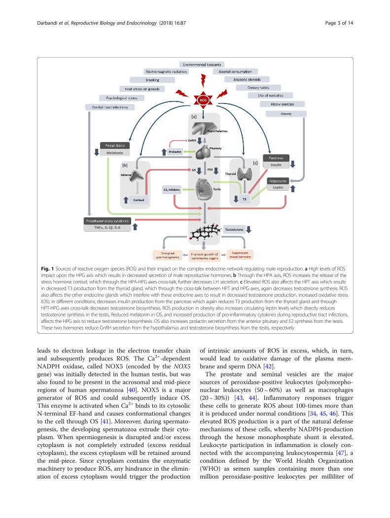
Fig. 1 Sources of reactive oxygen species (ROS) and their impact on the complex endocrine network regulating male reproduction. a High levels of ROSimpact upon the HPG axis which results in decreased secretion of male reproductive hormones. b Through the HPA axis, ROS increases the release of thestress hormone cortisol, which through the HPA-HPG axes cross-talk, further decreases LH secretion. c Elevated ROS also affects the HPT axis which resultsin decreased T3 production from the thyroid gland, which through the cross-talk between HPT and HPG axes, again decreases testosterone synthesis. ROSalso affects the other endocrine glands which interfere with these endocrine axes to result in decreased testosterone production. Increased oxidative stress(OS), in different conditions, decreases insulin production from the pancreas which again reduces T3 production from the thyroid gland and throughHPT-HPG axes cross-talk decreases testosterone biosynthesis. ROS production in obesity also increases circulating leptin levels which directly reducestestosterone synthesis in the testis. Reduced melatonin in OS, and increased production of pro-inflammatory cytokines during reproductive tract infections,affects the HPG axis to reduce testosterone biosynthesis. OS also increases prolactin secretion from the anterior pituitary and E2 synthesis from the testis.These two hormones reduce GnRH secretion from the hypothalamus and testosterone biosynthesis from the testis, respectively
Darbandi et al. Reproductive Biology and Endocrinology (2018) 16:87 Page 3 of 14
semen [48]. Varicocele, a condition caused by an abnor-mal dilation of veins in the pampiniform plexus sur-rounding the spermatic cord [49], is also associated withelevated levels of seminal ROS [50].
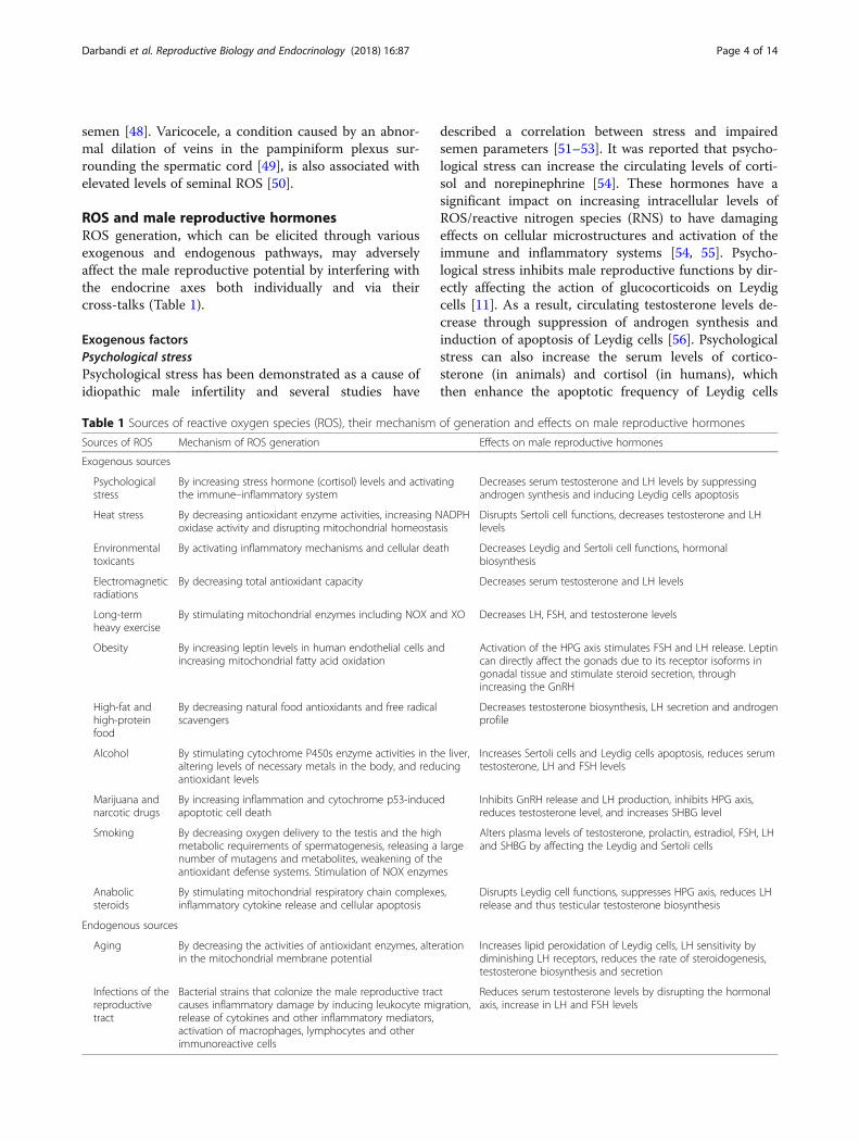
ROS and male reproductive hormonesROS generation, which can be elicited through variousexogenous and endogenous pathways, may adverselyaffect the male reproductive potential by interfering withthe endocrine axes both individually and via theircross-talks (Table 1).
Exogenous factorsPsychological stressPsychological stress has been demonstrated as a cause ofidiopathic male infertility and several studies have
described a correlation between stress and impairedsemen parameters [51–53]. It was reported that psycho-logical stress can increase the circulating levels of corti-sol and norepinephrine [54]. These hormones have asignificant impact on increasing intracellular levels ofROS/reactive nitrogen species (RNS) to have damagingeffects on cellular microstructures and activation of theimmune and inflammatory systems [54, 55]. Psycho-logical stress inhibits male reproductive functions by dir-ectly affecting the action of glucocorticoids on Leydigcells [11]. As a result, circulating testosterone levels de-crease through suppression of androgen synthesis andinduction of apoptosis of Leydig cells [56]. Psychologicalstress can also increase the serum levels of cortico-sterone (in animals) and cortisol (in humans), whichthen enhance the apoptotic frequency of Leydig cells
Table 1 Sources of reactive oxygen species (ROS), their mechanism of generation and effects on male reproductive hormones
Sources of ROS Mechanism of ROS generation Effects on male reproductive hormones
Exogenous sources
Psychologicalstress
By increasing stress hormone (cortisol) levels and activatingthe immune–inflammatory system
Decreases serum testosterone and LH levels by suppressingandrogen synthesis and inducing Leydig cells apoptosis
Heat stress By decreasing antioxidant enzyme activities, increasing NADPHoxidase activity and disrupting mitochondrial homeostasis
Disrupts Sertoli cell functions, decreases testosterone and LHlevels
Environmentaltoxicants
By activating inflammatory mechanisms and cellular death Decreases Leydig and Sertoli cell functions, hormonalbiosynthesis
Electromagneticradiations
By decreasing total antioxidant capacity Decreases serum testosterone and LH levels
Long-termheavy exercise
By stimulating mitochondrial enzymes including NOX and XO Decreases LH, FSH, and testosterone levels
Obesity By increasing leptin levels in human endothelial cells andincreasing mitochondrial fatty acid oxidation
Activation of the HPG axis stimulates FSH and LH release. Leptincan directly affect the gonads due to its receptor isoforms ingonadal tissue and stimulate steroid secretion, throughincreasing the GnRH
High-fat andhigh-proteinfood
By decreasing natural food antioxidants and free radicalscavengers
Decreases testosterone biosynthesis, LH secretion and androgenprofile
Alcohol By stimulating cytochrome P450s enzyme activities in the liver,altering levels of necessary metals in the body, and reducingantioxidant levels
Increases Sertoli cells and Leydig cells apoptosis, reduces serumtestosterone, LH and FSH levels
Marijuana andnarcotic drugs
By increasing inflammation and cytochrome p53-inducedapoptotic cell death
Inhibits GnRH release and LH production, inhibits HPG axis,reduces testosterone level, and increases SHBG level
Smoking By decreasing oxygen delivery to the testis and the highmetabolic requirements of spermatogenesis, releasing a largenumber of mutagens and metabolites, weakening of theantioxidant defense systems. Stimulation of NOX enzymes
Alters plasma levels of testosterone, prolactin, estradiol, FSH, LHand SHBG by affecting the Leydig and Sertoli cells
Anabolicsteroids
By stimulating mitochondrial respiratory chain complexes,inflammatory cytokine release and cellular apoptosis
Disrupts Leydig cell functions, suppresses HPG axis, reduces LHrelease and thus testicular testosterone biosynthesis
Endogenous sources
Aging By decreasing the activities of antioxidant enzymes, alterationin the mitochondrial membrane potential
Increases lipid peroxidation of Leydig cells, LH sensitivity bydiminishing LH receptors, reduces the rate of steroidogenesis,testosterone biosynthesis and secretion
Infections of thereproductivetract
Bacterial strains that colonize the male reproductive tractcauses inflammatory damage by inducing leukocyte migration,release of cytokines and other inflammatory mediators,activation of macrophages, lymphocytes and otherimmunoreactive cells
Reduces serum testosterone levels by disrupting the hormonalaxis, increase in LH and FSH levels
Darbandi et al. Reproductive Biology and Endocrinology (2018) 16:87 Page 4 of 14
[57]. Furthermore, during glucocorticoid productionby 11β-hydroxysteroid dehydrogenase-1 (11βHSD-1),NADPH was produced as a cofactor that is used forthe biosynthesis of steroidogenic enzymes and testos-terone [58].Stress adversely affects steroidogenesis, since changes
in the autonomic catecholaminergic activities duringstress may suppress Leydig cell functions, thus inhibitingsteroidogenic enzyme activities and testosterone produc-tion [11]. Stress-induced elevations of glucocorticoidlevels can directly decrease testosterone levels withoutaltering LH levels [59–61]. Further, in case of chronicstress, a decrease in LH and GnRH levels becomesapparent [62, 63].
Heat stress on gonadsIn males, testes are suspended in a scrotum outside thebody in order to keep the temperature 2 to 4 °C lowerthan that of core body temperature. This is a require-ment for normal spermatogenesis [64]. However, heatstress to the testes not only decreases semen quality butalso indirectly lowers embryo quality after fertilization asthe spermatozoa produced in overheated testis exhibitsdamage [65–67]. In this context, heat stress is respon-sible for enhancing ROS production as well as decreas-ing antioxidant enzyme activities, increasing NADPHoxidase activity and disrupting mitochondrial homeosta-sis [68, 69]. Numerous reports have documented thatfactors such as fever, sauna or steam room use, sleepingposture, long time sitting or driving, polyester-lined ath-letic supports, using a laptop on the lap and electricblankets impose negative effects on scrotal temperaturesand subsequently spermatogenesis [70, 71]. Studies havealso reported that clinical conditions such as cryptorchid-ism, varicocele, and acute febrile illness can increase tes-ticular temperature and suppress spermatogenesis [70].Activation of the hypothalamic–pituitary–adrenal (HPA)
axis and the consequent increase in plasma glucocorticoidconcentrations are two of the most important responses toheat stress. Heat stress imparts detrimental effects on malereproduction partly by disrupting the normal release ofGnRH from the hypothalamus as well as LH and FSH fromthe anterior pituitary gland [72]. Several studies have indi-cated that testicular heat stress leads to a decline in thecirculating levels of testosterone and LH but increasesserum cortisol levels [73, 74]. Testicular heat stress alsoleads to Leydig cell apoptosis and a reduction in testoster-one biosynthesis in adult rat testes [75]. Moreover, in-creased testicular temperature adversely affects Sertoli cellfunction, production of testicular androgen-binding pro-tein, spermatogenesis and semen parameters [76]. Thus,increased heat stress elevates the generation of ROS in themale reproductive tract by directly affecting cellular metab-olism [69] and by influencing stress hormone levels [77].
The resulting increase in ROS production, in turn, damagestesticular germ cells and other endocrine cells to disruptthe hormonal balance, thereby curbing male fertility [34].
Environmental toxicantsExposure to environmental contaminants adversely af-fects the male reproductive potential [78, 79]. Male in-fertility caused by exposure to environmental toxicantssuch as cadmium [80, 81], mercury [82, 83], bisphenol A(BPA) [84, 85] and dioxin [86] is a worldwide problem.Even chemical components of air pollution can induceOS by triggering redox-sensitive pathways subsequentlyleading to various malaise, such as inflammation and celldeath [87].These contaminations deteriorate semen parameters,
DNA integrity via disrupting Leydig and Sertoli cellfunction, hormone biosynthesis, gene expression andepigenetic modifications [12, 88, 89]. These toxicantscommonly act as ‘endocrine disrupting chemicals’(EDCs) that interfere with normal hormonal functions[90], enhance the level of circulating cortisol owing toOS induction [91] and reduces circulating testosteronelevels [92, 93]. Increased cortisol decreases LH secretionthrough crosstalk between the HPG-HPA axes. De-creased LH concentration fails to stimulate the Leydigcells resulting in decreased testosterone production,whereas decreased FSH affects normal Sertoli cell func-tions [94]. These toxicants also interfere with the cellularcommunications and adhesions between Sertoli–Sertolicells and Sertoli–germ cells via the phosphatidylinositol3-kinase (PI3K)/c-Src/focal adhesion kinase (FAK)signalling pathway which leads to reproductive dysfunc-tion [95] and disrupted hormonal secretion. Thus, thesetoxicants disrupt normal male reproductive hormonalbalance by their disruptive influence upon the endocrineand reproductive organs as well as by interfering in thecross-talk among different endocrine axes [96].
Electromagnetic radiationsSince the last few decades, it has been widely reportedthat long-term exposure to electromagnetic radiationscan generate ROS in reproductive organs, which notonly declines motility, viability, and normal morphologyof functional spermatozoa [97, 98], but also disorientsreproductive hormonal profiles. The use of cell phones[99], wireless internet [100] and other occupational orenvironmental radiations [101] are found to be majorcausative factors directly augmenting ROS generation inmale reproductive organs [102, 103]. Electromagneticradiation affects the HPA axis and increases adrenocorti-cotropic hormone (ACTH) secretion from the anteriorpituitary thereby increasing the production of cortisolfrom adrenal cortex [104]. These radiations can alsodecrease testosterone secretion from Leydig cells by
Darbandi et al. Reproductive Biology and Endocrinology (2018) 16:87 Page 5 of 14
disrupting the male reproductive hormonal axis [105].Electromagnetic radiation significantly affect LH levelsbut not FSH and PRL levels [106]. It has also been re-ported that exposure to electromagnetic waves directlyaffects the pineal gland, thereby deteriorating the bio-logical effect of melatonin on GnRH pulse in the hypo-thalamus [107]. Thus, altered GnRH levels influenceFSH and LH secretion and negatively affects testoster-one synthesis in the testis [108].
ExerciseContrary to regular exercise that enhances antioxidantdefences in the body, unaccustomed and/or exhaustiveexercise can lead to the undesirable generation of exces-sive ROS [109]. Although the exact redox mechanismsremain elusive, it seems that mitochondria, NADPH oxi-dase (NOX), and xanthine oxidase (XO) are the majorendogenous sources of ROS in skeletal muscle [109].Some studies showed that moderate physical activity canincrease FSH, LH, and testosterone levels [110], which iswidely associated with increased energy and musclestrength [111, 112]. Despite the impact of moderateexercise, data suggest that vigorous exercise may de-crease LH, FSH, and testosterone levels as well as semenparameters [113, 114]. However, other investigators havereported that testosterone levels remain unaltered fol-lowing heavy exercise [115, 116].
ObesityObesity is a complex health disorder that severely affectshormonal balance [117]. Obesity disrupts serum levels ofleptin [118], ghrelin [119], adiponectin [120], orexin[121], obestatin [122] and other metabolic hormone pro-files [117]. Reportedly, leptin correlates positively withbody fat mass [123, 124] and a leptin-induced generationof ROS in human endothelial cells result from increasedmitochondrial fatty acid oxidation [123, 124]. The activa-tion of the HPG axis could be enhanced by leptin andthus stimulate the release of GnRH, FSH and LH [125].Moreover, leptin can directly affect the gonads due to itsreceptor isoforms in gonadal tissue [125].Though the impact of ghrelin on serum testosterone
level is contentious [126–128], it is reported that ghrelinreceptors are present in the testis and that ghrelin playsa key role in testosterone production, but not directly inspermatogenesis [126]. Increased ROS levels appear tocause increased levels of ghrelin [129] which may, inturn, result in obesity and further ROS production.Serum adiponectin level is negatively correlated with
both testosterone [130] and ROS production [131].Orexin (hypocretin) is known to stimulate testosteroneproduction by enhancing the activities of steroidogenicenzymes in Leydig cells [132]. It is also reported to at-tenuate ROS-induced cell damage [133]. All these
metabolic hormones either directly or indirectly reducethe androgen profile in men.The complex cross-talk among these hormones is
interrupted in obesity, thus causing a massive annihila-tion of the hormonal milieu, which in turn affects malereproductive functions. Although there is a body of evi-dence highlighting the complexity and the multifactorialeffects that obesity has on certain male reproductivefunctions, the correlation between obesity and semenparameters is still debated [134, 135].
Food intakeThere is an inverse relationship between the dietary intakeof antioxidant-rich food and incidence of human diseases[136]. Many naturally-occurring antioxidant compoundsfrom plant sources have been identified as free radicals oractive oxygen scavengers [136]. Studies show that menwho consume high dietary fish, fruits, vegetables, legumes,whole grains and omega-3- and omega-6-fatty acids havebetter semen parameters compared with men con-suming high fat, caffeine (> 800 mg/day), red meat,processed meat, pizza, sugary drinks, and sweets intheir diet [137, 138]. Therefore, in order to compen-sate for poor nutritional vitamin intake, food andmedicine are routinely supplemented with syntheticand natural food antioxidants.It is well-known that chronic high-fat and
high-protein diets lead to an increase in ROS generationand subsequently OS [139, 140] by disrupting the anti-oxidant defence [140] and mitochondrial metabolism[139, 141]. This in turn negatively impacts semen qualitythrough alteration of hormone levels [142, 143]. Antioxi-dant therapies may possibly have a beneficial impact onsemen parameters, probably by protecting semen fromROS, reducing OS and improving basic sperm parame-ters. This improvement can be established by stimula-tion of testosterone biosynthesis, FSH and LH secretion,inhibin B and enhancement of androgen profile [144].Investigators have showed that mainly selenium, coen-zyme Q10 (CoQ10), and N-acetyl-cysteine can affectsemen parameters by increasing testosterone and inhibinB [145]. However, further research is warranted to deter-mine if there are any appropriate antioxidant com-pounds as well as suitable doses that could potentiallybe used in clinical practice.
AlcoholAlcohol consumption promotes the generation of ROSthrough its metabolism pathway in the liver by stimulat-ing the activity of cytochrome P450 enzymes, alterationof certain levels of metals (particularly free iron or cop-per ions) in the body, and finally, reduction in the anti-oxidant levels [146]. Due to the critical contribution ofcertain metals (particularly iron and copper) to the
Darbandi et al. Reproductive Biology and Endocrinology (2018) 16:87 Page 6 of 14
production of hydroxyl radical, anything that increasesthe levels of these metals can also promote ROS gener-ation and OS [147]. It has been reported that alcohol in-creases iron levels in the body not only by iron-richalcoholic beverages, such as red wine, but also by enhan-cing the absorption of iron from food [148].Evidences in both animals and humans show that alco-
hol is also associated with high levels of estradiol andthis finds relevance in the fact that estradiol enhancesbeta-endorphin release that is conventionally linked withthe effects of alcohol consumption [149]. Chronic alco-hol consumption can reduce serum testosterone, LH,and FSH levels by affecting the interactions between theneural and endocrine systems [149, 150]. Alcohol dis-rupts the cleavage of GnRH molecule from its precursorpre-pro GnRH and prevents the movement of proteinkinase C15 which is necessary for the GnRH-stimulationof LH and FSH [151, 152]. Eventually, this disrupts theendocrine balance and subsequently affects semenparameters [153].Among testicular cells, Sertoli cells are those that are
most affected by chronic alcohol consumption [154].Since Sertoli cells contribute the most to testicular size,chronic alcohol abuse eventually causes testicular atro-phy, degeneration of germ cells, decreased size of lumenof seminiferous tubules, an abundance of lipid droplets,vacuoles, dilatation of the blood vessels, variation inseminal vesicle diameter as well as apoptosis of Sertolicells. Due to the intratesticular cross-talk betweenSertoli and Leydig cells, Leydig cells are eventually alsoaffected by these changes [154, 155]. Though the correl-ation between alcohol consumption and infertility seemsto be dose-dependent, the threshold of alcohol con-sumption beyond which would affect male fertility re-mains ambiguous [156].
Opioids, narcotics and recreational drugsOpioids administration is associated with disruptedspermatogenesis and reduced sexual performance [157].Both endogenous and exogenous opioids inhibit GnRHsecretion, by disrupting the functions of HPG axis [158].They reportedly generate ROS [159], induce inflamma-tion as well as aid DNA/chromosomal damages andapoptosis in cells by p53 [160, 161]. Opioid consumptionleads to increase in serum concentrations of sex hor-mone binding globulin (SHBG), a protein which tightlybinds testosterone and E2 thus restricting the levels ofunbound testosterone [162, 163]. Therefore, for opioidusers, the level of total testosterone and E2 remain sub-normal [162, 163]. Consequently, decreased testosteronelevels also result in the decrease of LH levels. The lossof integrity of the HPG axis via opioid actions on sexhormones and LH levels, lead to clinical hypogonadism[162, 164]. The opioid methadone is also reported to
significantly reduce testosterone levels by directly affect-ing steroidogenesis [158].Marijuana contains the cannabinoid, delta-9-tetrahy-
drocannabinol (THC), which inhibits GnRH release andLH production [164]. Thus, THC, by imposing adversitiesupon the HPG axis and causing dose-dependent reductionin testosterone production, impairs spermatogenesis[164, 165] at different mitotic and meiotic stages,resulting in several morphogenetic sperm defects aswell as gynecomastia, impaired libido, erectile andejaculatory dysfunction [166].Studies showed that heroin can decrease gonadotropin
and testosterone levels by affecting the HPG axis [158].Similarly, cocaine exposure can also disrupt normal go-nadal functions and are associated with decreased testos-terone production and HPG axis dysregulation [167].Non-medical use of drug narcotics, such as hydroco-
done and oxycodone can interfere with spermatogenesisthrough their effects on the hypothalamus, and suppressLH release [164].
SmokingSmoking is a well-known cause of male subfertility/infer-tility [168]. A major mechanism for this effect appears tobe ROS production by the interference of oxygen deliveryto the testis which compromises the high metabolic re-quirements of spermatogenesis [168–170]. Smoking alsoreleases a large number of mutagens and metabolites (in-cluding radioactive polonium, cadmium, benzopyrene,carbon monoxide, tar, naphthalene, and aromatic hydro-carbons) which disrupt the normal structure and functionof the male reproductive organs [168, 169]. It mayenhance OS not only directly through the production ofreactive oxygen radicals in cigarette smoke, but also indir-ectly through the weakening of the antioxidant defencesystems [171–173]. Studies have indicated that exposureto smoke can change plasma levels of testosterone, PRL,E2, FSH, LH and SHBG by effects on Leydig and Sertolicells [171–173]. Studies have also shown that smoking isassociated with alterations in semen quality of both fertileand infertile men by affecting pituitary, thyroid, adrenaland testicular functions [174].
Anabolic steroidsRegular consumption of exogenous steroids can produceROS by disrupting mitochondrial respiratory chain com-plexes and lead to the release of inflammatory cytokinesand apoptosis [175]. Exogenous steroid hormones inhibitspermatogenesis by suppressing the HPG axis, thus lim-iting the release of FSH and LH and in turn decreasingtestosterone biosynthesis in the testis [176, 177]. Hypo-gonadism associated with anabolic androgenic steroid(AAS) abuse is usually reversible within 3–6 monthsafter discontinuation. However, complete recovery takes
Darbandi et al. Reproductive Biology and Endocrinology (2018) 16:87 Page 7 of 14
more than 3 years or may even be impossible to achieve[164]. AAS abuse primarily produces Leydig cell alter-ations which lead to a decrease in testosterone synthesis[177]. However, disruption in the end stage of sperm-atogenesis with a lack of mature spermatozoa (oligo-zoospermia/ azoospermia), testicular atrophy, andmorphologically-abnormal sperm have been reportedin AAS consumers [178]. Following AAS discontinu-ation, Leydig cells start further proliferation but cellu-lar counts generally remain less than normal,accounting for delayed recovery of testosterone levelsand the occasional irreversible effects of AAS [179].
Endogenous factorsThough endogenous ROS is necessary for normal malereproductive functions, its excessive production mayinterfere with the endocrine axes and their cross-talk.
AgingIn the aged male, Leydig cells are oxidatively damaged dueto excessive generation of endogenous ROS and decreasedconcentration and activity of antioxidant enzymes [180].As a result of excessive ROS generation, oxidative modifi-cations of DNA and alterations in the mitochondrialmembrane potential required for testosterone synthesistake place [181, 182]. Alongside these changes, an increasein LH sensitivity due to diminishing LH receptors per celland a reduced ability of LH to activate steroidogenic acuteregulatory (StAR) protein, which transport cholesterolfrom the outer mitochondrial membrane to the inner, oc-curs [183, 184]. Thus, overproduction of ROS may play arole in age-related testicular degeneration associated withmale infertility [185].The steroidogenic steps regulated by the P450 enzymes
are the most likely sites of ROS action [186, 187]. FSHand human chorionic gonadotropin (hCG) together havebeen reported to stimulate ROS-producing cellular metab-olisms affecting differentiation processes in germ cells[185, 188, 189]. Furthermore, following ROS production,the activities of several enzymes of the testosterone bio-synthetic pathway are reduced, resulting in further de-crease in testosterone synthesis and secretion [190, 191].
Reproductive tract infectionsReproductive tract infections is an important cause of dis-rupted male reproductive function and infertility [47].Many immunoregulatory and pro-inflammatory cytokinesare produced by testicular spermatogenic and somaticcells, both under normal conditions as well as during aninflammatory scenario [192]. Cytokines (such as IL–1, IL–6 or TNF-α) are even produced by non-immune cells likeLeydig cells and Sertoli cells, that appear as typical com-ponents of seminal plasma to maintain normal spermato-genesis [192, 193]. Reproductive tract infections can be
caused by ejaculatory duct inflammation, epididymitis,sexually transmitted infections (e.g. gonorrhoea, Chla-mydia trachomatis, Escherichia coli, mycobacteria andUreaplasma urealyticum), urethritis, testicular torsion,varicocele and several other causes like chronic prostatitis,inflammation of one or both testes (orchitis), and even bysome drug therapy (escitalopram, tramadol, levonorgestreletc.) [47, 194]. With the progression of inflammatorydamage and weakening of antioxidant defence, as a miti-gation strategy against the colonised bacterial strains,there can be increased ROS levels in the male genital tract,affecting the prostate gland, seminal vesicles or the epi-didymis [47, 195].Reproductive tract infections indirectly cause germ cell
degeneration and disruption of spermatogenesis througheither of the following occurrences [196]: (i) changes intesticular temperature following high fever; (ii) conges-tion of seminiferous tubule following interstitial oedema;or (iii) modification of testosterone production. Thoughstudies on male sex hormones and reproductive tract in-fections are scanty, some investigators observed the re-duction of testosterone together with an increase in LHand FSH levels in patients with reproductive tract infec-tions [196–198]. It has been reported that in patientswith chronic prostatitis, corticosterone level decreases,while testosterone level increases compared to normalcontrols [199]. Whereas in mumps orchitis, increasedcorticosterone level decreases both LH and FSH levelswhich results in reduced production of testosteronefrom Leydig cells [200].
Hormonal influence on the oxidative status ofmale reproductionOS that occurs due to either the enhanced production ofROS or reduced availability of antioxidants may causelipid peroxidation in Leydig cells and germ cells, damageto lipoproteins, protein aggregation and fragmentation,and steroidogenic enzyme inhibition [10]. Testicular OScauses a reduction in testosterone production, eitheras a result of the injury to the Leydig cells or toother endocrine structures like the anterior pituitary[201, 202]. Reportedly, normal steroidogenesis alsogenerates ROS, which are largely produced by mito-chondrial respiration and the catalytic reactions of thesteroidogenic cytochrome P450 enzymes [186]. ROSgenerated in this way, in turn, have been identified toinhibit subsequent steroid productions, and to damagemitochondrial membranes of spermatozoa [203]. OSis associated with increased numbers of immaturespermatozoa via an indirect effect on the malehormone production that is correlated with spermato-genesis [204, 205].It has been reported that systemic hormones (FSH,
LH, testosterone, E2, PRL) may regulate seminal total
Darbandi et al. Reproductive Biology and Endocrinology (2018) 16:87 Page 8 of 14
antioxidant capacity (TAC) [206, 207]. A positive rela-tionship between PRL or free T4 (fT4) and a negativecorrelation between gonadotropins or gonadal steroidswith TAC have also been shown [22]. It is evident thatsome hormones like testosterone and MLT may act asantioxidants to protect sperm and other testicular cellsfrom damage induced by ROS [208, 209]. Other metabo-lites of the steroidogenic pathway like DHEA arereported to enhance the level of cellular antioxidants,but the proper mechanism is still unclear [210]. Directand indirect relationships between testosterone and anti-oxidant levels like selenium and/or CoQ10 and betweentestosterone and zinc in infertile men, respectively, havebeen observed [207, 211]. CoQ10 can decrease FSH andLH levels [212]. A negative relationship has been foundbetween serum level of testosterone, E2, fT4 and spermDNA damage [213, 214]. Also, the antioxidant inhibitioncould affect triiodothyronine (T3), thyroxine (T4), neuro-transmitter noradrenaline and increase sperm DNA dam-age [215]. Intramuscular or subcutaneous injection ofhighly purified FSH to idiopathic infertile men reducesROS production [216] and the subsequent sperm DNAdamage [217]. Although it has been reported that testos-terone could produce DNA fragmentation in Sertoli andgerm cells by stimulating caspase activities in Sertoli cells[218], long-term effects of antioxidants can alter FSH, tes-tosterone, and inhibin B levels [219].
Mechanism of actionInnumerable exogenous and endogenous factors, as dis-cussed above, can produce ROS in the male reproductivesystem by disrupting the balance of oxidants and antiox-idants. Following the generation of ROS, the HPA axisbecomes activated and releases corticosterone (in ani-mals) and cortisol (in humans) in response to stress.These stress hormones, through the cross-talk betweenthe HPG and HPA axes, negatively affect LH secretionfrom the anterior pituitary. Decreased LH fails to stimu-late Leydig cells to produce enough testosterone.Decreased FSH diminishes the release of androgen-bind-ing protein (ABP) from the Sertoli cells, and thus, anoverall decline in circulating testosterone occurs duringsevere OS.ROS also affect HPT axis to reduce T3 and T4 secre-
tion. Decreased T3 reduces the levels of the StARmRNA and protein in Leydig cells, as well as testoster-one production [220]. Increased OS also decreases thesecretion of insulin from the pancreas which furthernegatively affects T3 release from the thyroid gland andthereby testosterone biosynthesis.Conditions such as obesity not only involve the HPA
and HPT axes, it also includes several metabolic hor-mones that manifest ROS-induced alterations in malereproductive functions. Obesity-induced ROS can affect
adipocytes to secrete more leptin, which together withinsulin, negatively regulate T3-release and thereby in-hibit testicular functions. Leptin, secreted by adipocytesalso inhibit GnRH release from the hypothalamus.Testicular E2 and inhibin are produced intensely dur-
ing OS, which then inhibit testosterone release. Follow-ing ROS exposure, aromatase activity increases whichresult in more E2 production. ROS exposure is alsoreported to increase PRL secretion from anterior pituit-ary which causes decreased GnRH release. Infections inthe reproductive tract can lead to the production ofpro-inflammatory cytokines (TNF-α, IL-1b, and IL-6)which again inhibit both GnRH release and testosteronesecretion.Thus, through its actions on an individual hormonal
axis and/or by disrupting the cross-talk among differentendocrine systems, ROS can lead to decreased testoster-one production as the outcome of endocrine disruption.Decreased testosterone fails to regulate spermatogenesisproperly to produce enough mature spermatozoa. It alsofails to maintain the normal growth of accessory repro-ductive organs which play crucial roles in sperm matur-ation. As a prime regulator of male reproductivebehaviour, testosterone deficiency may lead to sup-pressed sexual behaviour among men. Thus, by disrupt-ing the endocrine reproductive functions, ROS mayresult in male infertility (Fig. 1).
ConclusionThis review summarizes the alterations of the reproduct-ive endocrinological status by numerous endogenousand exogenous sources of ROS. Pivotal hormonal regula-tors of male reproductive functions can be affected bythe disruption of the balance between ROS productionand the antioxidant defence mechanism in the male re-productive system. Uncontrolled generation of ROS maydirectly damage reproductive tissues or can interferewith the normal regulatory mechanisms of the HPG axisand its crosstalk with other endocrine axes, to adverselyaffect male reproductive functioning, thereby inducingmale infertility.
Abbreviations11β-HSD: 11β-hydroxysteroid dehydrogenase; AAS: Anabolic androgenic steroid;AMH: Anti-Mullerian hormone; CORT: Corticosterone; delta-9-THC: Delta-9-tetrahydrocannabinol; DHEA: Dehydroepiandrosterone; E2: Estradiol; FSH: Follicle-stimulating hormone; fT4: Free T4; GC: Glucocorticoid; GnRH: Gonadotropinreleasing hormone; HPG: Hypothalamic-pituitary-gonadal; LH: Luteinizinghormone; MLT: Melatonin; NOX: NADPH oxidase; OS: Oxidative stress;PRL: Prolactin; ROS: Reactive oxygen species; SHBG: Sex hormone bindingglobulin; TAC: Total antioxidant capacity; XO: Xanthine oxidase
AcknowledgementsAuthors acknowledge the support by the American Center for ReproductiveMedicine, Cleveland Clinic, USA.
Darbandi et al. Reproductive Biology and Endocrinology (2018) 16:87 Page 9 of 14
Authors’ contributionsMD and SD drafted this article and contributed equally in the writing of themanuscript. AA conceived the original design for this study and supervisedthe project. PS, DD and RH revised the article critically for its scientificcontent and edited the manuscript and MRS helped supervise the writing ofthe manuscript. All authors read and approved the final manuscript.
Ethics approval and consent to participateNot applicable.
Consent for publicationNot applicable.
Competing interestsThe authors declare that they have no competing interests.
Publisher’s NoteSpringer Nature remains neutral with regard to jurisdictional claims inpublished maps and institutional affiliations.
Author details1Reproductive Biotechnology Research Center, Avicenna Research Institute,Academic Center for Education, Culture and Research, Tehran, Iran.2American Center for Reproductive Medicine, Cleveland Clinic, Cleveland,Ohio 44195, USA. 3Department of Physiology, Faculty of Medicine, MAHSAUniversity, Jalan SP2, Bandar Saujana Putra, 42610 Jenjarom, Selangor,Malaysia. 4Department of Physiology, Faculty of Medicine, UniversitiTeknologi MARA, Sungai Buloh Campus, Jalan Hospital, 47000 Sungai Buloh,Selangor, Malaysia. 5Department of Medical Biosciences, University of theWestern Cape, Bellville, Cape Town 7535, South Africa. 6ReproductiveImmunology Research Center, Avicenna Research Institute, Academic Centerfor Education, Culture and Research, Tehran, Iran.
Received: 14 June 2018 Accepted: 30 August 2018
References1. Carlsen E, Giwercman A, Keiding N, Skakkebaek NE. Evidence for decreasing
quality of semen during past 50 years. Bmj. 1992;305(6854):609–13.2. Swan SH, Elkin EP, Fenster L. The question of declining sperm density
revisited: an analysis of 101 studies published 1934-1996. Environ HealthPerspect. 2000;108(10):961.
3. Rolland M, Le Moal J, Wagner V, Royère D, De Mouzon J. Decline insemen concentration and morphology in a sample of 26 609 menclose to general population between 1989 and 2005 in France. HumReprod. 2012;28(2):462–70.
4. Sengupta P, Dutta S, Krajewska-Kulak E. The disappearing sperms: analysis ofreports published between 1980 and 2015. Am J Mens Health. 2017;11(4):1279–1304.
5. Sikka SC. Relative impact of oxidative stress on male reproductive function.Curr Med Chem. 2001;8(7):851–62.
6. Agarwal A, Prabakaran SA. Mechanism, measurement, and prevention ofoxidative stress in male reproductive physiology. Indian J Exp Biol. 2005;43(11):963–74.
7. Rakhit M, Gokul SR, Agarwal A, du Plessis SS. Antioxidant strategies toovercome OS in IVF-embryo transfer. In: Studies on Women’s Health.Editors: Agarwal, A., Aziz, N. and Rizk, B. Humana Press, Springer Science+Business Media, New York; 2013. p. 237–262.
8. Barazani Y, Katz BF, Nagler HM, Stember DS. Lifestyle, environment, andmale reproductive health. Urol Clin North Am. 2014;41(1):55–66.
9. Sullivan LB, Chandel NS. Mitochondrial reactive oxygen species and cancer.Cancer Metab. 2014;2:17.
10. Darbandi S, Darbandi M. Lifestyle modifications on further reproductiveproblems. Cresco J Reprod Sci. 2016;1(1):1–2.
11. Hardy MP, Gao H-B, Dong Q, Ge R, Wang Q, Chai WR, et al. Stress hormoneand male reproductive function. Cell Tissue Res. 2005;322(1):147–53.
12. Diamanti-Kandarakis E, Bourguignon J-P, Giudice LC, Hauser R, Prins GS,Soto AM, et al. Endocrine-disrupting chemicals: an Endocrine Societyscientific statement. Endocr Rev. 2009;30(4):293–342.
13. Spiers JG, Chen HJ, Sernia C, Lavidis NA. Activation of the hypothalamic-pituitary-adrenal stress axis induces cellular oxidative stress. Front Neurosci.2014;8:456.
14. Appasamy M, Muttukrishna S, Pizzey A, Ozturk O, Groome N, Serhal P, et al.Relationship between male reproductive hormones, sperm DNA damageand markers of oxidative stress in infertility. Reprod BioMed Online. 2007;14(2):159–65.
15. Baker H, Burger H, de Kretser D, Hudson B (1986) Relative incidence ofetiologic disorders in male infertility. In: Santen RJ, Swerdloff RS (eds) Malereproductive dysfunction: diagnosis and management of hypogonadism,infertility and impotence. Marcel Dekker, New York, pp 341–372.
16. Santen R, Paulsen C. Hypogonadotropic eunuchoidism. I. Clinical study ofthe mode of inheritance. J Clin Endocrinol Metab. 1973;36(1):47–54.
17. Kavoussi P, Costabile RA, Salonia A. Clinical urologic endocrinology:principles for Men’s health. London: Springer; 2012.
18. Jameson JL. Harrison’s endocrinology, 4E. New York: McGraw-Hill Education; 2016.19. Patton PE, Battaglia DE. Office andrology. New York: Humana Press; 2007.20. Byrd W, Bennett MJ, Carr BR, Dong Y, Wians F, Rainey W. Regulation of
biologically active dimeric inhibin a and B from infancy to adulthood in themale. J Clin Endocrinol Metab. 1998;83(8):2849–54.
21. Raivio T, Perheentupa A, McNeilly AS, Groome NP, Anttila R, Siimes MA, etal. Biphasic increase in serum inhibin B during puberty: a longitudinal studyof healthy Finnish boys. Pediatr Res. 1998;44(4):552–6.
22. Mancini A, Festa R, Silvestrini A, Nicolotti N, Di Donna V, La Torre G, et al.Hormonal regulation of total antioxidant capacity in seminal plasma. JAndrol. 2009;30(5):534–40.
23. Parker CR. Dehydroepiandrosterone and dehydroepiandrosterone sulfateproduction in the human adrenal during development and aging. Steroids.1999;64(9):640–7.
24. Jacob MH, DdR J, Belló-Klein A, Llesuy SF, Ribeiro MF. Dehydroepiandrosteronemodulates antioxidant enzymes and Akt signaling in healthy Wistar erat hearts.J Steroid Biochem Mol Biol. 2008;112(1):138–44.
25. Lu C, Yang W, Chen M, Liu T, Yang J, Tan P, et al. Inhibin a inhibits follicle-stimulating hormone (FSH) action by suppressing its receptor expression incultured rat granulosa cells. Mol Cell Endocrinol. 2009;298(1–2):48–56.
26. Li C, Zhou X. Melatonin and male reproduction. Clin Chim Acta. 2015;446:175–80.27. Awad H, Halawa F, Mostafa T, Atta H. Melatonin hormone profile in infertile
males. Int J Androl. 2006;29(3):409–13.28. La Marca A, Sighinolfi G, Radi D, Argento C, Baraldi E, Artenisio AC, et al.
Anti-Müllerian hormone (AMH) as a predictive marker in assistedreproductive technology (ART). Hum Reprod Update. 2010;16(2):113–30.
29. Holdcraft RW, Braun RE. Hormonal regulation of spermatogenesis. Int JAndrol. 2004;27(6):335–42.
30. Trigo RV, Bergadá I, Rey R, Ballerini MG, Bedecarrás P, Bergadá C, et al.Altered serum profile of inhibin B, pro-αC and anti-Müllerian hormone inprepubertal and pubertal boys with varicocele. Clin Endocrinol. 2004;60(6):758–64.
31. Castañeda Cortés DC, Langlois VS, Fernandino JI. Crossover of thehypothalamic pituitary–adrenal/Interrenal, –thyroid, and –gonadal axes intesticular development. Front Endocrinol. 2014;5:139.
32. Bisht S, Faiq M, Tolahunase M, Dada R. Oxidative stress and male infertility.Nat Rev Urol. 2017;14(8):470–85.
33. Gosalvez J, Tvrda E, Agarwal A. Free radical and superoxide reactivitydetection in semen quality assessment: past, present, and future. J AssistReprod Genet. 2017;34:697–707.
34. Agarwal A, Virk G, Ong C, du Plessis SS. Effect of oxidative stress on malereproduction. World J Men’s Health. 2014;32(1):1–17.
35. Ramalho-Santos J, Varum S, Amaral S, Mota PC, Sousa AP, Amaral A.Mitochondrial functionality in reproduction: from gonads and gametes toembryos and embryonic stem cells. Hum Reprod Update. 2009;15(5):553–72.
36. Kussmaul L, Hirst J. The mechanism of superoxide production by NADH:ubiquinone oxidoreductase (complex I) from bovine heart mitochondria.Proc Natl Acad Sci U S A. 2006;103(20):7607–12.
37. Vinogradov AD, Grivennikova VG. Generation of superoxide-radical by theNADH:ubiquinone oxidoreductase of heart mitochondria. Biochem Mosc.2005;70(2):120–7.
38. Kehrer JP. The Haber-Weiss reaction and mechanisms of toxicity. Toxicology.2000;149(1):43–50.
39. Blaylock MG, Cuthbertson BH, Galley HF, Ferguson NR, Webster NR. Theeffect of nitric oxide and peroxynitrite on apoptosis in humanpolymorphonuclear leukocytes. Free Radic Biol Med. 1998;25(6):748–52.
Darbandi et al. Reproductive Biology and Endocrinology (2018) 16:87 Page 10 of 14
40. Sabeur K, Ball B. Characterization of NADPH oxidase 5 in equine testis andspermatozoa. Reprod. 2007;134(2):263–70.
41. Petrushanko IY, Lobachev VM, Kononikhin AS, Makarov AA, Devred F,Kovacic H, et al. Oxidation of capital ES, Cyrillicsmall a, Cyrillic2+-bindingdomain of NADPH oxidase 5 (NOX5): toward understanding the mechanismof inactivation of NOX5 by ROS. PLoS One. 2016;11(7):e0158726.
42. Rengan AK, Agarwal A, van der Linde M, du Plessis SS. An investigation ofexcess residual cytoplasm in human spermatozoa and its distinction fromthe cytoplasmic droplet. Reprod Biol Endocrinol. 2012;10(1):92.
43. Saleh RA, Agarwal A, Nada EA, El-Tonsy MH, Sharma RK, Meyer A, et al.Negative effects of increased sperm DNA damage in relation to seminaloxidative stress in men with idiopathic and male factor infertility. FertilSteril. 2003;79:1597–605.
44. Gharagozloo P, Aitken RJ. The role of sperm oxidative stress in maleinfertility and the significance of oral antioxidant therapy. Hum Reprod.2011;26(7):1628–40.
45. Lavranos G, Balla M, Tzortzopoulou A, Syriou V, Angelopoulou R. InvestigatingROS sources in male infertility: a common end for numerous pathways. ReprodToxicol. 2012;34(3):298–307.
46. Agarwal A, Saleh RA, Bedaiwy MA. Role of reactive oxygen species in thepathophysiology of human reproduction. Fertil Steril. 2003;79(4):829–43.
47. Azenabor A, Ekun AO, Akinloye O. Impact of inflammation on malereproductive tract. J Reprod Infertil. 2015;16(3):123.
48. World Health Organization. WHO laboratory manual for the examinationand processing of human semen. Fifth Edition. WHO: Geneva, 2010.
49. Agarwal A, Prabakaran S, Allamaneni SS. Relationship between oxidativestress, varicocele and infertility: a meta-analysis. Reprod BioMed Online.2006;12(5):630–3.
50. Shiraishi K, Matsuyama H, Takihara H. Pathophysiology of varicocele in maleinfertility in the era of assisted reproductive technology. Int J Urol. 2012;19(6):538–50.
51. Clarke RN, Klock SC, Geoghegan A, Travassos DE. Relationship betweenpsychological stress and semen quality among in-vitro fertilization patients.Hum Reprod. 1999;14(3):753–8.
52. Lampiao F. Variation of semen parameters in healthy medical students dueto exam stress. Malawi Med J. 2009;21(4):166–7.
53. Gollenberg AL, Liu F, Brazil C, Drobnis EZ, Guzick D, Overstreet JW, et al.Semen quality in fertile men in relation to psychosocial stress. Fertil Steril.2010;93(4):1104–11.
54. Flaherty RL, Owen M, Fagan-Murphy A, Intabli H, Healy D, Patel A, et al.Glucocorticoids induce production of reactive oxygen species/reactivenitrogen species and DNA damage through an iNOS mediated pathway inbreast cancer. Breast Cancer Res. 2017;19(1):35.
55. Bakunina N, Pariante CM, Zunszain PA. Immune mechanisms linked todepression via oxidative stress and neuroprogression. Immunol. 2015;144(3):365–73.
56. O'Hara L, McInnes K, Simitsidellis I, Morgan S, Atanassova N, Slowikowska-Hilczer J, et al. Autocrine androgen action is essential for Leydig cellmaturation and function, and protects against late-onset Leydig cellapoptosis in both mice and men. FASEB J. 2015;29(3):894–910.
57. Gao HB, Tong MH, Hu YQ, Guo QS, Ge R, Hardy MP. Glucocorticoid inducesapoptosis in rat leydig cells. Endocrinol. 2002;143(1):130–8.
58. MacAdams MR, White RH, Chipps BE. Reduction of serum testosterone levelsduring chronic glucocorticoid therapy. Ann Intern Med. 1986;104(5):648–51.
59. Norman R. Effects of corticotropin-releasing hormone on luteinizinghormone, testosterone, and cortisol secretion in intact male rhesusmacaques. Biol Reprod. 1993;49(1):148–53.
60. Orr T, Taylor M, Bhattacharyya A, Collins D, Mann D. Acute immobilizationstress disrupts testicular steroidogenesis in adult male rats by inhibiting theactivities of 17α-hydroxylase and 17, 20-Lyase without affecting the bindingof LH/hCG receptors. J Androl. 1994;15(4):302–8.
61. Gao H-B, Tong M-H, Hu Y-Q, You H-Y, Guo Q-S, Ge R-S, et al. Mechanisms ofglucocorticoid-induced Leydig cell apoptosis. Mol Cell Endocrinol. 2003;199(1):153–63.
62. Almeida S, Anselmo-Franci J, Silva AR e, Carvalho TL. Chronic intermittentimmobilization of male rats throughout sexual development: a stressprotocol. Exp Physiol. 1998;83(05):701–4.
63. Wagenmaker ER, Breen KM, Oakley AE, Tilbrook AJ, Karsch FJ. Psychosocialstress inhibits amplitude of gonadotropin-releasing hormone pulsesindependent of cortisol action on the type II glucocorticoid receptor.Endocrinol. 2009;150(2):762–9.
64. Ivell R. Lifestyle impact and the biology of the human scrotum. Reprod BiolEndocrinol. 2007;5(1):15.
65. Paul C, Murray AA, Spears N, Saunders PT. A single, mild, transient scrotalheat stress causes DNA damage, subfertility and impairs formation ofblastocysts in mice. Reprod. 2008;136(1):73–84.
66. Paul C, Teng S, Saunders PT. A single, mild, transient scrotal heat stresscauses hypoxia and oxidative stress in mouse testes, which induces germcell death. Biol Reprod. 2009;80(5):913–9.
67. Yaeram J, Setchell BP, Maddocks S. Effect of heat stress on the fertility ofmale mice in vivo and in vitro. Reprod Fertil Dev. 2006;18(6):647–53.
68. Moon EJ, Sonveaux P, Porporato PE, Danhier P, Gallez B, Batinic-Haberle I,et al. NADPH oxidase-mediated reactive oxygen species productionactivates hypoxia-inducible factor-1 (HIF-1) via the ERK pathway afterhyperthermia treatment. Proc Natl Acad Sci. 2010;107(47):20477–82.
69. Belhadj Slimen I, Najar T, Ghram A, Dabbebi H, Ben Mrad M, Abdrabbah M.Reactive oxygen species, heat stress and oxidative-induced mitochondrialdamage. Rev Int J Hyperthermia. 2014;30(7):513–23.
70. Jung A, Schuppe HC. Influence of genital heat stress on semen quality inhumans. Andrologia. 2007;39(6):203–15.
71. Garolla A, Torino M, Sartini B, Cosci I, Patassini C, Carraro U, et al. Seminaland molecular evidence that sauna exposure affects humanspermatogenesis. Hum Reprod. 2013;28(4):877–85.
72. Aggarwal A, Upadhyay R. Heat stress and hormones, in heat stress andanimal productivity. India: Springer; 2013. p. 27–51.
73. Rhynes W, Ewing L. Testicular endocrine function in Hereford bulls exposedto high ambient temperature 1. Endocrinology. 1973;92(2):509–15.
74. Hansen PJ. Effects of heat stress on mammalian reproduction.Philosophical transactions of the Royal Society of London B. Biol Sci.2009;364(1534):3341–50.
75. Li Z, Tian J, Cui G, Wang M, Yu D. Effects of local testicular heat treatmenton Leydig cell hyperplasia and testosterone biosynthesis in rat testes.Reproduction, fertility. Development. 2016;28(9):1424–32.
76. Hagenas L, Ritzen EM, Svensson J, Hansson V, Purvis K. Temperaturedependence of Sertoli cell function. Int J Androl. 1978;1(Supplement 2):449–58.
77. Megahed G, Anwar M, Wasfy S, Hammadeh M. Influence of heat stress onthe cortisol and oxidant-antioxidants balance during Oestrous phase inbuffalo-cows (Bubalus bubalis): Thermo-protective role of antioxidanttreatment. Reprod Domest Anim. 2008;43(6):672–7.
78. Coutts SM, Fulton N, Anderson RA. Environmental toxicant-induced germcell apoptosis in the human fetal testis. Hum Reprod. 2007;22(11):2912–8.
79. Wong W, Yan H, Li W, Lie P, Mruk D, Cheng C. Cell junctions in the testis astargets for toxicants. In: Richburg J, Hoyer P, editors. Comprehensivetoxicology. Oxford: Elsevier; 2010. p. 167–88.
80. Benoff S, Hauser R, Marmar JL, Hurley IR, Napolitano B, Centola GM.Cadmium concentrations in blood and seminal plasma: correlations withsperm number and motility in three male populations (infertility patients,artificial insemination donors, and unselected volunteers). Mol Med. 2009;15(7–8):248–62.
81. Luparello C, Sirchia R, Longo A. Cadmium as a transcriptional modulator inhuman cells. Crit Rev Toxicol. 2011;41(1):75–82.
82. Choy CM, Yeung QS, Briton-Jones CM, Cheung CK, Lam CW, Haines CJ.Relationship between semen parameters and mercury concentrations inblood and in seminal fluid from subfertile males in Hong Kong. Fertil Steril.2002;78(2):426–8.
83. Mocevic E, Specht IO, Marott JL, Giwercman A, Jonsson BA, Toft G, et al.Environmental mercury exposure, semen quality and reproductivehormones in Greenlandic Inuit and European men: a cross-sectional study.Asian J Androl. 2013;15(1):97–104.
84. Welshons WV, Nagel SC, Vom Saal FS. Large effects from small exposures. III.Endocrine mechanisms mediating effects of bisphenol a at levels of humanexposure. Endocrinology. 2006;147(6 Suppl):S56–69.
85. Calafat AM, Ye X, Wong LY, Reidy JA, Needham LL. Exposure of the U.S.population to bisphenol a and 4-tertiary-octylphenol: 2003-2004. EnvironHealth Perspect. 2008;116(1):39–44.
86. Galimova EF, Amirova ZK, Galimov Sh N. Dioxins in the semen of men withinfertility. Environ Sci Pollut Res Int. 2015;22(19):14566–9.
87. Lodovici M, Bigagli E. Oxidative stress and air pollution exposure. Journal oftoxicology. 2011;2011:1–9.
88. Pacey A. Environmental and lifestyle factors associated with sperm DNAdamage. Hum Fertil. 2010;13(4):189–93.
Darbandi et al. Reproductive Biology and Endocrinology (2018) 16:87 Page 11 of 14
89. Skinner MK, Manikkam M, Guerrero-Bosagna C. Epigenetic transgenerationalactions of environmental factors in disease etiology. Trends Endocrinol Metab.2010;21(4):214–22.
90. Sengupta P, Dutta S. Metals. In M. K. Skinner (Ed.), Encyclopedia ofReproduction. vol. 1, pp. 579–587. Academic Press: Elsevier, Cambridge,Massachusetts, United States.
91. Güven M, Bayram F, Ünlühizarci K, Kelestimur F. Endocrine changes in patientswith acute organophosphate poisoning. Hum Exp Toxicol. 1999;18(10):598–601.
92. Herath CB, Jin W, Watanabe G, Arai K, Suzuki AK, Taya K. Adverse effects ofenvironmental toxicants, octylphenol and bisphenol a, on malereproductive functions in pubertal rats. Endocrine. 2004;25(2):163–72.
93. Meeker JD, Rossano MG, Protas B, Padmanahban V, Diamond MP,Puscheck E, et al. Environmental exposure to metals and malereproductive hormones: circulating testosterone is inversely associatedwith blood molybdenum. Fertil Steril. 2010;93(1):130–40.
94. Shimon I, Lubina A, Gorfine M, Ilany J. Feedback inhibition ofgonadotropins by testosterone in men with hypogonadotropichypogonadism: comparison to the intact pituitary-testicular axis inprimary hypogonadism. J Androl. 2006;27(3):358–64.
95. Sharma RP, Schuhmacher M, Kumar V. Review on crosstalk and commonmechanisms of endocrine disruptors: scaffolding to improve PBPK/PDmodel of EDC mixture. Environ Int. 2017;99:1–14.
96. Sengupta P, Banerjee R. Environmental toxins: alarming impacts ofpesticides on male fertility. Hum Exp Toxicol. 2014;33(10):1017–39.
97. Vignera S, Condorelli RA, Vicari E, D'Agata R, Calogero AE. Effects of theexposure to mobile phones on male reproduction: a review of theliterature. J Androl. 2012;33(3):350–6.
98. Darbandi M, Darbandi S, Agarwal A, Henkle R, Sadeghi MR. The effects ofexposure to low frequency electromagnetic fields on male fertility. AlternTher Health Med. 2017;23
99. Agarwal A, Singh A, Hamada A, Kesari K. Cell phones and male infertility: areview of recent innovations in technology and consequences. Int Braz JUrol. 2011;37(4):432–54.
100. Yildirim ME, Kaynar M, Badem H, Cavis M, Karatas OF, Cimentepe E. What isharmful for male fertility: cell phone or the wireless internet? Kaohsiung JMed Sci. 2015;31(9):480–4.
101. Al-Quzwini OF, Al-Taee HA, Al-Shaikh SF. Male fertility and its associationwith occupational and mobile phone towers hazards: an analytic study.Middle East Fertil Soc J. 2016;21(4):236–40.
102. Agarwal A, Deepinder F, Sharma RK, Ranga G, Li J. Effect of cell phoneusage on semen analysis in men attending infertility clinic: an observationalstudy. Fertil Steril. 2008;89(1):124–8.
103. Agarwal A, Desai NR, Makker K, Varghese A, Mouradi R, Sabanegh E, et al.Effects of radiofrequency electromagnetic waves (RF-EMW) from cellularphones on human ejaculated semen: an in vitro pilot study. Fertil Steril.2009;92(4):1318–25.
104. Mahdavi SM, Sahraei H, Yaghmaei P, Tavakoli H. Effects of electromagneticradiation exposure on stress-related behaviors and stress hormones in malewistar rats. Biomolecules Ther. 2014;22(6):570.
105. Meo SA, Al-Drees AM, Husain S, Khan MM, Imran MB. Effects of mobilephone radiation on serum testosterone in Wistar albino rats. Saudi Med J.2010;31(8):869–73.
106. Merhi ZO. Challenging cell phone impact on reproduction: a review. J AssistReprod Genet. 2012;29(4):293–7.
107. Stevens RG, Davis S. The melatonin hypothesis: electric power and breastcancer. Environ Health Perspects. 1996;104(Suppl 1):135.
108. Malpaux B, Daveau A, Maurice F, Gayrard V, Thiery J-C. Short-day effects ofmelatonin on luteinizing hormone secretion in the ewe: evidence for centralsites of action in the mediobasal hypothalamus. Biol Reprod. 1993;48(4):752–60.
109. Adefuye AO, Adeola HA, Sales KJ, Katz AA. Seminal fluid-mediatedinflammation in physiology and pathology of the female reproductive tract.J Immunol Res. 2016;2016:1–13.
110. Vaamonde D, Da Silva-Grigoletto ME, García-Manso JM, Barrera N, Vaamonde-Lemos R. Physically active men show better semen parameters and hormonevalues than sedentary men. Eur J Appl Physiol. 2012;112(9):3267–73.
111. Grandys M, Majerczak J, Duda K, Zapart-Bukowska J, Kulpa J, Zoladz J.Endurance training of moderate intensity increases testosteroneconcentration in young, healthy men. Int J Sports Med. 2009;30(07):489–95.
112. Fahrner C, Hackney AC. Effects of endurance exercise on free testosteroneconcentration and the binding affinity of sex hormone binding globulin(SHBG). Int J Sports Med. 1998;19(01):12–5.
113. Flynn M, Pizza F, Brolinson P. Hormonal responses to excessive training:influence of cross training. Int J Sports Med. 1997;18(03):191–6.
114. Safarinejad MR, Azma K, Kolahi AA. The effects of intensive, long-termtreadmill running on reproductive hormones, hypothalamus–pituitary–testisaxis, and semen quality: a randomized controlled study. J Endocrinol. 2009;200(3):259–71.
115. Kindermann W, Schnabel A, Schmitt W, Biro G, Cassens J, Weber F.Catecholamines, growth hormone, cortisol, insulin, and sex hormones inanaerobic and aerobic exercise. Eur J Appl Physiol Occup Physiol. 1982;49(3):389–99.
116. Jurimae J, Jurimae T. Responses of blood hormones to the maximal rowingergometer test in college rowers. J Sports Med Phys Fitness. 2001;41(1):73.
117. Kopelman PG. Hormones and obesity. Baillieres Clin Endocrinol Metab. 1994;8(3):549–75.
118. Ahima RS. Revisiting leptin’s role in obesity and weight loss. J Clin Invest.2008;118(7):2380.
119. Álvarez-Castro P, Pena L, Cordido F. Ghrelin in obesity, physiological andpharmacological considerations. Mini Rev Med Chem. 2013;13(4):541–52.
120. Kawano J, Arora R. The role of adiponectin in obesity, diabetes, andcardiovascular disease. J Cardiometab Syndr. 2009;4(1):44–9.
121. Perez-Leighton C, Butterick-Peterson T, Billington C, Kotz C. Role of orexinreceptors in obesity: from cellular to behavioral evidence. Int J Obes. 2013;37(2):167–74.
122. Ren A-J, Guo Z-F, Wang Y-K, Lin L, Zheng X, Yuan W-J. Obestatin, obesityand diabetes. Peptides. 2009;30(2):439–44.
123. Bouloumie A, Marumo T, Lafontan M, Busse R. Leptin induces oxidativestress in human endothelial cells. FASEB J. 1999;13(10):1231–8.
124. Yamagishi SI, Edelstein D, Du XL, Kaneda Y, Guzman M, Brownlee M. Leptininduces mitochondrial superoxide production and monocytechemoattractant protein-1 expression in aortic endothelial cells byincreasing fatty acid oxidation via protein kinase a. J Biol Chem. 2001;276(27):25096–100.
125. Wauters M, Considine RV, Van Gaal LF. Human leptin: from an adipocytehormone to an endocrine mediator. Eur J Endocrinol. 2000;143(3):293–311.
126. Ishikawa T, Fujioka H, Ishimura T, Takenaka A, Fujisawa M. Ghrelin expressionin human testis and serum testosterone level. J Androl. 2007;28(2):320–4.
127. Wang L, Fang F, Li Y, Zhang Y, Pu Y, Zhang X. Role of ghrelin ontestosterone secretion and the mRNA expression of androgen receptors inadult rat testis. Systems Biol Reprod Med. 2011;57(3):119–23.
128. Greenman Y, Rouach V, Limor R, Gilad S, Stern N. Testosterone is a strongcorrelate of ghrelin levels in men and postmenopausal women.Neuroendocrinology. 2009;89(1):79–85.
129. Suzuki H, Matsuzaki J, Hibi T. Ghrelin and oxidative stress in gastrointestinaltract. J Clin Biochem Nutr. 2010;48(2):122–5.
130. Page ST, Herbst KL, Amory JK, Coviello AD, Anawalt BD, Matsumoto AM,et al. Testosterone administration suppresses adiponectin levels in men.J Androl. 2005;26(1):85–92.
131. Yuan F, Li Y-N, Liu Y-H, Yi B, Tian J-W, Liu F-Y. Adiponectin inhibits thegeneration of reactive oxygen species induced by high glucose andpromotes endothelial NO synthase formation in human mesangial cells. MolMed Rep. 2012;6(2):449–53.
132. Zheng D, Zhao Y, Shen Y, Chang X, Ju S, Guo L. Orexin A-mediatedstimulation of 3β-HSD expression and testosterone production throughMAPK signaling pathways in primary rat Leydig cells. J Endocrinol Investig.2014;37(3):285–92.
133. Duffy CM, Nixon JP, Butterick TA. Orexin a attenuates palmitic acid-inducedhypothalamic cell death. Mol Cell Neurosci. 2016;75:93–100.
134. Aggerholm AS, Thulstrup AM, Toft G, Ramlau-Hansen CH, Bonde JP. Isoverweight a risk factor for reduced semen quality and altered serum sexhormone profile? Fertil Steril. 2008;90(3):619–26.
135. Al-Ali B M, Gutschi T, Pummer K, Zigeuner R, Brookman-May S, Wieland W,et al. Body mass index has no impact on sperm quality but on reproductivehormones levels. Andrologia. 2014;46(2):106–11.
136. Lobo V, Patil A, Phatak A, Chandra N. Free radicals, antioxidants and functionalfoods: impact on human health. Pharmacognosy Rev. 2010;4(8):118.
137. Chavarro JE, Toth TL, Sadio SM, Hauser R. Soy food and isoflavone intake inrelation to semen quality parameters among men from an infertility clinic.Hum Reprod. 2008;23(11):2584–90.
138. Mendiola J, Torres-Cantero AM, Moreno-Grau JM, Ten J, Roca M, Moreno-Grau S, et al. Food intake and its relationship with semen quality: a case-control study. Fertil Steril. 2009;91(3):812–8.
Darbandi et al. Reproductive Biology and Endocrinology (2018) 16:87 Page 12 of 14
139. Ruggiero C, Ehrenshaft M, Cleland E, Stadler K. High-fat diet induces aninitial adaptation of mitochondrial bioenergetics in the kidney despiteevident oxidative stress and mitochondrial ROS production. Am J PhysiolEndocrinol Metab. 2011;300(6):8.
140. Kolodziej U, Maciejczyk M, Niklinska W, Waszkiel D, Zendzian-Piotrowska M, Zukowski P, et al. Chronic high-protein diet inducesoxidative stress and alters the salivary gland function in rats. Arch OralBiol. 2017;84:6–12.
141. Kahle M, Schafer A, Seelig A, Schultheiss J, Wu M, Aichler M, et al. High fatdiet-induced modifications in membrane lipid and mitochondrial-membrane protein signatures precede the development of hepatic insulinresistance in mice. Mol Metab. 2014;4(1):39–50.
142. Chakraborty TR, Donthireddy L, Adhikary D, Chakraborty S. Long-term highfat diet has a profound effect on body weight, hormone levels, and estrouscycle in mice. Med Sci Monit. 2016;22:1601–8.
143. Attaman JA, Toth TL, Furtado J, Campos H, Hauser R, Chavarro JE. Dietary fatand semen quality among men attending a fertility clinic. Hum Reprod.2012; 27(5):1466–74.
144. Agarwal A, Sekhon LH. The role of antioxidant therapy in the treatment ofmale infertility. Hum Fertil (Camb). 2010;13(4):217–25.
145. Ahmadi S, Bashiri R, Ghadiri-Anari A, Nadjarzadeh A. Antioxidant supplementsand semen parameters: an evidence based review. Int J Reprod Biomed. 2016;14(12):729–36.
146. Wu D, Cederbaum AI. Alcohol, oxidative stress, and free radical damage.Alcohol Res Health. 2003;27:277–84.
147. Qureshi GA, Memon SA, Memon AB, Ghouri RA, Memon JM, Parvez SH. Theemerging role of iron, zinc, copper, magnesium and selenium and oxidativestress in health and diseases. Brill Online. 2005;19(2):147–69.
148. Whitfield JB, Zhu G, Heath AC, Powell LW, Martin NG. Effects of alcoholconsumption on indices of iron stores and of iron stores on alcohol intakemarkers. Alcohol Clin Exp Res. 2001;25(7):1037–45.
149. Emanuele MA, Emanuele N. Alcohol and the male reproductive system.Alcohol Res Health. 2001;25(4):282–7.
150. Maneesh M, Dutta S, Chakrabarti A, Vasudevan D. Alcohol abuse-durationdependent decrease in plasma testosterone and antioxidants in males.Indian J Physiol Pharmacol. 2006;50(3):291.
151. Uddin S, Wilson T, Emanuele M, Williams D, Kelley M, Emanuele N. Ethanol-induced alterations in the posttranslational processing, but not secretion ofluteinizing hormone-releasing hormone in vitro. Alcohol Clin Exp Res. 1996;20(3):556–60.
152. Kim JH, Kim HJ, Noh HS, Roh GS, Kang SS, Cho GJ, et al. Suppression byethanol of male reproductive activity. Brain Res. 2003;989(1):91–8.
153. Salonen I, Huhtaniemi I. Effects of chronic ethanol diet on pituitary-testicularfunction of the rat. Biol Reprod. 1990;42(1):55–62.
154. Zhu Q, Van Thiel DH, Gavaler JS. Effects of ethanol on rat Sertoli cell function:studies in vitro and in vivo. Alcohol Clin Exp Res. 1997;21(8):1409–17.
155. Zhu Q, Meisinger J, Emanuele NV, Emanuele MA, LaPaglia N, Thiel DH.Ethanol exposure enhances apoptosis within the testes. Alcohol Clin ExpRes. 2000;24(10):1550–6.
156. Pajarinen J, Karhunen PJ, Savolainen V, Lalu K, Penttilä A, Laippala P.Moderate alcohol consumption and disorders of human spermatogenesis.Alcohol Clin Exp Res. 1996;20(2):332–7.
157. Subiran N, Casis L, Irazusta J. Regulation of male fertility by the opioidsystem. Mol Med. 2011;17(7–8):846–53.
158. Brown TT, Wisniewski AB, Gonadal DAS. Adrenal abnormalities in drug users:cause or consequence of drug use behavior and poor health outcomes. AmJ Infect Dis. 2006;2(3):130–5.
159. Sarafian TA, Magallanes JAM, Shau H, Tashkin D, Roth MD. Oxidative stressproduced by marijuana smoke: an adverse effect enhanced bycannabinoids. Am J Respir Cell Mol Biol. 1999;20(6):1286–93.
160. Kim HR, Son BH, Lee SY, Chung KH, Oh SM. The role of p53 in marijuanasmoke condensates-induced genotoxicity and apoptosis. Environ HealthToxicol. 2012;27:e2012017.
161. Faux SP, Tai T, Thorne D, Xu Y, Breheny D, Gaca M. The role of oxidativestress in the biological responses of lung epithelial cells to cigarette smoke.Biomarkers. 2009;1:90–6.
162. Abs R, Verhelst J, Maeyaert J, Van Buyten J-P, Opsomer F, Adriaensen H, etal. Endocrine consequences of long-term intrathecal administration ofopioids. J Clin Endocrinol Metab. 2000;85(6):2215–22.
163. Daniell HW. Hypogonadism in men consuming sustained-action oralopioids. J Pain. 2002;3(5):377–84.
164. Fronczak CM, Kim ED, Barqawi AB. The insults of illicit drug use on malefertility. J Androl. 2012;33(4):515–28.
165. Park B, McPartland JM, Glass M. Cannabis, cannabinoids and reproduction.Prostaglandins Leukot Essent Fatty Acids. 2004;70(2):189–97.
166. Patra P, Wadsworth R. Quantitative evaluation of spermatogenesis in micefollowing chronic exposure to cannabinoids. Andrologia. 1991;23(2):151–6.
167. Heesch CM, Negus BH, Bost JE, Keffer JH, Snyder RW 2nd, Eichhorn EJ.Effects of cocaine on anterior pituitary and gonadal hormones. J PharmacolExp Ther. 1996;278(3):1195–200.
168. Meri ZB, Irshid IB, Migdadi M, Irshid AB, Mhanna SA. Does cigarette smokingaffect seminal fluid parameters? A comparative study. Oman Med J. 2013;28(1):12–6.
169. Sheynkin Y, Gioia K. Environmental and lifestyle considerations for theinfertile male. AUA Update Ser. 2013;32(4):30–8.
170. Tostes RC, Carneiro FS, Lee AJ, Giachini FR, Leite R, Osawa Y, et al. Cigarettesmoking and erectile dysfunction: focus on NO bioavailability and ROSgeneration. J Sex Med. 2008;5(6):1284–95.
171. Halmenschlager G, Rossetto S, Lara GM, Rhoden EL. Endocrinology:evaluation of the effects of cigarette smoking on testosterone levels inadult men. J Sex Med. 2009;6(6):1763–72.
172. Shiels MS, Rohrmann S, Menke A, Selvin E, Crespo CJ, Rifai N, et al.Association of cigarette smoking, alcohol consumption, and physical activitywith sex steroid hormone levels in US men. Cancer Causes Control. 2009;20(6):877–86.
173. Trummer H, Habermann H, Haas J, Pummer K. The impact of cigarettesmoking on human semen parameters and hormones. Hum Reprod. 2002;17(6):1554–9.
174. Kapoor D, Jones TH. Smoking and hormones in health and endocrinedisorders. Eur J Endocrinol. 2005;152(4):491–9.
175. Neri M, Bello S, Bonsignore A, Cantatore S, Riezzo I, Turillazzi E, et al.Anabolic androgenic steroids abuse and liver toxicity. Mini Rev MedChemist. 2011;11(5):430–7.
176. Buchanan JF, Davis LJ. Drug-induced infertility. Drug Intell Clin Pharm. 1984;18(2):122–32.
177. de Souza GL, Hallak J. Anabolic steroids and male infertility: a comprehensivereview. BJU Int. 2011;108(11):1860–5.
178. El Osta R, Almont T, Diligent C, Hubert N, Eschwege P, Hubert J. Anabolicsteroids abuse and male infertility. Basic Clin Androl. 2016;26:1–8.
179. Foster ZJ, Housner JA. Anabolic-androgenic steroids and testosteroneprecursors: ergogenic aids and sport. Curr Sports Med Rep. 2004;3(4):234–41.
180. Fujii J, Iuchi Y, Matsuki S, Ishii T. Cooperative function of antioxidant andredox systems against oxidative stress in male reproductive tissues. Asian JAndrol. 2003;5(3):231–42.
181. Allen JA, Shankara T, Janus P, Buck S, Diemer T, Held Hales K, et al.Energized, polarized, and actively respiring mitochondria are required foracute Leydig cell steroidogenesis. Endocrinology. 2006;147(8):3924–35.
182. Chen H, Zhou L, Lin C-Y, Beattie MC, Liu J, Zirkin BR. Effect of glutathioneredox state on Leydig cell susceptibility to acute oxidative stress. Mol CellEndocrinol. 2010;323(2):147–54.
183. Veldhuis JD. Recent insights into neuroendocrine mechanisms of aging of thehuman male hypothalamic-pituitary-gonadal Axis. J Androl. 1999;20(1):1–18.
184. Diemer T, Allen JA, Hales KH, Hales DB. Reactive oxygen disrupts mitochondriain MA-10 tumor Leydig cells and inhibits steroidogenic acute regulatory (StAR)protein and steroidogenesis. Endocrinology. 2003;144(7):2882–91.
185. Koksal I, Usta M, Orhan I, Abbasoglu S, Kadioglu A. Potential role of reactiveoxygen species on testicular pathology associated with infertility. Asian JAndrol. 2003;5(2):95–100.
186. Hanukoglu I. Antioxidant protective mechanisms against reactive oxygenspecies (ROS) generated by mitochondrial P450 systems in steroidogeniccells. Drug Metab Rev. 2006;38(1–2):171–96.
187. Peltola V, Huhtaniemi I, Metsa-Ketela T, Ahotupa M. Induction of lipidperoxidation during steroidogenesis in the rat testis. Endocrinology. 1996;137(1):105–12.
188. Perheentupa A, De Jong F, Huhtaniemi I. Biphasic effect of exogenoustestosterone on follicle-stimulating hormone gene expression and synthesisin the male rat. Mol Cell Endocrinol. 1993;93(2):135–41.
189. Perheentupa A, Huhtaniemi I. Gonadotropin gene expression and secretionin gonadotropin-releasing hormone antagonist-treated male rats: effect ofsex steroid replacement. Endocrinology. 1990;126(6):3204–9.
190. Aitken RJ, Roman SD. Antioxidant systems and oxidative stress in the testes.Oxidative Med Cell Longev. 2008;1(1):15–24.
Darbandi et al. Reproductive Biology and Endocrinology (2018) 16:87 Page 13 of 14
191. Chigurupati S, Son TG, Hyun D-H, Lathia JD, Mughal MR, Savell J, et al.Lifelong running reduces oxidative stress and degenerative changes in thetestes of mice. J Endocrinol. 2008;199(2):333–41.
192. Loveland KL, Klein B, Pueschl D, Indumathy S, Bergmann M, Loveland BE,et al. Cytokines in male fertility and reproductive pathologies:Immunoregulation and beyond. Front Endocrinol. 2017;8:1–16.
193. Maegawa M, Kamada M, Irahara M, Yamamoto S, Yoshikawa S, Kasai Y, et al.A repertoire of cytokines in human seminal plasma. J Reprod Immunol.2002;54(1–2):33–42.
194. Joki-Korpela P, Sahrakorpi N, Halttunen M, Surcel HM, Paavonen J, TiitinenA. The role of Chlamydia trachomatis infection in male infertility. FertilSteril. 2009;91(4 Suppl):1448–50.
195. Ochsendorf F. Infections in the male genital tract and reactive oxygenspecies. Hum Reprod Update. 1999;5(5):399–420.
196. Dejucq N, Jegou B. Viruses in the mammalian male genital tract and theireffects on the reproductive system. Microbiol Mol Biol Rev. 2001;65(2):208–31.
197. Aiman J, Brenner PF, MacDonald PC. Androgen and estrogen production inelderly men with gynecomastia and testicular atrophy after mumps orchitis.J Clin Endocrinol Metab. 1980;50(2):380–6.
198. Adamopoulos DA, Lawrence DM, Vassilopoulos P, Contoyiannis PA, SwyerGI. Pituitary-testicular interrelationships in mumps orchitis and other viralinfections. Br Med J. 1978;1(6121):1177–80.
199. Dimitrakov J, Joffe HV, Soldin SJ, Bolus R, Buffington CT, Nickel JC.Adrenocortical hormone abnormalities in men with chronic prostatitis/chronic pelvic pain syndrome. Urology. 2008;71(2):261–6.
200. Lane TM, Hines J. The management of mumps orchitis. BJU Int. 2006;97(1):1–2.201. Zirkin BR, Chen H. Regulation of Leydig cell steroidogenic function during
aging. Biol Reprod. 2000;63(4):977–81.202. Turner TT, Bang HJ, Lysiak JJ. Experimental testicular torsion: reperfusion
blood flow and subsequent testicular venous plasma testosteroneconcentrations. Urology. 2005;65(2):390–4.
203. Luo L, Chen H, Trush MA, Show MD, Anway MD, Zirkin BR. Aging and thebrown Norway rat leydig cell antioxidant defense system. J Androl. 2006;27(2):240–7.
204. Aitken RJ, Baker MA, Sawyer D. Oxidative stress in the male germ line andits role in the aetiology of male infertility and genetic disease. ReprodBioMed Online. 2003;7(1):65–70.
205. Agarwal A, Said TM. Role of sperm chromatin abnormalities and DNAdamage in male infertility. Hum Reprod Update. 2003;9(4):331–45.
206. Meucci E, Milardi D, Mordente A, Martorana GE, Giacchi E, De Marinis L,et al. Total antioxidant capacity in patients with varicoceles. Fertil Steril.2003;79:1577–83.
207. Mancini A, Leone E, Festa R, Grande G, Silvestrini A, Marinis L, et al. Effectsof testosterone on antioxidant systems in male secondary hypogonadism.J Androl. 2008;29(6):622–9.
208. Chainy G, Samantaray S, Samanta L. Testosterone-induced changes intesticular antioxidant system. Andrologia. 1997;29(6):343–9.
209. Shang X, Huang Y, Ye Z, Yu X, Gu W. Protection of melatonin againstdamage of sperm mitochondrial function induced by reactive oxygenspecies. Zhonghua Nan Ke Xue. 2004;10(8):604–7.
210. Lakpour N, Mahfouz RZ, Akhondi MM, Agarwal A, Kharrazi H, Zeraati H, et al.Relationship of seminal plasma antioxidants and serum male hormoneswith sperm chromatin status in male factor infertility. Syst Biol Reprod Med.2012;58(5):236–44.
211. Oluboyo A, Adijeh R, Onyenekwe C, Oluboyo B, Mbaeri T, Odiegwu C, et al.Relationship between serum levels of testosterone, zinc and selenium ininfertile males attending fertility clinic in Nnewi, south East Nigeria. Afr JMed Med Sci. 2012;41:51–4.
212. Safarinejad MR. Efficacy of coenzyme Q10 on semen parameters, spermfunction and reproductive hormones in infertile men. J Urol. 2009;182(1):237–48.
213. Richthoff J, Spano M, Giwercman Y, Frohm B, Jepson K, Malm J, et al. Theimpact of testicular and accessory sex gland function on sperm chromatinintegrity as assessed by the sperm chromatin structure assay (SCSA). HumReprod. 2002;17(12):3162–9.
214. Meeker JD, Singh NP, Hauser R. Serum concentrations of estradiol and freeT4 are inversely correlated with sperm DNA damage in men from aninfertility clinic. J Androl. 2008;29(4):379–88.
215. Dobrzyńska MM, Baumgartner A, Anderson D. Antioxidants modulatethyroid hormone-and noradrenaline-induced DNA damage in humansperm. Mutagenesis. 2004;19(4):325–30.
216. Palomba S, Falbo A, Espinola S, Rocca M, Capasso S, Cappiello F, et al.Effects of highly purified follicle-stimulating hormone on sperm DNAdamage in men with male idiopathic subfertility: a pilot study. J EndocrinolInvestig. 2011;34(10):747–52.
217. Colacurci N, Monti MG, Fornaro F, Izzo G, Izzo P, Trotta C, et al.Recombinant human FSH reduces sperm DNA fragmentation in men withidiopathic oligoasthenoteratozoospermia. J Androl. 2012;33(4):588–93.
218. Tesarik J, Martinez F, Rienzi L, Iacobelli M, Ubaldi F, Mendoza C, et al. In-vitroeffects of FSH and testosterone withdrawal on caspase activation and DNAfragmentation in different cell types of human seminiferous epithelium.Hum Reprod. 2002;17(7):1811–9.
219. Nematollahi-Mahani SN, Azizollahi GH, Baneshi MR, Safari Z, Azizollahi S.Effect of folic acid and zinc sulphate on endocrine parameters and seminalantioxidant level after varicocelectomy. Andrologia. 2014;46(3):240–5.
220. Manna PR, Tena-Sempere M, Huhtaniemi IT. Molecular mechanisms ofthyroid hormone-stimulated steroidogenesis in mouse Leydig tumor cellsinvolvement of the steroidogenic acute regulatory (StAR) protein. J BiolChem. 1999;274(9):5909–18.
Darbandi et al. Reproductive Biology and Endocrinology (2018) 16:87 Page 14 of 14