Embed Size (px)
Citation preview
The EMBO Journal vol.10 no.8 pp.2247-2258, 1991
Reactive oxygen intermediates as apparently widelyused messengers in the activation of the NF-xBtranscription factor and HIV-1
Ralf Schreck, Peter Rieberl andPatrick A.Baeuerle2Laboratorium far Molekulare Biologie der Ludwig-Maximilians-Universitiit Munchen, Genzentrum, Am Klopferspitz 18a, D-8033Martinsried and 'Institut fir Immunologie, Goethestrasse 31, D-8000Munchen 2, FRG
2Corresponding author
Communicated by I.W.Mattaj
Hydrogen peroxide and oxygen radicals are agentscommonly produced during inflammatory processes. Inthis study, we show that micromolar concentrations ofH202 can induce the expression and replication ofHIV-1 in a human T cell line. The effect is mediated bythe NF-xB transcription factor which is potently andrapidly activated by an H202 treatment of cells from itsinactive cytoplasmic form. N-acetyl-L-cysteine (NAC), awell characterized antioxidant which counteracts theeffects of reactive oxygen intermediates (ROI) in livingcells, prevented the activation of NF-xB by H202. NACand other thiol compounds also blocked the activationof NF-xB by cycloheximide, double-stranded RNA,calcium ionophore, TNF-a, active phorbol ester,interleukin-1, lipopolysaccharide and lectin. This suggeststhat diverse agents thought to activate NF-xB by distinctintracellular pathways might all act through a commonmechanism involving the synthesis of ROI. ROI appearto serve as messengers mediating directly or indirectlythe release of the inhibitory subunit IxB from NF-xB.Key words: N-acetyl-L-cysteine/activation of NF-xB/HIV-1/hydrogen peroxide/NF-xB
Introduction
NF-xB is a multisubunit transcription factor that can rapidlyactivate the expression of genes involved in inflammatory,immune and acute phase responses (for reviews see Baeuerleand Baltimore, 1991; Baeuerle, 1991). The protein is foundin many different cell types and tissues but has beencharacterized best in cells of the immune system such as
pre-B, B and T lymphocytes, macrophages and monocytes.Many target genes of the ubiquitous NF-xB show a tissue-or cell type-specific expression which might come from a
synergistic action of NF-xB with cell type-specific factorswithin enhancers and promoters. Most of the target genesfall into three classes: genes encoding (i) immunomodulatorycytokines such as TNF-a, IL-6, fl-interferon and GM-CSF,(ii) immunoregulatory cell surface receptors such as MHCclass I antigens, non-polymorphic subunits of MHC genesand the IL-2 cytokine receptor, and (iii) acute phase proteinssuch as serum amyloid A precursor and angiotensinogen.
In most cells, NF-xB is present in a non-DNA-bindingform in the cytoplasm (Baeuerle and Baltimore, 1988a,b).This complex is composed of three subunits: a DNA-binding
Oxford University Press
48-55 kd protein (p50) (Kawakami et al., 1988; Kieranet al., 1990; Ghosh et al., 1990), a DNA-binding 65-68kd protein (p65) (Baeuerle and Baltimore, 1989; Rubenet al., 1991; Urban et al., 1991) and a third inhibitorysubunit, called IxB, which is bound to p65 (Baeuerle andBaltimore, 1988b; Urban and Baeuerle, 1990; Urban et al.,1991). IxB inhibits DNA-binding of NF-xB and appears tobe responsible for the cytoplasmic localization of the complex(Baeuerle and Baltimore, 1988b). It is apparently the releaseof IxB which triggers the activation of the NF-xBtranscription factor.
Recent molecular cloning of the p50 (Bours et al., 1990;Ghosh et al., 1990; Kieran et al., 1990) and p65 subunits(Ruben et al., 1990) revealed that their DNA-binding/dimerization domains share high homology with thatof the rel proto-oncogene proteins. There is at least one morec-rel/NF-xB-like protein with a molecular size of 75 kd (p75)(Ballard et al., 1990). p50 can associate with c-rel and p75after a combined renaturation in vitro but it is unclear yetwhether this can occur in vivo as well.A characteristic of NF-xB is that many different agents
can induce its DNA-binding activity (reviewed in Baeuerleand Baltimore, 1991; Baeuerle, 1991). Among them areviruses (which can activate NF-xB either by the action ofviral transactivator proteins or double-stranded RNAintermediates), bacterial lipopolysaccharide, protein synthesisinhibitors, the cytokines TNF-a, TNF-, and interleukin-1,and T cell mitogens, such as phorbol 12-myristate 13-acetate(PMA), lectins, calcium ionophores and antibodies directedagainst T cell receptors. Very little is known of how suchdiverse agents can all cause the same reaction, i.e., therelease of IxB from p50-p65 in the cytoplasm.Some progress was recently made in understanding the
activation of NF-xB by PMA, an activator of protein kinaseC (PKC). The reaction of PKC with cytoplasm (Shirakawaand Mizel, 1989) or partially purified NF-xB-IxB complex(Ghosh and Baltimore, 1990) under kinasing conditionscaused an activation of the DNA-binding of NF-xB.Furthermore, treatment of purified IxB with PKC inactivatedthe inhibitor and the incorporation of radioactive phosphateinto a protein of the molecular size of IxB was observed(Ghosh and Baltimore, 1990). This suggested that a directphosphorylation of IxB through PKC released the inhibitorand thereby activated NF-xB. On the other hand, activationof NF-xB by TNF-a appears to be independent of PKC(Meichle et al., 1990). Although TNF induces a rapid andtransient activation of PKC (Schutze et al., 1990), depletionof the kinase by chronic PMA treatment and the use ofPKCinhibitors did not affect NF-xB activation by TNF. Also theNF-xB activation by protein synthesis inhibitors (Sen andBaltimore, 1986) and double-stranded RNA (Visvanathanand Goodbourn, 1989; Lenardo et al., 1989) are unlikelyto be mediated by PKC.Very recently, Roederer et al. (1990) reported that N-
qcetyl-L-cysteine (NAC) is a potent inhibitor of the PMA-
2247
R.Schreck, P.Rieber and P.A.Baeuerle
and TNF-a-induced activation of the HIV-1 LTR. While ourstudy was in progress, a second study from the Herzenberglaboratory (Staal et al., 1990) showed that NAC blockedspecifically the activation of NF-xB. The authors suggestedthat the intracellular glutathione (GSH) level, which isincreased by NAC, is an important regulator of the activityof NF-xB. Because GSH levels control the concentrationof ROI within cells via the GSH peroxidase (for review, seeHalliwell and Gutteridge, 1990), we were prompted to testthe possibility that oxygen radicals are involved in theactivation of NF-xB in the cytoplasm. We used H202 as amembrane-permeable reagent which allows studies of theeffects of oxygen radicals in living cells. Moreover, H202is physiologically produced in large amounts by granulocytesand macrophages during inflammatory processes and isimplicated, together with oxygen radicals, in manypathological situations (for reviews see Cerutti, 1985; Blakeet al., 1987; Halliwell and Gutteridge, 1989, 1990).
In this study, we report on the activation of NF-xB bytreatment of Jurkat T cells with micromolar amounts ofhydrogen peroxide. The same treatment could also potentlytransactivate the enhancer/promoter in the HIV-1 LTRdepending on intact binding sites for NF-xB, and increasethe production of new HIV-1 virus in latently infected Tcells. The antioxidant and radical scavenger NAC inhibitedthe activation of NF-xB by H202, strongly supporting theidea that oxygen radicals were involved in the activationprocess. We also found that the activation of NF-xB bycycloheximide, double-stranded RNA, interleukin-I (IL-1),bacterial lipopolysaccharide (LPS), calcium ionophore, lectinand, as reported earlier (Staal et al., 1990), by TNF-a andPMA, was strongly inhibited by NAC. Also other thiolcompounds blocked the activation of NF-xB and the effectwas not restricted to T cells. These findings could providea unifying concept of how many different agents can inducethe DNA-binding of the cytoplasmic form of NF-xB: ROI,which can transiently increase in cells by differentmechanisms, could serve as messengers that directly orindirectly cause the release of IxB from the p5O-p65 -IxBcomplex.
A
l~~~~~~~~~~~~~~~~~~~~~~~~~~~~~~~~~~~~FC.,
i.)
: "%:- ~ R 9 110 I2 13
B,:AAb
.a ODO<1,M H202
J 4..5 > >--
3C1~
_ 6 hrs16i hrs
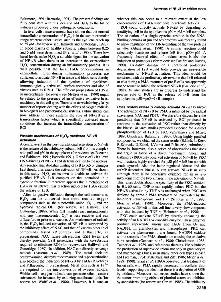
ResultsTreatment of T cells with H202 activates NF-xBJurkat T cells are widely used in the study ofT cell activationprocesses and provide a model system for studying theinduction of HIV-1 gene expression in latently infected cells(Nabel and Baltimore, 1987; Osbom et al., 1989). Thehuman T lymphoma cell line responds.-to treatments withthe active phorbol ester PMA, lectins and TNF with theactivation of NF-xB and NF-xB-controlled genes. Here wehave tested hydrogen peroxide, an agent produced duringinflammatory processes (for review see Halliwell andGutteridge, 1989), for its capability to activate NF-xB.Because H202 can permeate the plasma membrane and canbe converted intracellularly into more reactive oxygenintermediates, it allows investigation of the effects of H202and of oxygen radicals in living cells.
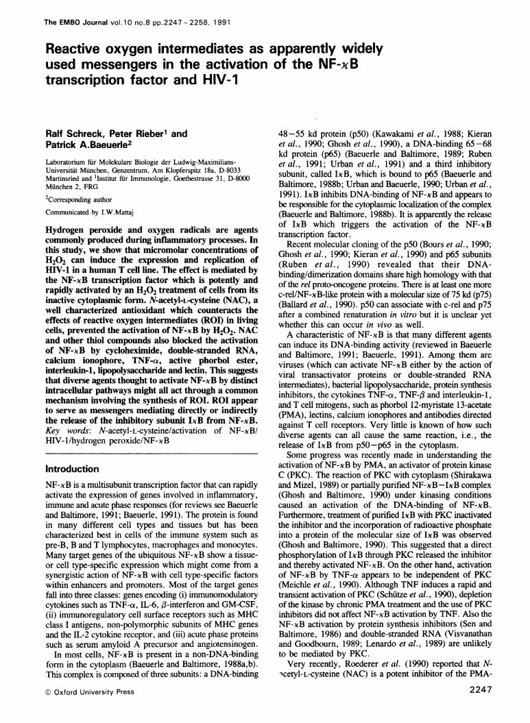
Jurkat T cells were incubated in the presence of 150 AMH202 (Figure lA). After various times, aliquots of the cellculture were harvested and cells fractionated into cytosol andnuclei. Nuclear salt extracts and cytosol were then preparedand analyzed for the specific DNA-binding of NF-xB using2248
Fig. 1. The effect of an H202 treatment on DNA-binding activities inJurkat T cells. (A) Rapid induction of a x enhancer-binding protein bytreatment of T cells with H202. Jurkat T cells were left untreated (Co,lanes 2 and 8) or incubated for various times with 150 AM H202.Nuclear extracts (lanes 2-7) and cytosolic fractions (lanes 8-13)were prepared and equal proportions (2-8 jig of protein) reacted witha 32P-labeled DNA probe encompassing the xB motif of the mouse xlight chain enhancer (Sen and Baltimore, 1986). A protein-DNAcomplex with purified NF-xB composed of p50 and p65 subunits(Baeuerle and Baltimore, 1989) was electrophoresed in lane 1. Sampleswere analyzed on a native 4% polyacrylamide gel. A fluorogram ofthe gel is shown. The filled arrowhead indicates the position of aNF-xB-DNA complex and the open arrowhead the position ofunbound DNA. (B) Dose dependence and the effect of long-termincubation by H202. Jurkat T cells were incubated for 4 and 16 h witheither 30, 50 or 100 AM H202. The radioactivity in the inducedprotein-DNA complex co-migrating with that of NF-xB wasdetermined by Cerenkov counting and the numbers corrected for thesame amount of protein.
electrophoretic mobility shift assays (EMSAs). As shownin Figure IA (lanes 2-7), H202 rapidly activated anactivity that retarded in native gels a 32P-labeled DNA
Activation of NF-xB by oxidant stress
A ;
2 34 r-~ 9
-~~ ~ -
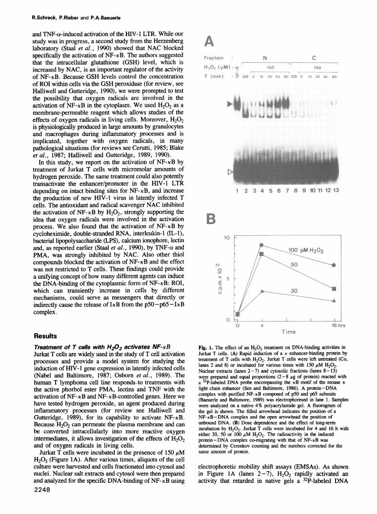
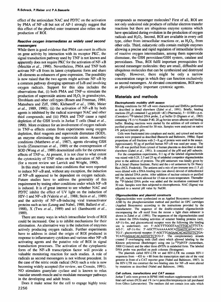
Fig. 2. Characterization of the H202-induced DNA-binding activity.
(A) Binding competition analysis. A nuclear extract from H202-treated(100 AtM; 3 h) Jurkat cells was used. 2.5-, 25- and 250-fold molar
excesses of unlabeled competitor oligonucleotides (described in Zabel
et al., 1991) were mixed with te 32P-labeled x enhancer probe and
the binding reaction started by the addition of the nuclear extract. The
competitor oligonucleotide 'IL-2R' encompassed the NF-xB binding
site from the interleukin 2 receptor ca chain promoter, 'IL-2R mu' is
the same oligonucleotide with two point mutations (see text), and 'unr'
is an unrelated DNA fragment described in Urban and Baeuerle
(1990). A fluorogram of a native gel is shown. The filled arrowhead
indicates the position of the xB-specific DNA-binding activity and
arrows the positions of two non-specific (n.s.) activities. The open
arrowhead shows the position of unbound DNA. (B) Imumunoreactivityof the H202-inducible protein-DNA complex. Purified human NF-xB
(Zabel et al., 1991) (lanes 1, 3, 5 and 7) and a nuclear extract from
Jurkat cells treated with 50 AM H202 for h (lanes 2, 4, 6 and 8)
were reacted with a mix of pre-immune sera (lanes 3 and 4), an
antiserum specific for the p50 subunit of NF-xB (no. 2; Kieran et al.,
1990) (lanes 5 and 6) or an antiserum reacting with the unique C-
terminus of the human c-rel protein (Brownell et al., 1988) (lanes 7
and 8). After inumunoreaction, the DNA probe was added together
with a DNA-binding mix and samples were electrophoresed on a
native gel. A fluorogram of a native gel is shown. Lanes 9-11I show
a slowly migrating binding activity from the serum labeled with an
arrow (S). Lane 12 shows the DNA-binding midx without additions. A
bracket on the left indicates the position of immune-complexed NF-xB.
(C) The effect of cycloheximide (CHX) on the induction of NF-xB by
H202. Jurkat T cells were left untreated (lane 2) or incubated with 10
Ag/nml (lane 3), 50 Atg/ln cyclohexiniide (lane 4) and 50 ItM H202(lane 5). Lanes 6 and 7 show a combined treatment of 50 AM H202with 10 and 50 Ag/ml CHX, respectively. The protein-DNA complex
of purifed NF-xB was electrophoresed in lane 1. A fluorogram of a
native gel is shown.
probe encompassing the decameric NF-xB motif from the
mouse x light chain enhancer. The newly activated
protein-DNA complex co-migrated with that formed by
purified human NF-xB composed of p50 and p65 protein
subunits (Figure IA, lane 1). More conclusive evidence for
the H202-activated factor being NF-xB came from a
binding competition analysis and an immunoreactivity
experiment shown below (see Figure 2).
In cytosolic fractions, no accumulation of theH202-activated protein-DNA complex was seen (FigureIB, lanes 9-13) indicating that the newly activated NF-xBwas rapidly translocated from the cytoplasm into the nucleus.None of the faster migrating factors binding to the xenhancer probe was affected by the H202-treatment of cellsindicating that the activation was specific for NF-xB anddid not reduce the DNA-binding of other proteins (see alsoFigure 3).Also concentrations < 150 14M H202 could efficiently
activate NF-xB (Figure iB). A kinetic analysis showed thata treatment of cells with 30 AM H202 accumulatedsignificant amounts of active NF-xB in nuclei. With 25 AtMH202, no significant activation was seen after 4 h (data notshown). However, we observed some variation betweenexperiments with respect to the minimal concentration ofH202 that induced NF-xB activity. This could be due todifferent extents of decomposition of H202 in the cellculture medium catalyzed by either serum components (Linkand Riley, 1988) or enzymes released from cells. In thepresence of 50 and 100 ,uM H202, more NF-xB wasactivated after 4 h than with 30 AtM. After 16 h of incubation,a slight reduction in the amount of active nuclear NF-xBwas seen (Figure 1B). There was only a small differencebetween the activation potential of 50 and 100 1tM H202,suggesting that a maximal stimulation of NF-xB was reachedbetween 50 and 100 AtM. Prolonged treatment of cells withH202 concentrations > 150 /AM significantly decreased thesurvival of Jurkat T cells. This was observed with other celllines too (Link and Riley, 1988). H202 could also activateNF-xB in various other cell lines tested, among them themouse fibroblast line Ltk- (see Table I) and the pre-B cellline 70Z/3 (data not shown).The possible identity of the H202-activated DNA-binding
protein with NF-xB was further investigated by a bindingcompetition analysis (Figure 2A) and by the use of antiseraspecific for the DNA-binding p50 subunit of NF-xB(Figure 2B). Nuclear extracts from cells treated with 100,uM H202 were reacted with a 32P-labeled x enhancer probein the absence (Figure 2A, lane 1) or presence of increasingamounts of various unlabeled competitor oligonucleotides(lanes 2-10). Competition with a 250-fold molar excess ofan oligonucleotide encompassing the NF-xB binding motif5'-GGGAATCTCC-3' from the IL-2R promoter completelyeliminated the formation of the radioactive protein-DNAcomplex induced by H202 treatment (Figure 2A, lane 4).At a 250-fold molar excess, the competition with increasingamounts of a mutant xB motif from the interleukin 2 receptorgene promoter (5'-GGGAATCTAA-3') showed only a weakeffect on binding (Figure 2A, lane 7). A DNA fragmentwhich does not contain sequences similar to xB motifsshowed no competition within the concentration range tested(lanes 8-10). These results demonstrate the xB-specificDNA-binding of the H202-activated factor. The binding ofthe two minor activities to the radioactive DNA probe wasnot strongly influenced by any of the competitoroligonucleotides demonstrating that their DNA-binding wasnot sequence-specific. These activities must have beenendogenous because they were not detectable in a reactionwithout nuclear extract (Figure 2B, lane 12).Next, we tested whether the H202-activated
protein-DNA complex could react with an antiserum raisedagainst the DNA-binding p50 subunit of NF-xB (Kieranet al., 1990). The serum did not cross-react with the related
2249
R.Schreck, P.Rieber and P.A.Baeuerle
c-rel protein in EMSAs (U.Zabel and P.Baeuerle,unpublished). The presence of anti-p50 serum during theDNA-binding reaction abolished the protein-DNA complexof purified NF-xB (Figure 2B, compare lanes 1 and 5). Also,the co-migrating inducible complex from a nuclear extractof H202-treated Jurkat cells was abolished by the antiserum(Figure 2B, compare lanes 2 and 6). The pre-immune serumhad no effect on the protein-DNA complex of NF-xB(Figure 2B, lanes 3 and 4). c-rel is another protein whichcan recognize xB sequence motifs (Kieran et al., 1990;Ballard et al., 1990) and shares high homology with p50NF-xB within a 300 amino acid long DNA-binding anddimerization domain (Bours et al., 1990; Kieran et al.,1990; Ghosh et al., 1990). An antiserum raised against theunique C-terminus of the human c-rel protein (Brownellet al., 1988) did not react with the purified NF-xB andH202-activated factor (Figure 2B, lanes 7 and 8). All threesera contained a factor which gave rise to a very slowlymigrating protein-DNA complex in EMSAs (Figure 2B,lanes 3-11) and was present in different amounts in the sera(lanes 9-11).A characteristic of NF-xB is its activation by a post-
translational mechanism involving the release of theinhibitory subunit IxB from a latent cytoplasmic form(Baeuerle and Baltimore, 1988a,b; for a review see Baeuerle,1991). In order to investigate whether the activation ofNF-xB by H202 was a post-translational event, weperformed the treatment with H202 in the presence of theprotein synthesis inhibitor cycloheximide (Figure 2C). IfJurkat T cells were treated with 10 or 50 A.g/mlcycloheximide alone, a weak activation of NF-xB was seen(Figure 2C, compare lane 2 with lanes 3 and 4). A treatmentwith 50 AxM H202 for 2 h was chosen to obtain only a sub-optimal activation of NF-xB (Figure 2C, lane 5). In thepresence of 10 jig/ml cycloheximide, the H202 treatmentcould further increase the amount of nuclear NF-xB (Figure2C, lane 6). As determined by Cerenkov counting of theprotein-DNA complexes, the effects of the protein synthesisinhibitor and H202 were additive. In a combined treatmentwith 50 .tg/ml cycloheximide and 50,AM H202, a slightsuperinduction was observed (Figure 2C, lane 7). Theseresults show that the activation of NF-xB by H202 occurredpost-translationally.
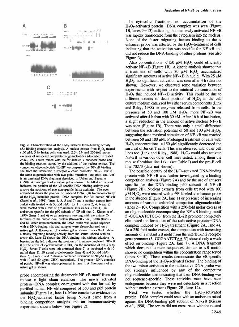
The activation of NF-xB by oxidant stress is specificWe tested whether treatment of cells with H202 influencesthe DNA-binding activity of other inducible and constitutivetranscription factors (Figure 3A). The nuclear extracts fromcontrol and H202-treated cells were incubated with 32p-labeled DNA probes which allow detection of the DNA-binding activities of NF-xB, AP-1/c-fos proteins,glucocorticoid receptor and various octamer-binding proteins(for a review, see Johnson and McKnight, 1989). Thespecificity of protein-DNA complexes was tested bycompetition with the respective unlabeled oligonucleotide.An induction of a DNA-binding activity was only seen withthe xB probe (Figure 3A, first panel). H202 did not induceactivities binding to the AP-1 probe or a glucocorticoidresponse element (Figure 3A, panels 2 and 3). Theconstitutive DNA-binding activities of the ubiquitous oct-Iand faster-migrating lymphoid-specific oct-2 proteins wereunchanged after treatment of cells with H202 (Figure 3A,last panel). In conclusion, the treatment of Jurkat cells with
A
44 4
AL.A
-.r,tx~
B
xB AP-I G :i(-i
"'reatmeni HS Cda rs.HL,'t ^
1 2 3 4 5
Fig. 3. The specificity of the H202 effect. (A) The effect of an H202treatment on the DNA-binding activity of other inducible andconstitutive transcription factors. Nuclear extracts from control (lanes1, 4, 7 and 10) and H202 treated Jurkat cells (100 AM; 3 h) werereacted with 32P-labeled DNA probes detecting NF-xB (xB; lanes1-3), jun/c-fos proteins (AP-1; lanes 4-5), the glucocorticoidreceptor (GR; lanes 7-9) and octamer-binding proteins (oct; lanes10-12). In lanes 3, 6, 9 and 12, a 100-fold molar excess of therespective unlabeled specific oligonucleotide was added as competitor.Samples were analyzed by EMSA. Fluorograms of native gels areshown. Filled arrowheads indicate the positions of presumably specificprotein-DNA complexes. The open arrowhead shows the position ofthe unbound DNA probes. (B) The effects of heat shock and chemicalstress factors on the activity of NF-xB. Jurkat cells were treated for1 h at 42°C (HS; lane 2) or for 4 h with 50 AM cadmium sulfate (Cd;lane 3), 50 AM sodium arsenite (Ars; lane 4) and 50 AM H202 (lane5). Lane 1 shows control cells. Nuclear extracts were analyzed byEMSA using a labeled x enhancer probe. A fluorogram of a nativegel is shown. The filled arrowhead indicates the position of theNF-xB-DNA complex and the open arrowhead the position ofunbound DNA probe.
H202 appears to activate specifically the NF-xBtranscription factor. Various DNA-binding activities detectedwith other DNA probes are either unchanged or show a slightdecrease in activity which might be indicative of someoxidative damage.Besides oxidant stress, also heat shock and chemical
2250
inducers of the cellular stress response can induce theexpression of genes (Ashbumer and Bonner, 1979; Ananthanet al., 1986; Zimarino and Wu, 1987; Ciavarra andSimeone, 1990). We therefore investigated whether exposureof Jurkat cells to heat shock (Figure 3B, lane 2), cadmiumsulfate (lane 3) and sodium arsenite (lane 4) under previouslyreported conditions (Geelen et al., 1988) and for the sameduration as the H202 treatment (1-4 h) can activateNF-xB. None of the treatments caused a rapid appearanceof detectable amounts of NF-xB binding activity in nuclearextracts from Jurkat T cells (Figure 3B). This suggests thatNF-xB is a transcription factor which is specifically activatedif T cells are exposed to oxidant stress.
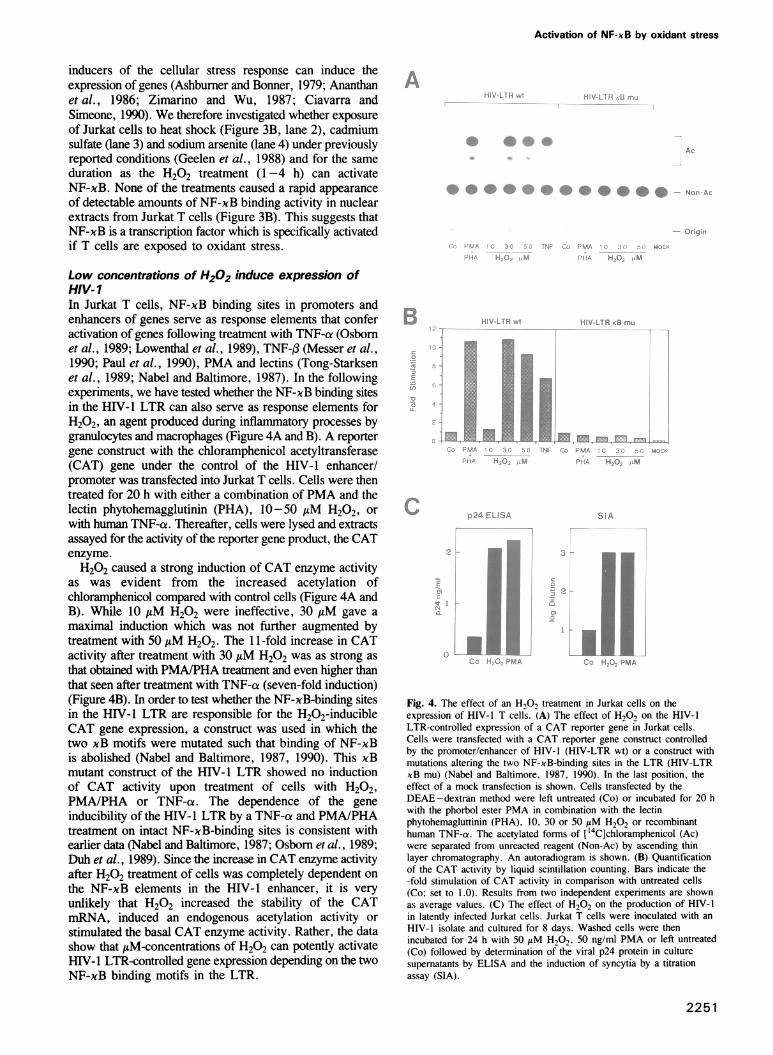
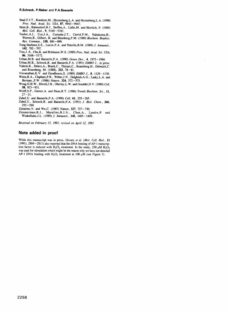
Low concentrations of H202 induce expression ofHIV-1In Jurkat T cells, NF-xB binding sites in promoters andenhancers of genes serve as response elements that conferactivation of genes following treatment with TNF-a (Osbomet al., 1989; Lowenthal et al., 1989), TNF-f (Messer et al.,1990; Paul et al., 1990), PMA and lectins (Tong-Starksenet al., 1989; Nabel and Baltimore, 1987). In the followingexperiments, we have tested whether the NF-xB binding sitesin the HIV-1 LTR can also serve as response elements forH202, an agent produced during inflammatory processes bygranulocytes and macrophages (Figure 4A and B). A reportergene construct with the chloramphenicol acetyltransferase(CAT) gene under the control of the HIV-1 enhancer/promoter was transfected into Jurkat T cells. Cells were thentreated for 20 h with either a combination of PMA and thelectin phytohemagglutinin (PHA), 10-50 gM H202, or
with human TNF-a. Thereafter, cells were lysed and extractsassayed for the activity of the reporter gene product, the CATenzyme.H202 caused a strong induction of CAT enzyme activity
as was evident from the increased acetylation ofchloramphenicol compared with control cells (Figure 4A andB). While 10 AM H202 were ineffective, 30 AM gave a
maximal induction which was not further augmented bytreatment with 50 MLM H202. The 1 1-fold increase in CATactivity after treatment with 30 AM H202 was as strong asthat obtained with PMA/PHA treatment and even higher thanthat seen after treatment with TNF-a (seven-fold induction)(Figure 4B). In order to test whether the NF-xB-binding sitesin the HIV-I LTR are responsible for the H202-inducibleCAT gene expression, a construct was used in which thetwo xB motifs were mutated such that binding of NF-xBis abolished (Nabel and Baltimore, 1987, 1990). This xBmutant construct of the HIV-I LTR showed no inductionof CAT activity upon treatment of cells with H202,PMA/PHA or TNF-a. The dependence of the geneinducibility of the HIV-l LTR by a TNF-ce and PMA/PHAtreatment on intact NF-xB-binding sites is consistent withearlier data (Nabel and Baltimore, 1987; Osbom et al., 1989;Duh et al., 1989). Since the increase in CAT enzyme activityafter H202 treatment of cells was completely dependent on
the NF-xB elements in the HIV-1 enhancer, it is veryunlikely that H202 increased the stability of the CATmRNA, induced an endogenous acetylation activity or
stimulated the basal CAT enzyme activity. Rather, the datashow that MLM-concentrations of H202 can potenfly activateHIV-1 LTR-controlled gene expression depending on the twoNF-xB binding motifs in the LTR.
...:[-,H
l.0_
Fig. 4. The effect of an H,O, treatment in Jurkat cells on theexpression of HIV-1 T cells. (A) The effect of H202 on the HIV-1LTR-controlled expression of a CAT reporter gene in Jurkat cells.Cells were transfected with a CAT reporter gene construct controlledby the promoter/enhancer of HIV-1 (HIV-LTR wt) or a construct withmutations altering the two NF-xB-binding sites in the LTR (HIV-LTRxB mu) (Nabel and Baltimore, 1987, 1990). In the last position, theeffect of a mock transfection is shown. Cells transfected by theDEAE-dextran method were left untreated (Co) or incubated for 20 hwith the phorbol ester PMA in combination with the lectinphytohemagluttinin (PHA), 10, 30 or 50 AM H202 or recombinanthuman TNF-cr. The acetylated forms of [14C]chloramphenicol (Ac)were separated from unreacted reagent (Non-Ac) by ascending thinlayer chromatography. An autoradiogram is shown. (B) Quantificationof the CAT activity by liquid scintillation counting. Bars indicate the-fold stimulation of CAT activity in comparison with untreated cells(Co; set to 1.0). Results from two independent experiments are shownas average values. (C) The effect of H202 on the production of HIV-1in latently infected Jurkat cells. Jurkat T cells were inoculated with an
HIV-1 isolate and cultured for 8 days. Washed cells were thenincubated for 24 h with 50 uM H202, 50 ng/mnl PMA or left untreated(Co) followed by determination of the viral p24 protein in culture
supematants by ELISA and the induction of syncytia by a titrationassay (SIA).
2251
Activation of NF-xB by oxidant stress
u:f,L
*4 .r.w ,.w
R.Schreck, P.Rieber and P.A.Baeuerle
We next tested whether treatment with H202 can activateHIV-1 replication in latently HIV-1 infected Jurkat T cells.Infected cells were treated with either 50 MM H202,50 ng/ml PMA or left untreated. Twenty-four hours later,the production of the p24 protein was determined by ELISAand the formation of syncytia was determined in a titrationassay using C8166 cells. Untreated cells exhibited a basallevel of p24 production and their cell culture supernatantscaused induction of syncytia only at a low dilution (Figure4C). Treatment of cells with 50 M H202 caused a 6.4-foldincrease in the production of the viral p24 protein and thecell culture supernatants from treated cells could induce theformation of syncytia at a 100-fold lower dilution than culturesupernatants from control cells (Figure 4C). Treatment ofcells with PMA showed effects very similar to those ofH202. The activation of the HIV- 1 LTR and viralreplication in T cells by H202 might be of great significancein the onset of HIV-1 production in AIDS patients that sufferfrom secondary infections and inflammatory processes.
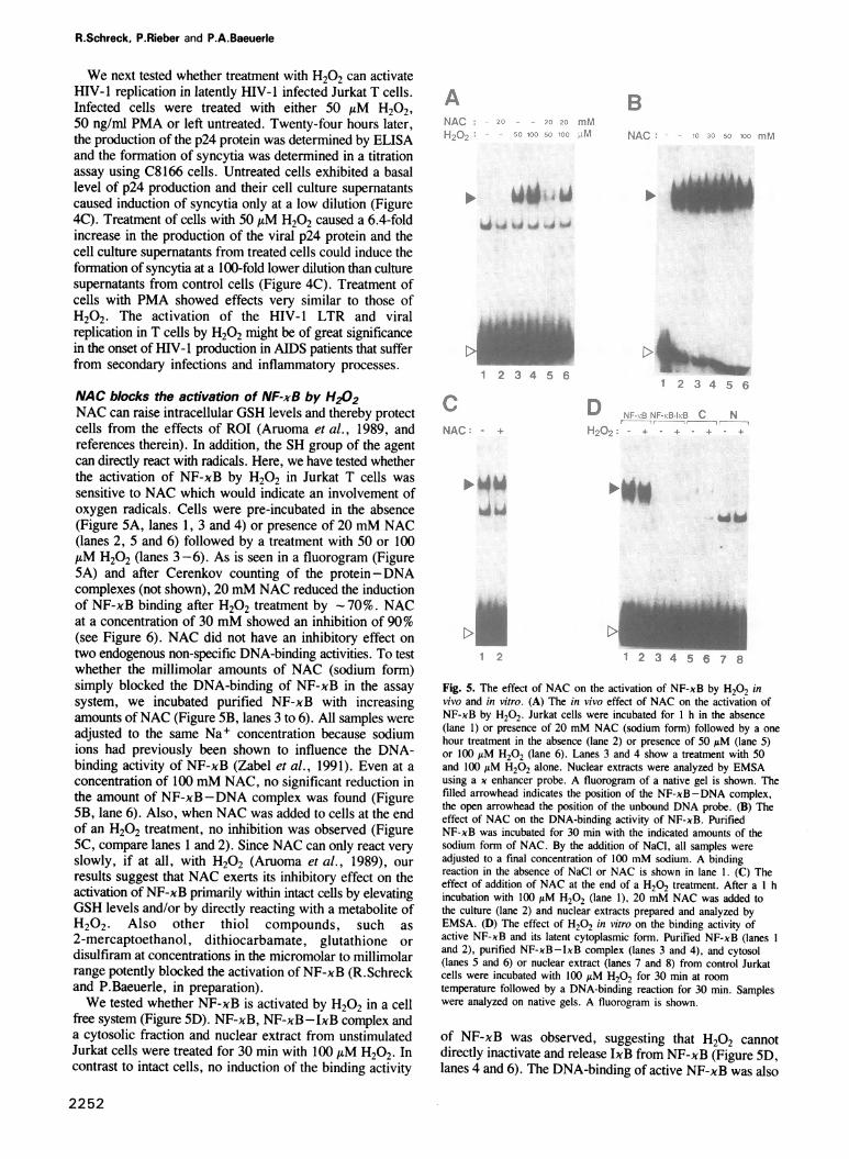
NAC blocks the activation of NF-xB by H202NAC can raise intracellular GSH levels and thereby protectcells from the effects of ROI (Aruoma et al., 1989, andreferences therein). In addition, the SH group of the agentcan directly react with radicals. Here, we have tested whetherthe activation of NF-xB by H202 in Jurkat T cells wassensitive to NAC which would indicate an involvement ofoxygen radicals. Cells were pre-incubated in the absence(Figure 5A, lanes 1, 3 and 4) or presence of 20 mM NAC(lanes 2, 5 and 6) followed by a treatment with 50 or 100AM H202 (lanes 3-6). As is seen in a fluorogram (Figure5A) and after Cerenkov counting of the protein-DNAcomplexes (not shown), 20 mM NAC reduced the inductionof NF-xB binding after H202 treatment by -70%. NACat a concentration of 30 mM showed an inhibition of 90%(see Figure 6). NAC did not have an inhibitory effect ontwo endogenous non-specific DNA-binding activities. To testwhether the millimolar amounts of NAC (sodium form)simply blocked the DNA-binding of NF-xB in the assaysystem, we incubated purified NF-xB with increasingamounts ofNAC (Figure SB, lanes 3 to 6). All samples wereadjusted to the same Na+ concentration because sodiumions had previously been shown to influence the DNA-binding activity of NF-xB (Zabel et al., 1991). Even at aconcentration of 100 mM NAC, no significant reduction inthe amount of NF-xB-DNA complex was found (FigureSB, lane 6). Also, when NAC was added to cells at the endof an H202 treatment, no inhibition was observed (FigureSC, compare lanes 1 and 2). Since NAC can only react veryslowly, if at all, with H202 (Aruoma et al., 1989), ourresults suggest that NAC exerts its inhibitory effect on theactivation of NF-xB primarily within intact cells by elevatingGSH levels and/or by directly reacting with a metabolite ofH202. Also other thiol compounds, such as2-mercaptoethanol, dithiocarbamate, glutathione ordisulfiram at concentrations in the micromolar to millimolarrange potently blocked the activation of NF-xB (R.Schreckand P.Baeuerle, in preparation).We tested whether NF-xB is activated by H202 in a cell
free system (Figure SD). NF-xB, NF-xB-IxB complex anda cytosolic fraction and nuclear extract from unstimulatedJurkat cells were treated for 30 min with 100 /AM H202. Incontrast to intact cells, no induction of the binding activity
r
;.j L
1*N 5 6-
D '4 -. I. Pi F ~.Je EL- N, -.
Si:0
;f 3 4 . 6 7 ki
Fig. 5. The effect of NAC on the activation of NF-xB by H202 invivo and in vitro. (A) The in vivo effect of NAC on the activation ofNF-xB by H202. Jurkat cells were incubated for 1 h in the absence(lane 1) or presence of 20 mM NAC (sodium form) followed by a onehour treatment in the absence (lane 2) or presence of 50 ,uM (lane 5)or 100 AM H202 (lane 6). Lanes 3 and 4 show a treatment with 50and 100 ttM H202 alone. Nuclear extracts were analyzed by EMSAusing a x enhancer probe. A fluorogram of a native gel is shown. Thefilled arrowhead indicates the position of the NF-xB-DNA complex,the open arrowhead the position of the unbound DNA probe. (B) Theeffect of NAC on the DNA-binding activity of NF-xB. PurifiedNF-xB was incubated for 30 min with the indicated amounts of thesodium form of NAC. By the addition of NaCl, all samples wereadjusted to a final concentration of 100 mM sodium. A bindingreaction in the absence of NaCI or NAC is shown in lane 1. (C) Theeffect of addition of NAC at the end of a H202 treatment. After a I hincubation with 100 yM H202 (lane 1), 20 mM NAC was added tothe culture (lane 2) and nuclear extracts prepared and analyzed byEMSA. (D) The effect of H202 in vitro on the binding activity ofactive NF-xB and its latent cytoplasmic form. Purified NF-xB (lanes 1
and 2), purified NF-xB-IxB complex (lanes 3 and 4), and cytosol(lanes 5 and 6) or nuclear extract (lanes 7 and 8) from control Jurkatcells were incubated with 100 AM 1-202 for 30 min at roomtemperature followed by a DNA-binding reaction for 30 min. Sampleswere analyzed on native gels. A fluorogram is shown.
of NF-xB was observed, suggesting that H202 cannotdirectly inactivate and release IxB from NF-xB (Figure SD,lanes 4 and 6). The DNA-binding of active NF-xB was also
2252
B
_.2
A..
Activation of NF-xB by oxidant stress
NAC ._ +, -rJF-.-B Activatig se--_ - I
Agent I - " 1. I
1 2 3 4 5 6 7 8 9 l. 13 14
Fig. 6. The effect of NAC on the activation of NF-xB by fivedifferent agents. Jurkat T cells were treated with PMA (50 ng/ml;lanes 2 and 6), human TNF-os (13 ng/ml; lanes 3 and 7), H,02 (100MM; lanes 4 and 8). poly(rI)-poly(rC) (rI rC, 0.1 mg/ml; lanes 10and 13) or cycloheximide (CHX, 50 Mg/ml; lanes 11 and 14) for 3 hin the absence (lanes 1-4 and 9-1 1) or presence of 30 mM NACadded 1 h prior to the activating treatment (lanes 5-8 and 12 - 14).Nuclear extracts were prepared and analyzed by EMSA using alabeled x enhancer probe. Fluorograms from two native gels areshown. The filled arrowhead indicates the position of theNF-xB-DNA complex and the open arrowhead the position ofunbound DNA probe.
not influenced by H202 (Figure 5D, lane 2). This findinglends further support to the idea that a metabolite of H202is involved in the activation of NF-xB.
In contrast to NF-xB, the activation of the AP-1 factorby PMA appears not to depend on ROI because the presenceof NAC did not effect the induction of AP- 1 DNA-bindingfollowing a PMA treatment of cells (R.Schreck andP.Baeuerle, in preparation). This also suggests that NACdid not interfere with the activity of PKC.
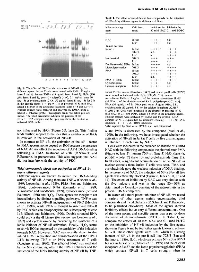
Thiol compounds block the activation of NF-xB bymany different agentsDifferent agents are known to induce the DNA-bindingactivity of NF-xB. Among them are TNF-ca (Osborn et al.,1989; Lowenthal et al., 1989), PMA (Sen and Baltimore,1986), double-stranded RNA (Lenardo et al., 1989;Visvanathan and Goodboum, 1989), cycloheximide (Sen andBaltimore, 1986) and H202. These agents are thought to actintracellularly by distinct signalling pathways. TNF-a wasshown to activate NF-xB independently of PKC (Meichleet al., 1990), while PMA is thought to inactivate NF-xBby PKC which can phosphorylate and subsequently releaseIxB (Ghosh and Baltimore, 1990). Double-stranded RNAcould act via the dl kinase (for review see London et al.,1987) and cycloheximide by preventing the synthesis of alabile inhibitor of NF-xB activation. Finally, H202 seemsto act via ROI as supported by the sensitivity of the inductiontowards NAC. However, NAC was recently shown to alsoinhibit HIV-1 replication and transactivation of the HIV-1LTR following TNF-a or PMA treatment of T cells(Roederer et al., 1990). The effect of NAC was mediatedby the NF-xB binding sites in the HIV-I enhancer and theinduction of the DNA-binding activity of NF-xB by TNF-
Table I. The effect of two different thiol compounds on the activationof NF-xB by different agents in different cell lines.
NF-x-activating Cell lines Inhibition by Inhibition byagent tested 30 mM NAC 0.1 mM PDTC
H,02 Jurkat + + + + n.d.Ltk- ++++ n.d.
Tumor necrosisfactor a Jurkat + + + +a + + + +
70Z/3 n.d. ++++Ltk- n.d. ++++
Interleukin- I 70Z/3 + + + + + +Ltk- +++ n.d.
Double-stranded RNA Jurkat ++ ++ n.d.Lipopolysaccharide 70Z/3 + + + + + + +PMA Jurkat ++++a ++++
70Z/3 +++ ++++Ltk- n.d. ++++
PMA + lectin Jurkat + + + + + + + +Cycloheximide Jurkat + + + n.d.Calcium ionophore Jurkat ++++ n.d.
Jurkat T cells, mouse fibroblasts (Ltk-) and mouse pre-B cells (70Z/3)were treated as indicated with H,02 (100 AM; 2 h), humanrecombinant TNF-a (13 ng/ml; 1-3 h), human recombinant IL-lI(10 U/ml; 1 -2 h), double-stranded RNA [poly(rI)-poly(rC); 4 h],PMA (50 ng/ml; 1-3 h), PMA plus lectin (5 Ag/ml PHA; 2 h),cycloheximide (10 Ag/ml; 3 h) or the calcium ionophore A23187(I AM; 3 h). Cells were incubated in the absence or presence of 30mM NAC or 0.1 mM PDTC added I h prior to the activating agents.Nuclear extracts were analyzed by EMSA and the protein - DNAcomplex of NF-xB quantified by Cerenkov counting. + + +, 50-75 %inhibition; + + + +, 75-100% inhibitionaAlso reported by Staal et al. (1990); n.d., not determined
a and PMA is decreased by the compound (Staal et al.,1990). In the following, we have investigated whether theactivation of NF-xB in Jurkat T cells by five distinct agentsis inhibited in each case by NAC.
Cells were incubated in the presence or absence of 30 mMNAC with the following compounds: the phorbol ester PMA(Figure 6, lane 2), human TNF-a, (lane 3) H202 (lane 4),poly(rI) -poly(rC) (lane 10) and cycloheximide (lane 1 1).In all cases, a significant accumulation of active NF-xB innuclear extracts from Jurkat T cells was seen. PMA andcycloheximide gave the weakest induction of DNA-binding.In the presence of NAC, the induction of NF-xB by all fiveagents was efficiently blocked (Figure 6, lanes 6-8, 13 and14). The extent of inhibition by NAC was very similar withthe five inducers and was in the range 80-90% asdetermined by Cerenkov counting of the radioactivity in theprotein-DNA complexes.
In search of a more potent inhibitor of NF-xB, we testeda variety of other agents mainly encompassing thiolcompounds and metal chelators (R.Schreck and P.Baeuerle,to be published elsewhere). Most of the agents showedinhibitory effects but at very different concentrations. Oneof the most potent and specific agents was a pyrrolidonederivative of dithiocarbamate (PDTC). In Table I, wecompare the effects of 30 mM NAC and 0.1 mM PDTCon the inhibition of NF-xB induction by the five agentsshown in Figure 6 and by four other agents known to activateNF-xB. These other agents were LPS, which is a stronginducer of NF-xB in the pre-B cell line 70Z/3 (Sen andBaltimore, 1986), IL-1, which can activate NF-xB in 70Z/3but not in Jurkat cells (Osborn et al., 1989) and the calciumionophore A23 187 and the lectin phytohemagglutinin (PHA)which activate NF-xB in T cells strongly when in
2253
,LA." iL AL.ii100, 4 .m .i IR --I li.l WIR
*4 44WW 6W .--4 bd 14 W tj &AWWI
R.Schreck, P.Rieber and P.A.Baeuerle
..r, ":
Y,... :. 1:11 F
I
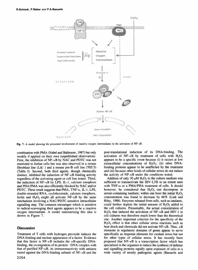
Fig. 7. A model showing the presumed involvement of reactive oxygen intermediates in the activation of NF-xB.
combination with PMA (Nabel and Baltimore, 1987) but onlyweakly if applied on their own (unpublished observation).First, the inhibition of NF-xB by NAC and PDTC was notrestricted to Jurkat cells but was also observed in a mousefibroblast line (Ltk-) and a mouse pre-B cell line (70Z/3)(Table I). Second, both thiol agents, though chemicallydistinct, inhibited the induction of NF-xB binding activityregardless of the activating agent or cell line tested. Third,the induction of NF-xB by LPS, IL-1, calcium ionophoreand PHA/PMA was also efficiently blocked by NAC and/orPDTC. These result suggests that PMA, TNF-at, IL-1, LPS,double-stranded RNA, cycloheximide, calcium ionophore,lectin and H202 might all activate NF-xB by the samemechanism involving a NAC/PDTC-sensitive intracellularsignalling step. The common messenger which is sensitiveto radical-scavenging thiol agents appears to be a reactiveoxygen intermediate. A model summarizing this idea isshown in Figure 7.
DiscussionTreatment of T cells with hydrogen peroxide induces theDNA-binding and nuclear appearance of a factor. Evidencethat this factor is NF-xB includes the xB-specific DNA-binding, the co-migration of its protein-DNA complex withthat of purified NF-xB, its immunoreactivity with antiseraraised against the DNA-binding subunit of NF-xB and the2254
post-translational induction of its DNA-binding. Theactivation of NF-xB by treatment of cells with H202appears to be a specific event because (i) it occurs at lowextracellular concentrations of H202; (ii) other DNA-binding proteins appear to be unaffected by the treatmentand (iii) because other kinds of cellular stress do not inducethe activity of NF-xB under the conditions tested.
Addition of only 30 AM H202 to the culture medium wassufficient to transactivate the HIV-LTR to an extent seenwith TNF-ae or a PMA/PHA treatment of cells. It shouldhowever, be considered that H202 can decompose inserum-containing medium; within one hour the initial H202concentration was found to decrease by 60% (Link andRiley, 1988). Enzymes released from cells, such as catalases,could further deplete the initial amount of H202 added tothe cell cultures. Presumably, the actual concentration ofH202 that induced the activation of NF-xB and HIV-1 incell cultures was therefore much lower than the theoreticalone. Another important criterion for the specificity of theH202 effect is that other cellular stress reactions such asheat shock and chemicals did not activate NF-xB. Thus, xBelements in regulatory domains of genes appear to servespecifically as response elements for oxidant stress but notfor other types of cellular stress. It has recently beenproposed that NF-xB is a transcription factor which hasspecialized in the organism to induce the synthesis of defenseand signalling proteins rapidly upon exposure of cells to awide variety of mostly pathogenic agents (Baeuerle and
ih.4. ., :1
Activation of NF-xB by oxidant stress
Baltimore, 1991; Baeuerle, 1991). The present findings arefully consistent with this idea and add H202 to the list ofinducers produced under pathogenic conditions.
In liver cells, measurements have shown that the normalintracellular concentration of H202 is in the sub-micromolarrange whereas other tissues such as the eye lens reach upto 25 AM (for review see Halliwell and Gutteridge, 1990).In blood plasma of healthy subjects, values between 0.25and 5 j4M were determined (Frei et al., 1988). These lowbasal levels make H202 a suitable signal for the activationof NF-xB when there is an increase in the extracellularH202 concentration during an inflammatory process. It iswell possible that the local H202 concentrations inextracellular fluids during inflammatory processes aresufficient to activate NF-xB in tissue and blood cells therebyallowing induction of the synthesis of cytokines,immunologically active cell surface receptors and also ofviruses such as HIV-1. The efficient propagation of HIV-1in macrophages (for review see Meltzer et al., 1990) mightbe related to the presence of an effective oxidative bt rstmachinery in this cell type. There is an overwhelmingly la ,enumber of reports dealing with the effects of oxygen radicalsin biological and pathobiological systems. Future studies cannow address in these systems the role of NF-xB as atranscription factor which is specifically activated underconditions that increase the intracellular concentration ofROI.
Possible mechansims of H202-mediated NF-xBactivationA central event in the post-translational activation of NF-xBis the release of the inhibitory subunit IxB from its complexwith p65 and p50 in the cytoplasm (for reviews see Baeuerleand Baltimore, 1991; Baeuerle 1991). Release of IxB allowsDNA-binding of NF-xB and its translocation to the nucleus.Any reaction that abolishes binding of IxB to p65 or damagesIxB should thus enable the activation of NF-xB. As shownin this study, H202 on its own is unable to activate thepurified NF-xB-IxB complex or that contained in acytosolic fraction. It therefore appears that a metabolite ofH202 or an intracellular reaction induced by H202 causedthe release of IxB.
After its passive diffusion through the cell membrane,H202 can be converted into more reactive oxygencompounds such as the superoxide anion, 02-, and thehydroxyl radical OH- (for review, see Halliwell andGutteridge, 1989). While OH- might react instantanouslywith any macromolecule, 02- is less reactive and candiffuse further prior to a reaction. An involvement of radicalsin the H202-induced activation of NF-xB is supported bythe inhibitory effect of NAC and that of various other thiolcompounds tested (R.Schreck and P.Baeuerle, inpreparation). NAC raises intracellular GSH levels andthereby provides GSH peroxidase with the co-substraterequired to eliminate ROI (for review, see Halliwell andGutteridge, 1989). In addition, NAC can directly scavengeradicals (Aruoma et al., 1989). The metal chelatorsdesferroxamine, diethyldithiocarbamate and o-phenanthrolinealso blocked the induction of NF-xB by H202 (R.Schreckand P.Baeuerle, in preparation). Metal ions such as Fe2+are required for the interconversion of oxygen radicals.Within cells, oxygen radicals can generate other reactivesubstances, for instance, by oxidizing membrane lipids (forreview see Wolff et al., 1986). However, it is unclear
whether this can occur to a relevant extent at the lowconcentrations of H202 used here to activate NF-xB.ROI could directly activate NF-xB by degrading or
modifying IxB in the cytoplasmic p50-p65 -IxB complex.The oxidation of a single cysteine residue in the DNA-binding domains ofjun and fos proteins was recently foundto allow regulation of the DNA-binding of the two proteinsin vitro (Abate et al., 1990). A similar reaction couldselectively inactivate and release IxB from p50-p65. Afrequently observed effect of oxidant stress is also theinduction of proteolysis (for review see Pacifici and Davies,1990). Oxidative damage or a controlled proteolyticdegradation of IxB would both provide an irreversiblemechansim of NF-xB activation. This idea would beconsistent with the preliminary observation that IxB releasedin vivo during a PMA treatment of pre-B cells can apparentlynot be reused to inhibit the activated NF-xB (Baeuerle et al.,1988). In vitro studies are in progress to understand theprecise role of ROI in the release of IxB from thecytoplasmic pSO-p65-IxB complex.
Does protein kinase C directly activate NF-xB in vivo?The activation of NF-xB by PMA is inhibited by the radicalscavengers NAC and PDTC. We therefore discuss here thepossibility that NF-xB is activated by ROI produced inresponse to the activation of PKC rather than directly bythe kinase. In vitro studies provided evidence for a directphosphorylation of IxB by PKC (Shirakawa and Mizel,1989; Ghosh and Baltimore, 1990). Using highly purifiedcomponents, we obtained the same results (E.Link, L.Kerr,R.Schreck, U.Zabel, I.Vetma and P.Bauerle, submitted).There is, however, also a series of observations that doesnot argue in favor of a direct mechanism. Ghosh andBaltimore (1990) only observed activation of NF-xB by PKCwith fractions highly enriched for p50-p65 -IxB but not withcrude cytosol. Also the heme-regulated kinase and thecAMP-dependent kinase A can activate NF-xB in vitroalthough there is no conclusive evidence for an in vivoinvolvement of the two kinases in the activation of NF-xB.This raises doubts about the specificity of the PKC reaction.In HL-60 cells, TNF-a can rapidly induce PKC but theNF-xB activation by TNF-a is unchanged when PKC wasdepleted by chronic PMA treatment or inactivated by theinhibitors staurosporine and H-7 (Schutze et al., 1990;Meichle et al., 1990). Moreover, the PMA-inducedactivation of NF-xB in this cell line is very slow comparedwith that induced by TNF-ca (Hohmann et al., 1990).PKC could activate NF-xB by directly enhancing the
activity of an NADPH oxidase-like enzyme. These enzymesproduce superoxide anions and H202 from 02 andNADPH. In granulocytes and macrophages, PKC canactivate the plasma-membrane bound NADPH oxidasewithin seconds after PMA stimulation causing the oxidativeburst reaction (Gennaro et al., 1986; Christiansen, 1988;Tauber et al., 1989, and references therein). PMA inducesthe production of superoxide anions not only in granulocytesand macrophages but also in many other cell types (Rosenand Freeman, 1984; Matsubara and Ziff, 1986; Meier et al.,1989, 1990). Staal et al. (1990) observed that treatment ofJurkat cells with PMA rapidly decreased intracellular thiollevels, supporting the idea that there is a depletion of GSHby oxidants. Moreover, numerous studies have shown thatthe tumor-promoting effects of phorbol esters are inhibitedby antioxidants (for review see Cerutti, 1985). The inhibitory
2255
R.Schreck, P.Rieber and P.A.Baeuerle
effect of the antioxidant NAC and PDTC on the activationby PMA of NF-xB but not of AP-l strongly suggest thatthis effect of the phorbol ester treatment also relies on theproduction of ROI.
Reactive oxygen intermediates as widely used secondmessengers
While there is good evidence that PMA can exert its effectson gene activity by interaction with its receptor PKC, thesignal transduction pathway used by TNF is not known andapparently does not require PKC for the activation of NF-xB(Meichle et al., 1990). Nevertheless, PMA and TNF bothactivate NF-xB from its latent cytoplasmic form and sharexB elements as enhancers of gene expression. The possibilityis now raised that the two agents might activate NF-xB bya common pathway diverging upstream of IxB and involvingoxygen radicals. Support for this idea includes theobservations that, (i) both PMA and TNF-a stimulate theproduction of superoxide anions and H202 in granulocytes,fibroblasts and other cell types (Rosen and Freeman, 1984;Matsubara and Ziff, 1986; Klebanoff et al., 1986; Meieret al., 1989, 1990); (ii) the activation of NF-xB by bothagents is inhibited by the radical scavenger NAC and otherthiol compounds; and (iii) PMA and TNF cause a rapiddepletion of the GSH levels in Jurkat T cells (Staal et al.,1990). More evidence for an involvement of oxygen radicalsin TNF-a effects comes from experiments using oxygendepletion, thiol reagents and superoxide dismutase (SOD),an enzyme eliminating the superoxide anion. Anaerobicconditions (Matthews et al., 1987), agents elevating GSHlevels (Zimmerman et al., 1989) or the overexpression ofSOD (Wong et al., 1989) desensitized cells for the cytotoxiceffects of TNF-a. At present, it is not clear to what extentthe cytotoxicity of TNF relies on the activation of NF-xB(for a recent review see Larrick and Wright, 1990).
In this study we tested nine conditions that were reportedto induce NF-xB and, without any exception, the inductionof NF-xB appeared to be dependent on oxygen radicals.Future studies have to address the generality of thisrequirement by testing other conditions under which NF-xBis induced. It is of great interest to see whether NAC andPDTC inhibit the effect of UV light on the induction ofHIV-1 and NF-xB (Valerie et al., 1988; Stein et al., 1989)and the activity of NF-xB-inducing viral transactivatorproteins such as tax (Leung and Nabel, 1988; Ballard et al.,1988), X (Twu et al., 1989) and iel (Sambucetti et al.,1989).There are many ways in which intracellular levels of ROI
could be increased. One is to inhibit mechanisms for theirelimination. An alternative way is the induction of enzymesactively producing oxygen radicals. Further experimentshave to address in detail the origin of ROI produced inresponse to inflammatory cytokines and various other NF-xBactivating agents and the putative role of ROI in signaltransduction processes. The activation of the cytoplasmicform of the NF-xB transcription factor will provide a
valuable monitoring reaction for such studies. A role ofradicals as second messengers is not without precedent. Inthe case of the nitric oxide radical (NO) such a role is nowbecoming widely accepted (for review, see Crossin, 1991).NO stimulates guanylate cyclase and is known to relaxvascular smooth muscle and to modulate messenger pathwaysin the developing and adult brain.Does it make sense for the cell to engage highly toxic
2256
compounds as messenger molecules? First of all, ROI arenot only undesired side products of cellular electron transferreactions but there are ubiquitous enzyme systems whichhave specialized during evolution in the production of oxygenradicals and H202. Second, ROI are available in every celltype, either from intracellular reactions or, as H202, fromother cells. Third, eukaryotic cells contain multiple enzymesallowing a precise and rapid regulation of intracellular levelsof reactive oxygen intermediates, among them superoxidedismutase, the GSH peroxidase/GSH system, catalase andperoxidases. Thus, ROI fulfil important prerequisites forsecond messenger molecules: they are small, diffusible andubiquitous molecules that can be synthesized and destroyedrapidly. However, there might be only a narrowconcentration range in which they can function exclusivelyas second messengers. At elevated concentrations, ROI serveas physiologically important cytotoxic agents.
Materials and methodsElectrophoretic mobility shift assaysBinding conditions for NF-xB were characterized and EMSAs performedas described in detail elsewhere (Zabel et al., 1991). Briefly, bindingreactions (20 11) contained 2 Ag poly(dI-dC) (Pharmacia), 5-10 000 c.p.m.(Cerenkov) 32P-labeled DNA probe, 2 1tl buffer D (Dignam et al., 1983)containing 1% (v/v) Nonidet P-40, 20 1g bovine serum albumin and bindingbuffer. Binding reactions were started by the addition of cell extracts orpurified protein and allowed for 30 min. Samples were analyzed on native4% polyacrylamide gels.
Cells were fractionated into cytoplasm and nuclei, and cytosol and nuclearextracts were prepared as described (Baeuerle and Baltimore, 1988a). Equalproportions of cell fractions with 2-8 ytg of protein were used in the assays.Approximately 50 pg of purified human NF-xB was used per assay. TheNF-xB was purified from cytosol of human placenta as described in detailelsewhere (Zabel et al., 1991). It consisted of the p50 and p65 subunits.
For the competition experiments, 0.1 ng of the labeled oligonucleotidewas mixed with 0.25, 2.5 and 25 ng of unlabeled competitor oligonucleotidesprior to the addition of proteins. The p50 antiserum was kindly given byDr A.Israel (Pasteur Institute, Paris) and the c-rel antiserum by Dr NancyRice (NCI, Frederick). The antisera and a mix of pre-immune sera (1.5 Al)were diluted with a DNA-binding mix (see above) devoid of dithiothreitoland the labeled DNA probe. After addition of nuclear extracts or purifiedNF-xB, reactions were allowed for 15 min at room temperature. Thereafterthe 32P-labeled DNA probe was added and the incubation continued for30 min. Samples were then subjected to electrophoresis. NAC (Sigma) wasadjusted to a neutral pH value by NaOH.
Oligonucleotides and plasmid constructsOligonucleotides were synthesized on an Applied Biosystems synthesizerA380 by the phosphoroamidate method and purified on OPC cartridges(Applied Biosystems) according to the instructions provided by themanufacturer. The sequence of the double-stranded oligonucleotideencompassing the xB motif from the mouse x light chain enhancer isshown in Zabel et al. (1991). The sequences of the oligonucleotides usedto detect the DNA-binding activities of octamer binding proteins (oct),AP-l/c-fos, and glucocorticoid receptor were the following (the bindingsite is underlined). oct: 5'-AGCTTTGGGTAATTTGCATTTCTA-AG-3'; AP-1/c-fos: 5'-AGCTTAAAAAAGCATGAGTCAGACACC-TG-3'; glucocorticoid receptor: 5'-AGCTTGAGAACACAGTGTTCTGA-TCATGAGAACACAGTGTTCTCG-3'. The complementary strandscreated 5'-overhanging ends (SalI sites) which allowed labeling by theKlenow polymerase (Boehringer) using one [U-32P]dNTP (Amersham,3000 Ci/mmol) and the other three dNTPs in unlabeled form. The labeledDNA probe was purified on push columns (Stratagene).The plasmids called HIV-LTR wt and HIV-LTR mu contain HIV-1
sequences from -453 to +80 from the transcription start site of the viralgenome in front of a CAT reporter gene (Nabel and Baltimore, 1987). Inthe HIV-LTR mu construct the two binding sites for NF-xB were alteredby mutations as described (Nabel and Baltimore, 1990).
Cell culture, transfections and CAT assaysJurkat T cells were grown in RPMI 1640 medium supplemented with 10%fetal calf serum (FCS) and 1% (w/v) penicillin/streptomycin (all purchasedfrom Gibco Laboratories). The medium did not contain iron salts which
Activation of NF-xB by oxidant stress
are known to promote the decomposition of H202 into hydroxyl radicals.IL-1,8 was purchased from Genzyme (Boston), LPS, PHA, PDTC, NAC,A23187, cycloheximide and PMA from Sigma and poly(rI) -poly(rC) fromPharmacia.
Transfections were performed by the DEAE -dextran method accordingto Pomerantz et al. (1990). Briefly, 1 x 10⁷ cells were washed with PBSand resuspended in Tris-buffered saline containing DEAE-dextran(Pharmacia) at 2001tg/ml and 15 Ag/ml of plasmid DNA in a total volumeof 1 ml. Incubations were continued for 90 min at 37°C with frequentagitation. After a shock with 10% (v/v) DMSO for 2 min at roomtemperature, cells were washed with PBS and resuspended in 20ml of RPMI1640 medium supplemented with 10% FCS. Twenty-four hours aftertransfection, cells were stimulated for 20 h with recombinant human TNF-cx(30 ng/ml, a kind gift from Hofmann LaRoche, Basel), a combination ofPMA (50 ng/ml) and PHA (5 ALg/ml; both Sigma) or H202 (Merck). Cellextracts were prepared by three freeze -thaw cycles and proteinconcentrations determined by the method of Bradford (Biorad).CAT activity was determined essentially as described (Gorman et al.,
1983) using samples of the same protein content. In a reaction mix of 150 A1containing 20 mM acetyl CoA (Sigma) and 0.3 ItCi [14C]chloramphenicol(Amersham), 100 Ag of protein was incubated for 4 h at 37°C. Reactionproducts were analyzed by thin-layer chromatography followed byautoradiography and liquid scintillation counting. Transfections wereperformed in duplicate. Mock transfections showed a chloramphenicolacetylation of 0.3%.
p24 ELISA and syncytia induction assayJurkat cells (3 x 10⁵ cells/ml) were infected with 10 IE of the HIV-1 isolateM899 (kindly provided by Prof. Dr Gurtler, Pettenkofer Institut, Munich).On day 8 post-infection, cells were washed. On day eleven, cells were treatedwith 50 ng/ml PMA, 50 ltM H202 or left untreated. The next day, cellculture supernatants were harvested. The amount of p24 protein in thesupernatants was determined by ELISA (HIVAG-1 test from Abott GmbH,Wiesbaden-Delkenheim) and the amount of newly produced virus quantifiedby a syncytia induction assay using C1866 cells (Weiss et al., 1986).
AcknowledgmentsWe are indebted to Claudia Winter and Christine Federle for excellenttechnical assistance, Dr Georg Arnold and Inge Leitner for synthesizingoligonucleotides, Cathy Schindewolf for helpful comments on the manuscriptand Prof. Dr E.-L.Winnacker for his continuous support. This work is partof the doctoral thesis of R.S. and was supported by grants from the BMFTand DFG (SFB 217; Ba 957/1-2).
ReferencesAbate,C., Patel,L., Rauscher,F.J. III and Curran,T. (1990) Science, 249,
1157-1161.Ananthan,J., Goldberg,A.L. and Voellmy,G.R. (1986) Science, 232,
522-524.Aruoma,O.I., Halliwell,B., Hoey,B.M. and Butler,J. (1989) Free Radical
Biol. Med., 6, 593-597.Ashburner,M., and Bonner,J.J. (1979) Cell, 17, 241-254.Baeuerle,P.A. (1991) Biochim. Biophys. Acta, in press.Baeuerle,P.A. and Baltimore,D. (1988a) Cell, 53, 211-217.Baeuerle,P.A. and Baltimore,D. (1988b) Science, 242, 540-546.Baeuerle,P.A. and Baltimore,D. (1989) Genes Dev., 3, 1689-1698.Baeuerle,P.A. and Baltimore,D. (1991) In Cohen,P. and Foulkes,J.G. (eds),
Molecular Aspects of Cellular Regulation, Hormonal Control Regulationof Gene Transcription. Elsevier/North Holland Biomedical Press,Amsterdam pp. 409-432.
Baeuerle,P.A. Lenardo,M., Pierce,J.W. and Baltimore,D. (1988) ColdSpring Harbor Symp. Quant. Biol., 53, 789 -798.
Ballard,D.W., Bohnlein,E., Lowenthal,J.W., Wano,Y., Franza,B.R. andGreene,W.C. (1988) Science, 241, 1652-1655.
Ballard,D.W., Walker,W.H., Doerre,S., Sista,P., Molitor,J.A.,Dixon,E.P., Peffer,N.J., Hannink,M. and Greene,W.C. (1990) Cell, 63,803-814.
Blake,D.R., Allen,R.E. and Lunec,J. (1987) Brit. Med. Bull., 43, 371 -385.Bours,V., Villalobos,J., Burd,P.R., Kelly,K. and Siebenlist,U. (1990)
Nature, 348, 76-80.Bradford,M. (1976) Anal. Biochem., 72, 248.Brownell,E., Ruscetti,F.W., Smith,R.G. and Rice,N.R. (1988) Oncogene,
3, 93-98.Cerutti,P.A. (1985) Science, 227, 375-381.
Christiansen,N.O. (1988) FEBS Lett., 239, 195-198.Ciavarra,R.P. and Simeone,A. (1990) Cellular Immunol., 131, 11-26.Crossin,K.L. (1991) Trends Biochem. Sci., 16, 81-82.Dignam,J.P., Lebovitz,R.M. and Roeder,R.G. (1983) Nucleic Acids Res.,
11, 1475-1489.Duh,E.J., Maury,W.J., Folks,T.M., Fauci,A.S. and Rabson,A.B. (1989)
Proc. Natl. Acad. Sci. USA, 86, 5974-5978.Frei,B., Yamamoto,Y., Niclas,D. and Ames,B.N. (1988) Anal. Biochem.,
175, 120-130.Geelen,J.L.M.C., Minnaar,R.P., Boom,R., van der Nordaa,J. and
Goudsmit,J. (1988) J. Gen. Virol., 69, 2913-2917.Gennaro,R., Florio,C. and Romeo,D. (1986) Biochem. Biophys. Res.
Commun., 134, 305 -312.Ghosh,S. and Baltimore,D. (1990) Nature, 344, 678-682.Ghosh,S., Gifford,A.M., Riviere,L.R., Tempst,P., Nolan,G.P. and
Baltimore,D. (1990) Cell, 62, 1019-1029.Gorman,C.M., Merlino,G.T., Willingham,M.C., Pastan,I. and
Howard,B.H. (1982) Proc. Natl. Acad. Sci. USA, 79, 6777-6781.Halliwell,B. and Gutteridge,J.M.C. (1989). Free Radicals in Biology and
Medicine. Second Edition. Clarendon Press, Oxford.Halliwell,B. and Gutteridge,J.M.C. (1990) Methods Enzymol., 186, 1-85.Hohmann,H.-P., Brockhaus,M., Baeuerle,P.A., Remy,R., Kolbeck,R. and
van Loon,A.P.G.M. (1990) J. Biol. Chem., 265, 22409-22417.Johnson,P.F. and McKnight,S.L. (1989) Annu. Rev. Biochem., 58,
799-839.Kawakami,K., Scheidereit,C. and Roeder,R.G. (1988) Proc. Natl. Acad.
Sci. USA, 85, 4700-4704.Kieran,M., Blank,V., Logeat,F., Vandekerckhove,J., Lottspeich,F.,
LeBail,O., Urban, M.B., Kourilsky,P., Baeuerle,P.A. and Israel,A.(1990) Cell, 62, 1007-1018.
Klebanoff,S.J., Vadas,M.A., Harlan,J.M., Sparks,L.H., Gamble,J.R.,Agosti,J.M. and Waltersdorph,A.M. (1986) J. Immunol., 136,4220-4225.
Larrick,J.W. and Wright,S.C. (1990) FASEB J., 4, 3215-3223.Lenardo,M.J., Fan,C.-M., Maniatis,T. and Baltimore,D. (1989) Cell, 57,287-294.
Leung,K. and Nabel,G.J. (1988) Nature, 333, 776-778.Link,E.M. and Riley,P.A. (1988) Biochem. J., 249, 391-199.London,I.M., Levin,D.H., Matts,R.L., Thomas,N.S.B., Pteryshyn,R., and
Chen,J.-J. (1987) In Boyer,P.D. and Krebs,E.G. (eds), The Enzymes.Academic Press, New York. Vol. 18, pp. 359-380.
Lowenthal,J.W., Ballard,D.W., Bohnlein,W. and Greene,W.C. (1989)Proc. Natl. Acad. Sci. USA, 86, 2331-2335.
Matthews,N., Neale,M.L., Jackson,S.K. and Stark,J.M. (1987)Immunology, 62, 153-155.
Matsubara,T. and Ziff,M. (1986) J. Cell. Physiol., 127, 207-210.Meichle,A., Schutze,S., Hensel,G., Brunsing,D. and Kronke,M. (1990)
J. Biol. Chem., 265, 8339-8343.Meier,B., Radeke,H.H., Selle,S., Younes,M., Sies,H., Resch,K. and
Habermehl, G.G. (1989) Biochem. J., 263, 539-545.Meier,B., Radeke,H.H., Selle,S., Habermehl,G.G., Resch,K. and Sies,H.
(1990) Biol. Chem. Hoppe Seyler, 371, 1021-1025.Meltzer,M.S., Skillman,D.R., Hoover,D.L., Hanson,B.D., Turpin,J.A.,
Kalter,D.C. and Gendelman,H.E. (1990) Immunology Today, 11,217-223.
Messer,G., Weiss,E.H. and Baeuerle,P.A. (1990) Cytokine, 2, 389-397.Nabel,G. and Baltimore,D. (1987) Nature, 326, 711-713.Nabel,G.J. and Baltimore,D. (1990) Nature, 344, 178.Osborn,L., Kunkel,S. and Nabel,G.J. (1989) Proc. Natl. Acad. Sci. USA,
86, 2336-2340.Pacifici,R.E. and Davies,K.J.A. (1990) Methods Enzymol., 186, 485-502.Paul,N.L., Lenardo,M.J., Novak,K.D., Sarr,T., Tang,W.-L. and
Ruddle,N.H. (1990) J. Virol., 64, 5412-5419.Pomerantz,R.J., Feinberg,M.B., Trono,D. and Baltimore,D. (1990) J. Exp.
Med., 172, 253-261.Roederer,M., Staal,F.J.T., Raju,P.A., Ela,S.W., Herzenberg,L.A. and
Herzenberg,L.A. (1990) Proc. Natl. Acad. Sci. USA, 87, 4884-4888.Rosen,G.M. and Freeman,B.A. (1984) Proc. Natl. Acad. Sci. USA, 81,
7269-7273.Ruben,S., Dillon,P.J., Schreck,R., Henkel,T., Chen,C.-H., Maher,M.,
Baeuerle,P.A. and Rosen,C. (1991) Science, 251, 1490-1493.Sambucetti,L.C., Cherrington,J.M., Wilkinson,G.W.G. and Mocarski,E.S.
(1989) EMBO J., 8, 4251-4258.Schutze,S., Nottrott,S., Pfizenmaier,K. and Kronke,M. (1990) J. Immunol.,
144, 2604-2608.Sen,R. and Baltimore,D. (1986) Cell, 47, 921-928.Shirakawa,F. and Mizel,S.B. (1989) Mol. Cell. Biol., 9, 2424-2430.
2257
R.Schreck, P.Rieber and P.A.Baeuerle
Staal,F.J.T., Roederer,M., Herzenberg,L.A. and Herzenberg,L.A. (1990)Proc. Natl. Acad. Sci. USA, 87, 9943 -9947.
Stein,B., Rahmsdorf,H.J., Steffen,A., Litfin,M. and Herrlich, P. (1989)Mol. Cell. Biol., 9, 5169-5181.
Tauber,A.I., Cox,J.A., Curnutte,J.T., Carrol,P.M., Nakakuma,H.,Warren,B., Gilbert, H. and Blumberg,P.M. (1989) Biochem. Biophys.Res. Commun., 158, 884-890.
Tong-Starksen,S.E., Luciw,P.A. and Peterlin,B.M. (1989) J. Immunol.,142, 702-707.
Twu,J.-S., Chu,K. and Robinson,W.S. (1989) Proc. Natl. Acad. Sci. USA,86, 5168-5172.
Urban,M.B. and Baeuerle,P.A. (1990) Genes Dev., 4, 1975-1984.Urban,M.B., Schreck,R. and Baeuerle,P.A. (1991) EMBO J., in press.Valerie,K., Delers,A., Bruck,C., Thiriart,C., Rosenberg,H., Debouck,C.
and Rosenberg, M. (1988), 333, 78-81.Visvanathan,K.V. and Goodbourn,S. (1989) EMBO J., 8, 1129-1138.Weiss,R.A., Clapham,P.R., Weber,J.N., Dalgleish,A.G., Lasky,L.A. and
Berman.,P.W. (1986) Nature, 324, 572-575.Wong,G.H.W., Elwell,J.H., Oberley,L.W. and Goeddel,D.V. (1989) Cell,
58, 923-931.Wolff,S.P., Garner,A. and Dean,R.T. (1986) Trends Biochem. Sci., 11,
27-31.Zabel,U. and Baeuerle,P.A. (1990) Cell, 61, 255-265.Zabel,U., Schreck,R. and Baeuerle,P.A. (1991) J. Biol. Chem., 266,252-260.
Zimarino,V. and Wu,C. (1987) Nature, 327, 727-730.Zimmerman,R.J., Marafino,B.J.Jr., Chan,A., Landre,P. and
Winkelhake,J.L. (1989) J. lInmunol., 142, 1405-1409.
Received on February 15, 1991; revised on April 12, 1991
Note added in proofWhile this manuscript was in press, Devary et al. (Mol. Cell. Biol., 11(1991), 2804-2811) also reported that the DNA binding of AP-l transcrip-tion factor is induced with H202 treatment. In the study, 250 AM H202was used for stimulation which might be the reason why we have not detectedAP-1 DNA binding with H202 treatment at 100 /AM (see Figure 3).
2258