Embed Size (px)
Citation preview
Jour
nal o
f Cel
l Sci
ence
COMMENTARY
In vivo cell biology in zebrafish – providing insights into vertebratedevelopment and disease
Ana M. Vacaru1,2, Gokhan Unlu3, Marie Spitzner4, Marina Mione4, Ela W. Knapik3 and Kirsten C. Sadler1,2,5,*
ABSTRACT
Over the past decades, studies using zebrafish have significantly
advanced our understanding of the cellular basis for development
and human diseases. Zebrafish have rapidly developing transparent
embryos that allow comprehensive imaging of embryogenesis
combined with powerful genetic approaches. However, forward
genetic screens in zebrafish have generated unanticipated findings
that are mirrored by human genetic studies: disruption of genes
implicated in basic cellular processes, such as protein secretion or
cytoskeletal dynamics, causes discrete developmental or disease
phenotypes. This is surprising because many processes that were
assumed to be fundamental to the function and survival of all cell
types appear instead to be regulated by cell-specific mechanisms.
Such discoveries are facilitated by experiments in whole animals,
where zebrafish provides an ideal model for visualization and
manipulation of organelles and cellular processes in a live
vertebrate. Here, we review well-characterized mutants and newly
developed tools that underscore this notion. We focus on the
secretory pathway and microtubule-based trafficking as illustrative
examples of how studying cell biology in vivo using zebrafish has
broadened our understanding of the role fundamental cellular
processes play in embryogenesis and disease.
KEY WORDS: Zebrafish, Protein secretion, Vesicular transport,
Microtubule transport
IntroductionClassical cell biologists studied cells in organisms abundantly
available in their environments, such as algae, plants and marineanimals. As the field evolved, cultured cells became de rigueur,and thus in vitro studies have generated much of the modern cell
biology lexicon. The genetic advantages and expanding tools forstudying subcellular structures in the small transparent zebrafishembryo offer the opportunity for cell biologists to return to our
roots and to address fundamental cell biological questions in thecontext of a whole organism.
Forward genetic screens to identify genes underlyingdevelopmental events are a mainstay of zebrafish research.These screens have generated an extensive repertoire of mutants
where a gene implicated in a basic cell biological process has been
disrupted. Surprisingly, many of these mutants have specificphenotypes that involve only a few tissues or cell types.This is reminiscent of human genetic disorders such asmitochondriopathies (Schapira, 2006), ciliopathies (Hildebrandt
et al., 2011) and diseases of protein trafficking (De Matteis andLuini, 2011) where a defect in a protein involved in a fundamentalorganelle function results in a discrete clinical syndrome.
Similarly, researchers were surprised by the finding that manygenes thought to be essential for the function or survival of cells inculture, and that were assumed to be ubiquitously expressed, were
instead revealed to have spatio-temporally restricted expressionpatterns during zebrafish development. These findings led to thehypothesis that fundamental cellular processes are regulated by
cell-type-specific mechanisms.The use of zebrafish and other animals – both invertebrate and
vertebrate – (see Box 1) provides valuable insights into how basiccellular processes are regulated during development and howdisrupting these processes can impact on embryogenesis. This is
elegantly illustrated by a recent investigation into the formationof the notochord, which forms the embryonic axial skeleton (Elliset al., 2013). Notochord cells appear hollow owing to a large
cytoplasmic vacuole found to be a lysosomal-derived organellegenerated by the endosomal trafficking machinery. Rab32a is aGTPase which, in cultured cells, has been found to be involved inmitochondrial dynamics (Alto et al., 2002; Bui et al., 2010),
trafficking to autophagosomes (Hirota and Tanaka, 2009) andother lysosome-related functions (Bultema et al., 2012; Wasmeieret al., 2006), and, in zebrafish, Rab32a has been found to be
essential for vacuole formation and hence for notochorddevelopment. This exemplifies how pairing traditional cellbiological approaches with advances in microscopy, genetics
and pharmacology in zebrafish leads to unprecedentedunderstanding of how basic cellular processes drive specificdevelopmental events.
Here, we review the advantages of using zebrafish to study theintersection between cell biology, vertebrate development and
pathology, and highlight some well-established imaging andgenetic tools. We focus on the protein secretory pathway andmicrotubule-directed vesicular traffic to exemplify how merging
cell biology, genetics and embryology has provided uniqueinsight into how cells work in the context of tissues and organs ina living organism.
The zebrafish toolbox – microscopes, genetics and drugsZebrafish overviewZebrafish produce large numbers of externally fertilized eggs,
which rapidly develop into transparent embryos and progress tofree-swimming feeding larvae within 5 days post fertilization(dpf). Gastrulation is complete within hours of fertilization and by
1 dpf, embryonic axes are established and neural development is
1Department of Developmental and Regenerative Biology, Icahn School ofMedicine at Mount Sinai, 1 Gustave L. Levy Place, Box 1020, New York, NY10029, USA. 2Department of Medicine/Division of Liver Diseases, Icahn School ofMedicine at Mount Sinai, 1 Gustave L. Levy Place, Box 1020, New York, NY10029, USA. 3Division of Genetic Medicine, Department of Medicine, andDepartment of Cell and Developmental Biology, Vanderbilt University MedicalCenter, Nashville, TN 37232, USA. 4Institute of Toxicology and Genetics,Karlsruhe Institute of Technology, 76131 Karlsruhe, Germany. 5Graduate Schoolof Biomedical Sciences, Icahn School of Medicine at Mount Sinai, 1 Gustave L.Levy Place, Box 1020, New York, NY 10029, USA.
*Author for correspondence ([email protected])
� 2014. Published by The Company of Biologists Ltd | Journal of Cell Science (2014) 127, 485–495 doi:10.1242/jcs.140194
485
Jour
nal o
f Cel
l Sci
ence
underway. An exquisite map of cell division and movementduring these early developmental events has been achieved bytracking individually labeled nuclei using advanced microscopy
(Keller et al., 2008; Schmid et al., 2013). By the end of 2 dpf,organogenesis is underway throughout the embryo and, by 5 dpf,most organs carry out specialized functions. Although zebrafish
have traditionally been used to study embryogenesis, areas suchas behavior, pathology, infectious diseases and drug screening areactively investigated. Here, we focus on zebrafish tools usedto study cell biology in vivo, with the aim to understand
development and disease.
Labeling subcellular structures in zebrafishHigh-resolution studies of embryonic development and diseasemodels require analysis of the behaviors of individual cells,subcellular structures and proteins in the context of an intact
tissue or organism. Fluorescent proteins have revolutionized cellbiology. However, the relative small size and dynamic nature ofsubcellular components present challenges for imaging. This isexacerbated by the cellular complexity of whole animals. Thus, in
contrast to the elegant studies describing organelle dynamics incultured cells or yeast, relatively little is known about these
processes in vertebrates.Developing a zebrafish toolkit that labels, tracks and measures
intracellular structures comparable to that available in mammalsis a work in progress. A recent and concerted effort by the
zebrafish community and several companies has improved thelibrary of antibodies recognizing zebrafish proteins, which arecataloged in the zebrafish model organism database (http://zfin.
org). However, specific antibodies for many structures remainunavailable, and antibody staining does not allow live imaging, astrength of the zebrafish system. Developing transgenics
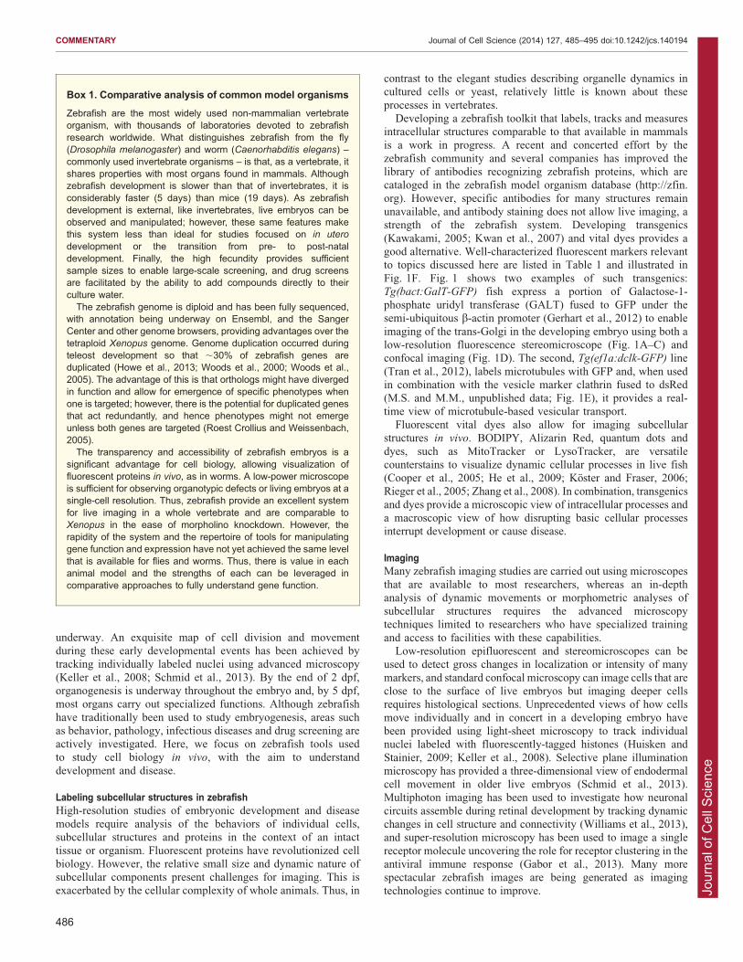
(Kawakami, 2005; Kwan et al., 2007) and vital dyes provides agood alternative. Well-characterized fluorescent markers relevantto topics discussed here are listed in Table 1 and illustrated in
Fig. 1F. Fig. 1 shows two examples of such transgenics:Tg(bact:GalT-GFP) fish express a portion of Galactose-1-phosphate uridyl transferase (GALT) fused to GFP under thesemi-ubiquitous b-actin promoter (Gerhart et al., 2012) to enable
imaging of the trans-Golgi in the developing embryo using both alow-resolution fluorescence stereomicroscope (Fig. 1A–C) andconfocal imaging (Fig. 1D). The second, Tg(ef1a:dclk-GFP) line
(Tran et al., 2012), labels microtubules with GFP and, when usedin combination with the vesicle marker clathrin fused to dsRed(M.S. and M.M., unpublished data; Fig. 1E), it provides a real-
time view of microtubule-based vesicular transport.Fluorescent vital dyes also allow for imaging subcellular
structures in vivo. BODIPY, Alizarin Red, quantum dots and
dyes, such as MitoTracker or LysoTracker, are versatilecounterstains to visualize dynamic cellular processes in live fish(Cooper et al., 2005; He et al., 2009; Koster and Fraser, 2006;Rieger et al., 2005; Zhang et al., 2008). In combination, transgenics
and dyes provide a microscopic view of intracellular processes anda macroscopic view of how disrupting basic cellular processesinterrupt development or cause disease.
ImagingMany zebrafish imaging studies are carried out using microscopes
that are available to most researchers, whereas an in-depthanalysis of dynamic movements or morphometric analyses ofsubcellular structures requires the advanced microscopytechniques limited to researchers who have specialized training
and access to facilities with these capabilities.Low-resolution epifluorescent and stereomicroscopes can be
used to detect gross changes in localization or intensity of many
markers, and standard confocal microscopy can image cells that areclose to the surface of live embryos but imaging deeper cellsrequires histological sections. Unprecedented views of how cells
move individually and in concert in a developing embryo havebeen provided using light-sheet microscopy to track individualnuclei labeled with fluorescently-tagged histones (Huisken and
Stainier, 2009; Keller et al., 2008). Selective plane illuminationmicroscopy has provided a three-dimensional view of endodermalcell movement in older live embryos (Schmid et al., 2013).Multiphoton imaging has been used to investigate how neuronal
circuits assemble during retinal development by tracking dynamicchanges in cell structure and connectivity (Williams et al., 2013),and super-resolution microscopy has been used to image a single
receptor molecule uncovering the role for receptor clustering in theantiviral immune response (Gabor et al., 2013). Many morespectacular zebrafish images are being generated as imaging
technologies continue to improve.
Box 1. Comparative analysis of common model organisms
Zebrafish are the most widely used non-mammalian vertebrateorganism, with thousands of laboratories devoted to zebrafishresearch worldwide. What distinguishes zebrafish from the fly(Drosophila melanogaster) and worm (Caenorhabditis elegans) –commonly used invertebrate organisms – is that, as a vertebrate, itshares properties with most organs found in mammals. Althoughzebrafish development is slower than that of invertebrates, it isconsiderably faster (5 days) than mice (19 days). As zebrafishdevelopment is external, like invertebrates, live embryos can beobserved and manipulated; however, these same features makethis system less than ideal for studies focused on in utero
development or the transition from pre- to post-nataldevelopment. Finally, the high fecundity provides sufficientsample sizes to enable large-scale screening, and drug screensare facilitated by the ability to add compounds directly to theirculture water.The zebrafish genome is diploid and has been fully sequenced,
with annotation being underway on Ensembl, and the SangerCenter and other genome browsers, providing advantages over thetetraploid Xenopus genome. Genome duplication occurred duringteleost development so that ,30% of zebrafish genes areduplicated (Howe et al., 2013; Woods et al., 2000; Woods et al.,2005). The advantage of this is that orthologs might have divergedin function and allow for emergence of specific phenotypes whenone is targeted; however, there is the potential for duplicated genesthat act redundantly, and hence phenotypes might not emergeunless both genes are targeted (Roest Crollius and Weissenbach,2005).The transparency and accessibility of zebrafish embryos is a
significant advantage for cell biology, allowing visualization offluorescent proteins in vivo, as in worms. A low-power microscopeis sufficient for observing organotypic defects or living embryos at asingle-cell resolution. Thus, zebrafish provide an excellent systemfor live imaging in a whole vertebrate and are comparable toXenopus in the ease of morpholino knockdown. However, therapidity of the system and the repertoire of tools for manipulatinggene function and expression have not yet achieved the same levelthat is available for flies and worms. Thus, there is value in eachanimal model and the strengths of each can be leveraged incomparative approaches to fully understand gene function.
COMMENTARY Journal of Cell Science (2014) 127, 485–495 doi:10.1242/jcs.140194
486
Jour
nal o
f Cel
l Sci
ence
GeneticsThe zebrafish genome has been sequenced and annotated, andmost zebrafish genes are highly conserved in mammals, with azebrafish ortholog identified for ,70% of human genes (Howe
et al., 2013). Transgenesis and forward genetic screens areimportant widely used methods in zebrafish, and recent advancesin reverse genetic approaches are having a major impact on thefield. Box 2 details methods of generating transgenics and
targeted mutagenesis using TALENs and the Crispr/Cas systems.Forward genetic screens allow the unbiased discovery of genes
that contribute to a specific phenotype and have generated thousands
of mutants in the zebrafish genome. One drawback, however, is thatgenerating and maintaining stable lines can be laborious and anotheris that the genome duplication that occurred during teleost evolution
(Postlethwait et al., 1998) can generate genes that act redundantly,
necessitating that both are targeted before revealing a phenotype. Inaddition, maternal stores of mRNA and proteins can delay theemergence of mutant phenotypes until after these are exhausted, and
thus mutant phenotypes often reflect the impact of gene loss on laterdevelopmental stages.
Morpholinos provide a complementary approach, wherebyoligonucleotides that are injected into the fertilized egg block
target mRNA translation or splicing. Although the use ofmorpholinos is plagued by fears of off-target effects andtransient effects, translation-blocking morpholinos deplete
proteins derived from both maternal and zygotic mRNA, so thatthe effects of target gene loss on early embryonic events can bestudied. Additionally, by titrating the amount of morpholino
Table 1. List of markers and tools of intracellular and extracellular structures used in vivo in zebrafish
Cellular structure or process Tool type Tool name Application References
Nucleus Vital dye Acridine orange Visualize the nuclei of dying cells Paquet et al., 2009; Peri andNusslein-Volhard, 2008
Transgenic H2B–mRFP Visualize nuclei in vivo Distel et al., 2011Transgenic H2B–EGFP Visualize nuclei in vivo Keller et al., 2008Transgenic NLS–EGFP Visualize the nuclei in vivo Kim et al., 2011Transgenic NLS–mCherry Visualize the nuclei in vivo Lam et al., 2010
Chromosomes Transgenic H2afz–GFP Visualize chromosomes during mitosis andtrack single nuclei during early development
Laguerre et al., 2009
Cytoplasm Vital dye BODIPY TR methylester dye
Cooper et al., 2005
Secretion Transgenic Dbp–EGFP Monitoring glycoprotein secretion fromhepatocytes
Xie et al., 2010
Endoplasmic reticulum stress Transgenic huORFZ huORFchop–GFP [expresses GFP fused tothe upstream open reading frame (uORF)from the 5’UTR of the human CHOP ]*
Lee et al., 2011
Golgi apparatus Transgenic GalT–GFP Trans-Golgi marker Gerhart et al., 2012Endosomes Vital dye F4-64 Endosomal marker Fischer-Parton et al., 2000
Transgenic Rab3–YFP Secretory endosomal marker Campbell et al., 2007Transgenic Rab5c–GFP Early endosomal marker Clark et al., 2011Transgenic Rab5c-Q81L–mCherry Constitutively active form of Rab5 Clark et al., 2011Transgenic Rab5c-S36N–mCherry Dominant-negative form of Rab5 Clark et al., 2011Transgenic Rab7–GFP Late endosomal marker Clark et al., 2011Transgenic Rab7-Q67L–mCherry Constitutively active form of Rab7 Clark et al., 2011Transgenic Rab7-T22N–mCherry Dominant-negative form of Rab7 Clark et al., 2011Transgenic Rab11a–GFP Recycling endosomal marker Clark et al., 2011Transgenic Rab11a-Q70L–mCherry Constitutively active form of Rab11 Clark et al., 2011Transgenic Rab11a-S25N-mCherry Dominant-negative form of Rab11 Clark et al., 2011Transgenic Rab32a–GFP Endosomal marker Ellis et al., 2013
Rab32a-T27N–GFP Dominant-negative form of Rab32 Ellis et al., 2013Transgenic Rab38b-T23N-GFP Dominant-negative form of Rab38 Ellis et al., 2013
Plasma membrane Vital dye Annexin–Cy5 Visualize early membrane of apoptotic cells Peri and Nusslein-Volhard, 2008Transgenic GFP–CaaX
(mem–GFP)Plasma membrane marker Ellis et al., 2013
Transgenic Mem–CFP Plasma membrane marker Distel et al., 2011Microtubules and motor
proteinsTransgenic Dclk–GFP Microtubule marker Tran et al., 2012
Transgenic Tau–GFP Marker for plus-end of microtubules in axons Yanicostas et al., 2009; Yoshidaet al., 2002
Transgenic a-tubulin–GFP Microtubule marker Asakawa and Kawakami, 2010Transgenic Kif17–GFP Marker for plus-end directed vesicle trafficking Bader et al., 2012; Insinna
et al., 2009Transgenic EB1–GFP Labels microtubules plus-end to visualize
growth directionTran et al., 2012
Transgenic EB3–GFP Labels microtubules plus-end to visualizegrowth direction
Tran et al., 2012
Lysosomes Transgenic Lamp2–GFP Lysosomal marker Ellis et al., 2013
*This regulates GFP translation to reflect endogenous Chop levels and monitor the ER stress response. GFP is found in neural tissues during ER and thermalstress.
COMMENTARY Journal of Cell Science (2014) 127, 485–495 doi:10.1242/jcs.140194
487
Jour
nal o
f Cel
l Sci
ence
injected, the degree of knockdown can be finely tuned. This hasproved particularly useful for our work to model one type of
congenital disorder of glycosylation (CDG), which, in humans iscaused by a hypomorphic mutation in one of the genes requiredfor N-linked protein glycosylation (Freeze, 2007). Homozygous
null mutations of these genes are lethal in mammals (Thiel andKorner, 2011) and injecting high morpholino concentrations causessevere embryonic phenotypes and high mortality in zebrafish (Chuet al., 2013; Cline et al., 2012). However, fine-tuning of the
knockdown was facilitated by injecting lower morpholinoconcentrations so that residual enzyme activity in the zebrafishmorphants matched that measured in samples from patients,
improving survival and revealing novel phenotypes (Chu et al.,2013; Cline et al., 2012). Although the breadth and speed ofzebrafish genetic approaches do not match those available in
invertebrates, they are accomplished at a fraction of the costs of, andwith sample sizes exceeding, typical rodent experiments (see Box 1).
DrugsZebrafish have a rich history in toxicology research, as
compounds can be simply added to their water. For example,zebrafish are proving useful for alcohol research (Jang et al.,2012; Monk et al., 2013; North et al., 2010; Passeri et al., 2009;
Tsedensodnom et al., 2013; Yin et al., 2012). By using transgeniczebrafish expressing a GFP-tagged secreted glycoprotein inhepatocytes (Howarth et al., 2013; Tsedensodnom et al., 2013;Xie et al., 2010), and zebrafish that express fluorescent protein
markers of the hepatocyte secretory organelles and of other cellsin the liver (Yin et al., 2012), the mechanisms by which alcoholand other drugs cause organ-specific and organelle-mediated
toxicity can be uniquely addressed. Moreover, large-scale drugscreens exploit the ease of treating zebrafish with drugs (Petersonand Macrae, 2012) and have identified compounds modulating
processes ranging from metabolism (Nath et al., 2013) to sleep(Rihel and Schier, 2012).
200 µm
200 µm
2 dpfTg(bact:GalT-GFP)
A
B
C
Muscle cells
DAPITg(bact:GalT-GFP)
10 µm
10 µm
4 dpfHepatocytes
clathrin-DS-RedTg(ef1a:dclk-GFP)
1 dpf
D
E
COPII
ER
ERGIC
Golgi
COPI
LE
EE
PM
Lysosome
NLS–cherry h2afz–mRFP
EEEEEEEEERab5–GFP
Microtubules
GFP–α–tubulin dclk–GFP Tau–GFP
Kif17–GFP EB1–GFP EB2–GFP
Rab7–GFP
Rab11–GFP
GFP–CaaX mem–CFP
Lamp2–GFP
NucleusRE
GalT–EGFP
NLS–EGFP
F
Fig. 1. Fluorescent protein markers of subcellularstructures in zebrafish. (A–C) Live 2 dpf Tg(bact:GalT-
GFP) embryo highlighting the trans-Golgi complex. Theboxed region in B is magnified in the C. (D) Confocalprojection through a cryosection of the liver from 5 dpfTg(bact:GalT-GFP) larva. Nuclei are stained with DAPI(gray). (E) Muscle in a live 1 dpf Tg(ef1a:dclk-GFP)
embryo with GFP-tagged microtubule-associated protein(DCK) and vesicles marked with clathrin-DS-Red(magenta). (F) Zebrafish fluorescent-protein-taggedorganelle markers that can be expressed withtransgenics. PM, plasma membrane; LE, late endosomes;EE, early endosomes; RE, recycling endosomes.
COMMENTARY Journal of Cell Science (2014) 127, 485–495 doi:10.1242/jcs.140194
488
Jour
nal o
f Cel
l Sci
ence
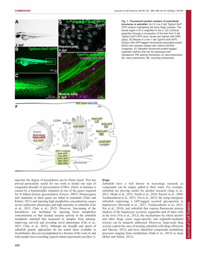
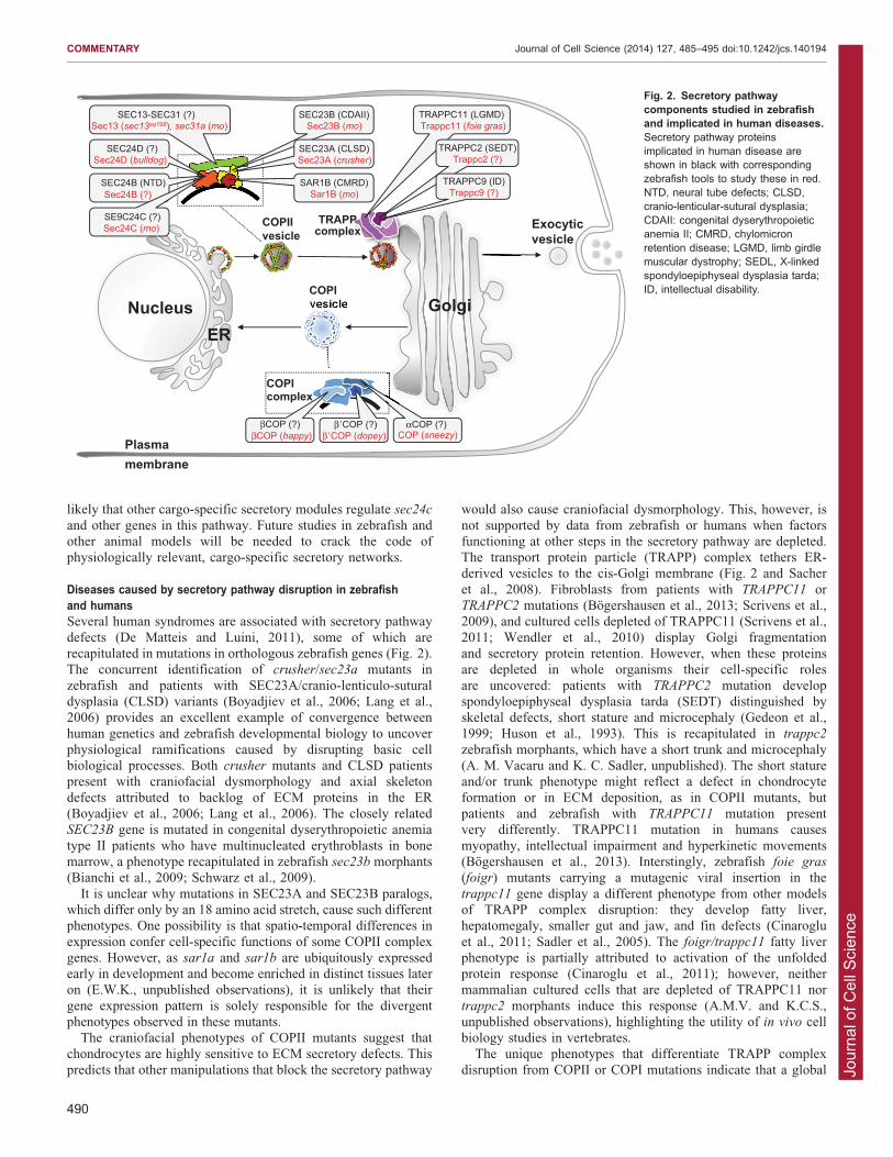
The protein secretory pathwayThe secretory pathway generates, trafficks and processes proteinsdestined for the extracellular space or the plasma membrane. Itcomprises the endoplasmic reticulum (ER), the ER-Golgiintermediate compartment (ERGIC), the Golgi complex and the
vesicles that carry cargo between them (Fig. 2). Protein synthesisand glycosylation occur in the ER and Golgi, respectively. Thecoat protein II (COPII) complex facilitates cargo selection,
vesicle formation and anterograde trafficking from the ER to theGolgi, whereas retrograde transport occurs in COPI vesicles(Fig. 1F; Fig. 2). Diverse protein complexes function at each step
of this pathway to recruit Rab GTPases and SNARE proteins,which direct and tether vesicles to target organelles and facilitatemembrane fusion.
Work in unicellular organisms and cultured cells created anassumption that protein secretion is uniformly regulated across celltypes. Studies in human, zebrafish and other vertebrates, however,revealed that this pathway is regulated in a spatio-temporal and
paralog-specific manner (Melville and Knapik, 2011; Unlu et al.,2013). Although all cells secrete proteins, some – including B-
cells, chondrocytes, hepatocytes and the endocrine and exocrinepancreatic cells – are considered ‘professional’ secretory cellsand these cells were assumed to be particularly sensitive tosecretory pathway disruption. Zebrafish mutants in secretory
pathway genes both supported and refuted this hypothesis: somemutants show phenotypes in most highly secretory cells,whereas others have phenotypes that are restricted to only a
subset of cells.The emerging view is that the secretory machinery is integral to
morphogenesis and organ function in a cell-specific fashion. The
availability of zebrafish genetic mutants and fluorescent in vivo
reporters provides novel insight into organismal functions of thesecretory pathway. The effects of secretory pathway disruption
relevant to development and disease are discussed below.
Developmental consequences of secretory pathway disruptionThe COPII complex comprises the Sar1 GTPase, Sec23–Sec24
dimers of the inner coat, and Sec13–Sec31 heterotetramers of theouter coat (Fig. 2; Kaiser and Schekman, 1990; Novick et al.,1980). Zebrafish mutants of sec23a (crusher) and sec24d
(bulldog) develop craniofacial dysmorphology, kinked pectoralfins and short body length (Lang et al., 2006; Sarmah et al.,2010). These are attributed to a failure of extracellular matrix
(ECM) secretion during chondrocyte differentiation. Sec23A- andSec24D-deficient animals fail to export collagen and other N-glycosylated proteins from the chondrocyte ER, arresting
differentiation and ultimately causing cell death (Lang et al.,2006; Sarmah et al., 2010; Unlu et al., 2013), whereas collagensecretion and skeletal development are intact upon depletion ofthe close paralog Sec24C (Sarmah et al., 2010). Sec23B
mutations in humans and zebrafish disrupt erythropoesis(Bianchi et al., 2009; Schwarz et al., 2009), a differentphenotype from the chondrocyte defects observed in crusher
and bulldog mutants. These COPII phenotypes are distinct fromthe dwarf mutants sneezy, happy and dopey, which disrupt genesthat encode the a, b and b9 subunits of the COPI complex,
respectively (Fig. 2), which are characterized by defects innotochord and melanosome formation (Coutinho et al., 2004).These data suggest that although COPI and COPII are requiredfor efficient secretion and membrane recycling in all cells, loss of
specific members of each complex have profound and disparateeffects on a subset of cells. Mutations in individual COPIIcomponents cause an array of phenotypes in highly secretory cell
types in organs such as cartilage, notochord, eye and gut (Niuet al., 2012; Schmidt et al., 2013; Townley et al., 2008; Townleyet al., 2012), and in erythrocytes (Bianchi et al., 2009; Schwarz
et al., 2009; Unlu et al., 2013), whereas cells that depend onvacuole formation are most sensitive to COPI depletion.
So how do transcriptional regulatory mechanisms direct the
secretory pathway to assure a timely availability of cargo-specificcoats? A large-scale screen in zebrafish identified the feelgood
mutant, which carries a missense variant in the creb3L2 gene(Driever et al., 1996; Knapik, 2000; Neuhauss et al., 1996) – the
first known transcription factor that regulates availability of theCOPII components sec24d and sec23a, but not sec24c (Melvilleet al., 2011). Similarities between feelgood, crusher and bulldog
mutant phenotypes suggest that a ‘secretory module’ consisting ofCreb3L2–Sec23A–Sec24D specializes in procollagen secretion.Given that zebrafish depleted of Sec24C do not manifest skeletal
dysmorphology and the gene is not a target of Creb3L2, it is
Box 2. Genetic approaches – a summary
Transgenics are a common means to overexpress genes inzebrafish and, now, both transient knockdown by morpholinosand gene targeting are routine methods to induce gene loss.Random targeting of a transgene and promoter in the genome isfacilitated using the tol2 transposon (Suster et al., 2009). Cell-specific promoters are available for many cell types to directtransgene expression and bacterial artificial chromosomerecombineering can recapitulate endogenous gene expressionpatterns and levels; the heat shock promoter (hsp70) is also awidely used inducible system (Halloran et al., 2000). Multiple linesof fish expressing the same transgene in different tissues can bequickly generated by crossing fish in which the transgene is underthe UAS promoter, to lines that express the Gal4 transcriptionalactivator under a tissue-specific promoter (Distel et al., 2009;Halpern et al., 2008).Chemical and gene-breaking transposons (http://zfishbook.org/)
have generated a large library of non-targeted mutants.Homologous recombination to generate targeted gene mutationshas eluded the zebrafish community for years. However, use ofTALENs (transcription activator-like effector nucleases) (Bedell etal., 2012; Huang et al., 2011; Sander et al., 2011) and the type IIprokaryotic CRISPR/Cas (clustered regularly interspaced shortpalindromic repeats/CRISPR-associated) system (Hwang et al.,2013; Jao et al., 2013; Xiao et al., 2013) has recently beensuccessful in specific gene targeting to generate germ-linemutants. These systems use chimeric nucleases composed ofprogrammable sequence-specific DNA-binding modules linked tononspecific DNA nucleases to induce double-stranded DNA breaksthat are inefficiently repaired by error-prone non-homologous end-joining or homology-directed repair (Gaj et al., 2013). TALEN pairsthat have been engineered by a zebrafish consortium arecommercially available from http://www.addgene.com for manygenes. Resources for TALEN design include https://groups.google.com/group/talengineering, http://www.addgene.org/talengineering/TALENzebrafish/ and http://www.TALengineering.org TheCRISPR/Cas system introduces single nucleotide substitutions,insertions and deletions ranging from 1 to 20 bp. The relative easeof synthesis makes the CRISPR/Cas systems an attractiveapproach for genome editing (see Hwang et al., 2013; http://www.addgene.org; http://www.crispr-cas.org). It is anticipated thatwithin the near future, nearly all protein-coding genes in zebrafishwill have been disrupted by either forward genetic screens or oneof these reverse genetic approaches.
COMMENTARY Journal of Cell Science (2014) 127, 485–495 doi:10.1242/jcs.140194
489
Jour
nal o
f Cel
l Sci
ence
likely that other cargo-specific secretory modules regulate sec24c
and other genes in this pathway. Future studies in zebrafish andother animal models will be needed to crack the code ofphysiologically relevant, cargo-specific secretory networks.
Diseases caused by secretory pathway disruption in zebrafishand humansSeveral human syndromes are associated with secretory pathway
defects (De Matteis and Luini, 2011), some of which arerecapitulated in mutations in orthologous zebrafish genes (Fig. 2).The concurrent identification of crusher/sec23a mutants in
zebrafish and patients with SEC23A/cranio-lenticulo-suturaldysplasia (CLSD) variants (Boyadjiev et al., 2006; Lang et al.,2006) provides an excellent example of convergence between
human genetics and zebrafish developmental biology to uncoverphysiological ramifications caused by disrupting basic cellbiological processes. Both crusher mutants and CLSD patientspresent with craniofacial dysmorphology and axial skeleton
defects attributed to backlog of ECM proteins in the ER(Boyadjiev et al., 2006; Lang et al., 2006). The closely relatedSEC23B gene is mutated in congenital dyserythropoietic anemia
type II patients who have multinucleated erythroblasts in bonemarrow, a phenotype recapitulated in zebrafish sec23b morphants(Bianchi et al., 2009; Schwarz et al., 2009).
It is unclear why mutations in SEC23A and SEC23B paralogs,which differ only by an 18 amino acid stretch, cause such differentphenotypes. One possibility is that spatio-temporal differences in
expression confer cell-specific functions of some COPII complexgenes. However, as sar1a and sar1b are ubiquitously expressedearly in development and become enriched in distinct tissues lateron (E.W.K., unpublished observations), it is unlikely that their
gene expression pattern is solely responsible for the divergentphenotypes observed in these mutants.
The craniofacial phenotypes of COPII mutants suggest that
chondrocytes are highly sensitive to ECM secretory defects. Thispredicts that other manipulations that block the secretory pathway
would also cause craniofacial dysmorphology. This, however, is
not supported by data from zebrafish or humans when factorsfunctioning at other steps in the secretory pathway are depleted.The transport protein particle (TRAPP) complex tethers ER-derived vesicles to the cis-Golgi membrane (Fig. 2 and Sacher
et al., 2008). Fibroblasts from patients with TRAPPC11 orTRAPPC2 mutations (Bogershausen et al., 2013; Scrivens et al.,2009), and cultured cells depleted of TRAPPC11 (Scrivens et al.,
2011; Wendler et al., 2010) display Golgi fragmentationand secretory protein retention. However, when these proteinsare depleted in whole organisms their cell-specific roles
are uncovered: patients with TRAPPC2 mutation developspondyloepiphyseal dysplasia tarda (SEDT) distinguished byskeletal defects, short stature and microcephaly (Gedeon et al.,
1999; Huson et al., 1993). This is recapitulated in trappc2
zebrafish morphants, which have a short trunk and microcephaly(A. M. Vacaru and K. C. Sadler, unpublished). The short statureand/or trunk phenotype might reflect a defect in chondrocyte
formation or in ECM deposition, as in COPII mutants, butpatients and zebrafish with TRAPPC11 mutation presentvery differently. TRAPPC11 mutation in humans causes
myopathy, intellectual impairment and hyperkinetic movements(Bogershausen et al., 2013). Interstingly, zebrafish foie gras
(foigr) mutants carrying a mutagenic viral insertion in the
trappc11 gene display a different phenotype from other modelsof TRAPP complex disruption: they develop fatty liver,hepatomegaly, smaller gut and jaw, and fin defects (Cinaroglu
et al., 2011; Sadler et al., 2005). The foigr/trappc11 fatty liverphenotype is partially attributed to activation of the unfoldedprotein response (Cinaroglu et al., 2011); however, neithermammalian cultured cells that are depleted of TRAPPC11 nor
trappc2 morphants induce this response (A.M.V. and K.C.S.,unpublished observations), highlighting the utility of in vivo cellbiology studies in vertebrates.
The unique phenotypes that differentiate TRAPP complexdisruption from COPII or COPI mutations indicate that a global
COPI
SEC24D (?)Sec24D (bulldog)
SEC13-SEC31 (?)Sec13 (sec13sq198), sec31a (mo)
SAR1B (CMRD)Sar1B (mo)
Exocyticvesicle
SEC23B (CDAII)Sec23B (mo)
NucleussER
COPIIvesicle
αCOP (?)COP (sneezy)
β�COP (?)βCOP (?)βCOP (happy)
TRAPPC11 (LGMD)Trappc11 (foie gras)
Golgi
TRAPPC2 (SEDT)Trappc2 (?)
TRAPPC9 (ID)Trappc9 (?)
SEC24B (NTD)Sec24B (?)
SEC23A (CLSD)Sec23A (crusher)
SE9C24C (?)Sec24C (mo)
TRAPP complex
COPI complex
Plasmamembrane
β�COP (dopey)
Fig. 2. Secretory pathwaycomponents studied in zebrafishand implicated in human diseases.Secretory pathway proteinsimplicated in human disease areshown in black with correspondingzebrafish tools to study these in red.NTD, neural tube defects; CLSD,cranio-lenticular-sutural dysplasia;CDAII: congenital dyserythropoieticanemia II; CMRD, chylomicronretention disease; LGMD, limb girdlemuscular dystrophy; SEDL, X-linkedspondyloepiphyseal dysplasia tarda;ID, intellectual disability.
COMMENTARY Journal of Cell Science (2014) 127, 485–495 doi:10.1242/jcs.140194
490
Jour
nal o
f Cel
l Sci
ence
block in protein secretion is not the only mechanism thatunderlies their associated phenotypes. Moreover, although
depleting individual TRAPP or COP complex factors hassimilar effects in isolated cells, the physiological consequencescould not be predicted without use of whole animals. Thesefindings point to cell- and developmental-specific roles for each
gene involved in protein secretion and underscore the need forcomparative whole animal models to decipher the cellular andphysiological functions of this pathway.
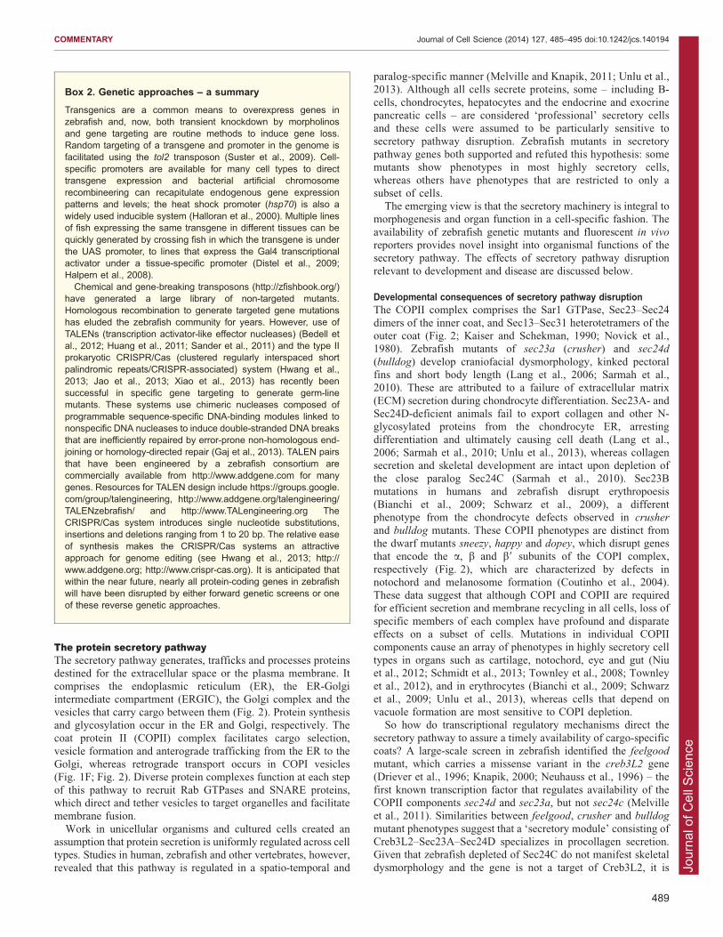
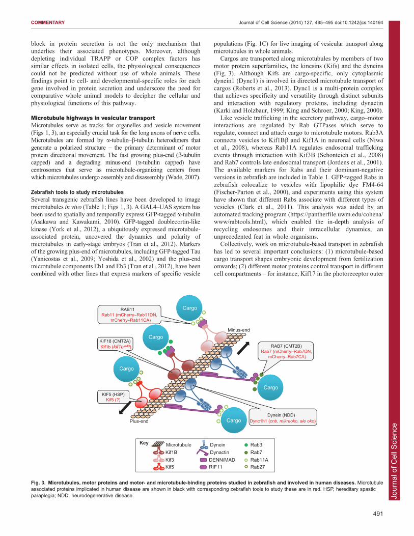
Microtubule highways in vesicular transportMicrotubules serve as tracks for organelles and vesicle movement
(Figs 1, 3), an especially crucial task for the long axons of nerve cells.Microtubules are formed by a-tubulin–b-tubulin heterodimers thatgenerate a polarized structure – the primary determinant of motor
protein directional movement. The fast growing plus-end (b-tubulincapped) and a degrading minus-end (a-tubulin capped) havecentrosomes that serve as microtubule-organizing centers fromwhich microtubules undergo assembly and disassembly (Wade, 2007).
Zebrafish tools to study microtubulesSeveral transgenic zebrafish lines have been developed to image
microtubules in vivo (Table 1; Figs 1, 3). A GAL4–UAS system hasbeen used to spatially and temporally express GFP-tagged a-tubulin(Asakawa and Kawakami, 2010). GFP-tagged doublecortin-like
kinase (York et al., 2012), a ubiquitously expressed microtubule-associated protein, uncovered the dynamics and polarity ofmicrotubules in early-stage embryos (Tran et al., 2012). Markers
of the growing plus-end of microtubules, including GFP-tagged Tau(Yanicostas et al., 2009; Yoshida et al., 2002) and the plus-endmicrotubule components Eb1 and Eb3 (Tran et al., 2012), have beencombined with other lines that express markers of specific vesicle
populations (Fig. 1C) for live imaging of vesicular transport alongmicrotubules in whole animals.
Cargos are transported along microtubules by members of twomotor protein superfamilies, the kinesins (Kifs) and the dyneins(Fig. 3). Although Kifs are cargo-specific, only cytoplasmicdynein1 (Dync1) is involved in directed microtubule transport of
cargos (Roberts et al., 2013). Dync1 is a multi-protein complexthat achieves specificity and versatility through distinct subunitsand interaction with regulatory proteins, including dynactin
(Karki and Holzbaur, 1999; King and Schroer, 2000; King, 2000).Like vesicle trafficking in the secretory pathway, cargo–motor
interactions are regulated by Rab GTPases which serve to
regulate, connect and attach cargo to microtubule motors. Rab3Aconnects vesicles to Kif1Bb and Kif1A in neuronal cells (Niwaet al., 2008), whereas Rab11A regulates endosomal trafficking
events through interaction with Kif3B (Schonteich et al., 2008)and Rab7 controls late endosomal transport (Jordens et al., 2001).The available markers for Rabs and their dominant-negativeversions in zebrafish are included in Table 1. GFP-tagged Rabs in
zebrafish colocalize to vesicles with lipophilic dye FM4-64(Fischer-Parton et al., 2000), and experiments using this systemhave shown that different Rabs associate with different types of
vesicles (Clark et al., 2011). This analysis was aided by anautomated tracking program (https://pantherfile.uwm.edu/cohena/www/rabtools.html), which enabled the in-depth analysis of
recycling endosomes and their intracellular dynamics, anunprecedented feat in whole organisms.
Collectively, work on microtubule-based transport in zebrafish
has led to several important conclusions: (1) microtubule-basedcargo transport shapes embryonic development from fertilizationonwards; (2) different motor proteins control transport in differentcell compartments – for instance, Kif17 in the photoreceptor outer
Minus-end
Plus-end
RAB7 (CMT2B)Rab7 (mCherry–Rab7DN,
mCherry–Rab7CA)
KIF18 (CMT2A)Kif1b (kif1bst43)
Dynein (NDD) Dync1h1 (cnb, mikreoko, ale oko)
Cargo
KIF5 (HSP)Kif5 (?)
Cargo
CargoRAB11Rab11 (mCherry–Rab11DN,
mCherry–Rab11CA)
Cargo
Cargo
Microtubule Dynein Rab3 Kif1B Dynactin Rab7 Kif3 DENN/MAD Rab11A Kif5 RIF11 Rab27
Key
Fig. 3. Microtubules, motor proteins and motor- and microtubule-binding proteins studied in zebrafish and involved in human diseases. Microtubuleassociated proteins implicated in human disease are shown in black with corresponding zebrafish tools to study these are in red. HSP, hereditary spasticparaplegia; NDD, neurodegenerative disease.
COMMENTARY Journal of Cell Science (2014) 127, 485–495 doi:10.1242/jcs.140194
491
Jour
nal o
f Cel
l Sci
ence
segment and Kif3 in the cytoplasm; and (3) zebrafish Rabhomologs recapitulate the behavior of their counterparts in
mammalian cells. Thus, the expanding set of tools in zebrafish isenabling organism-wide functional analysis of proteins involvedin directed vesicular transport.
Off-track – microtubule-based diseases in humans and zebrafishAlthough most cells rely on microtubules for transporting theircargo, disruption of this process profoundly affects the central
and peripheral nervous system due to the long-distance thatcargos travel from cell bodies to axonal tips. Alzheimer,Huntington disease (HD) and Charcot–Marie–Tooth disease
(CMT) (De Vos et al., 2008) are some neurodegenerativediseases (NDDs) caused by defects in vesicular transport. Fig. 3illustrates the orthologous genes underlying NDDs in humans and
mutant phenotypes in zebrafish.CMT type 2A is an axonal sensorimotor neuropathy
characterized by severe peripheral muscle weakness andatrophy induced by loss of function mutations in the kinesin-3
family member, Kif1B (Zhao et al., 2001). In zebrafish, the pointmutation kif1bst43 affects the microtubule interaction site and hasbeen used to study kif1b functions in the developing central
nervous system. The use of chimeric embryos, generated bytransplanting kif1b-deficient neuronal cells into wild-type hosts(and vice versa), has revealed that there is a reduced growth
potential in mutant neuronal axons (Lyons et al., 2009). Thus,kif1b is required in a cell autonomous manner for axonaldevelopment in the central and peripheral nervous system.
Shprintzen–Goldberg syndrome is characterized by central andenteric nervous system defects and caused by homozygousmutations of the Kif1-binding protein Kbp (Brooks et al., 2005).The role of kbp in axonal outgrowth and maintenance, microtubule
organization and localization of axonal mitochondria and vesicleswas elucidated using kbpst23 zebrafish line that expresses a mutatedversion of kbp (Lyons et al., 2008). These studies uncovered a
reduced number of axons in the enteric nervous system of kbpst23
mutants, providing a new animal model for this syndrome.Several mutations in cytoplasmic dynein 1 heavy chain 1
(DYNC1H1) are linked to hereditary motor neuropathiesincluding CMT and spinal muscular atrophy with lowerextremity predominance (Harms et al., 2012; Weedon et al.,2011). Mutations in axonemal dynein or cytoplasmic dynein 2
have been linked to a large number of ciliopathies (Leigh et al.,2009; Huber and Cormier-Daire, 2012). Zebrafish morphants foraxonemal dyneins are used to study left–right asymmetry
(Kawakami et al., 2005); (Essner et al., 2005), and morphantsof cytoplasmic dynein 2 subunits revealed ciliary abnormalities inzebrafish kidney, eye and nose (Krock et al., 2009).
Although the mechanism of pathogenesis caused by dync1h1
mutations remains unclear, experiments in mice have suggestedthat there are defects in axonal transport of mitochondria
(Eschbach et al., 2013). This is also reflected in the zebrafishdync1h1 (cannonball) mutant, which displays abnormal organellepositioning and accumulation of Golgi-associated vesicles in theinner segment of the retina (Insinna et al., 2010) and Schwann
cell deficient myelination (Langworthy and Appel, 2012), asymptom common to NDDs. Additionally, dynactin mutationshave been found in patients with amyotrophic lateral sclerosis and
Perry syndrome (Stockmann et al., 2013). Death of sensoryneurons and axonal degeneration is also reported in zebrafishwith morpholino knockdown of dctn1a, dctn1b and dctn2a
(Insinna et al., 2010) and the mutants dctn1a (mikre oko)
(Tsujikawa et al., 2007) and dctn2 (ale oko) (Jing and Malicki,2009). This suggests that depletion of Dync1 or Dync2 has a
dose-dependent effect on photoreceptor cell organization and onvesicle transport.
Mutations in Rab GTPases can also lead to NDDs. It has beenshown that Rab11-dependent vesicle formation and transport is
influenced by the huntingtin (Htt) protein, which causes HD (Liet al., 2009a; Li et al., 2009b). Zebrafish expressing a GFP-taggedHtt mutant protein (Williams et al., 2008) are a valuable tool to
identify the Rabs and other factors that are required for Htttrafficking. The transgenics described above with dominant-negative and constitutively active versions of fluorescently
tagged Rab5, Rab7 and Rab11 proteins (Clark et al., 2011; Elliset al., 2013) will be valuable systems for dissecting specificcontributions of Dync1, dynactin and Rab GTPases in NDDs.
All cells are not created equal – conclusions and futuredirectionsIdentifying novel genes that regulate embryogenesis is a common aim
of zebrafish forward genetic screens, which continue to supplylibraries of mutants with disease-related phenotypes. Historically,researchers have focused on those mutants caused by disrupting genes
encoding transcription factors or signaling molecules that are thoughtto serve key roles in directing specific developmental processes.However, similar phenotypes result from mutations in genes that serve
basic cellular functions. These have been largely overlooked. Thevacuolar sorting protein 18 (vps18a) mutant serves to illustrate thispoint: VPS18 is required for endosomal and lysosomal trafficking in
yeast (Poupon et al., 2003) but in zebrafish, vps18a mutation causesspecific defects in the hepatobiliary system, notochord andmelanocytes (Ellis et al., 2013; Maldonado et al., 2006; Sadleret al., 2005). This recurring theme – whereby a gene is assumed to
serve a universal and essential cellular function based on studies inyeast or isolated cells but instead is revealed to play cell-specific rolesduring development or in a pathology – is an important and largely
underexplored topic in cell biology, and can be addressed by usingzebrafish.
Other lines of evidence that challenge the widely held assumption
that genes that carry out basic cellular functions must beubiquitously expressed and function equivalently in all cellsinclude an in situ screen to document the expression patterns ofthe zebrafish transcriptome during development, with many such
genes found to be expressed in a spatio-temporally specific fashion.For instance, the zebrafish orthologs of members of theheterotrimeric Sec61 translocon complex – sec61al1, sec61al2,
sec61b and sec61 – which threads newly synthesized proteins fromthe ribosome to ER lumen, all have distinct in situ expressionpatterns [Thisse, B. and Thisse, C. (2004) Fast Release Clones:
A High Throughput Expression Analysis. ZFIN Direct DataSubmission (http://zfin.org/)]. Interestingly, mutation in sec61al1
causes defects in the jaw (Nissen et al., 2006), liver and gut (K.C.S.,
unpublished observations) and brain ventricle laterality (Doll et al.,2011), corresponding to the organs where sec61al1 is highly expressed,indicating tissue-specific roles for this translocon component.
Screens typically yield more mutants than can be examined in
detail. Although much has been learned from mutants that haveobvious ties to well-studied transcriptional or signaling networks, wepropose that focusing instead on those mutants resulting from defects
in genes that have been previously classified as fundamental tocellular homeostasis are equally valuable because such mutants canclarify how cells are integrated into the function and formation of the
organism.
COMMENTARY Journal of Cell Science (2014) 127, 485–495 doi:10.1242/jcs.140194
492
Jour
nal o
f Cel
l Sci
ence
In summary, in vivo cell biology in zebrafish is uncoveringtissue- and organ-specific functions for genes that previously
have been assumed to serve the same function across cell types.The multicolor imaging tools and multiple genetic approachesallow zebrafish researchers to address both developmental anddisease-related questions pertinent to cell biology. Because of
these advances, we are coming full circle back to the topics thatwere of interest to the cell biology founders who pioneeredstudies of cells in situ.
AcknowledgementsWe are grateful to Jaime Chu and Michel Bagnat for critical reading of themanuscript.
Competing interestsThe authors declare no competing interests.
FundingThe work of our laboratories is supported by the Association for InternationalCancer Research (to M.M.); the National Institutes of Health (to K.C.S. andE.W.K.); the Zebrafish Initiative of the Vanderbilt University Academic VentureCapital Fund (to E.W.K.); and the Vanderbilt International Scholar Program (toG.U.). Deposited in PMC for release after 12 months.
ReferencesAlto, N. M., Soderling, J. and Scott, J. D. (2002). Rab32 is an A-kinaseanchoring protein and participates in mitochondrial dynamics. J. Cell Biol. 158,659-668.
Asakawa, K. and Kawakami, K. (2010). A transgenic zebrafish for monitoring invivo microtubule structures. Dev. Dyn. 239, 2695-2699.
Bedell, V. M., Wang, Y., Campbell, J. M., Poshusta, T. L., Starker, C. G., Krug,R. G., I. I, Tan, W., Penheiter, S. G., Ma, A. C., Leung, A. Y. et al. (2012). Invivo genome editing using a high-efficiency TALEN system. Nature 491, 114-118.
Bianchi, P., Fermo, E., Vercellati, C., Boschetti, C., Barcellini, W., Iurlo, A.,Marcello, A. P., Righetti, P. G. and Zanella, A. (2009). Congenitaldyserythropoietic anemia type II (CDAII) is caused by mutations in theSEC23B gene. Hum. Mutat. 30, 1292-1298.
Bogershausen, N., Shahrzad, N., Chong, J. X., von Kleist-Retzow, J. C.,Stanga, D., Li, Y., Bernier, F. P., Loucks, C. M., Wirth, R., Puffenberger, E. G.et al. (2013). Recessive TRAPPC11 mutations cause a disease spectrum oflimb girdle muscular dystrophy and myopathy with movement disorder andintellectual disability. Am. J. Hum. Genet. 93, 181-190.
Boyadjiev, S. A., Fromme, J. C., Ben, J., Chong, S. S., Nauta, C., Hur, D. J.,Zhang, G., Hamamoto, S., Schekman, R., Ravazzola, M. et al. (2006). Cranio-lenticulo-sutural dysplasia is caused by a SEC23A mutation leading to abnormalendoplasmic-reticulum-to-Golgi trafficking. Nat. Genet. 38, 1192-1197.
Brooks, A. S., Bertoli-Avella, A. M., Burzynski, G. M., Breedveld, G. J.,Osinga, J., Boven, L. G., Hurst, J. A., Mancini, G. M., Lequin, M. H., de Coo,R. F. et al. (2005). Homozygous nonsense mutations in KIAA1279 areassociated with malformations of the central and enteric nervous systems.Am. J. Hum. Genet. 77, 120-126.
Bui, M., Gilady, S. Y., Fitzsimmons, R. E., Benson, M. D., Lynes, E. M.,Gesson, K., Alto, N. M., Strack, S., Scott, J. D. and Simmen, T. (2010). Rab32modulates apoptosis onset and mitochondria-associated membrane (MAM)properties. J. Biol. Chem. 285, 31590-31602.
Bultema, J. J., Ambrosio, A. L., Burek, C. L. and Di Pietro, S. M. (2012). BLOC-2, AP-3, and AP-1 proteins function in concert with Rab38 and Rab32 proteinsto mediate protein trafficking to lysosome-related organelles. J. Biol. Chem. 287,19550-19563.
Chu, J., Mir, A., Gao, N., Rosa, S., Monson, C., Sharma, V., Steet, R., Freeze, H. H.,Lehrman, M. A. and Sadler, K. C. (2013). A zebrafish model of congenital disordersof glycosylation with phosphomannose isomerase deficiency reveals an earlyopportunity for corrective mannose supplementation. Dis. Model. Mech. 6, 95-105.
Cinaroglu, A., Gao, C., Imrie, D. and Sadler, K. C. (2011). Activating transcriptionfactor 6 plays protective and pathological roles in steatosis due to endoplasmicreticulum stress in zebrafish. Hepatology 54, 495-508.
Clark, B. S., Winter, M., Cohen, A. R. and Link, B. A. (2011). Generation of Rab-based transgenic lines for in vivo studies of endosome biology in zebrafish. Dev.Dyn. 240, 2452-2465.
Cline, A., Gao, N., Flanagan-Steet, H., Sharma, V., Rosa, S., Sonon, R., Azadi,P., Sadler, K. C., Freeze, H. H., Lehrman, M. A. et al. (2012). A zebrafish modelof PMM2-CDG reveals altered neurogenesis and a substrate-accumulationmechanism for N-linked glycosylation deficiency. Mol. Biol. Cell 23, 4175-4187.
Cooper, M. S., Szeto, D. P., Sommers-Herivel, G., Topczewski, J., Solnica-Krezel, L., Kang, H. C., Johnson, I. and Kimelman, D. (2005). Visualizingmorphogenesis in transgenic zebrafish embryos using BODIPY TR methyl esterdye as a vital counterstain for GFP. Dev. Dyn. 232, 359-368.
Coutinho, P., Parsons, M. J., Thomas, K. A., Hirst, E. M., Saude, L., Campos,I., Williams, P. H. and Stemple, D. L. (2004). Differential requirements for COPItransport during vertebrate early development. Dev. Cell 7, 547-558.
De Matteis, M. A. and Luini, A. (2011). Mendelian disorders of membranetrafficking. N. Engl. J. Med. 365, 927-938.
De Vos, K. J., Grierson, A. J., Ackerley, S. and Miller, C. C. (2008). Role ofaxonal transport in neurodegenerative diseases. Annu. Rev. Neurosci. 31, 151-173.
Distel, M., Wullimann, M. F. and Koster, R. W. (2009). Optimized Gal4 geneticsfor permanent gene expression mapping in zebrafish. Proc. Natl. Acad. Sci.USA 106, 13365-13370.
Doll, C. A., Burkart, J. T., Hope, K. D., Halpern, M. E. and Gamse, J. T. (2011).Subnuclear development of the zebrafish habenular nuclei requires ERtranslocon function. Dev. Biol. 360, 44-57.
Driever, W., Solnica-Krezel, L., Schier, A. F., Neuhauss, S. C., Malicki, J.,Stemple, D. L., Stainier, D. Y., Zwartkruis, F., Abdelilah, S., Rangini, Z. et al.(1996). A genetic screen for mutations affecting embryogenesis in zebrafish.Development 123, 37-46.
Ellis, K., Bagwell, J. and Bagnat, M. (2013). Notochord vacuoles are lysosome-related organelles that function in axis and spine morphogenesis. J. Cell Biol.200, 667-679.
Eschbach, J., Sinniger, J., Bouitbir, J., Fergani, A., Schlagowski, A. I., Zoll, J.,Geny, B., Rene, F., Larmet, Y., Marion, V. et al. (2013). Dynein mutationsassociated with hereditary motor neuropathies impair mitochondrial morphologyand function with age. Neurobiol. Dis. 58, 220-230.
Essner, J. J., Amack, J. D., Nyholm, M. K., Harris, E. B. and Yost, H. J. (2005).Kupffer’s vesicle is a ciliated organ of asymmetry in the zebrafish embryo thatinitiates left-right development of the brain, heart and gut. Development 132,1247-1260.
Fischer-Parton, S., Parton, R. M., Hickey, P. C., Dijksterhuis, J., Atkinson,H. A. and Read, N. D. (2000). Confocal microscopy of FM4-64 as a tool foranalysing endocytosis and vesicle trafficking in living fungal hyphae. J. Microsc.198, 246-259.
Freeze, H. H. (2007). Congenital disorders of glycosylation: CDG-I, CDG-II, andbeyond. Curr. Mol. Med. 7, 389-396.
Gabor, K. A., Stevens, C. R., Pietraszewski, M. J., Gould, T. J., Shim, J., Yoder,J. A., Lam, S. H., Gong, Z., Hess, S. T. and Kim, C. H. (2013). Super resolutionmicroscopy reveals that caveolin-1 is required for spatial organization of CRFB1and subsequent antiviral signaling in zebrafish. PLoS ONE 8, e68759.
Gaj, T., Gersbach, C. A. and Barbas, C. F., III (2013). ZFN, TALEN, and CRISPR/Cas-based methods for genome engineering. Trends Biotechnol. 31, 397-405.
Gedeon, A. K., Colley, A., Jamieson, R., Thompson, E. M., Rogers, J.,Sillence, D., Tiller, G. E., Mulley, J. C. and Gecz, J. (1999). Identification of thegene (SEDL) causing X-linked spondyloepiphyseal dysplasia tarda. Nat. Genet.22, 400-404.
Gerhart, S. V., Eble, D. M., Burger, R. M., Oline, S. N., Vacaru, A., Sadler, K. C.,Jefferis, R. and Iovine, M. K. (2012). The Cx43-like connexin protein Cx40.8 isdifferentially localized during fin ontogeny and fin regeneration. PLoS ONE 7,e31364.
Halloran, M. C., Sato-Maeda, M., Warren, J. T., Su, F., Lele, Z., Krone, P. H.,Kuwada, J. Y. and Shoji, W. (2000). Laser-induced gene expression in specificcells of transgenic zebrafish. Development 127, 1953-1960.
Halpern, M. E., Rhee, J., Goll, M. G., Akitake, C. M., Parsons, M. and Leach,S. D. (2008). Gal4/UAS transgenic tools and their application to zebrafish.Zebrafish 5, 97-110.
Harms, M. B., Ori-McKenney, K. M., Scoto, M., Tuck, E. P., Bell, S., Ma, D.,Masi, S., Allred, P., Al-Lozi, M., Reilly, M. M. et al. (2012). Mutations in the taildomain of DYNC1H1 cause dominant spinal muscular atrophy. Neurology 78,1714-1720.
He, C., Bartholomew, C. R., Zhou, W. and Klionsky, D. J. (2009). Assayingautophagic activity in transgenic GFP-Lc3 and GFP-Gabarap zebrafishembryos. Autophagy 5, 520-526.
Hildebrandt, F., Benzing, T. and Katsanis, N. (2011). Ciliopathies. N. Engl. J.Med. 364, 1533-1543.
Hirota, Y. and Tanaka, Y. (2009). A small GTPase, human Rab32, is required forthe formation of autophagic vacuoles under basal conditions. Cell. Mol. Life Sci.66, 2913-2932.
Howarth, D. L., Yin, C., Yeh, K. and Sadler, K. C. (2013). Defining hepaticdysfunction parameters in two models of Fatty liver disease in zebrafish larvae.Zebrafish 10, 199-210.
Howe, K., Clark, M. D., Torroja, C. F., Torrance, J., Berthelot, C., Muffato, M.,Collins, J. E., Humphray, S., McLaren, K., Matthews, L. et al. (2013). Thezebrafish reference genome sequence and its relationship to the humangenome. Nature 496, 498-503.
Huang, P., Xiao, A., Zhou, M., Zhu, Z., Lin, S. and Zhang, B. (2011). Heritablegene targeting in zebrafish using customized TALENs. Nat. Biotechnol. 29, 699-700.
Huber, C. and Cormier-Daire, V. (2012). Ciliary disorder of the skeleton. Am. J.Med. Genet. C. Semin. Med. Genet. 160C, 165-174.
Huisken, J. and Stainier, D. Y. (2009). Selective plane illumination microscopytechniques in developmental biology. Development 136, 1963-1975.
Huson, S. M., Crowley, S., Hall, C. M., Supramaniam, G. and Winter, R. M.(1993). Previously unrecognized form of familial spondyloepiphyseal dysplasiatarda with characteristic facies. Clin. Dysmorphol. 2, 20-27.
COMMENTARY Journal of Cell Science (2014) 127, 485–495 doi:10.1242/jcs.140194
493
Jour
nal o
f Cel
l Sci
ence
Hwang, W. Y., Fu, Y., Reyon, D., Maeder, M. L., Tsai, S. Q., Sander, J. D.,Peterson, R. T., Yeh, J. R. and Joung, J. K. (2013). Efficient genome editing inzebrafish using a CRISPR-Cas system. Nat. Biotechnol. 31, 227-229.
Insinna, C., Baye, L. M., Amsterdam, A., Besharse, J. C. and Link, B. A. (2010).Analysis of a zebrafish dync1h1 mutant reveals multiple functions for cytoplasmicdynein 1 during retinal photoreceptor development. Neural Dev. 5, 12.
Jang, Z. H., Chung, H. C., Ahn, Y. G., Kwon, Y. K., Kim, J. S., Ryu, J. H., Ryu,H., Kim, C. H. and Hwang, G. S. (2012). Metabolic profiling of an alcoholic fattyliver in zebrafish (Danio rerio). Mol. Biosyst. 8, 2001-2009.
Jao, L. E., Wente, S. R. and Chen, W. (2013). Efficient multiplex biallelic zebrafishgenome editing using a CRISPR nuclease system. Proc. Natl. Acad. Sci. USA110, 13904-13909.
Jing, X. and Malicki, J. (2009). Zebrafish ale oko, an essential determinant ofsensory neuron survival and the polarity of retinal radial glia, encodes the p50subunit of dynactin. Development 136, 2955-2964.
Jordens, I., Fernandez-Borja, M., Marsman, M., Dusseljee, S., Janssen, L.,Calafat, J., Janssen, H., Wubbolts, R. and Neefjes, J. (2001). The Rab7effector protein RILP controls lysosomal transport by inducing the recruitment ofdynein-dynactin motors. Curr. Biol. 11, 1680-1685.
Kaiser, C. A. and Schekman, R. (1990). Distinct sets of SEC genes governtransport vesicle formation and fusion early in the secretory pathway. Cell 61,723-733.
Karki, S. and Holzbaur, E. L. (1999). Cytoplasmic dynein and dynactin in celldivision and intracellular transport. Curr. Opin. Cell Biol. 11, 45-53.
Kawakami, K. (2005). Transposon tools and methods in zebrafish. Dev. Dyn. 234,244-254.
Kawakami, Y., Raya, A., Raya, R. M., Rodrıguez-Esteban, C. and Izpisua Belmonte,J. C. (2005). Retinoic acid signalling links left-right asymmetric patterning andbilaterally symmetric somitogenesis in the zebrafish embryo. Nature 435, 165-171.
Keller, P. J., Schmidt, A. D., Wittbrodt, J. and Stelzer, E. H. (2008).Reconstruction of zebrafish early embryonic development by scanned lightsheet microscopy. Science 322, 1065-1069.
King, S. M. (2000). The dynein microtubule motor. Biochim. Biophys. Acta 1496,60-75.
King, S. J. and Schroer, T. A. (2000). Dynactin increases the processivity of thecytoplasmic dynein motor. Nat. Cell Biol. 2, 20-24.
Knapik, E. W. (2000). ENU mutagenesis in zebrafish – from genes to complexdiseases. Mamm. Genome 11, 511-519.
Koster, R. W. and Fraser, S. E. (2006). FGF signaling mediates regeneration ofthe differentiating cerebellum through repatterning of the anterior hindbrain andreinitiation of neuronal migration. J. Neurosci. 26, 7293-7304.
Krock, B. L., Mills-Henry, I. and Perkins, B. D. (2009). Retrograde intraflagellartransport by cytoplasmic dynein-2 is required for outer segment extension invertebrate photoreceptors but not arrestin translocation. Invest. Ophthalmol.Vis. Sci. 50, 5463-5471.
Kwan, K. M., Fujimoto, E., Grabher, C., Mangum, B. D., Hardy, M. E.,Campbell, D. S., Parant, J. M., Yost, H. J., Kanki, J. P. and Chien, C. B.(2007). The Tol2kit: a multisite gateway-based construction kit for Tol2transposon transgenesis constructs. Dev. Dyn. 236, 3088-3099.
Lang, M. R., Lapierre, L. A., Frotscher, M., Goldenring, J. R. and Knapik, E. W.(2006). Secretory COPII coat component Sec23a is essential for craniofacialchondrocyte maturation. Nat. Genet. 38, 1198-1203.
Langworthy, M. M. and Appel, B. (2012). Schwann cell myelination requiresDynein function. Neural Dev. 7, 37.
Leigh, M. W., Pittman, J. E., Carson, J. L., Ferkol, T. W., Dell, S. D., Davis,S. D., Knowles, M. R. and Zariwala, M. A. (2009). Clinical and geneticaspects of primary ciliary dyskinesia/Kartagener syndrome. Genet. Med. 11,473-487.
Li, X., Sapp, E., Chase, K., Comer-Tierney, L. A., Masso, N., Alexander, J.,Reeves, P., Kegel, K. B., Valencia, A., Esteves, M. et al. (2009a). Disruption ofRab11 activity in a knock-in mouse model of Huntington’s disease. Neurobiol.Dis. 36, 374-383.
Li, X., Standley, C., Sapp, E., Valencia, A., Qin, Z. H., Kegel, K. B., Yoder, J.,Comer-Tierney, L. A., Esteves, M., Chase, K. et al. (2009b). Mutant huntingtinimpairs vesicle formation from recycling endosomes by interfering with Rab11activity. Mol. Cell. Biol. 29, 6106-6116.
Lyons, D. A., Naylor, S. G., Mercurio, S., Dominguez, C. and Talbot, W. S.(2008). KBP is essential for axonal structure, outgrowth and maintenance inzebrafish, providing insight into the cellular basis of Goldberg-Shprintzensyndrome. Development 135, 599-608.
Lyons, D. A., Naylor, S. G., Scholze, A. and Talbot, W. S. (2009). Kif1b isessential for mRNA localization in oligodendrocytes and development ofmyelinated axons. Nat. Genet. 41, 854-858.
Maldonado, E., Hernandez, F., Lozano, C., Castro, M. E. and Navarro, R. E.(2006). The zebrafish mutant vps18 as a model for vesicle-traffic relatedhypopigmentation diseases. Pigment Cell Res. 19, 315-326.
Melville, D. B. and Knapik, E. W. (2011). Traffic jams in fish bones: ER-to-Golgiprotein transport during zebrafish development. Cell Adh. Migr. 5, 114-118.
Melville, D. B., Montero-Balaguer, M., Levic, D. S., Bradley, K., Smith, J. R.,Hatzopoulos, A. K. and Knapik, E. W. (2011). The feelgood mutation inzebrafish dysregulates COPII-dependent secretion of select extracellular matrixproteins in skeletal morphogenesis. Dis. Model. Mech. 4, 763-776.
Monk, K. R., Voas, M. G., Franzini-Armstrong, C., Hakkinen, I. S. and Talbot,W. S. (2013). Mutation of sec63 in zebrafish causes defects in myelinated axonsand liver pathology. Dis. Model. Mech. 6, 135-145.
Nath, A. K., Roberts, L. D., Liu, Y., Mahon, S. B., Kim, S., Ryu, J. H., Werdich,A., Januzzi, J. L., Boss, G. R., Rockwood, G. A. et al. (2013). Chemical andmetabolomic screens identify novel biomarkers and antidotes for cyanideexposure. FASEB J. 27, 1928-1938.
Neuhauss, S. C., Solnica-Krezel, L., Schier, A. F., Zwartkruis, F., Stemple,D. L., Malicki, J., Abdelilah, S., Stainier, D. Y. and Driever, W. (1996). Mutationsaffecting craniofacial development in zebrafish. Development 123, 357-367.
Nissen, R. M., Amsterdam, A. and Hopkins, N. (2006). A zebrafish screen forcraniofacial mutants identifies wdr68 as a highly conserved gene required forendothelin-1 expression. BMC Dev. Biol. 6, 28.
Niu, X., Gao, C., Jan Lo, L., Luo, Y., Meng, C., Hong, J., Hong, W. and Peng, J.(2012). Sec13 safeguards the integrity of the endoplasmic reticulum andorganogenesis of the digestive system in zebrafish. Dev. Biol. 367, 197-207.
Niwa, S., Tanaka, Y. and Hirokawa, N. (2008). KIF1Bbeta- and KIF1A-mediatedaxonal transport of presynaptic regulator Rab3 occurs in a GTP-dependentmanner through DENN/MADD. Nat. Cell Biol. 10, 1269-1279.
North, T. E., Babu, I. R., Vedder, L. M., Lord, A. M., Wishnok, J. S.,Tannenbaum, S. R., Zon, L. I. and Goessling, W. (2010). PGE2-regulatedwnt signaling and N-acetylcysteine are synergistically hepatoprotective inzebrafish acetaminophen injury. Proc. Natl. Acad. Sci. USA 107, 17315-17320.
Novick, P., Field, C. and Schekman, R. (1980). Identification of 23 complementationgroups required for post-translational events in the yeast secretory pathway. Cell21, 205-215.
Paquet, D., Bhat, R., Sydow, A., Mandelkow, E. M., Berg, S., Hellberg, S.,Falting, J., Distel, M., Koster, R. W., Schmid, B. et al. (2009). A zebrafishmodel of tauopathy allows in vivo imaging of neuronal cell death and drugevaluation. J. Clin. Invest. 119, 1382-1395.
Passeri, M. J., Cinaroglu, A., Gao, C. and Sadler, K. C. (2009). Hepatic steatosisin response to acute alcohol exposure in zebrafish requires sterol regulatoryelement binding protein activation. Hepatology 49, 443-452.
Peterson, R. T. and Macrae, C. A. (2012). Systematic approaches to toxicology inthe zebrafish. Annu. Rev. Pharmacol. Toxicol. 52, 433-453.
Postlethwait, J. H., Yan, Y. L., Gates, M. A., Horne, S., Amores, A., Brownlie,A., Donovan, A., Egan, E. S., Force, A., Gong, Z. et al. (1998). Vertebrategenome evolution and the zebrafish gene map. Nat. Genet. 18, 345-349.
Poupon, V., Stewart, A., Gray, S. R., Piper, R. C. and Luzio, J. P. (2003). Therole of mVps18p in clustering, fusion, and intracellular localization of lateendocytic organelles. Mol. Biol. Cell 14, 4015-4027.
Rieger, S., Kulkarni, R. P., Darcy, D., Fraser, S. E. and Koster, R. W. (2005).Quantum dots are powerful multipurpose vital labeling agents in zebrafishembryos. Dev. Dyn. 234, 670-681.
Rihel, J. and Schier, A. F. (2012). Behavioral screening for neuroactive drugs inzebrafish. Dev. Neurobiol. 72, 373-385.
Roberts, A. J., Kon, T., Knight, P. J., Sutoh, K. and Burgess, S. A. (2013).Functions and mechanics of dynein motor proteins. Nat. Rev. Mol. Cell Biol. 14,713-726.
Roest Crollius, H. and Weissenbach, J. (2005). Fish genomics and biology.Genome Res. 15, 1675-1682.
Sacher, M., Kim, Y. G., Lavie, A., Oh, B. H. and Segev, N. (2008). The TRAPPcomplex: insights into its architecture and function. Traffic 9, 2032-2042.
Sadler, K. C., Amsterdam, A., Soroka, C., Boyer, J. and Hopkins, N. (2005). Agenetic screen in zebrafish identifies the mutants vps18, nf2 and foie gras asmodels of liver disease. Development 132, 3561-3572.
Sander, J. D., Cade, L., Khayter, C., Reyon, D., Peterson, R. T., Joung, J. K.and Yeh, J. R. (2011). Targeted gene disruption in somatic zebrafish cells usingengineered TALENs. Nat. Biotechnol. 29, 697-698.
Sarmah, S., Barrallo-Gimeno, A., Melville, D. B., Topczewski, J., Solnica-Krezel, L. and Knapik, E. W. (2010). Sec24D-dependent transport ofextracellular matrix proteins is required for zebrafish skeletal morphogenesis.PLoS ONE 5, e10367.
Schapira, A. H. (2006). Mitochondrial disease. Lancet 368, 70-82.Schmid, B., Shah, G., Scherf, N., Weber, M., Thierbach, K., Campos, C. P.,Roeder, I., Aanstad, P. and Huisken, J. (2013). High-speed panoramic light-sheet microscopy reveals global endodermal cell dynamics. Nat. Commun. 4,2207.
Schmidt, K., Cavodeassi, F., Feng, Y. and Stephens, D. J. (2013). Early stagesof retinal development depend on Sec13 function. Biol. Open 2, 256-266.
Schonteich, E., Wilson, G. M., Burden, J., Hopkins, C. R., Anderson, K.,Goldenring, J. R. and Prekeris, R. (2008). The Rip11/Rab11-FIP5 andkinesin II complex regulates endocytic protein recycling. J. Cell Sci. 121,3824-3833.
Schwarz, K., Iolascon, A., Verissimo, F., Trede, N. S., Horsley, W., Chen, W.,Paw, B. H., Hopfner, K. P., Holzmann, K., Russo, R. et al. (2009). Mutationsaffecting the secretory COPII coat component SEC23B cause congenitaldyserythropoietic anemia type II. Nat. Genet. 41, 936-940.
Scrivens, P. J., Shahrzad, N., Moores, A., Morin, A., Brunet, S. and Sacher, M.(2009). TRAPPC2L is a novel, highly conserved TRAPP-interacting protein.Traffic 10, 724-736.
Scrivens, P. J., Noueihed, B., Shahrzad, N., Hul, S., Brunet, S. and Sacher, M.(2011). C4orf41 and TTC-15 are mammalian TRAPP components with a role atan early stage in ER-to-Golgi trafficking. Mol. Biol. Cell 22, 2083-2093.
Stockmann, M., Meyer-Ohlendorf, M., Achberger, K., Putz, S., Demestre, M.,Yin, H., Hendrich, C., Linta, L., Heinrich, J., Brunner, C. et al. (2013). Thedynactin p150 subunit: cell biology studies of sequence changes found in ALS/MND and Parkinsonian syndromes. J. Neural Transm. 120, 785-798.
COMMENTARY Journal of Cell Science (2014) 127, 485–495 doi:10.1242/jcs.140194
494
Jour
nal o
f Cel
l Sci
ence
Suster, M. L., Kikuta, H., Urasaki, A., Asakawa, K. and Kawakami, K. (2009).Transgenesis in zebrafish with the tol2 transposon system. Methods Mol. Biol.561, 41-63.
Thiel, C. and Korner, C. (2011). Mouse models for congenital disorders ofglycosylation. J. Inherit. Metab. Dis. 34, 879-889.
Townley, A. K., Feng, Y., Schmidt, K., Carter, D. A., Porter, R., Verkade, P. andStephens, D. J. (2008). Efficient coupling of Sec23-Sec24 to Sec13-Sec31drives COPII-dependent collagen secretion and is essential for normalcraniofacial development. J. Cell Sci. 121, 3025-3034.
Townley, A. K., Schmidt, K., Hodgson, L. and Stephens, D. J. (2012). Epithelialorganization and cyst lumen expansion require efficient Sec13-Sec31-drivensecretion. J. Cell Sci. 125, 673-684.
Tran, L. D., Hino, H., Quach, H., Lim, S., Shindo, A., Mimori-Kiyosue, Y.,Mione, M., Ueno, N., Winkler, C., Hibi, M. et al. (2012). Dynamic microtubulesat the vegetal cortex predict the embryonic axis in zebrafish. Development 139,3644-3652.
Tsedensodnom, O., Vacaru, A. M., Howarth, D. L., Yin, C. and Sadler, K. C.(2013). Ethanol metabolism and oxidative stress are required for unfoldedprotein response activation and steatosis in zebrafish with alcoholic liverdisease. Dis. Model. Mech. 6, 1213-1226.
Tsujikawa, M., Omori, Y., Biyanwila, J. and Malicki, J. (2007). Mechanism ofpositioning the cell nucleus in vertebrate photoreceptors. Proc. Natl. Acad. Sci.USA 104, 14819-14824.
Unlu, G., Levic, D. S., Melville, D. B. and Knapik, E. W. (2013). Traffickingmechanisms of extracellular matrix macromolecules: Insights from vertebratedevelopment and human diseases. Int. J. Biochem. Cell Biol. 47, 57-67.
Wade, R. H. (2007). Microtubules: an overview. Methods Mol. Med. 137, 1-16.Wasmeier, C., Romao, M., Plowright, L., Bennett, D. C., Raposo, G. andSeabra, M. C. (2006). Rab38 and Rab32 control post-Golgi trafficking ofmelanogenic enzymes. J. Cell Biol. 175, 271-281.
Weedon, M. N., Hastings, R., Caswell, R., Xie, W., Paszkiewicz, K., Antoniadi,T., Williams, M., King, C., Greenhalgh, L., Newbury-Ecob, R. et al. (2011).Exome sequencing identifies a DYNC1H1 mutation in a large pedigree withdominant axonal Charcot-Marie-Tooth disease. Am. J. Hum. Genet. 89, 308-312.
Wendler, F., Gillingham, A. K., Sinka, R., Rosa-Ferreira, C., Gordon, D. E.,Franch-Marro, X., Peden, A. A., Vincent, J. P. and Munro, S. (2010). Agenome-wide RNA interference screen identifies two novel components of themetazoan secretory pathway. EMBO J. 29, 304-314.
Williams, A., Sarkar, S., Cuddon, P., Ttofi, E. K., Saiki, S., Siddiqi, F. H.,Jahreiss, L., Fleming, A., Pask, D., Goldsmith, P. et al. (2008). Novel targets
for Huntington’s disease in an mTOR-independent autophagy pathway. Nat.Chem. Biol. 4, 295-305.
Williams, P. R., Morgan, J. L., Kerschensteiner, D. and Wong, R. O. (2013). Invivo imaging of zebrafish retina. Cold Spring Harb. Protoc. 2013,pdb.prot072652.
Woods, I. G., Kelly, P. D., Chu, F., Ngo-Hazelett, P., Yan, Y. L., Huang, H.,Postlethwait, J. H. and Talbot, W. S. (2000). A comparative map of thezebrafish genome. Genome Res. 10, 1903-1914.
Woods, I. G., Wilson, C., Friedlander, B., Chang, P., Reyes, D. K., Nix, R.,Kelly, P. D., Chu, F., Postlethwait, J. H. and Talbot, W. S. (2005). Thezebrafish gene map defines ancestral vertebrate chromosomes. Genome Res.15, 1307-1314.
Xiao, A., Wang, Z., Hu, Y., Wu, Y., Luo, Z., Yang, Z., Zu, Y., Li, W., Huang, P.,Tong, X. et al. (2013). Chromosomal deletions and inversions mediated byTALENs and CRISPR/Cas in zebrafish. Nucleic Acids Res. 41, e141.
Xie, J., Farage, E., Sugimoto, M. and Anand-Apte, B. (2010). A novel transgeniczebrafish model for blood-brain and blood-retinal barrier development. BMCDev. Biol. 10, 76.
Yanicostas, C., Herbomel, E., Dipietromaria, A. and Soussi-Yanicostas, N.(2009). Anosmin-1a is required for fasciculation and terminal targeting ofolfactory sensory neuron axons in the zebrafish olfactory system. Mol. Cell.Endocrinol. 312, 53-60.
Yin, C., Evason, K. J., Maher, J. J. and Stainier, D. Y. (2012). The basic helix-loop-helix transcription factor, heart and neural crest derivatives expressed transcript 2,marks hepatic stellate cells in zebrafish: analysis of stellate cell entry into thedeveloping liver. Hepatology 56, 1958-1970.
York, A. G., Parekh, S. H., Dalle Nogare, D., Fischer, R. S., Temprine, K.,Mione, M., Chitnis, A. B., Combs, C. A. and Shroff, H. (2012). Resolutiondoubling in live, multicellular organisms via multifocal structured illuminationmicroscopy. Nat. Methods 9, 749-754.
Yoshida, T., Ito, A., Matsuda, N. and Mishina, M. (2002). Regulation by proteinkinase A switching of axonal pathfinding of zebrafish olfactory sensory neuronsthrough the olfactory placode-olfactory bulb boundary. J. Neurosci. 22, 4964-4972.
Zhang, Y. Z., Ouyang, Y. C., Hou, Y., Schatten, H., Chen, D. Y. and Sun, Q. Y.(2008). Mitochondrial behavior during oogenesis in zebrafish: a confocalmicroscopy analysis. Dev. Growth Differ. 50, 189-201.
Zhao, C., Takita, J., Tanaka, Y., Setou, M., Nakagawa, T., Takeda, S., Yang,H. W., Terada, S., Nakata, T., Takei, Y. et al. (2001). Charcot-Marie-Toothdisease type 2A caused by mutation in a microtubule motor KIF1Bbeta. Cell105, 587-597.
COMMENTARY Journal of Cell Science (2014) 127, 485–495 doi:10.1242/jcs.140194
495


![Engineering Cell Fate for Tissue Regeneration by In Vivo ... · eration in zebrafish, as well as lens regeneration in axolotls [16]. Endogenous transdifferentiation as a means of](https://img.pdfslide.us/doc/110x75/5ec37f622929061fb3185de5/engineering-cell-fate-for-tissue-regeneration-by-in-vivo-eration-in-zebrafish.jpg)