Embed Size (px)
Citation preview

23
Abstract: This study was done to assess the influenceof the topical application of two different desensitizingagents on dentin permeability and dentinal tubuleocclusion. Twenty-one rats provided 84 teeth: 36 for thein vitro and 48 for the in vivo investigation. The followingagents were tested: Group 1, 2% potassium nitrateplus 2% sodium fluoride gel; Group 2, 5% sodiumfluoride varnish; Group 3, 3% hydroxyethylcellulosegel (control). Cervical cavities were prepared andEDTA was applied to expose the dentinal tubules. Aftereach treatment, Evans blue dye was applied to theteeth. Dentin permeability, scanning electronmicroscope (SEM) sections, and energy dispersive X-ray (EDX) were analyzed. One-way ANOVA was usedto compare the data. There were significant differences(P < 0.05) among groups for dentin permeability,number of tubules/mm2, tubule area and tubulardiameter. Groups 1 and 2 (both in vitro and in vivo)showed open and partially occluded tubules. Group 3had the most open tubules. EDX revealed similarcomposition for both experimental conditions. Withinthe limits of the study, 2% nitrate potassium plus 2%sodium fluoride gel and 5% fluoride varnish decreasedthe dentin permeability, resulting in partial tubularocclusion. (J Oral Sci 52, 23-32, 2010)
Keywords: dentin hypersensitivity; dentinpermeability; desensitizing agents;occluding tubules; dentin sensitivity.
IntroductionLife expectancy is increasing and patients are retaining
their natural teeth for a longer time due to effectivetreatment strategies for caries and periodontal disease.Consequently, there is a higher risk of cervical dentinhypersensitivity (DH) as a result of physiological gingivalrecession with aging (1,2).
DH is defined as pain arising from exposed cervicaldentin caused by chemical (erosive foods and drinks),thermal (hot and cold), mechanical (brushing), evaporativeor osmotic stimuli applied to opened dentinal tubules.This is a common problem found in many adult populations,with a prevalence ranging from 8 to 57% (2-5).
Dentin exposure may be the result of abfraction, abrasionor erosion, and denudation of the root surface. It can occuras a result of gingival recession or non-surgical and surgicalperiodontal treatment (3-8).
The dentinal tubules play an important role in transferringstimuli and irritants to the pulp. The hydrodynamic theoryof dentin sensitivity postulates a flow of fluid through thetubule as the transducing mechanism for hydrodynamicstimuli (5). It has been demonstrated that hypersensitivedentin has a larger number of wide, open dentinal tubuleson the surface than does non-sensitive dentin (9). A rationalapproach to the control of pain arising from exposed dentinwould thus be to block or reduce the diameter of thetubules.
Journal of Oral Science, Vol. 52, No. 1, 23-32, 2010
Correspondence to Dr. Fábio André Santos, Department ofDentistry, Ponta Grossa State University, Ave. Carlos Cavalcanti,n.4748, CEP: 84030-900, Uvaranas, Ponta Grossa, PR BrazilTel: +55-42-3220-3741Fax: +55-42-3220-3101E-mail: [email protected]
In vitro and in vivo analyses of the effects of desensitizingagents on dentin permeability and dentinal tubule occlusion
Shelon Cristina S. Pinto1), Márcia T. Pochapski2), Denise S. Wambier1), Gibson L. Pilatti1) and Fábio A. Santos1)
1)Department of Dentistry, Ponta Grossa State University, Paraná, Brazil2)Department of Pharmacology, Anesthesiology and Therapeutics, Piracicaba Dental School,
University of Campinas, Piracicaba, SP, Brazil
(Received 17 April and accepted 14 November 2009)
Original
24
Dentin desensitization may sometimes occurspontaneously as a natural decrease of dentin permeability.However, in most cases, treatment is still necessary. Thereare numerous treatments for DH. Application of anti-inflammatory agents, occluding dentinal tubular agents aswell as root covering by periodontal surgery are treatmentapproaches to DH that reduce the excitability of the nervefibers within the pulp (10-15).
Occluding dentinal tubular agents can create a barrierby precipitating proteins and calcium/phosphate ions onthe surface or within the tubule orifices. The mechanismof action of the various chemical desensitizing agents isstill not well understood, as these agents might havedifferent behavior when applied to in vitro and in vivoconditions. Therefore, it is necessary to evaluate theireffect in both conditions. Research with a variety ofproducts has often shown contradictory results (1,2,12,13,16-18). Moreover, the so-called “placebo effect” hasto be taken into consideration for its significant role inclinical investigations. These reported treatments includea wide range of procedures depending on the nature of thedentin exposure and the associated symptoms (1).
Most of the studies on the effectiveness of importantdesensitizing agents were performed in vitro. However,animal models can reproduce oral characteristics, such as
saliva, oral biofilm, temperature and diet. Dye diffusionin dentinal tubules is an additional method of analyzingthe effect of desensitizing agents on dentin permeability.The purpose of this study was to compare the effects ofthe topical application of two different desensitizingproducts (potassium nitrate plus sodium fluoride gel versussodium fluoridated varnish) on dentin permeability anddentinal tubule occlusion.
Materials and MethodsThis research was approved by the Ethics Committee
for Teaching and Research in Animals of Ponta Grossa StateUniversity (protocol #6290/06) and followed the guidelinesof the Brazilian College of Animal Experimentation(COBEA).
Twenty-one male rats (Rattus norvegicus; Wistar) (2-3months old), weighing 200 to 300 grams, provided 84 teeth(upper and lower incisors) for the studies. For the in vitrostudy, 36 teeth were taken from 9 rats used for otherlaboratory experiments in the Department of Dentistry,Ponta Grossa State University. For the in vivo study, 48teeth were obtained from 12 additional rats.
The teeth were randomly divided into three groups foreach experimental condition (in vitro and in vivo) accordingto the desensitizing agents used: Group 1, (KNO3 + NaF)2% potassium nitrate plus 2% sodium fluoride gel(Dessensibilize® KF2%, FGM Produtos OdontológicosLtda, Joinville, SC, Brazil); Group 2, (NaF) 5% sodiumfluoride varnish (Fluorniz® , S.S. White, Rio de Janeiro,RJ, Brazil); Group 3, 3% hydroxyethylcellulose gel(control).
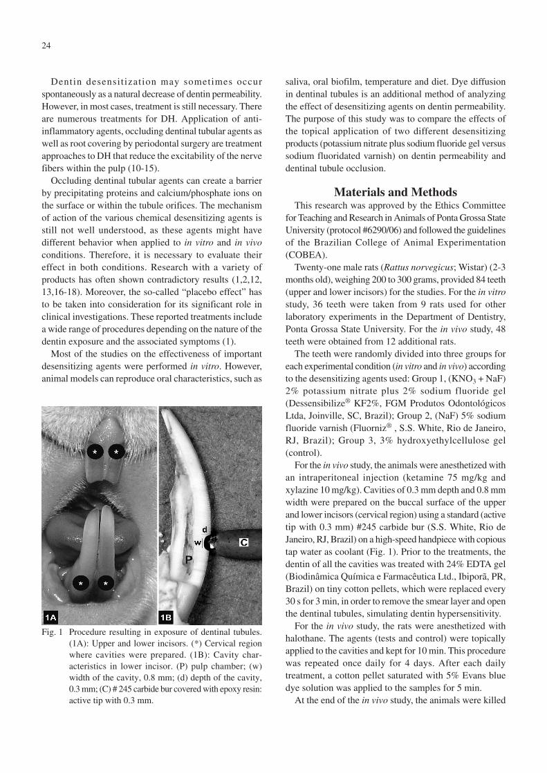
For the in vivo study, the animals were anesthetized withan intraperitoneal injection (ketamine 75 mg/kg andxylazine 10 mg/kg). Cavities of 0.3 mm depth and 0.8 mmwidth were prepared on the buccal surface of the upperand lower incisors (cervical region) using a standard (activetip with 0.3 mm) #245 carbide bur (S.S. White, Rio deJaneiro, RJ, Brazil) on a high-speed handpiece with copioustap water as coolant (Fig. 1). Prior to the treatments, thedentin of all the cavities was treated with 24% EDTA gel(Biodinâmica Química e Farmacêutica Ltd., Ibiporã, PR,Brazil) on tiny cotton pellets, which were replaced every30 s for 3 min, in order to remove the smear layer and openthe dentinal tubules, simulating dentin hypersensitivity.
For the in vivo study, the rats were anesthetized withhalothane. The agents (tests and control) were topicallyapplied to the cavities and kept for 10 min. This procedurewas repeated once daily for 4 days. After each dailytreatment, a cotton pellet saturated with 5% Evans bluedye solution was applied to the samples for 5 min.
At the end of the in vivo study, the animals were killed
Fig. 1 Procedure resulting in exposure of dentinal tubules.(1A): Upper and lower incisors. (*) Cervical regionwhere cavities were prepared. (1B): Cavity char-acteristics in lower incisor. (P) pulp chamber; (w)width of the cavity, 0.8 mm; (d) depth of the cavity,0.3 mm; (C) # 245 carbide bur covered with epoxy resin:active tip with 0.3 mm.

25
by cervical dislocation after previous sedation withhalothane, and the teeth were carefully extracted to avoiddamage to their surface. The teeth were kept frozen (-20°C)until analyzed (24 h).
The same protocol was used for the application of theagents (tests and control) in the in vitro study; however,the teeth previously extracted were stored in saline solution(0.9% NaCl) at room temperature during the 4-dayexperimental period. The saline solution was changeddaily in order to avoid bacterial contamination.
Dentin permeability analysisFive percent (5%) by weight Evans blue (Merck
Chemical Ltda, Darmstadt, Germany) solution wasemployed to analyze the dentin permeability of 42 teeth(6 per group in vitro; 8 per group in vivo). The sampleswere embedded in acrylic resin and sectioned longitudinally(bucco-lingual direction) using a water-cooled diamondsaw (ISOMET 1000- Precision Saw, Buheler, model112180, Lake Bluff, IL, USA). In order to remove thesludge and dentin within dentinal tubules, the specimenswere ultrasonically cleaned for 10 min at 47°C.
The samples were photographed (Olympus™ BX41,Olympus, Tokyo, Japan) at ×40 magnification and imagesof 5.1 megapixels (digital camera, Olympus Camedia C-5060, Tokyo, Japan) were obtained. These stored digitalizedimages were not enhanced and no transformationprocedures were carried out. The image analysis softwareused was Image Pro Plus™ Version 4.5.0.29 (MediaCybernetics, Silver Spring, MD, USA). Each image wascalibrated individually with a standard scale (µm). Threemeasurements (upper, middle and lower parts) were takenfor each image indicating the depth of the dye infiltration,and finally, the mean was calculated for each specimen.The same examiner performed all the measurements, aftertesting the reproducibility of the data.
Scanning electron microscopy (SEM)A total of 6 teeth/group (in vitro) and 8 teeth/group (in
vivo) were evaluated by SEM. All specimens weretransversely sectioned using a water-cooled diamond saw(ISOMET 1000, Precision Saw Buehler, Lake Bluff, IL,USA). The sectioned specimens were washed with 20 mlof distilled water and ultrasonicated for 10 min. Dehydrationwas achieved to critical point dryness using a graded seriesof ethanol (25%, 50%, 70%, 90% and 100% for 10min/each). Specimens were mounted on metal stubs, keptin a 37 °C stove for 24 h and stored in a vacuum silica-gel desiccator for 48 h. In order to perform the SEManalysis, the samples were sputter coated with 25 nm ofgold for 10 min.
Nine images from each sample (central area) wereobtained by SEM (Shimadzu SSX 550™, Shimadzu doBrasil Comércio Ltd, São Paulo, Brazil), operated at 20kV at ×2,000 and ×5,000 magnification. The SEMphotomicrographs were evaluated quantitatively andqualitatively. Quantitative analysis was performed bycounting the number of dentinal tubules and obtaining anestimate per mm2 (×2,000 magnification) considering a totalarea of 1,600 µm2, the diameter of dentinal tubules (×5,000magnification) and opened tubular area (×2,000magnification) using the software Image Pro Plus™ Version4.5.0.29. Qualitative evaluation (×2,000 magnification)considered dentin surface characteristics, intertubular andperitubular dentin, dentinal tubules and smear layer deposits.All the analyses were performed by one trained examiner.
Energy-dispersive X-ray (EDX)Samples were examined by energy dispersive X-ray
microanalysis (Shimadzu SSX 550™, Shimadzu do BrasilComércio Ltd) in order to determine the presence ofchemical elements in deposits found next to the dentinaltubules of each specimen. The spectrum was obtained at20 Kv, spot size of 5 nm and the counting time was 300 s.This provided qualitative information of the presence offluorine (F), sodium (Na), magnesium (Mg), silicon (Si),phosphorus (P), potassium (K) and calcium (Ca).
Statistical analysisIntra-examiner reproducibility was assessed twice within
48 h to check the reproducibility of dentin permeabilityand tubular diameter measurements. Tubule number andtubule area were repeatedly compared using both manualand automatic methods (digital computer image analysis).The reproducibility was tested using the Bland and Altmanprocedure (19).
Comparisons among groups according to experimentalcondition (in vitro and in vivo) for tubule number, diameterand area as well as dentin permeability were tested by one-way ANOVA and Bonferroni post hoc test. The normalityof the distribution of data was confirmed using the Shapiro-Wilks test. An alpha value of ≤0.05 was used to indicatestatistically significant differences among the groups. Allanalyses were performed using a software program(GraphPad Prism version 5.00 for Windows, GraphPadSoftware, San Diego, CA, USA).
Results
ReproducibilityReproducibility for dentin permeability, tubule diameter
(intra-examiner), dentin tubule number, and total tubule

26
area (comparing manual and automatic methods) werewithin the limits of agreement (Fig. 2).
Dentin permeabilityThe mean and standard error (SE) of the dentin
permeability to 5% Evans blue dye is shown in Fig. 3. Therewas a significant difference among the groups in bothexperimental conditions (in vitro, P < 0.0001 and in vivo,P = 0.0107).
Scanning electron microscope (SEM)The number of dentinal tubules per mm2 (mean and SE)
was significantly different among the groups (in vitro, P= 0.0195 and in vivo, P = 0.0064) according to one-wayANOVA and Bonferroni post hoc tests (Fig. 4).
The mean and SE of the dentinal tubule diameter areshown in Fig. 5. A statistically significant difference wasfound (in vitro, P < 0.0012 and in vivo, P = 0.0022).
The dentinal tubule area exhibited statistically significantdifferences among groups (in vitro, P = 0.0001 and in vivo,
P = 0.0011), using one-way ANOVA and Bonferroni posthoc tests (Fig. 6).
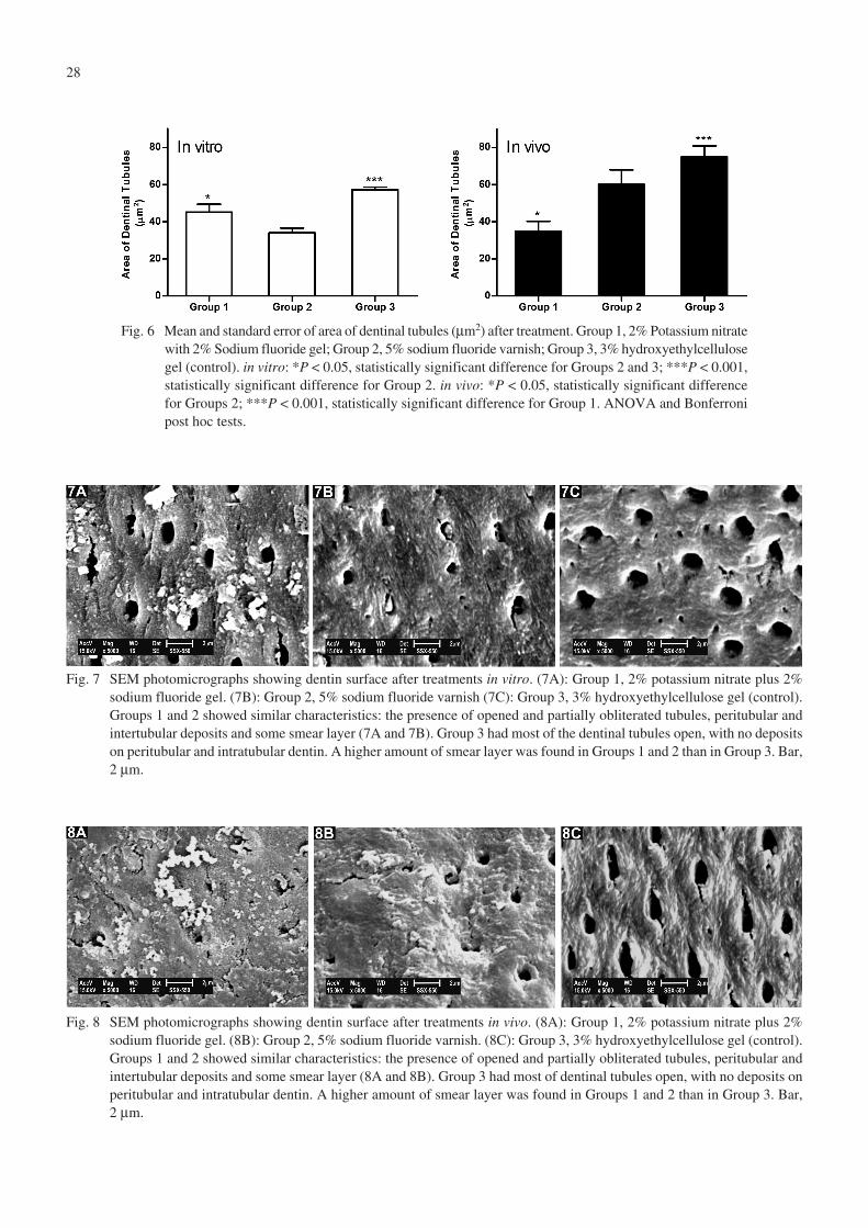
The qualitative analysis showed the presence of openedand partially occluded tubules, peritubular and intertubulardeposits and some smear layer in Group 1 and Group 2(Figs. 7A, 7B, 8A and 8B). In Group 3, most of the dentinaltubules were opened, with no deposits on peritubular andintratubular dentin. A higher amount of smear layer wasfound in Group 1 and Group 2 than in Group 3 (Figs. 7Cand 8C).
Energy-dispersive X-ray (EDX)Energy-dispersive X-ray (EDX) analysis revealed peaks
of chemical elements found on experimental and controlsamples. High peaks of calcium and phosphorus wereobserved in all groups (both experimental conditions).Group 1 showed traces of sodium, magnesium, silicon, andpotassium; Group 2 showed fluorine, sodium and silicon;and Group 3 exhibited traces of sodium, magnesium andsilicon (Figs. 9 and 10).
Fig. 2 Intra-examiner reproducibility. Bland-Altman plots of the data obtained by repeated analysis. (2A): Dentinal permeabilitymeasurements at two different time points. (2B): Tubule number and tubules (SEM) were repeatedly compared using bothmanual and automatic methods (digital computer image analysis). (2C): Tubule diameter (SEM) measurements at twodifferent time points. (2D): Tubule area (SEM) was repeatedly compared using both manual and automatic methods. Intra-examiner reproducibility was within the limits of agreement. SEM, scanning electron microscope.

27
Fig. 4 Mean and standard error of the number of dentinal tubules (×1,000/mm2) after treatment. Group 1, 2%Potassium nitrate with 2% Sodium fluoride gel; Group 2, 5% sodium fluoride varnish; Group 3, 3%hydroxyethylcellulose gel (control). in vitro: *P < 0.05, statistically significant difference for Group 1.in vivo: (**P < 0.01) statistically significant difference for Group 1. ANOVA and Bonferroni post hoctests
Fig. 3 Mean and standard error of dentin permeability (5% Evans blue dye) after treatment. Group 1, 2% Potassiumnitrate with 2% Sodium fluoride gel; Group 2, 5% sodium fluoride varnish; Group 3, 3%hydroxyethylcellulose gel (control). in vitro: ***P < 0.001, statistically significant difference for Groups1 and 2. in vivo: *P < 0.05, statistically significant difference for Groups 1 and 2. ANOVA andBonferroni post hoc tests.
Fig. 5 Mean and standard error of diameter of dentinal tubules (µm) after treatment. Group 1, 2% Potassiumnitrate with 2% Sodium fluoride gel; Group 2, 5% sodium fluoride varnish; Group 3, 3%hydroxyethylcellulose gel (control). in vitro: ***P < 0.001, statistically significant difference for Group1. in vivo: **P < 0.01, statistically significant difference for Group 1. ANOVA and Bonferroni posthoc tests.
28
Fig. 6 Mean and standard error of area of dentinal tubules (µm2) after treatment. Group 1, 2% Potassium nitratewith 2% Sodium fluoride gel; Group 2, 5% sodium fluoride varnish; Group 3, 3% hydroxyethylcellulosegel (control). in vitro: *P < 0.05, statistically significant difference for Groups 2 and 3; ***P < 0.001,statistically significant difference for Group 2. in vivo: *P < 0.05, statistically significant differencefor Groups 2; ***P < 0.001, statistically significant difference for Group 1. ANOVA and Bonferronipost hoc tests.
Fig. 7 SEM photomicrographs showing dentin surface after treatments in vitro. (7A): Group 1, 2% potassium nitrate plus 2%sodium fluoride gel. (7B): Group 2, 5% sodium fluoride varnish (7C): Group 3, 3% hydroxyethylcellulose gel (control).Groups 1 and 2 showed similar characteristics: the presence of opened and partially obliterated tubules, peritubular andintertubular deposits and some smear layer (7A and 7B). Group 3 had most of the dentinal tubules open, with no depositson peritubular and intratubular dentin. A higher amount of smear layer was found in Groups 1 and 2 than in Group 3. Bar,2 µm.
Fig. 8 SEM photomicrographs showing dentin surface after treatments in vivo. (8A): Group 1, 2% potassium nitrate plus 2%sodium fluoride gel. (8B): Group 2, 5% sodium fluoride varnish. (8C): Group 3, 3% hydroxyethylcellulose gel (control).Groups 1 and 2 showed similar characteristics: the presence of opened and partially obliterated tubules, peritubular andintertubular deposits and some smear layer (8A and 8B). Group 3 had most of dentinal tubules open, with no deposits onperitubular and intratubular dentin. A higher amount of smear layer was found in Groups 1 and 2 than in Group 3. Bar,2 µm.

29
DiscussionThe present study used an animal model (rat) because
of the ease of obtaining samples and reproducing oralcharacteristics similar to those of humans. The cervical areaof the incisors was analyzed because it was moreappropriate (higher number of dentinal tubules) thanstudying dentin permeability in the incisal tip (lowpermeability due to atubular tissue and pulp remnants), ashas been demonstrated previously (20,21). The results ofexperiments concerning the permeability and tubularocclusion carried out on this animal model were similarto the results of previously published work (20).
The analysis of dentin permeability in this study usinga blue dye showed that in both studies (in vitro and in vivo)there was greater dentin permeability in Group 3 (control).In the tooth specimens which received topical treatmentwith dentin desensitizing agents (Groups 1 and 2), there
was lower permeability in vitro than in vivo. The reasonsfor different results are likely related to factors such as oraltemperature, saliva flow, dental biofilm and mastication.These could interfere with the action of the desensitizingproducts. The higher dye diffusion in the control groupsconfirms these data. In the current study, dentin permeabilitywas higher in vivo than in vitro specimens that weretopically treated with desensitizing products (Groups 1 and2). The results of the present study were different from thoseobtained by Vongsavan et al. (21), who observed minimalpenetration of the dye in vivo and a higher penetration invitro. They eliminated the external factors that couldinfluence the dye diffusion when the dye was allowed tosit for 15 min. In the present study, the dye remained for5 min daily for 4 days (a total of 20 min). Vongsavan andMatthews (22) showed better dye penetration in vitro thanin vivo, probably due to pulpal pressure.
Fig. 9 Energy-dispersive X-ray (EDX) spectrum of in vitro samples. (9A): Group 1, 2% potassium nitrate plus 2% sodium fluoride.(9B): Group 2, 5% sodium fluoride varnish. (9C): Group 3, 3% hydroxyethylcellulose gel (control). EDX plots representingpeaks of elemental ion concentration showed for Group 1 traces of Na, Mg, Si and K, Group 2 revealed F, Na and Si, andGroup 3 exhibited Na, Mg and Si. Presence of C, O, P and Ca due to the background noise produced by mineral contentof the underlying dentine.
Fig. 10 Energy-dispersive X-ray (EDX) spectrum of in vivo samples. (10A): Group 1, 2% potassium nitrate plus 2% sodiumfluoride. (10B): Group 2, 5% sodium fluoride varnish. (10C): Group 3, 3% hydroxyethylcellulose gel (control). EDXplots representing peaks of elemental ion concentration showed for Group 1 traces of Na, Mg, Si and K, Group 2 revealedF, Na, Mg and Si, and Group 3 exhibited Na, Mg and Si. Presence of C, O, P and Ca due to the background noise producedby mineral content of the underlying dentine.

30
Considering the methodology used in this study,continuous eruption of the rat incisor could be a limitationfor longer periods of experimentation. Therefore, treatmentwas done for only 4 days and the dentin was exposed onthe buccal surfaces of the cervical area of rat incisors forDH analysis in this study (20). Rat incisors can thus beused to evaluate dentin permeability and dentinal tubuleocclusion by desensitizing agents.
In this study, the number of dentinal tubules/mm2
exhibited similar results for both experimental conditions(in vitro and in vivo). Teeth in Group 1 (in vivo) showedlower dentin tubule counts compared with the controlgroup.
In the present study, a 0.3-mm cervical cavity was made,equivalent to one-fourth of the distance between the pulpand enamel. Forssell-Ahlberg et al. (20) observed a highernumber of dentinal tubules than was found in the presentstudy. This may have been due to the presence ofdesensitizing agents occluding tubules in the currentresults. Garberoglio and Brännström (23), Olsson et al. (24)and Dourda et al. (25) showed that the number of dentinaltubules ranged from 30,900/mm2 to 37,000/mm2 in humandentin, at a position halfway between pulp and enamel.Paes Leme et al. (8) observed 19,333 ± 2,770 tubules/mm2
and 16,433 ± 782 tubules/mm2, respectively, for potassiumnitrate or strontium chloride containing dentifrice-treateddentin and those treated with fluoride varnish. Humancervical dentin has been reported to have 19,000 tubulesper mm2 in superficial dentin. As the halfway point betweensuperficial dentin and the pulp is reached, the number oftubules increases to 30,000 tubules per mm2 (23). Thus,the results found for desensitizing agent treatments inanimals can be extrapolated to humans.
Dentin tubule diameter was smallest in teeth that weretopically treated with 2% potassium nitrate plus 2% sodiumfluoride gel (Group 1), followed by Groups 2 and 3.Similar results were found for in vitro and in vivo groups.These results obtained in Group 1 were probably due tothe action of the desensitizing agents, creating a highernumber of partially obliterated tubules. Rat incisors inthe current study showed a mean dentin tubule diameterof 1.0 µm. These values were similar to those previouslyreported (20). Dentin tubule diameter, in the middle dentin,is similar for humans and rats according to Garberoglioand Brännström (23). Similar results were found in otherstudies (20, 23), although it is necessary to consider thatin these studies, the dentin was not submitted todesensitizing agent treatment. Therefore, in the currentstudy, dentin tubule diameter is expected to be lower intreated groups. The difference among studies may berelated to the different chemical agents applied to the
dentin.Dentinal tubule area was evaluated by SEM at 2,000
times magnification (total area of 1,600 µm2). It waspossible to observe a significant difference among groups.These findings represent 3%, 2% and 5% (in vitro) and2%, 4% and 6% (in vivo) of the dentin area occupied bytubules in Groups 1, 2 and 3, respectively. In humans,Dourda et al. (25) found 6% of the dentin area occupiedby tubules at a distance halfway between pulp and enamel.Rat incisors showed 4% of the area occupied by tubules(20). The percentage of the area occupied by dentinaltubules in this study was probably lower due to the use ofdesensitizing agents, which occluded many tubule orifices.
Quantitative analysis of dentinal tubule number, diameterand area could be prejudiced due to changes in directionof the dentinal tubules and in the position of the SEMsamples. The presence of the smear layer and the smallanalyzed area could also introduce some bias.
Similar characteristics were found in teeth that weretopically treated with 2% potassium nitrate plus 2% sodiumfluoride gel and 5% fluoride varnish. Partial to total dentintubule occlusion could be seen. Paes Leme et al. (8)obtained similar results, in that potassium nitrate was notable to completely obliterate dentin tubules. However,Knight et al. (26) reported that 5% potassium nitratetoothpaste was able to occlude dentin tubules with crystalsof different sizes and shapes. Although these studies didnot analyze the chemical composition of the deposits,these precipitates could be due to the toothpaste abrasiveagents (calcium carbonate) and not related to potassiumnitrate.
The samples treated with fluoride varnish (Group 2) haddentin tubules that were partially occluded, and there werefewer deposits and less smear layer on peritubular andintertubular dentin. These data are in agreement with otherreports (8,11,26). In those studies, fluorides were unableto occlude dentin tubules after a single application. Knightet al. (26), Pashley (11) and Paes Leme et al. (8) could notobserve calcium fluoride crystals in dentin tubules, andconsequently no reduction in dentin hypersensitivity. Themajority of dentin tubules were open and there were nosmear layer deposits in the Group 3 specimens that weretreated with 3% hydroxyethylcellulose gel (control).
According to these results, both potassium nitrate andfluoride varnish were able to reduce dentin permeabilitywhen compared to the control group, suggesting thatrepeated applications could contribute to crystal formation,not only on the dentin surface but also inside dentinaltubules. This statement is supported by Mukai et al. (27)and Arrais et al. (14), who showed crystal precipitation onperitubular dentine and inside dentin tubules after acidulated

31
sodium fluoride application for 4 min.The EDX microanalysis of samples showed only traces
of desensitizing chemical active agents. However, Arraiset al. (7) did not find chemical evidence of the activeingredients of desensitizing agents after EDX analysis. Thereasons for these different results are probably the variationsin product formulation and concentration used in thepresent research.
The results of the present study should be interpretedwith caution before extrapolating them to dental practicein humans, considering the nature and limitations of animaland in vitro experimentation.
In spite of the study limitations, the results showedsignificant difference among desensitizing agents incomparison with the control group. In conclusion, 2%nitrate potassium plus 2% sodium fluoride gel and 5%fluoride varnish decreased the dentin permeability, althoughthey produced only partial dentin tubule surface occlusion.Repeated application of the desensitizing agents maypossibly contribute to higher clinical effectiveness indentin tubule occlusion.
AcknowledgmentsThis work was supported by CAPES (Coordenação de
Aperfeiçoamento de Pessoal de Nível Superior), Brazil.
References1. Rees JS, Addy M (2002) A cross-sectional study of
dentine hypersensitivity. J Clin Periodontol 29, 997-1003.
2. Ritter AV, de L Dias W, Miguez P, Caplan DJ, SwiftEJ Jr (2006) Treating cervical dentin hypersensitivitywith fluoride varnish: a randomized clinical study.J Am Dent Assoc 137, 1013-1020.
3. Ahmed TR, Mordan NJ, Gilthorpe MS, Gillam DG(2005) In vitro quantification of changes in humandentine tubule parameters using SEM and digitalanalysis. J Oral Rehabil 32, 589-597.
4. Wara-aswapati N, Krongnawakul D, Jiraviboon D,Adulyanon S, Karimbux N, Pitiphat W (2005) Theeffect of a new toothpaste containing potassiumnitrate and triclosan on gingival health, plaqueformation and dentine hypersensitivity. J ClinPeriodontol 32, 53-58.
5. Brännström M, Lindén LA, Aström A (1967) Thehydrodynamics of the dental tubule and of pulpfluid. A discussion of its significance in relation todentinal sensitivity. Caries Res 1, 310-317.
6. Pashley DH, O’Meara JA, Kepler EE, GallowaySE, Thompson SM, Stewart FP (1984) Dentinpermeability. Effects of desensitizing dentifrices in
vitro. J Periodontol 55, 522-525.7. Arrais CA, Micheloni CD, Giannini M, Chan DC
(2003) Occluding effect of dentifrices on dentinaltubules. J Dent 31, 577-584.
8. Paes Leme AF, dos Santos JC, Giannini M, WadaRS (2004) Occlusion of dentin tubules bydesensitizing agents. Am J Dent 17, 368-372.
9. Absi EG, Addy M, Adams D (1987) Dentinehypersensitivity. A study of the patency of dentinaltubules in sensitive and non-sensitive cervicaldentine. J Clin Periodontol 14, 280-284.
10. Kerns DG, Scheidt MJ, Pashley DH, Horner JA,Strong SL, Van Dyke TE (1991) Dentinal tubuleocclusion and root hypersensitivity. J Periodontol62, 421-428.
11. Pashley DH (1992) Dentin permeability and dentinsensitivity. Proc Finn Dent Soc 88, Suppl 1, 31-37.
12. West NX, Hughes JA, Addy M (2001) The effectof pH on the erosion of dentine and enamel bydietary acids in vitro. J Oral Rehabil 28, 860-864.
13. Frechoso SC, Menéndez M, Guisasola C, ArreguiI, Tejerina JM, Sicilia A (2003) Evaluation of theefficacy of two potassium nitrate bioadhesive gels(5% and 10%) in the treatment of dentinehypersensitivity. A randomised clinical trial. J ClinPeriodontol 30, 315-320.
14. Arrais CA, Chan DC, Giannini M (2004) Effects ofdesensitizing agents on dentinal tubule occlusion.J Appl Oral Sci 12, 144-148.
15. Zandim DL, Corrêa FO, Sampaio JE, Rossa JúniorC (2004) The influence of vinegars on exposure ofdentinal tubules: a SEM evaluation. Braz Oral Res18, 63-68.
16. Pereira JC, Segala AD, Gillam DG (2005) Effect ofdesensitizing agents on the hydraulic conductanceof human dentin subjected to different surface pre-treatments – an in vitro study. Dent Mater 21, 129-138.
17. Absi EG, Addy M, Adams D (1995) Dentinehypersensitivity: uptake of toothpastes onto dentineand effects of brushing, washing and dietary acid –SEM in vitro study. J Oral Rehabil 22, 175-182.
18. Oberg C, Pochapski MT, Farago PV, Grando CJF,Pilatti GL, Santos FA (2009) Evaluation ofdesensitizing agents on dentin permeability anddentinal tubule occlusion: an in vitro study. Gen Dent57, 497-501.
19. Bland JM, Altman DG (1999) Measuring agreementin method comparison studies. Stat Methods MedRes 8, 135-160.
20. Forssell-Ahlberg K, Brännström M, Edwall L (1975)

32
The diameter and number of dentinal tubules in rat,cat, dog and monkey. A comparative scanningelectron microscopic study. Acta Odontol Scand33, 243-250.
21. Vongsavan N, Matthews RW, Matthews B (2000)The permeability of human dentine in vitro and invivo. Arch Oral Biol 45, 931-935.
22. Vongsavan N, Matthews B (1991) The permeabilityof cat dentine in vivo and in vitro. Arch Oral Biol36, 641-646.
23. Garberoglio R, Brännström M (1976) Scanningelectron microscopic investigation of human dentinaltubules. Arch Oral Biol 21, 355-362.
24. Olsson S, Oilo G, Adamczak E (1993) The structureof dentin surfaces exposed for bond strength
measurements. Scand J Dent Res 101, 180-184.25. Dourda AO, Moule AJ, Young WG (1994) A
morphometric analysis of the cross-sectional areaof dentine occupied by dentinal tubules in humanthird molar teeth. Int Endod J 27, 184-189.
26. Knight NN, Lie T, Clark SM, Adams DF (1993)Hypersensitive dentin: testing of procedures formechanical and chemical obliteration of dentinaltubuli. J Periodontol 64, 366-373.
27. Mukai Y, Tomiyama K, Okada S, Mukai K, NegishiH, Fujihara T, Teranaka T (1998) Dentinal tubuleocclusion with lanthanum fluoride and powderedapatite glass ceramics in vitro. Dent Mater J 17, 253-263.