Embed Size (px)
Citation preview
ANTIMICROBIAL AGENTS AND CHEMOTHERAPY, Feb. 2007, p. 551–559 Vol. 51, No. 20066-4804/07/$08.00�0 doi:10.1128/AAC.00642-06Copyright © 2007, American Society for Microbiology. All Rights Reserved.
In Vitro and In Vivo Activities of Chios Mastic Gum Extracts andConstituents against Helicobacter pylori�
Sotirios Paraschos,1 Prokopios Magiatis,1 Sofia Mitakou,1* Kalliopi Petraki,2 Antonios Kalliaropoulos,2Petros Maragkoudakis,2 Andreas Mentis,2 Dionyssios Sgouras,2* and Alexios-Leandros Skaltsounis1
Laboratory of Pharmacognosy and Natural Products Chemistry, Department of Pharmacy, University of Athens,Panepistimiopolis Zografou, Athens 15771, Greece,1 and Laboratory of Medical Microbiology,
Hellenic Pasteur Institute, 127 Vassilisis Sofias Avenue, Athens 11521, Greece2
Received 25 May 2006/Returned for modification 1 August 2006/Accepted 10 November 2006
The extracts and pure major constituents of Chios mastic gum (resin of Pistacia lentiscus var. chia) weretested for their activities against Helicobacter pylori. A total mastic extract without polymer (TMEWP) wasprepared after removal of the contained insoluble polymer in order to ameliorate solubility and enhance in vivoactivity. Administration of TMEWP to H. pylori SS1-infected mice over the period of 3 months with an averagedose of 0.75 mg/day led to an approximately 30-fold reduction in the H. pylori colonization (1.5 log CFU/g oftissue). However, no attenuation in the H. pylori-associated chronic inflammatory infiltration and the activityof chronic gastritis was observed. To further characterize potential active mastic constituents, the TMEWP wasseparated into an acidic and a neutral fraction. Both were extensively characterized by nuclear magneticresonance and mass spectroscopy to elucidate the structure of the components contained within each fraction.After chromatographic separation, the acid fraction gave the major triterpenic acids, while the neutral fractiongave several triterpenic alcohols and aldehydes. Mastic extracts and isolated pure triterpenic acids were testedfor in vitro activity against a panel of 11 H. pylori clinical strains. The acid fraction was found to be the mostactive extract (minimum bactericidal concentration [MBC], 0.139 mg/ml), and the most active pure compoundwas isomasticadienolic acid (MBC, 0.202 mg/ml [0.443 mM]). Our results show that administration ofTMEWP may be effective in reducing H. pylori colonization and that the major triterpenic acids in the acidextract may be responsible for such an activity.
Pistacia lentiscus L. is an evergreen shrub of the Anacardi-aceae family, very common in the eastern Mediterranean area.The variety chia (Duham) is uniquely cultivated in southernChios, a Greek island in the Aegean. The resin of that plant,mastic gum, is obtained as an exudate after “hurting” the trunkand branches.
Mastic gum has been used in traditional Greek medicine forvarious gastrointestinal disorders like gastralgia, dyspepsia,and peptic ulcer for more than 2,500 years. Ancient Greekphysicians, such as Hippocrates, Dioscorides, Theophrastos,and Galenos, mentioned its properties and recommended itsuse. In modern times, it is used as a seasoning in Mediterra-nean cuisine, in the production of chewing gum, in perfumery,in dentistry, and by the local population of Chios for the reliefof gastralgia and protection against peptic ulcer. Early studiesinvolving mastic administration to rats with experimentally in-duced gastric and duodenal ulcers indicated a significant de-crease of free acidity (2). Moreover, a double-blind clinicaltrial carried out on patients with symptomatic and endoscop-ically proven duodenal ulcer showed increased symptomaticrelief in patients on mastic (1 g daily) compared to patients on
placebo, while endoscopically proven healing occurred in 70%of the patients on mastic (1).
In 1983, Barry Marshall and Robin Warren suggested thatgastric inflammation and peptic ulceration were the result ofan infection caused by Helicobacter pylori (34). In the years tofollow, the presence of H. pylori infection was shown to be theetiologic determinant of chronic active gastritis and a majorrisk factor for the development of peptic ulcer disease, gastricatrophy, gastric adenocarcinoma, and mucosa-associated lym-phoid tissue lymphoma (23). Antibiotic eradication schemeshave proved very effective in clearing the infection; however,low patient compliance and the development of antibiotic re-sistance have created the need for new H. pylori eradicationstrategies. Mastic gum has been reported to possess consider-able in vitro antibacterial and antifungal activities (32). A fewyears ago, it was specifically reported to be effective againstHelicobacter pylori in vitro (7, 12, 21). However, in a morerecent in vivo study of H. pylori infection, the activity of masticgum was compared with antibiotic eradication schemes, andafter a 7-day therapy no eradication of the bacterium from thestomachs of mice receiving mastic was observed (17). Finally,H. pylori-positive patients were treated with mastic capsules for7 days, and they all remained H. pylori positive after the ad-ministration (6). The last two studies concluded that no “an-tibiotic-like” activity should be expected from crude mastic.
The crude resin that was used in all previous studies con-tained a high percentage (30%) of an insoluble and stickypolymer (poly-�-myrcene) (33) that obviously hinders its oraladministration and reduces the bioavailability of the containedactive compounds. To bypass such problems, we prepared a
* Corresponding author. Mailing address for D. Sgouras: Depart-ment of Medical Microbiology, Hellenic Pasteur Institute, 127 VassilisisSofias Avenue, Athens 115 21, Greece. Phone: 30210-6478824. Fax:30210-6440171. E-mail: [email protected]. Mailing address for S.Mitakou: Laboratory of Pharmacognosy and Natural Products Chemistry,Department of Pharmacy, University of Athens, PanepistimiopolisZografou, Athens 15771, Greece. Phone: 30210-7274290. Fax: 30210-7274594. E-mail: [email protected].
� Published ahead of print on 20 November 2006.
551
on June 29, 2018 by guesthttp://aac.asm
.org/D
ownloaded from
total mastic extract without polymer (TMEWP) and tested itsactivity against H. pylori in mice infected with the H. pylori SS1strain. In addition, well-characterized mastic gum fractions aswell as isolated pure compounds were tested in vitro against H.pylori in an effort to identify the most active constituents.
MATERIALS AND METHODS
General procedures. Optical rotations were measured with a Perkin-Elmer 341polarimeter. Nuclear magnetic resonance (NMR) spectra were recorded onBruker DRX 400 and Bruker AC 200 spectrometers (1H [400 and 200 MHz] and13C [50 MHz]); chemical shifts are expressed in ppm downfield from tetramethylsilane. The 1H-1H and the 1H-13C NMR experiments (distortionless enhance-ment by polarization transfer, correlated spectroscopy [COSY], COSY for long-range couplings, heteronuclear multiple quantum coherence, heteronuclear mul-tiple-bond correlation, and nuclear Overhauser effect spectroscopy [NOESY])were performed using standard Bruker microprograms. Gas chromatography-mass spectroscopy (MS) analysis was performed on a Finnigan GCQ Plus massspectrometer. Electron impact and chemical ionization-MS spectra were deter-mined on a Finnigan GCQ Plus mass spectrometer using CH4 as the chemicalionization reagent and high-resolution mass spectrometry on an AEI MS-90spectrometer. Medium-pressure liquid chromatography (MPLC) was performedwith a Buchi model 688 apparatus on columns containing Si gel (type 60; 20 to40 �m; Merck).
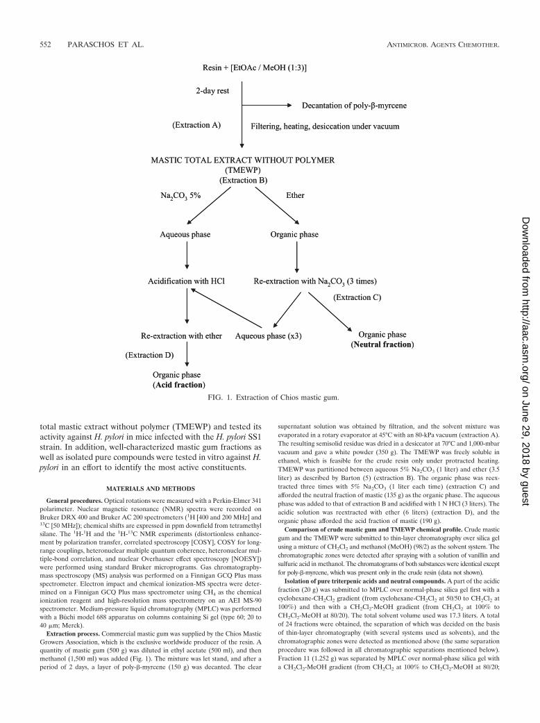
Extraction process. Commercial mastic gum was supplied by the Chios MasticGrowers Association, which is the exclusive worldwide producer of the resin. Aquantity of mastic gum (500 g) was diluted in ethyl acetate (500 ml), and thenmethanol (1,500 ml) was added (Fig. 1). The mixture was let stand, and after aperiod of 2 days, a layer of poly-�-myrcene (150 g) was decanted. The clear
supernatant solution was obtained by filtration, and the solvent mixture wasevaporated in a rotary evaporator at 45°C with an 80-kPa vacuum (extraction A).The resulting semisolid residue was dried in a desiccator at 70°C and 1,000-mbarvacuum and gave a white powder (350 g). The TMEWP was freely soluble inethanol, which is feasible for the crude resin only under protracted heating.TMEWP was partitioned between aqueous 5% Na2CO3 (1 liter) and ether (3.5liter) as described by Barton (5) (extraction B). The organic phase was reex-tracted three times with 5% Na2CO3 (1 liter each time) (extraction C) andafforded the neutral fraction of mastic (135 g) as the organic phase. The aqueousphase was added to that of extraction B and acidified with 1 N HCl (3 liters). Theacidic solution was reextracted with ether (6 liters) (extraction D), and theorganic phase afforded the acid fraction of mastic (190 g).
Comparison of crude mastic gum and TMEWP chemical profile. Crude masticgum and the TMEWP were submitted to thin-layer chromatography over silica gelusing a mixture of CH2Cl2 and methanol (MeOH) (98/2) as the solvent system. Thechromatographic zones were detected after spraying with a solution of vanillin andsulfuric acid in methanol. The chromatograms of both substances were identical exceptfor poly-�-myrcene, which was present only in the crude resin (data not shown).
Isolation of pure triterpenic acids and neutral compounds. A part of the acidicfraction (20 g) was submitted to MPLC over normal-phase silica gel first with acyclohexane-CH2Cl2 gradient (from cyclohexane-CH2Cl2 at 50/50 to CH2Cl2 at100%) and then with a CH2Cl2-MeOH gradient (from CH2Cl2 at 100% toCH2Cl2-MeOH at 80/20). The total solvent volume used was 17.3 liters. A totalof 24 fractions were obtained, the separation of which was decided on the basisof thin-layer chromatography (with several systems used as solvents), and thechromatographic zones were detected as mentioned above (the same separationprocedure was followed in all chromatographic separations mentioned below).Fraction 11 (1.252 g) was separated by MPLC over normal-phase silica gel witha CH2Cl2-MeOH gradient (from CH2Cl2 at 100% to CH2Cl2-MeOH at 80/20;
FIG. 1. Extraction of Chios mastic gum.
552 PARASCHOS ET AL. ANTIMICROB. AGENTS CHEMOTHER.
on June 29, 2018 by guesthttp://aac.asm
.org/D
ownloaded from
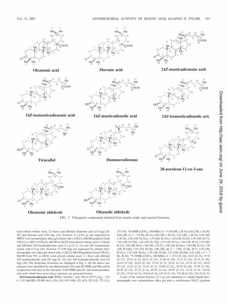
total solvent volume used, 2.1 liters) and afforded oleanonic acid (515 mg) (29,30) and moronic acid (338 mg) (13). Fraction 12 (2.431 g) was separated byMPLC over normal-phase silica gel eluted with a CH2Cl2-MeOH gradient (fromCH2Cl2 at 100% to CH2Cl2-MeOH at 80/20; total solvent volume used, 3.3 liters)and afforded 24Z-masticadienonic acid (1.1 g) (5, 8, 22) and 24Z-isomasticadi-enonic acid (1.0 g) (26). Fraction 17 (198 mg) was separated by column chro-matography over silica gel eluted with a CH2Cl2-MeOH gradient (from CH2Cl2-MeOH from 99/1 to 80/20; total solvent volume used, 1.1 liter) and afforded24Z-masticadienolic acid (85 mg) (9, 22) and 24Z-isomasticadienolic acid (92mg) (24). The molecular structures are displayed in Fig. 2. All the above con-stituents were identified by one-dimensional (1D) and 2D NMR and MS and bycomparison with data in the literature. Full NMR data for 24Z-isomasticadien-onic acid, which have never been reported, are presented below.
24Z-Isomasticadienonic acid. White “powder,” m.p. 166 to 167°C; [�]D: �34°(c 1.10, MeOH), EI-MS (m/z): 454 (34), 439 (100), 421 (62), 393 (13), 271 (11),
257 (19). 1H-NMR (CDCl3, 400 MHz): � � 0.70 (3H, s, H-18), 0.82 (3H, s, H-30),0.86 (3H, d, J � 5.8 Hz, H-21), 0.98 (3H, s, H-28), 1.02 (3H, s, H-19), 1.04 (3H,s, H-29), 1.08 (1H, H-22�), 1.19 (1H, H-15�), 1.38 (1H, H-20), 1.45 (1H, H-17),1.46 (1H, H-22�), 1.48 (1H, H-15�), 1.57 (1H, H-1�), 1.64 (1H, H-5), 1.67 (2H,H-12), 1.80 (2H, H-6), 1.84 (3H, s, H-27), 1.88 (1H, H-16�), 1.90 (2H, H-11), 1.99(1H, H-16�), 1.93 (1H, H-1�), 2.06 (2H, dd, J � 7 Hz, 14 Hz, H-7), 2.39 (1H,H-23�), 2.42 (1H, H-2�), 2.49 (1H, H-2�), 2.51 (1H, H-23�), 6.01 (1H, t, J � 7Hz, H-24). 13C-NMR (CDCl3, 100 MHz): � � 15.51 (C-18), 18.53 (C-21), 19.74(C-27), 20.22 (C-6), 20.51 (C-19), 21.06 (C-28), 21.32 (C-16), 24.15 (C-30),26.65 (C-29), 26.83 (C-23), 27.42 (C-7), 28.01 (C-11), 29.74 (C-15), 30.65(C-12), 34.55 (C-2), 35.51 (C-1), 35.80 (C-22), 36.39 (C-20), 37.09 (C-10),49.99 (C-13), 47.23 (C-4), 49.99 (C-14), 49.99 (C-17), 51.42 (C-5), 125.81(C-25), 132.61 (C-9), 134.63 (C-8), 147.35 (C-24), 173.40 (C-26), 218.32 (C-3).
A part of the neutral fraction (17.2 g) was submitted to column liquid chro-matography over normal-phase silica gel with a cyclohexane-CH2Cl2 gradient
FIG. 2. Triterpenic compounds isolated from mastic acidic and neutral fractions.
VOL. 51, 2007 ANTIMICROBIAL ACTIVITY OF MASTIC GUM AGAINST H. PYLORI 553
on June 29, 2018 by guesthttp://aac.asm
.org/D
ownloaded from

(from cyclohexane at 100% to CH2Cl2 at 100%) to afford 22 fractions. The totalsolvent volume used was 21 liters. Fraction 5 (839 mg) was separated by MPLCover normal-phase silica gel with a cyclohexane-CH2Cl2 gradient (from 90/10 to100% CH2Cl2; total volume used, 2 liters) and afforded tirucallol (110 mg) (23)and dammaradienone (128 mg) (20). Fraction 8 (533 mg) was separated byMPLC over normal-phase silica gel eluted with a cyclohexane-CH2Cl2 gradient(from 80/20 to 100% CH2Cl2; total volume used, 1.5 liters) and afforded 28-norolean-12-en-3-one (206 mg) (20). Fraction 10 (396 mg) was separated byMPLC over normal-phase silica gel eluted with a cyclohexane-CH2Cl2 gradient(from 80/20 to 100% CH2Cl2; total volume used, 1.4 liters) and affordedoleanonic aldehyde (152 mg) (25) and oleanolic aldehyde (98 g) (3). The mo-lecular structures are displayed in Fig. 2. All the above constituents were iden-tified by 1D and 2D NMR and MS and by comparison with data in the publishedliterature.
The neutral fraction was also submitted to analytical separation by gas chro-matography-MS. Comparison of mass spectra with MS data library Wiley 275.land data in the literature (4) resulted in the identification of the five compoundsmentioned above as the major neutral ones, while several minor diterpenes ortriterpenes with aldehyde, ketone, or hydroxyl groups were detected on the basisof molecular weight but not thoroughly identified.
In vitro test of mastic total extract, fractions, and pure compounds. Minimumbactericidal concentrations (MBC) were evaluated utilizing H. pylori referencestrain CCUG 38771 and another 10 clinical strains belonging to the HellenicPasteur Institute collection, isolated from gastric antrum biopsies from patientssuffering from gastritis or duodenal or gastric ulcer (LAVHP-1 to LAVHP-10).H. pylori isolates were stored in brain heart infusion broth (BHIB) supplementedwith 20% glycerol at �80°C. All strains prior to use were cultured twice undermicroaerophilic conditions (CampyPak Plus; Becton-Dickinson, Cockeysville,MD) for 24 h at 37°C, in Chalgren’s-Wilkins (CHW) agar plates supplementedwith 7% (vol/vol) horse blood and 1% (vol/vol) Vitox (Oxoid, Basingstoke,United Kingdom). Liquid cultures of H. pylori bacteria to a density of 109
CFU/ml were prepared by suspension in BHIB (Oxoid) supplemented with 10%fetal calf serum (Flow Laboratories, Irvine, Scotland) and 0.25% yeast extract(Oxoid). Successive twofold dilutions of each mastic extract or pure compound inBHIB medium were placed in sterilized 96-well flat-bottom microplates(Sarstedt, Numbrecht, Germany) within a total volume of 100 �l. All extractswere tested at a final concentration range of 0.049 to 1.560 mg/ml, with theexception of the acidic fraction, for which successive twofold dilutions rangedfrom 0.060 to 1.920 mg/ml. To each well containing the mastic dilutions, approx-imately 107 CFU of H. pylori bacteria were added within a 100-�l volume, and themicroplates were incubated at 37°C for 24 h with continuous agitation undermicroaerophilic conditions. Thereafter, viability of H. pylori was evaluated bydetermination of viable CFU in CHW agar plates following incubation at 37°Cfor 48 h under microaerophilic conditions. The MBC was defined as the lowestconcentration of mastic extract or pure compound that killed at least 99.9% ofthe CFU contained in the original inoculum. The mean MBC for each masticpreparation was determined as the average of three independent experiments.
Infection of mice with H. pylori strain SS1. Specific-pathogen-free 6- to8-week-old female C57BL/6 mice were obtained from the Central Animal Fa-cility of the Hellenic Pasteur Institute. They were housed according to relevantGreek national legislation, fed a commercial diet, and given water ad libitum,except as otherwise stated. H. pylori infections by the SS1 strain were carried outas described before (27, 28). Briefly, freshly prepared aliquots (100 �l; 108 CFU)of H. pylori strain SS1 in BHIB (Oxoid) were administered to mice via orogastricinoculation, three times within a week. Accordingly, all noninfected control micewere inoculated with the same volume of plain BHIB.
Administration of TMEWP in vivo. TMEWP was diluted into ethanol at aconcentration of 180 mg/ml and then dissolved into a final aqueous solution of180 �g/ml. The extract was administered through the animals’ drinking water,starting 1 month following initial H. pylori infection and for 3 more months. Thefollowing groups of animals were included in the study: H. pylori-infected miceadministered TMEWP (SMH; n � 10); uninfected mice administered TMEWP(SM; n � 10); and H. pylori-infected mice left untreated (SH; n � 10) as a controlgroup. Animal weight was monitored throughout the whole observation periodas a measure of health of the animals. Daily water consumption was measured,and a mean daily TMEWP consumption per animal was calculated to be 0.75 mgthroughout the whole administration period. In addition to the above therapeuticin vivo protocol, we conducted a preliminary prophylactic study where C57BL/6mice (n � 5) were administered TMEWP for a week prior to H. pylori infection,and we assessed H. pylori colonization 2 weeks after the initial infection.
Assessment of H. pylori colonization levels. At end of the 3-month observationperiod, blood samples were collected via the tail vein and animals were sacrificedby cervical dislocation. Excised stomachs were dissected along the lesser curva-
ture, and H. pylori detection in the gastric tissue was accomplished by H. pyloriquantitative culture and PCR. For H. pylori SS1 quantitative culturing, pre-weighed half-stomach samples were homogenized in thioglycolate medium(Oxoid), serially diluted in phosphate-buffered saline, and plated on CHW agarplates with antibiotics (vancomycin, 10 �g/ml; trimethoprim, 10 �g/ml; polymyxinB, 104 IU/liter; amphotericin B, 2 �g/ml; nalidixic acid, 10 �g/ml; bacitracin, 30�g/ml; fluorocytosine, 5 �g/ml; all from Sigma, St. Louis, Mo.). The cultures wereincubated under microaerophilic conditions at 37°C for up to 6 days. H. pyloricolonies were visualized on the basis of urease activity, and results were ex-pressed as log CFU per gram of gastric tissue. The minimum bacterial densitydetected by this method was 100 CFU per gram. Qualitative H. pylori detectionin the gastric samples was performed by H. pylori-specific PCR utilizing primersfor the ureC (glmM) gene as described before (18). Genomic DNA for thedetection of H. pylori by PCR in tissue samples or bacterial colonies was isolatedby using a DNeasy tissue kit (QIAGEN).
Histopathologic analysis of gastric tissue samples. Excised stomachs wereopened along the lesser curvature, and the longitudinal half was fixed in 10%neutral buffered formalin solution, embedded in paraffin, and processed forhistopathologic analysis. Antral, body, and cardioesophageal mucosa sampleswere examined in the same section. Eleven serial longitudinal 4-�m sectionswere cut from each specimen; 9 of them were stained with hematoxylin-eosin forevaluation of gastric inflammation, and 2 were stained by the May-GrunwaldGiemsa stain method for the assessment of H. pylori colonization. The bacterialdensity and the pathological characteristics of the gastric mucosa were assessedaccording to the updated Sydney system (11). Histopathologic evaluation wasperformed with no prior knowledge of the identity of the samples by the his-topathologist.
Determination of serum anti-H. pylori immunoglobulin G levels. Immunoglob-ulin G (IgG) levels of anti-H. pylori antibodies were detected in the serumsamples collected by an in-house enzyme-linked immunosorbent assay method.Briefly, 15 �g of H. pylori SS1 antigen produced by sonication and subsequentdialysis (SpectraPor; cutoff pore size, 8 kDa) was used to coat 96-well plates.Collected mouse serum samples (diluted 1/50) were primarily incubated in theplates for 24 h at 4°C, and then rabbit anti-mouse IgG (whole molecule)–peroxidase conjugate (Sigma) was used for the secondary incubation (2 h at37°C). Color was developed by addition of o-phenylenediamine (Sigma), andoptical density at 492 nm was measured in a Sunrise microtiter plate reader(Tekan).
Statistical analysis. Analysis of the results from the in vivo experiments wasperformed with respect to H. pylori colonization by two-tailed unpaired t test withWelch correction and with respect to the associated gastritis by the Wilcoxonrank sum test due to the ordinal nature of the data. A P value of �0.05 wasconsidered significant in both tests.
RESULTS
Isolation of TMEWP, acid and neutral fractions, and triter-penic compounds. The TMEWP was obtained from crude mas-tic gum in a 70% proportion using a modification of themethod described by Barton (5). Ether was replaced by ethylacetate, a more convenient and nonexplosive solvent, in orderto minimize the risk when manipulating large quantities ofresin.
The TMEWP was further divided into two fractions, anacidic and a neutral one. The acidic fraction of TMEWP afterseveral chromatographic separations afforded the major triter-penic acids (Fig. 2) oleanonic acid (515 mg), moronic acid (338mg), 24Z-masticadienonic acid (1.1 g), 24Z-isomasticadienonicacid (1.0 g), 24Z-masticadienolic acid (95 mg), and 24Z-iso-masticadienolic acid (102 mg). The neutral fraction, after sim-ilar treatment, afforded five neutral triterpenic compounds:tirucallol (110 mg), dammaradienone (128 mg), 28-norolean-12-en-3-one (206 mg), oleanonic aldehyde (152 mg), andoleanolic aldehyde (98 mg).
All the above constituents were identified by NMR (1H, 13C,distortionless enhancement by polarization transfer, COSY,heteronuclear multiple quantum coherence, heteronuclearmultple-bond correlation, and NOESY) and MS and by com-
554 PARASCHOS ET AL. ANTIMICROB. AGENTS CHEMOTHER.
on June 29, 2018 by guesthttp://aac.asm
.org/D
ownloaded from
parison with data in the literature. The 1H and 13C NMR datafor 24Z-isomasticadienonic acid have never been reported, andthe stereochemistry of the double bond at position 24 of mas-ticadienonic acid, isomasticadienonic acid, masticadienolicacid, and isomasticadienolic acid has never been studied. In allcases, the NOESY correlation of Me-27 with the double bondproton H-24 confirmed the stereochemistry as Z. The 1H and13C NMR data of 24Z-isomasticadienonic acid are reportedherein for the first time.
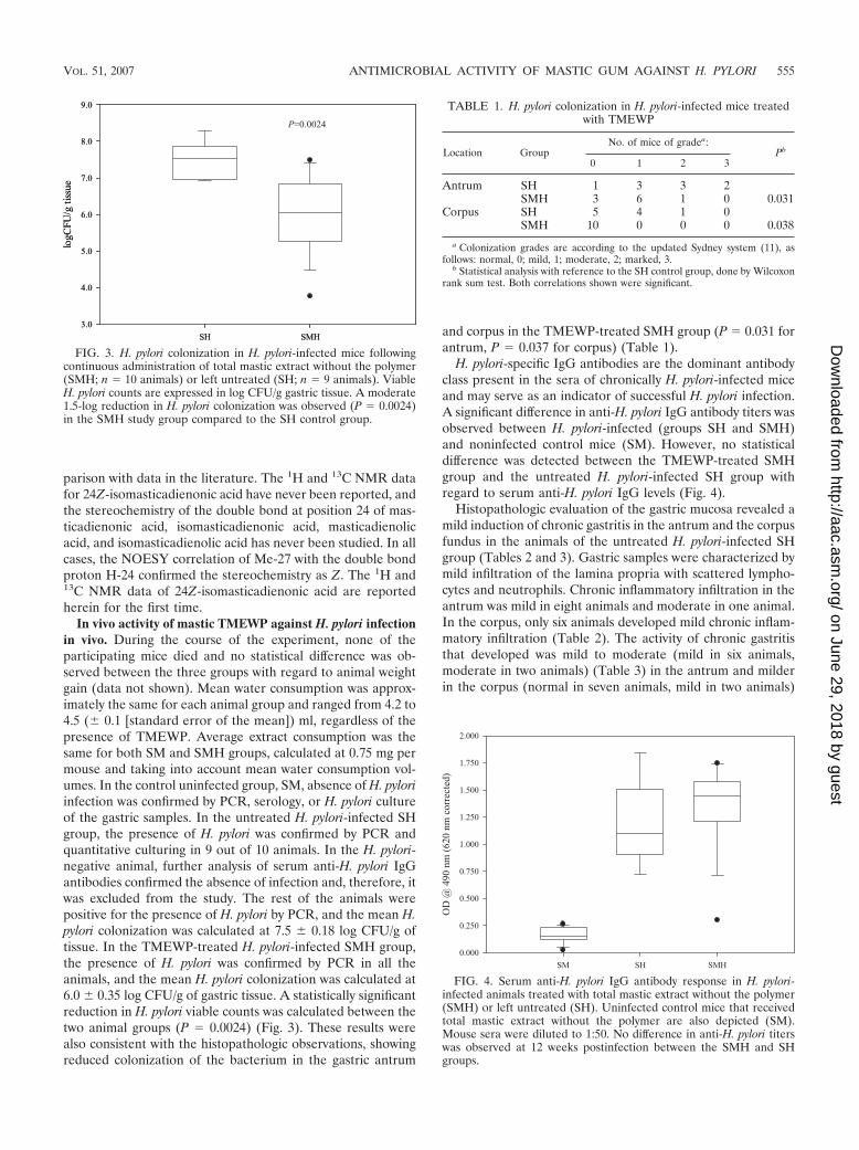
In vivo activity of mastic TMEWP against H. pylori infectionin vivo. During the course of the experiment, none of theparticipating mice died and no statistical difference was ob-served between the three groups with regard to animal weightgain (data not shown). Mean water consumption was approx-imately the same for each animal group and ranged from 4.2 to4.5 ( 0.1 [standard error of the mean]) ml, regardless of thepresence of TMEWP. Average extract consumption was thesame for both SM and SMH groups, calculated at 0.75 mg permouse and taking into account mean water consumption vol-umes. In the control uninfected group, SM, absence of H. pyloriinfection was confirmed by PCR, serology, or H. pylori cultureof the gastric samples. In the untreated H. pylori-infected SHgroup, the presence of H. pylori was confirmed by PCR andquantitative culturing in 9 out of 10 animals. In the H. pylori-negative animal, further analysis of serum anti-H. pylori IgGantibodies confirmed the absence of infection and, therefore, itwas excluded from the study. The rest of the animals werepositive for the presence of H. pylori by PCR, and the mean H.pylori colonization was calculated at 7.5 0.18 log CFU/g oftissue. In the TMEWP-treated H. pylori-infected SMH group,the presence of H. pylori was confirmed by PCR in all theanimals, and the mean H. pylori colonization was calculated at6.0 0.35 log CFU/g of gastric tissue. A statistically significantreduction in H. pylori viable counts was calculated between thetwo animal groups (P � 0.0024) (Fig. 3). These results werealso consistent with the histopathologic observations, showingreduced colonization of the bacterium in the gastric antrum
and corpus in the TMEWP-treated SMH group (P � 0.031 forantrum, P � 0.037 for corpus) (Table 1).
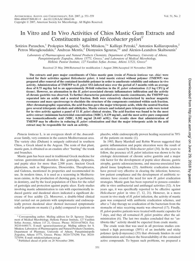
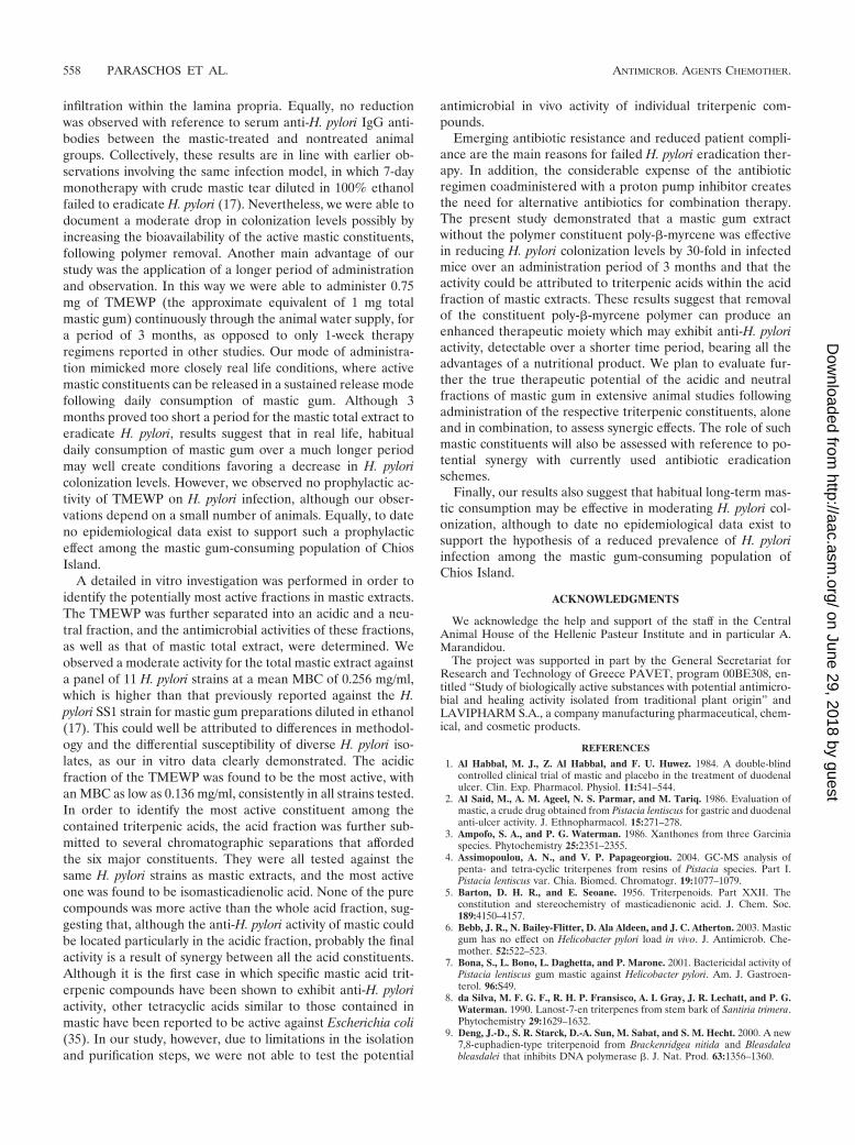
H. pylori-specific IgG antibodies are the dominant antibodyclass present in the sera of chronically H. pylori-infected miceand may serve as an indicator of successful H. pylori infection.A significant difference in anti-H. pylori IgG antibody titers wasobserved between H. pylori-infected (groups SH and SMH)and noninfected control mice (SM). However, no statisticaldifference was detected between the TMEWP-treated SMHgroup and the untreated H. pylori-infected SH group withregard to serum anti-H. pylori IgG levels (Fig. 4).
Histopathologic evaluation of the gastric mucosa revealed amild induction of chronic gastritis in the antrum and the corpusfundus in the animals of the untreated H. pylori-infected SHgroup (Tables 2 and 3). Gastric samples were characterized bymild infiltration of the lamina propria with scattered lympho-cytes and neutrophils. Chronic inflammatory infiltration in theantrum was mild in eight animals and moderate in one animal.In the corpus, only six animals developed mild chronic inflam-matory infiltration (Table 2). The activity of chronic gastritisthat developed was mild to moderate (mild in six animals,moderate in two animals) (Table 3) in the antrum and milderin the corpus (normal in seven animals, mild in two animals)
FIG. 3. H. pylori colonization in H. pylori-infected mice followingcontinuous administration of total mastic extract without the polymer(SMH; n � 10 animals) or left untreated (SH; n � 9 animals). ViableH. pylori counts are expressed in log CFU/g gastric tissue. A moderate1.5-log reduction in H. pylori colonization was observed (P � 0.0024)in the SMH study group compared to the SH control group.
TABLE 1. H. pylori colonization in H. pylori-infected mice treatedwith TMEWP
Location GroupNo. of mice of gradea:
Pb
0 1 2 3
Antrum SH 1 3 3 2SMH 3 6 1 0 0.031
Corpus SH 5 4 1 0SMH 10 0 0 0 0.038
a Colonization grades are according to the updated Sydney system (11), asfollows: normal, 0; mild, 1; moderate, 2; marked, 3.
b Statistical analysis with reference to the SH control group, done by Wilcoxonrank sum test. Both correlations shown were significant.
FIG. 4. Serum anti-H. pylori IgG antibody response in H. pylori-infected animals treated with total mastic extract without the polymer(SMH) or left untreated (SH). Uninfected control mice that receivedtotal mastic extract without the polymer are also depicted (SM).Mouse sera were diluted to 1:50. No difference in anti-H. pylori titerswas observed at 12 weeks postinfection between the SMH and SHgroups.
VOL. 51, 2007 ANTIMICROBIAL ACTIVITY OF MASTIC GUM AGAINST H. PYLORI 555
on June 29, 2018 by guesthttp://aac.asm
.org/D
ownloaded from

(Table 3). No development of glandular atrophy or intestinalmetaplasia was observed, as the time interval from the onset ofinfection was too short. In the extract-treated H. pylori-infectedSMH group, the development of chronic gastritis was similar tothe SH group (Tables 2 and 3), with marginally reduced num-bers of neutrophils infiltrating the lamina propria in somecases. However, statistical analysis with regards to chronic in-flammatory infiltration or the development of chronic activegastritis revealed no significant differences between the twoanimal groups. The collected data from the animal studiessuggested that continuous administration of 0.75 mg TMEWPto H. pylori-infected mice may moderately reduce H. pyloricolonizing numbers without a profound effect on the associatedgastritis. However, prophylactic administration of TMEWP didnot prevent H. pylori colonization (data not shown).
In vitro activity of mastic total extract, fractions, and com-pounds against H. pylori strain SS1. Having observed a mod-erate antimicrobial effect in vivo against H. pylori, we pro-ceeded to investigate the potential in vitro anti-H. pyloriactivity of TMEWP and its acidic and neutral fractions againsta panel of 10 clinical isolates of H. pylori and the CCUG 38771reference strain (Table 4). Figure 5 depicts characteristic killcurves for strains LAVHP-6 (one of the least susceptiblestrains) and LAVHP-7 (the most susceptible strain). Masticextracts exhibited concentration- and strain-dependent bacte-ricidal activities. More specifically, in all strains tested, withoutexception the acidic fraction exhibited the highest activity, witha mean MBC of 0.136 mg/ml, followed by the TMEWP (MBC,0.256 mg/ml). Reduced activity was observed for the neutralfraction of the TMEWP (0.638 mg/ml). Up to twofold differenceswere observed in the MBC between individual strains tested, and
only in the case of LAVHP-7 strain was a higher susceptibilityagainst the TMEWP and its acidic fraction observed.
Having obtained the highest activity with the acidic fractionof the TMEWP, we proceeded to test the isolated pure acidiccompounds for anti-Helicobacter activity. Highest overall activ-ity was obtained consistently and for all 11 of the H. pyloristrains tested with isomasticadienolic acid, with a mean MBCof 0.202 mg/ml (0.443 mM), followed by masticadienolic (0.220mg/ml [0.482 mM]), oleanonic (0.292 mg/ml [0.643 mM]), andmoronic acid (0.310 mg/ml [0.683 mM]) (Table 5). Interest-ingly, the 3-oxo derivatives, isomasticadienonic and masticadi-enonic acids, showed reduced activity compared to the corre-sponding 3-hydroxyl derivatives.
DISCUSSION
All previous in vivo studies evaluating activity of mastic gumagainst H. pylori have used a crude mastic preparation whichcontained a high percentage (30%) of an insoluble and stickypolymer. We speculated that the presence of the polymer hin-dered potential in vivo activity of mastic during oral adminis-tration, and for this reason we prepared TMEWP. We haveverified that the chemical consistency of TMEWP was virtuallyidentical to that of crude mastic gum, except for the absence ofthe polymer, and it also presented better solubility propertiesand increased concentration of active constituents. Previousanimal studies for the determination of the potential anti-Helicobacter activity of mastic gum were organized on a short-term administration schedule. However, we have extended ouradministration and hence tested the TMEWP activity over aperiod of 3 months.
In the present study we utilized an established H. pyloriinfection model to evaluate the potential therapeutic effect ofcontinuous TMEWP administration on H. pylori colonizationand development of associated gastritis. This model involvesthe mouse-adapted H. pylori Sydney strain 1 (SS1), which col-onizes the C57BL/6 mouse heavily and leads to the develop-ment of appreciable levels of gastritis closely mimicking humanH. pylori infection (16, 19, 31). The particular infection model
TABLE 2. Chronic inflammatory infiltrationa in H. pylori-infectedmice treated with TMEWP
Location GroupNo. of mice of gradeb:
Pc
0 1 2a
Antrum SH 0 8 1SMH 0 8 2 0.713
Corpus SH 3 6 0SMH 7 3 0 0.569
a Lymphocyte infiltration.b Histopathology grades are according to the updated Sydney system (11) as
follows: normal, 0; mild, 1; moderate, 2.c Statistical analysis with reference to the SH control group, done by Wilcoxon
rank sum test.
TABLE 3. Activity of chronic gastritisa in H. pylori-infected micetreated with TMEWP
Location GroupNo. of mice of gradeb:
Pc
0 1 2a
Antrum SH 1 6 2SMH 5 2 3 0.369
Corpus SH 7 2 0SMH 8 1 1 0.967
a Neutrophil infiltration.b Histopathology grades are according to the updated Sydney system (11) as
follows: normal, 0; mild, 1; moderate, 2.c Statistical analysis with reference to the SH control group, done by Wilcoxon
rank sum test.
TABLE 4. Minimum bactericidal concentrations of mastic extractsa
on H. pylori strains
Strain
MBC (mg/ml)
Totalextract
Acidicfraction
Neutralfraction
CCUG 38771 0.390 0.240 1.560LAVHP-1 0.195 0.120 0.390LAVHP-2 0.195 0.120 0.780LAVHP-3 0.195 0.120 0.780LAVHP-4 0.390 0.120 0.780LAVHP-5 0.195 0.120 0.390LAVHP-6 0.390 0.120 0.780LAVHP-7 0.090 0.060 0.390LAVHP-8 0.390 0.120 0.390LAVHP-9 0.195 0.240 0.390LAVHP-10 0.195 0.120 0.390
a With the exception of the acidic fraction, for which successive twofold dilu-tions ranged from 0.060 to 1.920 mg/ml, all other extracts were tested at a finalconcentration range of 0.049 to 1.560 mg/ml.
556 PARASCHOS ET AL. ANTIMICROB. AGENTS CHEMOTHER.
on June 29, 2018 by guesthttp://aac.asm
.org/D
ownloaded from

has been successfully utilized in the past to evaluate potentialanti-H. pylori activity of a number of agents, such as antibiotics(14, 15) or lactic acid-producing bacteria (27, 28).
Our experiments showed that the mastic total extract couldmoderately reduce H. pylori colonization in the antrum andcorpus of the stomach. The reduction in colonization levelscalculated was approximately 30-fold, in the range of 1.5 logCFU/g of tissue. These results were in concurrence with thevisible reduction in H. pylori colonization observed in the his-topathology evaluations. However, such a moderate fall in H.pylori colonizing numbers could not support any attenuation ofthe H. pylori-associated chronic active gastritis. We have doc-umented no such decline in either neutrophilic or lymphocytic
FIG. 5. Bactericidal activity of mastic gum extracts against H. pylori in a liquid medium. H. pylori strains LAVHP-6 (more resistant strain) andLAVHP-7 (most susceptible strain) were cultured under microaerophilic conditions in BHI as described in Materials and Methods and exposedto acidic, TMEWP, and neutral fractions at the concentrations depicted in the legend. After further incubation, viability was determined at eachtime point.
TABLE 5. Mean MBC of mastic triterpenic acid compounds withinthe mastic acidic fraction against H. pyloria
Substance Mean MBC inmg/ml (mM)
24Z-Isomasticadienonic acid................................................0.333 (0.733)24Z-Masticadienonic acid ....................................................0.350 (0.770)24Z-Isomasticadienolic acid.................................................0.202 (0.443)24Z-Masticadienolic acid......................................................0.220 (0.482)Oleanonic acid.......................................................................0.292 (0.643)Moronic acid..........................................................................0.310 (0.683)
a H. pylori strains are described in Table 4.
VOL. 51, 2007 ANTIMICROBIAL ACTIVITY OF MASTIC GUM AGAINST H. PYLORI 557
on June 29, 2018 by guesthttp://aac.asm
.org/D
ownloaded from
infiltration within the lamina propria. Equally, no reductionwas observed with reference to serum anti-H. pylori IgG anti-bodies between the mastic-treated and nontreated animalgroups. Collectively, these results are in line with earlier ob-servations involving the same infection model, in which 7-daymonotherapy with crude mastic tear diluted in 100% ethanolfailed to eradicate H. pylori (17). Nevertheless, we were able todocument a moderate drop in colonization levels possibly byincreasing the bioavailability of the active mastic constituents,following polymer removal. Another main advantage of ourstudy was the application of a longer period of administrationand observation. In this way we were able to administer 0.75mg of TMEWP (the approximate equivalent of 1 mg totalmastic gum) continuously through the animal water supply, fora period of 3 months, as opposed to only 1-week therapyregimens reported in other studies. Our mode of administra-tion mimicked more closely real life conditions, where activemastic constituents can be released in a sustained release modefollowing daily consumption of mastic gum. Although 3months proved too short a period for the mastic total extract toeradicate H. pylori, results suggest that in real life, habitualdaily consumption of mastic gum over a much longer periodmay well create conditions favoring a decrease in H. pyloricolonization levels. However, we observed no prophylactic ac-tivity of TMEWP on H. pylori infection, although our obser-vations depend on a small number of animals. Equally, to dateno epidemiological data exist to support such a prophylacticeffect among the mastic gum-consuming population of ChiosIsland.
A detailed in vitro investigation was performed in order toidentify the potentially most active fractions in mastic extracts.The TMEWP was further separated into an acidic and a neu-tral fraction, and the antimicrobial activities of these fractions,as well as that of mastic total extract, were determined. Weobserved a moderate activity for the total mastic extract againsta panel of 11 H. pylori strains at a mean MBC of 0.256 mg/ml,which is higher than that previously reported against the H.pylori SS1 strain for mastic gum preparations diluted in ethanol(17). This could well be attributed to differences in methodol-ogy and the differential susceptibility of diverse H. pylori iso-lates, as our in vitro data clearly demonstrated. The acidicfraction of the TMEWP was found to be the most active, withan MBC as low as 0.136 mg/ml, consistently in all strains tested.In order to identify the most active constituent among thecontained triterpenic acids, the acid fraction was further sub-mitted to several chromatographic separations that affordedthe six major constituents. They were all tested against thesame H. pylori strains as mastic extracts, and the most activeone was found to be isomasticadienolic acid. None of the purecompounds was more active than the whole acid fraction, sug-gesting that, although the anti-H. pylori activity of mastic couldbe located particularly in the acidic fraction, probably the finalactivity is a result of synergy between all the acid constituents.Although it is the first case in which specific mastic acid trit-erpenic compounds have been shown to exhibit anti-H. pyloriactivity, other tetracyclic acids similar to those contained inmastic have been reported to be active against Escherichia coli(35). In our study, however, due to limitations in the isolationand purification steps, we were not able to test the potential
antimicrobial in vivo activity of individual triterpenic com-pounds.
Emerging antibiotic resistance and reduced patient compli-ance are the main reasons for failed H. pylori eradication ther-apy. In addition, the considerable expense of the antibioticregimen coadministered with a proton pump inhibitor createsthe need for alternative antibiotics for combination therapy.The present study demonstrated that a mastic gum extractwithout the polymer constituent poly-�-myrcene was effectivein reducing H. pylori colonization levels by 30-fold in infectedmice over an administration period of 3 months and that theactivity could be attributed to triterpenic acids within the acidfraction of mastic extracts. These results suggest that removalof the constituent poly-�-myrcene polymer can produce anenhanced therapeutic moiety which may exhibit anti-H. pyloriactivity, detectable over a shorter time period, bearing all theadvantages of a nutritional product. We plan to evaluate fur-ther the true therapeutic potential of the acidic and neutralfractions of mastic gum in extensive animal studies followingadministration of the respective triterpenic constituents, aloneand in combination, to assess synergic effects. The role of suchmastic constituents will also be assessed with reference to po-tential synergy with currently used antibiotic eradicationschemes.
Finally, our results also suggest that habitual long-term mas-tic consumption may be effective in moderating H. pylori col-onization, although to date no epidemiological data exist tosupport the hypothesis of a reduced prevalence of H. pyloriinfection among the mastic gum-consuming population ofChios Island.
ACKNOWLEDGMENTS
We acknowledge the help and support of the staff in the CentralAnimal House of the Hellenic Pasteur Institute and in particular A.Marandidou.
The project was supported in part by the General Secretariat forResearch and Technology of Greece PAVET, program 00BE308, en-titled “Study of biologically active substances with potential antimicro-bial and healing activity isolated from traditional plant origin” andLAVIPHARM S.A., a company manufacturing pharmaceutical, chem-ical, and cosmetic products.
REFERENCES
1. Al Habbal, M. J., Z. Al Habbal, and F. U. Huwez. 1984. A double-blindcontrolled clinical trial of mastic and placebo in the treatment of duodenalulcer. Clin. Exp. Pharmacol. Physiol. 11:541–544.
2. Al Said, M., A. M. Ageel, N. S. Parmar, and M. Tariq. 1986. Evaluation ofmastic, a crude drug obtained from Pistacia lentiscus for gastric and duodenalanti-ulcer activity. J. Ethnopharmacol. 15:271–278.
3. Ampofo, S. A., and P. G. Waterman. 1986. Xanthones from three Garciniaspecies. Phytochemistry 25:2351–2355.
4. Assimopoulou, A. N., and V. P. Papageorgiou. 2004. GC-MS analysis ofpenta- and tetra-cyclic triterpenes from resins of Pistacia species. Part I.Pistacia lentiscus var. Chia. Biomed. Chromatogr. 19:1077–1079.
5. Barton, D. H. R., and E. Seoane. 1956. Triterpenoids. Part XXII. Theconstitution and stereochemistry of masticadienonic acid. J. Chem. Soc.189:4150–4157.
6. Bebb, J. R., N. Bailey-Flitter, D. Ala Aldeen, and J. C. Atherton. 2003. Masticgum has no effect on Helicobacter pylori load in vivo. J. Antimicrob. Che-mother. 52:522–523.
7. Bona, S., L. Bono, L. Daghetta, and P. Marone. 2001. Bactericidal activity ofPistacia lentiscus gum mastic against Helicobacter pylori. Am. J. Gastroen-terol. 96:S49.
8. da Silva, M. F. G. F., R. H. P. Fransisco, A. I. Gray, J. R. Lechatt, and P. G.Waterman. 1990. Lanost-7-en triterpenes from stem bark of Santiria trimera.Phytochemistry 29:1629–1632.
9. Deng, J.-D., S. R. Starck, D.-A. Sun, M. Sabat, and S. M. Hecht. 2000. A new7,8-euphadien-type triterpenoid from Brackenridgea nitida and Bleasdaleableasdalei that inhibits DNA polymerase �. J. Nat. Prod. 63:1356–1360.
558 PARASCHOS ET AL. ANTIMICROB. AGENTS CHEMOTHER.
on June 29, 2018 by guesthttp://aac.asm
.org/D
ownloaded from
10. de Pascual Teresa, J., J. G. Urones, P. Basabe, M. J. Sexmero Cuadrado, andR. Fernandez Moro. 1986. Triterpens from Euphorbia broteri. Phytochemis-try 26:1767–1776.
11. Dixon, M. F., R. M. Genta, J. H. Yardley, and P. Correa. 1996. Classificationand grading of gastritis. The updated Sydney system. Am. J. Surg. Pathol.20:1161–1181.
12. Huwez, F. U., D. Thirlwell, A. Cockayne, and D. A. Ala’Aldeen. 1998. Masticgum kills Helicobacter pylori. N. Engl. J. Med. 339:1946.
13. Ito, J., F.-R. Chang, H.-K. Wang, Y. K. Park, M. Ikegaki, N. Kilgore, andK.-H. Lee. 2001. Anti-AIDS agents. 48. Anti-HIV activity of moronic acidderivatives and the new melliferone-related trierpenoid isolated from Bra-zilian propolis. J. Nat. Prod. 64:1278–1281.
14. Jenks, P. J., A. Labigne, and R. L. Ferrero. 1999. Exposure to metronidazolein vivo readily induces resistance in Helicobacter pylori and reduces theefficacy of eradication therapy in mice. Antimicrob. Agents Chemother.43:777–781.
15. Jenks, P. J., R. L. Ferrero, J. Tankovic, J. M. Thiberge, and A. Labigne. 2000.Evaluation of nitrofurantoin combination therapy of metronidazole-sensitiveand -resistant Helicobacter pylori infections in mice. Antimicrob. AgentsChemother. 44:2623–2629.
16. Lee, A., J. O’Rourke, M. C. de Ungria, B. Robertson, G. Daskalopoulos, andM. F. Dixon. 1997. A standardized mouse model of Helicobacter pylori in-fection: introducing the Sydney strain. Gastroenterology 112:1386–1397.
17. Loughlin, M. F., D. A. Ala’Aldeen, and P. J. Jenks. 2003. Monotherapy withmastic does not eradicate Helicobacter pylori infection from mice. J. Antimi-crob. Chemother. 51:367–371.
18. Lu, J. J., C. L. Perng, R. Y. Shyu, C. H. Chen, Q. Lou, S. K. Chong, and C. H.Lee. 1999. Comparison of five PCR methods for detection of Helicobacter-pylori DNA in gastric tissues. J. Clin. Microbiol. 37:772–774.
19. Mahler, M., S. Janke, S. Wagner, and H. J. Hedrich. 2002. Differentialsusceptibility of inbred mouse strains to Helicobacter pylori infection. Scand.J. Gastroenterol. 37:267–278.
20. Marner, F.-J., A. Freyer, and J. Lex. 1991. Triterpenoids from gum mastic,the resin of Pistacia lentiscus. Phytochemistry 30:3709–3712.
21. Marone, P., L. Bono, E. Leone, S. Bona, E. Carretto, and L. Perversi. 2001.Bactericidal activity of Pistacia lentiscus mastic gum against Helicobacterpylori. J. Chemother. 13:611–614.
22. Mulholland, D. A., and J. J. Nair. 1994. Triterpenoids from Disoxylumpettigrewianum. Phytochemistry 37:1409–1411.
23. Peek, R. M., and J. E. Crabtree. 2006. Helicobacter infection and gastricneoplasia. J. Pathol. 208:233–248.
24. Pozzo-Balbi, T., L. Nobile, G. Scapini, and M. Cini. 1978. The triterpenoidacids of Schinus molle. Phytochemistry 17:2107–2110.
25. Reddy, G. C., S. Rangaswami, and R. Sunder. 1977. Triterpenoids of thestem bark of Gardenia gummifera. Planta Med. 32:206–211.
26. Seoane, E. 1956. Further crystalline constituents of gum mastic. J. Am.Chem. Soc. 189:4158–4160.
27. Sgouras, D. N., E. G. Panayotopoulou, B. Martinez-Gonzalez, K. Petraki, S.Michopoulos, and �. Mentis. 2005 Lactobacillus johnsonii La1 attenuatesHelicobacter pylori-associated gastritis and reduces the levels of proinflam-matory chemokines in C57BL/6 mice. Clin. Diagn. Lab. Immunol. 12:1378–1386.
28. Sgouras, D., P. Maragkoudakis, K. Petraki, B. Martinez-Gonzalez, E. Eriotou,S. Michopoulos, G. Kalantzopoulos, E. Tsakalidou, and A. Mentis. 2004. Invitro and in vivo inhibition of Helicobacter pylori by Lactobacillus casei strainShirota. Appl. Environ. Microbiol. 70:518–526.
29. Shirane, N., Y. Hashimoto, K. Ueda, H. Takenaka, and K. Katoh. 1996.Ring-A cleavage of 3-oxo-olean-12-3n-28-oic acid by the fungus Chaetomiumlongirostre. Phytochemistry 43:99–104.
30. Sung, T. V., J. Peter-Katalinic, and G. Adam. 1991. A bidesmosidic triter-penoid saponin from Schefflera octophylla. Phytochemistry 30:3717–3720.
31. Sutton, P., J. Wilson, and A. Lee. 2000. Further development of the Helico-bacter pylori mouse vaccination model. Vaccine 18:2677–2685.
32. Tassou, C. C., and G. J. E. Nychas. 1995. Antimicrobial activity of theessential oil of mastic gum (Pistacia lentiscus var. chia) on gram positive andgram negative bacteria in broth and model food system. Int. Biodeter. Bio-degradation 36:411–420.
33. van den Berg, K. J., J. van der Horst, J. J. Boon, and O. O. Sudmeijer. 1998.cis-1,4-Poly-�-myrcene: the structure of the polymeric fraction of masticresin (Pistacia lentiscus L.) elucidated. Tetrahedron Lett. 39:2645–2648.
34. Warren, J. R., and B. Marshall. 1983. Unidentified curved bacilli on gastricepithelium in active chronic gastritis. Lancet 321:1273–1275.
35. Yang, S.-P., and J.-M. Yue. 2001. Two novel cytotoxic and antimicrobialtriterpenoids from Pseudolarix kaempferi. Bioorg. Med. Chem. Lett. 11:3119–3122.
VOL. 51, 2007 ANTIMICROBIAL ACTIVITY OF MASTIC GUM AGAINST H. PYLORI 559
on June 29, 2018 by guesthttp://aac.asm
.org/D
ownloaded from