Embed Size (px)
Citation preview
Manjunathan and Ragunathan Biological Research (2015) 48:29 DOI 10.1186/s40659-015-0021-z
RESEARCH ARTICLE Open Access
In ovo administration of humanrecombinant leptin shows dose dependentangiogenic effect on chickenchorioallantoic membrane
Reji Manjunathan and Malathi Ragunathan*Abstract
Background: Leptin, the cytokine produced by white adipose tissue is known to regulate food energy homeostasisthrough its hypothalamic receptor. In vitro studies have demonstrated that leptin plays a major role in angiogenesisthrough binding to the receptor Ob-R present on ECs by stimulating and initiating new capillary like structures fromECs. Various in vivo studies indicate that leptin has diverse effect on angiogenesis. A few reports have showed thatleptin exerts pro angiogenic effects while some suggested that it has antiangiogenic potential. It is theoreticallyhighly important to understand the effect of leptin on angiogenesis to use as a therapeutic molecule in variousangiogenesis related pathological conditions. Chicken chorio allantoic membrane (CAM) on 9th day of incubationwas incubated with 1, 3 and 5 μg concentration of HRL for 72 h using gelatin sponge. Images where taken afterevery 24 h of incubation and analysed with Angioguant software. The treated area was observed under microscopeand histological evaluation was performed for the same. Tissue thickness was calculated morphometrically fromhaematoxylin and eosin stained cross sections. Reverse transcriptase PCR and immunohistochemistry were alsoperformed to study the gene and protein level expression of angiogenic molecules.
Results: HRL has the ability to induce new vessel formation at the treated area and growth of the newly formedvessels and cellular morphological changes occur in a dose dependent manner. Increase in the tissue thickness atthe treated area is suggestive of initiation of new capillary like structures. Elevated mRNA and protein levelexpression of VEGF165 and MMP2 along with the activation of ECs as demonstrated by the presence of CD34expression supports the neovascularization potential of HRL.
Conclusion: Angiogenic potential of HRL depends on the concentration and time of incubation and is involvedin the activation of ECs along with the major interaction of VEGF 165 and MMP2. It is also observed that 3 μg ofHRL exhibits maximum angiogenic potential at 72 h of incubation. Thus our data suggest that dose dependentangiogenic potential HRL could provide a novel role in angiogenic dependent therapeutics such as ischemia andwound healing conditions.
Keywords: Angiogenesis, HRL, Human Recombinant Leptin, CAM, chorioallantoic membrane, ECs, endothelial cells,VEGF, vascular endothelial growth factor, MMP, matrix metalloproteinase, Blood vessels, CD34
* Correspondence: [email protected] of Genetics, Dr. ALM PG IBMS, Taramani Campus, University ofMadras, Chennai 600 113, Tamilnadu, India
© 2015 Manjunathan and Ragunathan. This is an Open Access article distributed under the terms of the Creative CommonsAttribution License (http://creativecommons.org/licenses/by/4.0), which permits unrestricted use, distribution, andreproduction in any medium, provided the original work is properly credited. The Creative Commons Public DomainDedication waiver (http://creativecommons.org/publicdomain/zero/1.0/) applies to the data made available in this article,unless otherwise stated.

Manjunathan and Ragunathan Biological Research (2015) 48:29 Page 2 of 13
BackgroundThe Ob-gene product leptin is 16 kDa peptide hormone,produced mainly by white adipose tissue is proposed toplay a key role in the regulation of body weight andthermogenesis through the receptor found in the hypo-thalamus [1]. Accumulating evidences from in vitro andin vivo assays suggest that leptin promote endothelialcell (EC) proliferation and survival in favour of angio-genesis [2, 3]. In vivo and in vitro results from Hyunget al. study indicates that angiogenic potential of leptinis mediated by matrix metalloproteinase (MMPs) [4].However, the actual mechanism behind the involvementof matrix metalloproteinase in leptin mediated angiogen-esis is not clear.The effect of leptin on angiogenesis is still not under-
stood as some reports have suggested its potential to in-duce angiogenesis while some have indicated of its antiangiogenic effect [5–9]. Recently it has been reportedthat in ovo administration of leptin inhibited angiogen-esis on chicken chorio allantoic membrane (CAM) [10].Leptin has been administered as a therapeutic moleculein various angiogenesis related pathological conditionsespecially in wound healing [11]. However a quantitativein vivo evaluation on the effect of leptin on angiogenesisis important because of its therapeutic application po-tential in the pathology of angiogenesis dependent con-ditions. Therefore in the present study we examined theangiogenic potential of human recombinant leptin (HRL)using well vascularized CAM of developing chicken em-bryo (Gallus gallus domesticus). The advantage of CAMassay is that it is a highly vascularized structure with po-tential growth and this in ovo vivo system is highly usefulto understand the physiological angiogenesis and hencewidely used for the screening of various compounds fortheir angiogenic activity [12].In the present study we analysed and compared the
angiogenic ability of HRL of varying concentrations suchas 1, 3 and 5 μg for an incubation period of 72 h using lateCAM. Gelatin sponges soaked with leptin were placed onthe membrane at 9th day of post incubation so as to allowslow delivery of the chemical at the treated area with lessor no inflammation [13, 14]. The ability of HRL to inducenew vessel growth at the treated area is directly visualizedfrom the CAM images taken at different time period of in-cubation and growth of these vessels measured with ImageJ and Angioquant MATLAB softwares [14–16]. Angio-genic response of HRL at the treated area is further ana-lysed in detail from the histological sections. We alsoexamined the expression of major angiogenic molecules atthe molecular and protein level to understand the involve-ment of these factors on HRL induced angiogenesis. Ourfindings suggest that the potential of HRL to induceangiogenesis depends on various physiological factorsespecially dose and time of incubation. The result have
demonstrated that HRL favours neovascularizationthrough sprouting of vessels which is accelerated by theexpression of VEGF165 and MMP2 in a dose dependentway in chicken CAM vasculature.
MethodsMaterialsFertilized white leghorn chicken eggs were purchasedfrom Tamil Nadu Poultry Research Station, Madras Vet-erinary University, Nandanam, Tamil Nadu, India. Gel-atin sponges purchased from Jhonson & Jhonson PvtLmtd, India, Paraffin film and wax were purchased fromSigma, Aldrich, USA. Haematoxylin and Eosin stain pur-chased from Medox, India and Human recombinant lep-tin from MP Biomedicals, Inc. France. TRIzol reagent,agarose and EtBr were from Sigma, Aldrich, USA.ImProm-11™ Reverse Transcriptase kit and GoTaq GreenMaster Mix PCR amplification kit were from Promega,USA, Oligo (dt) of length 18-meres from eurofins, mwgoperon, Germany, Random hexamers from MP Biomedi-cals, USA. All primers were purchased from Bioserve,India. DNA ladders purchased from Invitrogen, USA.DAB system purchased from Bangalore Genei, India.CD34 antibody (Endothelial Cell Marker, Cluster desig-nation 34) from US Biological, USA and VEGF A fromCALBIOCHEM, EMD. Bradford reagent and FITC (Goatant-rabbit IgG were from Bangalore Genei, India. SDS-PAGE Standards marker was from BIO-RAD, CANADAand gelatin from Medox, India. Rabbit polyclonal MMP2is a kind gift from Dr. Li Haiqing, MD, Ph.D, −Technologytransfer specialist, National Cancer Institute, Rockville,USA. Unless otherwise specified all other common re-agents and chemicals were purchased from Sigma,Aldrich, USA.
The in ovo CAM modelFertilized white leghorn chicken eggs weighing 50 ± 2 g,were incubated at 37 °C in a ‘humidified atmosphere(>60 % relative humidity) as per the protocol describedin Hen’s Egg Test - Chorioallantoic Membrane (HET-CAM) method adapted from ZEBET (The GermanCentre for the Documentation and validation of Alterna-tive Methods, Republic of Germany). At day 3 of postincubation, 2 to 3 ml of albumin was withdrawn, using a21 gauge needle, through the large blunt edge of the eggin order to minimize the adhesion of the shell membranewith CAM. A square window of 2 cm2 was opened in theegg shell and sealed with paraffin film to preventdehydration and the eggs were incubated further. At day 9of post incubation, gelatin sponges of size of 1 mm3 wereplaced on top of the growing CAM under sterile condition[14, 17] and were soaked with 15 μl volume of 1, 3 and5 μg concentration of HRL. Control eggs were incubatedwith 1X PBS (pH-7.3). The window was closed with a
Manjunathan and Ragunathan Biological Research (2015) 48:29 Page 3 of 13
transparent adhesive tape and eggs were incubated for72 h until it reached post incubation day 12. CAM werephotographed at 0, 24, 48 and 72 h using Canon digitalcamera and images were analysed with Image J and Angio-quant Toolbox, MATLAB 6.5 software to measure totallength and size of the blood vessels (micrometre) from thearea of treatment.
Light microscope analysisAfter 72 h of incubation the area of the CAM treated withHRL was detached carefully. The excised membrane waskept on glass slides and images were taken using lightmicroscope both at 4 and 10× magnification to view thegrowth of capillaries [18].
HistologyAfter 72 h of incubation area of the CAM treated withHRL was flooded with Bouin’s fixative solution. Around1 cm2 of the membrane around the treated area was re-moved carefully using forceps and surgical scissors anddehydrated through graded series of alcohol (50 %, 70 %,90 % and absolute) and embedded in paraffin wax. Verti-cal cross tissue sections (7 μm in thickness) were takenusing Rotary Microtome (Weswicox, Japan). Sections weretreated with alcohol in ascending order (absolute, 90 %,70 %, and 50 %) and cleared with xylene before stainingwith haematoxylin and eosin. After mounting with DPX,the histological sections were observed under light micro-scope at 40× magnification for qualitative assessment andimages were recorded using Nikon Camera attached withlight microscope at 10× magnification [14].
Morphometric analysis of CAM tissue thickness (DCAM)Thickness of the CAM for all the groups treated withHRL were measured from haematoxylin and eosinstained vertical cross sections using a calibrated object-ive at 40× magnification with 10 × 10 calibrated grid at10× ocular. Distances between the chorionic and allan-toic epithelial layers were measured in micrometre at 6different locations from the same sample and is repeatedfor six serial cross sections of the same. Average tissuethickness was calculated from each tissue sample of thesame and obtained a mean DCAM thickness [14, 19].
Semi-Quantitative Reverse Transcriptase–PolymeraseChain Reaction (RT-PCR)Total RNA was isolated from CAM treated with HRLusing TRIzol reagent (100 mg/1 ml). The quantity andthe purity of the isolated RNA were checked using UV-visible spectrophotometer. cDNA of 20 μl volume wassynthesized using ImProm-11™ Reverse Transcriptase kitwith Oligo (dt) of length 18-meres and random hexam-ers. PCR amplification was performed using GoTaqGreen Master Mix kit and changes in the level of mRNA
expression of VEGF165 [20], VEGF121 [21], MMP2 [20],MMP9 [22] and GAPDH [23] were evaluated using PCRwith 100 pico moles of specific primers. The relative ex-pression level of each mRNA transcript was normalizedwith that of control. PCR products were subjected toelectrophoresis.
Gelatin zymographyCAM tissues at the treated area from each group werehomogenized (100 mg/ml) using Tris buffer (0.5 MTris–HCl (pH-6.8), 10 % SDS, glycerol and 0.01 %bromophenol blue) and centrifuged at 12,000 rpm/4 °C/10 min. The concentration of protein from the super-natant was determined using Bradford reagent and thegelatinase activity was examined on a 10 % SDS-PAGEelectrophoresis containing 1.0 mg/ml of gelatin. Proteinsamples of 25 μg/40 μl were loaded per well along with20 μl pertained SDS-PAGE Standards marker. Afterelectrophoresis, the gels were washed with 2.5 % ofTriton-X-100 and incubated in digestion buffer (50 mMTris HCl-pH-7.5,100 mM CaCl2, 1 μM ZnCl2, 1 % Tri-ton X-100, 0.02 % NaN3-100 ml) for 16 to 18 h at 37 °Cwith gentle agitation. The gel was stained with stainingsolution (0.05 % (w/v, Coomassie blue in 50 % methanoland 10 % acetic acid) for 1 h and de stained with metha-nol/acetic acid mixture. Gelatinase activity of MMP2was detected as clear white bands against background.The gels were scanned and images were recorded usingBIO-RAD Calibrated Densitometer Software (GS 800,USA). The density of the bands was calculated with PDQuest Advances Software and normalized with controlvalue [24].
ImmunohistochemistryThree different concentration namely 1, 3 and 5 μg ofHRL have been used of which 3 μg of HRL yielded max-imum angiogenic response when compared to other twoconcentrations. The deparafinized and dehydrated CAMof 5 μM thickness after treating with 3 μg of HRL wasallowed to undergo antigen retrieval process using So-dium Citrate (10 mM-pH 6.0) in a microwave oven for20 min followed by washing in DDH2O for 3X5 min in1X PBS (pH 7.3). Normal Goat Serum Blocking Solution(2 % goat serum,1 % BSA, 0.1 % cold fish skin gelatin,0.1 % Triton X-100, 0.05 % Tween- 20, 0.05 % SodiumAzide, 0.01 M PBS (pH 7.2) of 50 to 75 μl was added im-mediately on the sections and incubated for 1 h in a hu-midified chamber. After washing with 1X PBS, primaryantibodies of MMP2 (1:200), VEGF (VEGF A) (1:100)and CD34 (1:200) diluted in blocking serum were ap-plied on the sections and after overnight incubationrinsed with 1X PBS with 0.05 % of Tween-20. DilutedFITC (Goat ant-rabbit IgG) and HRP (both Goat anti-rabbit and Goat anti- mouse IgG) secondary antibodies
Manjunathan and Ragunathan Biological Research (2015) 48:29 Page 4 of 13
of 1:40 dilution was applied for 1 h according to manufac-turer’s instruction. For HRP conjugated secondary anti-bodies DAB system was used for colour development. Theslides were finally counterstained with Mayer’s haema-toxylin and mounted with 90 % of glycerol. For HRP con-jugated system the images were recorded using lightmicroscope and for FITC conjugation BX51 OlympusFluorescence Microscope at a wavelength of 515 nm withASI FISH View 5.5 software at 40× magnification [25].
Data analysis and statistical analysisAll experiments were independently performed in tripli-cate. Data were analysed using one way ANOVA analysisof variance test and Tukey post hoc test as appropriate(Sigma stat 2.0). Data were expressed as means + S.E.Mand P-values of *p = < 0.001 and #p = 0.001 were selectedfor showing statistically significant difference.
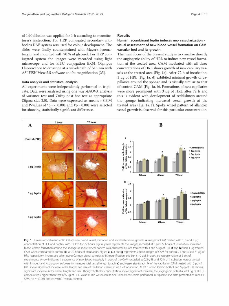
Fig. 1 Human recombinant leptin initiate new blood vessel formation and acconcentration of HRL and control with 1X PBS for 72 hours. Figure panel reprblood vessels formation around the sponge as spoke wheel pattern was obseCAM when compared to control (b) at 72 hours of incubation. Figure a, c, e aHRL respectively. Images are taken using Cannon digital camera at 4X magnifexperiments. Arrow indicates the presence of new blood vessels. b Images ofwith Image J and Angioquant software to measure total vessel length (graphHRL shows significant increase in the length and size of the blood vessels at 4significant increase in the vessel length and size. Though both the concentratcomparatively higher than that of 5 μg of HRL. Value at 0 h was taken as oneSEM, (*p = <0.001 and #p = 0.001 versus control)
ResultsHuman recombinant leptin induces neo vascularization -visual assessment of new blood vessel formation on CAMvascular bed and its growthThe main focus of the present study is to visualize directlythe angiogenic ability of HRL to induce new vessel forma-tion at the treated area. CAM incubated with all threeconcentrations of HRL shows growth of new capillary ves-sels at the treated area (Fig. 1a). After 72 h of incubation,1 μg of HRL (Fig. 1a. d) exhibited minimal growth of ca-pillaries around the sponge and is visually similar to thatof control CAM (Fig. 1a. b). Formations of new capillarieswere more prominent with 3 μg of HRL after 72 h andthis is evident with development of reddishness aroundthe sponge indicating increased vessel growth at thetreated area (Fig. 1a. f ). Spoke wheel pattern of allantoicvessel growth is observed for this particular concentration.
celerate vessel growth. a Images of CAM treated with 1, 3 and 5 μgesents the images recorded at 0 and 72 hours of incubation. Increasedrved in CAM treated with 3 and 5 μg of HRL (f and h) than 1 μg treatednd g represents 0 hour images of CAM for control , 1 and 3 and 5 μg ofication and bar is 10 μM. Images are representative of 3 set ofthe CAM recorded at 0, 24, 48 and 72 h of incubation were analyseda) and vessel size (graph b) of the capillaries. CAM treated with 3 μg of8 h of incubation. At 72 h of incubation both 3 and 5 μg of HRL showsion shows significant increase, the angiogenic potential of 3 μg of HRL is. Experiments were performed in triplicate and data presented as mean +

Manjunathan and Ragunathan Biological Research (2015) 48:29 Page 5 of 13
HRL of 5 μg also shows numerous scattered allantoic ves-sels in and around sponge at 72 h but with lesser vesselsgrowth when compared to that of 3 μg (Fig. 1a. h).From the above observation it can be inferred that,
both 3 and 5 μg concentration of HRL has the ability toinduce angiogenesis through new capillary formation at72 h of incubation. In order to conform this and also toarrive at the optimum working concentration for HRLwe measured the growth of the vessels in terms of itslength and size, from the images taken at 0, 24, 48 and72 h of incubation with Image J and AngioquantMATLAB software. This software is specially made forquantifying angiogenesis using in vitro assays and hasbeen employed for CAM with some modifications usingImage J software [11, 13]. Of the three different concentra-tions, used 3 μg of HRL demonstrates a significant in-crease in the vessel length (Fig. 1b. b) and size (Fig. 1b. b)at 48 h of incubation which is 1.99 fo1d greater than thatof control (#p = 0.001, *p = <0.001). At 72 h of incubationboth 3 and 5 μg of HRL induces a significant increase inthe vessel length and size (*p = <0.001) and of these two,3 μg of leptin shows 2.1 fold increases in vessel growth
Fig. 2 Human recombinant leptin able initiate sprouting of new vessels froand 5 μg of HRL and control with 1X PBS for 72 h (Figures b, d, f, h are thwith 3 and 5 μg of HRL (e, f, g and h) shows the presences of branches anblood vessel formation by sprouting and CAM treated with 1 μg of HRL (cof beginning of sprouting of blood vessels when compared to control (a abranches and sub branches at the treated area. Figure represents the imagmagnification of bar is 20 μM and are representative of 3 set of experimenthe main vessel
which is higher than that of 5 μg that showed which hasonly 1.8 fold increase. HRL of 1 μg also shows angiogenicpotential with increase in the vessel growth up to 1.5 fold,the value is not significant when compared to control thathas 1.2 fold increase in the vessel growth from 0 to 72 hof incubation. Thus it is obvious that HRL can induce newvessel formation at the treated area in a dose dependentmanner with 3 μg concentration having maximum angio-genic response.
Human recombinant leptin initiate growth of vessels bymeans of sprouting – visualising the morphology ofblood vessels on CAM vascular bedWe analysed the morphology of blood vessels in orderto confirm the sprouting angiogenic ability of HRL atthe treated area. Images of the CAMs were recorded im-mediately after 72 h of incubation using microscope. Itwas found that HRL is able to induce growth of newblood vessels from the existing one (Fig. 2). CAM incu-bated with 3 μg of HRL shows more number of sproutingvessels along with branches and sub branches at thetreated area (Fig. 2e). An enlarged view of the same
m existing one. Light microscopic images of CAM treated with 1, 3e enlarged version of a, c, e, g respectively). Images of CAM treatedd sub branches from the main blood vessels as an indication of newand d) shows the presence of bulging like structures as an indicationnd b). Among 3 and 5 μg of HRL 3 μg shows the presences of morees recorded at 72 h of incubation using light microscope at 20Xts. Arrow indicates the presence of branches and sub branches from

Manjunathan and Ragunathan Biological Research (2015) 48:29 Page 6 of 13
treated area shows the presence of more branches formthe main vessel and sub branches from the newly formedvessels, a feature directly suggesting the fact that HRL ofthis particular concentration and time of incubation exertsits maximum angiogenic activity (Fig. 2f). CAM treatedwith 5 μg of HRL also shows the presence of sprouting ofvessels from the main one with appearances of branchesand sub branches (Fig. 2g and h), but its potential to in-duce new vessel formation at the treated area is compara-tively lesser than that of 3 μg HRL. HRL of 1 μg alsoshows the presence of sprouting structures from the mainvessel, but is not remarkably strong when compared toother concentrations (Fig. 2c). An enlarged view of thetreated area for the same shows roughness throughout theouter surface of the main blood vessel along with bulgingappearance suggestive of its potential to initiate sproutingof new blood vessels at the treated area. Control CAMdoesn’t show any such sprout formation from the existingvessels (Fig. 2a) and the enlarged view of the same indi-cates stable blood vessel without any rough appearance orbud like structures from that of main one (Fig. 2b). Thus,HRL is able to induce sprouting of new vessels from theexisting one with maximum sprouting efficacy at 3 μgconcentration.
Fig. 3 Human recombinant leptin induces morphological changes in favoimages of CAM treated with 1, 3 and 5 μg of HRL and control with 1X PB(ae) epithelial layers with sub epithelial capillary network (SEC), CAM treatallantoic epithelial layer along with small vessels at the stroma region. CAlarge vessels at the surrounding of large one with increased tissue thickncomparatively lesser number of small vessels around the large one than Cwound or inflammation. Representative sections are 7 μM in thickness anlbv for large blood vessel and sbv for small blood vessel)
Human recombinant leptin induces morphologicalchanges leading to neo vascularization – histologicalobservation of the CAM vasculatureTo conform the angiogenic potential of HRL, we alsoanalysed the changes in the cellular morphology of theCAM vasculature from cross sections of the treated areastained with haematoxylin and eosin. Changes in themorphology of the CAM vasculature has been changedobserved for all three concentrations of HRL and is indi-cative of the positive angiogenic response due to HRL(Fig. 3). CAM incubated with 1 μg of HRL (Fig. 3b) showsirregular appearance of thin stratum (which contains bothchorionic (ce) and allantoic (ae) epithelial layers) with thepresences of few blood vessels at the primary stratum.CAM treated with 3 μg of HRL (Fig. 3c) shows vessels en-larged in size with remarkable irregular growth pattern atthin stratum. Morphology of ECs are altered those ECswhich are present at the capillary plexus/blood sinus area(which lies in between the primary and thin stratum)shows sprouting appearance typical for angiogenesis.Thickness of the primary stratum is also increased prob-ably due to cellular accumulation at the sub epithelialcapillary network (SEC). Presences of a number of smallblood vessels around the main vessels is suggestive of the
ur of angiogenesis. Haematoxylin and eosin stained histologicalS for 72 h. Control CAM (a) shows thin chorionic (ce) and allantoiced with 1 μg of HRL (b) shows comparatively thicker chorionic andM treated with 3 μg of HRL (c) shows the presences of numerousess at the primary stratum. CAM treated with 5 μg of HRL (d)AM treated with 3 μg of HRL. Gelatin sponges has not causedd were recorded at 10X magnification and bar is 50 μM. (v for vein,

Manjunathan and Ragunathan Biological Research (2015) 48:29 Page 7 of 13
angiogenic potential of leptin either by forming new ves-sels or by inducing the sprouting from the main one.CAM treated with 5 μg of HRL (Fig. 3d) also shows ir-regular appearances at thin stratum due to increase in thenumber of blood vessels and diminished accumulation ofcells were observed beneath SEC. Very few small vesselswere found around the large one as an indication of min-imal sprouting which is lesser than that observed with3 μg. Control CAM (Fig. 3a) shows a few scattered bloodvessels at primary stratum with uniform growth at thethin stratum due to growth of less number of blood ves-sels at this area. These observations demonstrates thatHRL is able to induce formation of new vessel growth byinducing positive morphological changes at the cellularlevel in favour of angiogenesis. Leptin of 3 μg concentra-tion is capable of eliciting promising cellular response thatcould lead to more angiogenic growth.
Human recombinant leptin increases the size of tissue bymeans of inducing new vessel growth. Quantitativemeasurement of the angiogenic potential of humanrecombinant leptin by morphometric measurement ofCAM tissue thickness (DCAM)The ability of 1, 3 and 5 μg of HRL to induce new ves-sels growth was further confirmed by measuring the sizeof the tissue from the from haematoxylin and eosinstained paraffin embedded vertical cross sections and isplotted as average tissue thickness (Fig. 4). Formation ofnew blood vessels or enlargement or growth of the exist-ing blood vessels will cause an increase in the thicknessof the CAM at the stroma and will push both the
Fig. 4 Human recombinant leptin increases the size of tissue bymeans of inducing new vessel growth. Total thickness of the tissueat the treated area with 1, 3 and 5 μg of HRL was measured fromhaematoxylin and eosin stained vertical cross section after 72 h.Control CAM was treated with 1X PBS. The distance between chorionicand allantoic epithelial layers was calculated morphometrically in μM.CAM treated with 3 μg of HRL shows significantly increased tissuethickness than other two concentrations. Material shrinkage is estimatedto be ~25 % relative to the fresh material in all cases. Experimentswere performed in triplicate and data presented as mean + SEM,(*p = <0.001versus control)
chorionic and allantoic epithelial layer a part. In thepresent study we calculated the distance between thesetwo layers (thin stratum) morphometrically in μM after72 h of incubation. It is found that thickness of the CAMis considerably increased significantly for 3 μg of HRL(*p = <0.001) when compared to other two studied con-centrations suggestive of its ability of to induce angiogen-esis under this concentration. In paraffin-embeddedtissues, material shrinkage is estimated to be ~25 % rela-tive to the fresh material and since all tissues were pre-pared similarly, tissue shrinkage is assumed to be same forall CAM zones. Thus, shrinkage corrections are not ne-cessary while comparing tissue thickness [26].
Angiogenic potential of human recombinant leptindepends on the activation of major angiogenic growthfactors - molecular profiling of VEGF 165, VEGF 121,bFGF2, MMP2 and MMP9The expression of primary angiogenic growth factorssuch as VEGF, bFGF2, MMP2 and MMP9 during leptininduced angiogenesis is analysed by measuring the vari-ation in the mRNA level. In our study it is observed thatmRNA level expression of these specified angiogenicgrowth factors increased when treated with HRL (Fig. 5).The intensity of the bands were measured as relative ODand given in Fig. 5b. There was a consistent increase inthe expression pattern for 3 μg HRL which is signifi-cantly higher (*p = <0.001) than that of other two con-centrations. It was also observed that for 5 μg of HRLthe relative mRNA level of VEGF165 and MMP2 in-creased significantly (*p = <0.001) owing to the involve-ment of these two main angiogenic growth factors onHRL induced angiogenesis. The gene profiling data indi-cates that the angiogenic ability of HRL mainly dependson the activation VEGF165 and MMP2 since the expres-sion value have increased significantly when treated with3 and 5 μg of HRL. Thus, HRL at 3 μg concentration isable to induce maximum angiogenic growth by increas-ing the gene expression of VEGF121, bFGF2, MMP9along with VGEF165 and MMP2.
Human recombinant leptin accelerates the gelatinaseactivity of MMP2The potential of HRL to induce neo vascularization wasmeasured intern by its ability to accelerate gelatinase ac-tivity of MMP2 to degrade ECM in order to favour ECsproliferation and migration. Figure 6a indicates that thegelatinase activity of MMP2 is increased indicating ECsproliferation and activation when treated with HRL. Theintensity value measured in terms of OD and the graphplotted (Fig. 6b) shows that the gelatinase activity ofMMP2 is increased significantly (*p = <0.001) whentreated with 3 and 5 μg of HRL. The gelatin zymogramanalysis of MMP2 activity demonstrates that HRL has

Fig. 5 Human recombinant leptin enhances the mRNA level expression of VEGF165, VEGF121, bFGF2, MMP2 and MMP9. Images of ReverseTranscriptase- PCR products of (a) VEGF165 (b) VEGF121 (c) bFGF2 (d) MMP2 and (e) MMP9 from CAM after treated with 1, 3 and 5 μg of HRLand control with 1X PBS for 72 h. GAPDH was used as internal loading control. The transcripts were confirmed by 100bp DNA ladder. Graphrepresents the OD value ratio of mRNA transcripts after normalizing with GADPH OD value of the same. The OD value was measured using ImageJ software. The relative level expression of specified genes were increased significantly for 3 μg of HRL and under 5 μg of HRL the relative expressionof VEGF 165 and MMP2 increased significantly after 72 h of incubation.. Each value is the mean ± SEM, *p= <0.001 versus control, n = 3.Experiments were performed in triplicate and data presented as mean + SEM. (Lane 1 - control, lane 2 – 1 μg HRL, lane 3 – 3 μg HRL andlane 4- 5 μg HRL)
Manjunathan and Ragunathan Biological Research (2015) 48:29 Page 8 of 13
the potential to accelerate neo vascularization by meansof increasing the degradation of ECM. This data also in-dicates that irrespective of the concentration, HRL is
able to accelerate the gelatinase activity of MMP2 whichin turn signifies that MMP2 plays a major role in HRLinduced angiogenesis.

Fig. 6 Human recombinant leptin enhances the gelatinase activityof MMP2. a Image represents the gelatin zymography of MMP2activity from CAM after treated with 1, 3 and 5 μg of HRL andcontrol with 1X PBS for 72 h. The image shows that the gelatinaseactivity was increased under 3 and 5 μg of HRL which is visible aswhite band against black background. Image is the result of 3 setof experiments. b MMP2 gelatinase activity was measured as ODvalue using Image J software. Percentage of MMP2 gelatinaseactivity increased significantly under 3 and 5 μg of HRL with higherpercentage under 3 μg of HRL. Each value is the mean ± SEM,*p = <0.001 versus control, n = 3. (Lane 1- protein weight marker,Lane 2 - control, lane 3–1 μg HRL, lane 4–3 μg HRL and lane5–5 μg HRL)
Manjunathan and Ragunathan Biological Research (2015) 48:29 Page 9 of 13
Human recombinant leptin enhances activation ofendothelial cellsFrom our data it is clear that the angiogenic potential ofHRL is maximum for 3 μg concentration enabling theformation of new blood vessels at the treated area. Acti-vation of ECs is considered as one of the major step inangiogenic cascades especially during sprouting angio-genesis [27]. The result obtained from the study confirmthe potential of HRL to activate ECs by identifying thepresence of CD34 (activated endothelial cell marker) ex-pression on ECs. The result indicates that the CD34 ex-pression is observed at capillary plexus or blood sinusarea in which ECs were pooled out along with angioblastcells (Fig. 7b) and the pattern of staining resembles morelike coiled structures with elongated protrusions suggest-ive of the sprouting of ECs under HRL (Fig. 7d). Alsothe ECs present at the luminal side of the vessels showshigher staining for CD34 than those present in the con-trol (Fig. 7a). Presence of these elongated ECs is suggest-ive of its enhanced proliferative capacity when treatedwith HRL which in turn lead to the formation of capillarylike structure during the process of angiogenesis especiallywhen sprouting angiogenesis occurs (Fig. 7c and e). Incontrol, those ECs which were present at the lumen of themain vessel shows CD34 expression and no staining was
observed at the blood sinus area (Fig. 7a). Thus it can beconcluded that HRL has the ability to enhance EC activa-tion which in turn can favour these cells to participateactively in angiogenesis.
Angiogenic potential of human recombinant leptinmainly depends on the activation of VEGF A and MMP2 –protein expressionMolecular profiling of angiogenic factors after treatmentwith HRL indicated that angiogenic potential of leptinmainly depends on the functional activation of VEGF A(VEGF 165) and MMP2. Hence we attempted to under-stand the same by scoring the protein level expression ofVEGF A and MMP2 using immunohistochemical method.Results show that with 3 μg concentration of HRL the ex-pression of VEGF A protein (Fig. 8b) is higher at thelumen of main vessels as well as at the surrounding smallones as an indicating enhanced ECs activation while con-trol shows the expression only at the luminal surface ofthe main vessel (Fig. 8a). Expression of MMP2 proteinafter HRL treatment seems to be more concentrated atthe vessel surface particularly at the lumen and also at thethin epithelial layer which contains peptidoglycan extracellular matrix (Fig. 8d) and this is higher than that ofcontrol (Fig. 8c). Presence of increased level of MMP2protein at the lumen of the vessel is suggestive of the ac-tive breakdown of extra cellular matrix in favour of ECsmigration and proliferation, support the occurrence ofsprouting angiogenesis after HRL treatment. Thus the ex-pression pattern illustrates that HRL able can activate thepool of ECs in such a way that these cells can actively par-ticipate in the angiogenic cascade process. The results alsodemonstrates that 3 μg of HRL is capable of inducing neo-vascularization by increasing the degradation ECM whichin turn allows the micro vascular endothelial cells to mi-grate, proliferate and differentiate. Presence of VEGF A atthe endothelium also indicates that 3 μg of HRL couldfavour endothelial cell activation followed by sprouting,initiating new blood vessel formation.
DiscussionAngiogenesis is defined as the formation of new bloodvessels from existing ones which plays an important rolein many physiological and pathological events [28]. Lep-tin, the Ob gene product is known to exert its biologicalactivity through binding with it receptors termed Ob-Ridentified in hypothalamus and also in peripheral vascu-lar tissue such as ECs [1, 29]. Reports from variousin vivo analyses have suggested both angiogenic and antiangiogenic property of leptin [3, 10]. But no reports haveyielded conclusion results. Reports have also suggested thatthis dual function of leptin depends on multiple factorssuch as dose, time of incubation, mode of administrationand to certain extent the type of species [3, 4, 8–10, 30].

Fig. 7 Human recombinant leptin activate endothelial cells. Images of CAM stained for CD34 under 3 μg leptin for 72 h. Control CAM treatedwith 1X PBS for 72 h. Control (a) shows the expression of CD34 protein at the lumen of the blood vessel and CAM treated with 3 μg leptinshows the expression at the capillary plexus or blood sinus area as coiled structure where endothelial cells pooled out (b, d) and also at thoseendothelial cell which is present at the lumen of the blood vessel (c, e). Images were recorded using light microscope. Arrow indicates thepresence of CD34 protein, magnification 40X and bar is 50 μM
Manjunathan and Ragunathan Biological Research (2015) 48:29 Page 10 of 13
In this context we analysed the angiogenic potential ofHRL using in vivo CAM assay. The main advantage ofthe assay is that it can be used as a rapid method of de-termining the angiogenic responses because it is consid-ered as an intermediate step between a single model(cell culture) and a more complex system (mammalianmodel). The other important advantage is that CAM canbe used to score the tissue responses towards the angio-genic activity of bio materials accurately and are similarto that of mammalian model responses [31] and alsohelps in the maintenances of the test materials at the siteof administration. To overcome this technical problemwe used gelatin sponges for the delivery of the chemicalwhich can held and adhere firmly to the CAM surfacewith no or less inflammatory reactions [13, 32].In the present work we found that in ovo administra-
tion of HRL is able to induce neovascularization at thetreated area in a dose dependent manner. Among thoseanalysed concentration (1, 3 and 5 μg) only 3 μg of HRLshows significant angiogenic response as observed theincrease in vessel growth at an early incubation periodof 48 h. The results are in accordance with the earlier re-search report where the effect was prominent even at24 h of incubation [5]. But this shorter time period of in-cubation on CAM is considered as vasodilation periodrather than compound effect [13]. Hence in the presentstudy we performed the experiments for 72 h to analyse
the angiogenic potential of HRL because at this timepoint newly formed vessels will become stabilized. Inter-estingly we found that at 72 h of incubation both 3 and5 μg of HRL shows significant angiogenic effect indicatingthat the angiogenic potential of HRL not only depends onthe concentration but also on the duration of incubation.Microscopic analysis of the growth pattern of the vesselsimplicates that 3 μg of leptin is able to induce moresprouting of new vessels from the existing one when com-pared to 5 μg concentration. Thus, HRL has the potentialto induce neovascularization in a dose dependent mannerand that 3 μg concentration could be more appropriateresulting in earlier and stable angiogenesis.Placing any foreign material onto CAM can develop
inflammatory reaction which can cause secondary vaso-proliferative response leading to false positive conclusion[13]. To avoid this issue we have performed a thoroughhistological evaluation after adding HRL. Changes in thehistological structure and altered cellular morphology ofthe treated area together indicated that HRL is able toinitiate new blood vessel formation without local inflam-mation. HRL of 3 μg shows comparatively higher angio-genic responses by accelerating the formation of newvessel through sprouting from the major one. Also theangiogenic or antiangiogenic effect of compound can beeffectively analysed by measuring the thickness of theCAM at the treated area as formation and growth of the

Fig. 8 Human recombinant leptin induces the expression of VEGF165 and MMP2. Immunohistochemical images of CAM for VEGF165 and MMP2after treated with 3 μg of HRL and control with 1X PBS for 72 h. CAM treated with 3 μg of HRL shows more expression of VEGF A at the lumenof the blood vessels (fig b) and MMP2 at the stroma (fig d) than control (fig a and c). Images are representative of 3 set of experiments and wererecorded by light and BX51 Olympus fluorescence microscopes. Arrow indicates the presence of protein, magnification is 60X and bar 50 μM
Manjunathan and Ragunathan Biological Research (2015) 48:29 Page 11 of 13
new blood vessels will result in an increased tissue thick-ness at the stroma region. Any changes related to thegrowth at the stroma will push chorion and allantoicepithelium a part [15]. In our study we found that 3 μgof HRL is able to exert maximum angiogenic response atthe stroma region very near to chorionic epithelium ra-ther than mesodermal layer by means of forming capil-lary like tubes and structures leading to increased tissuethickness.Results from rat corneal assay as also from various
in vitro analyses showed that angiogenic effect of leptinis mediated by VEGF [33] and is supported by matrix re-modelling [34]. Molecular profiling of VEGF and its iso-forms such as VEGF165 and VEGF121 indicates thatHRL induced angiogenesis mainly depends on the ex-pression of VEGF165 rather than VEGF121. IncreasedmRNA level of MMPs especially MMP2 confirmed themajor role of MMP2 in matrix remodelling during HRLinduced angiogenesis and is in agreement with the earl-ier report [29, 34, 35]. Various reports have suggestedthat the physiological functions of leptin get initiatedonce it binds to its receptor Ob-R present in various cel-lular systems. During angiogenesis leptin is shown toexert its activity by binding with Ob-R receptor presenton ECs followed by up regulation of the downstream
signalling [29]. Here also it is possible that the angio-genic response of HRL on CAM is mediated by thereceptor Ob-R considering the fact that human andchicken leptin receptor has 62 % of homology and thestructural domains of the receptor are highly conservedin both [36]. The present data suggests that 3 μg of HRLhas more angiogenic potential by inducing the increasedmRNA level expression of VEGF165, VEGF121, bFGF2,MMP2 and MMP9 and also the protein level expressionof VEGF165 and MMP2. Thus of HRL could up regulatethe expression of VEGF165 and MMP2 to induce neo-vascularization resulting in the observed sprouting at thearea of treatment.Activation of EC is considered as one of the most im-
portant and preliminary step in the cascade of angiogen-esis [28]. In vitro analysis suggested that leptin couldinitiate the activation of ECs to form tube like structureswhile inducing angiogenesis [29]. In this work we alsoanalysed the potential effect of HRL on the activation ofECs by studying the immuno localization of CD34 ex-pression on CAM ECs. Presence of more activated ECsat the blood sinus region along with coiled like struc-tures formation of ECs at the sinus area together highlysupport the ability of HRL to activate ECs during angio-genesis. Presences of elongated ECs at the lumen of the
Manjunathan and Ragunathan Biological Research (2015) 48:29 Page 12 of 13
vessel also indicates that 3 μg of HRL could be theoptimum concentration that can initiate the formationof new capillary like structures which is remarkable atthe first phase of sprouting angiogenic process.
ConclusionThe present work demonstrates that HRL has the poten-tial to induce neovascularization by means of sproutingat the treated area. Expression of CD34 on activated ECsindicates that HRL can initiate the activation ECs andcould favour the formation of tube like structure fromthe pool of ECs. Increased tissue thickness and alteredcellular morphology of CAM with HRL treatment sup-ports dose dependent angiogenic ability of HRL. It wasalso observed that HRL induced angiogenesis mainly de-pends on the activation of VEGF165 and MMP2. Directvisualization and growth of newly formed vessels at thetreated area demonstrates that the angiogenic ability es-pecially sprouting effect of HRL depends on the concen-tration and time of incubation. It is found that 3 μg ofHRL exhibits significant angiogenic response at 72 h ofincubation. Altogether our findings suggest that HRLcould be a useful angiogenic therapeutic molecule andcan be more effectively used in the future especially forthe treatment of ischemic disorders and wound healingwhich requires new vessel formation.
Competing interestsThe authors declare that they have no competing interests.
Authors’ contributionsR M carried out the experimental studies and made manuscript. M Rcoordinated and helped in interpreting the data and reviewing themanuscript. Both authors read and approved the final manuscript.
AcknowledgementWe would like to thank Dr. Shanthi, MBBS, MD (Pathology), Department ofPathology for immunohistochemical study and Mr. Thankaraj for microscopetechnical assistant. We would like to thank Dr. Li Haiqing, MD, Ph.D, −Technologytransfer specialist, National Cancer Institute, Rockville, USA for the kind gift ofRabbit polyclonal MMP2 antibody. We thank the funding agency, UniversityGrant commission of India for giving merit fellowship and also forsupporting the department under SAP program.
Received: 18 February 2015 Accepted: 2 June 2015
References1. Zhang Y, Proenca R, Maffei M, Barone M, Leopold L, Friedman JM. Positional
cloning of the mouse obese gene and its human homologue. Nature.1994;372:425–32.
2. Goetze S, Bungenstock A, Czupalla C, Eilers F, Stawowy P, Kintscher U, et al.Leptin induces endothelial cell migration through akt, which is inhibited byppargamma-ligands. Hypertension. 2002;40:748–54.
3. Stavros A, Anastasios J, Karayiannakis, Maria L, Anna E, Alexandros P, et al.Human leptin induces angiogenesis in vivo. Cytokine. 2008;42:353–7.
4. Hyun-YP, Hyuck MK, Hyun JL, Bum KH, Ju YL, Byoung EP, et al. Potential roleof leptin in angiogenesis: leptin induces endothelial cell proliferation andexpression of matrix metalloproteinases in vivo and in vitro. Exp Mol Med.2002;33:95–102.
5. Bouloumie A, Drexler HC, Lafontan M, Busse R. Leptin, the product of Obgene, promotes angiogenesis. Circ Res. 1998;83:1059–66.
6. Sierra- Honigmann MR, Nath AK, Murakami C, Garcia- Cardena G, Papape-tropoulos A, Sessa WC, et al. Biological action of leptin as an angiogenicfactor. Science. 1998;281:1683–6.
7. Islami D, Bischof P, Chardonnes D. Modulation of placental vascular growthendothelial growth factor by leptin and hCG. Mol Hum Reprod. 2003;9:395–8.
8. Vyboh P, Zeman M, Bilcik B, Sarnikova B, Kostal L. Angiogenic effect ofleptin in the quail chorioallantoic membrane. Acta Vet Brno. 2010;79:13–7.
9. Ribatti D, Nico B, Belloni AS, Vacca A, Ronacali L, Nussdoefer GG. Angiogenicactivity of leptin in the chick embryo chorioallantoic membrane is in partmediated by endogenous fibroblast growth factor-2. Int J Mol Med.2001;8:265–8.
10. Sua L, Raoa K, Guoa F, Lia X, Ahmeda AA, Nia Y, et al. In ovo leptinadministration inhibits chorioallantoic membrane angiogenesis in femalechicken embryos through the STAT3- mediated vascular endothelial growthfactor (VEGF) pathway. Domest Anim Endocrinol. 2012;43:26–36.
11. Stefan F, Birgit S, Heiko K, Nicole K, Josef P. Leptin enhances woundre-epithelialization and constitutes a direct function of leptin in skinrepair. J Clin Invest. 2000;106:501–9.
12. Nowak SP, van Beijnum JR, van Berkel M, van den Bergh H, Griffioen AW.Vascular regrowth following photodynamic therapy in the chicken embryochorioallantoic membrane. Angiogenesis. 2010;13:281–92.
13. Ribatti D, Nico B, Vacca A, Roncali L, Burri PH, Djonov V. Chorioallantoicmembrane capillary bed: A useful target for studying angiogenesis andanti-angiogenesis in vivo. Anat Rec. 2001;264:317–24.
14. Reji BR, Karthick R, Malathi R. Angiogenic efficacy of Heparin on chickchorioallantoic membrane. Vascular Cell. 2012;4:1–8.
15. Niemisto A, Dunmire V, Yli-Harja O, Zhang W, Shmulevich I. Robust quantificationof in vitro angiogenesis through image analysis. IEEE Trans Med Imaging.2005;24:549–53.
16. Verma K, Gu J, Werner E. Tumor Endothelial Marker 8 Amplifies CanonicalWnt Signaling in Blood Vessels. PLoS One. 2011;6:223–34.
17. Ribatti D. Chick embryo chorioallantoic membrane as a useful tool to studyangiogenesis. Int Rev Cell Mol Biol. 2008;270:181–224.
18. Ribatti D, Presta M. The role of fibroblast growth factor-2 in thevascularization of the chick embryo chorioallantoic membrane. J Cell MolMed. 2002;6:439–46.
19. Yang EY, Moses HE. Transforming Growth factor Beta −1 induced changesin cell migration, Proliferation and Angiogenesis in the Chickenchorioallantoic Membrane. J Cell Biol. 1990;111:731–41.
20. Giannopoulou E, Papadimitriou E. Amifostine has antiangiogenic propertiesin vitro by changing the redox status of human endothelial cells. Free RadicRes. 2003;37:1191–9.
21. Larger E, Marre M, Corvol P, Gasc JM. Hyperglycemia-induced defects inangiogenesis in the chicken chorioallantoic membrane model. Diabetes.2004;53:752–61.
22. Kim DH, Lilliehook C, Roides B, Chen Z, Chang M, Mobashery S, et al.Testosterone-induced matrix metalloproteinase activation is a checkpointfor neuronal addition to the adult song bird brain. J Neurosci. 2008;28:208–16.
23. Zijlstra A, Aimes RT, Zhu D, Regazzoni K, Kupriyanova T, Seandel M, et al.Collagenolysis-dependent angiogenesis mediated by matrixmetalloproteinase-13 (collagenase-3). J Biol Chem. 2004;279:27633–45.
24. Liekens S, De Clercq E, Neyts J. Angiogenesis: regulators and clinicalapplications. Biochem Pharmacol. 2001;61:253–70.
25. Ozyigit MO, Kahraman MM, Sonmez G. The identification of matrixmetalloproteinase and their tissue inhibitors in broiler chickens byimmunohistochemistry. Avian Pathol. 2005;34:509–16.
26. Arava R, Ilan H, Amos A. Regional and developmental variations of bloodvessel morphometry in the chick embryo chorioallantoic membrane. J ExpBiol. 2005;208:2483–8.
27. Ha Y, Tsukada A, Saito N, Shimada K. Changes in mRNA expression ofmmp-2 in the mullerian duct of chicken embryo. Gen Comp Endocrinol.2004;139:131–6.
28. Klagsbrun M, D’Amore PA. Regulators of angiogenesis. Annu Rev Physiol.1991;53:217–39.
29. Park HY, Kwon HM, Lim HJ, Hong BK, Lee JY, Park BE, et al. Potential role ofleptin in angiogenesis, Leptin induces endothelial cell proliferation andexpression of matrix metalloproteinases in vivo and in vitro. Exp Mol Med.2001;33:95–102.
30. Aronis KN, Diakopoulos KN, Firenzia CG, Chanberland JL, Mantzoros CS.Leptin administered in physiological or pharmacological doses does notregulate circulatory angiogenic factors in human. Diabetol. 2011;54:2358–67.

Manjunathan and Ragunathan Biological Research (2015) 48:29 Page 13 of 13
31. Valdes TI, Kreutzer D, Moussy F. The chick chorioallantoic membrane as anovel in vivo model for the testing of biomaterials. J Biomed Mater Res.2002;62:273–82.
32. Ribatti D, Gualandris A, Bastaki M, Vacca A, Iurlaro M, Roncali L, et al. Newmodel for the study of angiogenesis and antiangiogenesis in the chickembryo chorioallantoic membrane: The gelatin sponge/chorioallantoicmembrane assay. J Vasc Res. 1997;34:455–63.
33. Rahmouni K, Haynes WG. Endothelial effects of leptin: Implications in healthand diseases. Curr Diab Rep. 2005;5:260–6.
34. Cao R, Brakenhielm E, Wahlestedt C, Thyberg J, Cao Y. Leptin inducesvascular permeability and synergistically stimulates angiogenesis with fgf-2and vegf. Proc Natl Acad Sci U S A. 2001;98:6390–5.
35. Newby AC. Dual role of matrix metalloproteinases (matrixins) in intimalthickening and atherosclerotic plaque rupture. Physiol Rev. 2005;85:1–31.
36. Horev G, Einat P, Aharoni T, Eshdat Y, Friedman EM. Molecular cloning andproperties of the chicken leptin-receptor (CLEPR) gene. Mol Cell Endocrinol.2000;162:95–106.
Submit your next manuscript to BioMed Centraland take full advantage of:
• Convenient online submission
• Thorough peer review
• No space constraints or color figure charges
• Immediate publication on acceptance
• Inclusion in PubMed, CAS, Scopus and Google Scholar
• Research which is freely available for redistribution
Submit your manuscript at www.biomedcentral.com/submit