Embed Size (px)
Citation preview
YU ET AL . VOL. 5 ’ NO. 7 ’ 5717–5728 ’ 2011
www.acsnano.org
5717
June 01, 2011
C 2011 American Chemical Society
Impact of Silica NanoparticleDesign on Cellular Toxicity andHemolytic ActivityTian Yu,†,§ Alexander Malugin,†,§ and Hamidreza Ghandehari†,‡,§,*
†Department of Pharmaceutics and Pharmaceutical Chemistry, ‡Department of Bioengineering, and §Utah Center for Nanomedicine, Nano Institute of Utah,University of Utah, Salt Lake City, Utah, 84108, United States
Silica-based nanomaterials have at-tracted much attention in biomedicalapplications as cell markers, gene
transfection agents, imaging moieties, anddrug carriers.1�5 They possess a variety ofunique properties, such as ease of synthesis,availability of surface modification, robustmechanical properties, and relatively inertchemical composition.6,7 Synthetic strate-gies in template fabrication have furtherenabled the production of silica nanomater-ials with distinct shape features, with in-creasing interest in their evaluation inbiological systems.8�11 Despite these ad-vantages, the influence of physicochemicalfactors such as geometry, pore size, andsurface functional groups of SiO2 still needsto be carefully examined for successful uti-lity of these constructs in nanomedicine.12
Emerging literature suggests that nano-and microparticle shape can influence cel-lular uptake and biodistribution.13�15 Forexample it has been reported that high-aspect-ratio cationic hydrogel particles(150 � 450 nm) were internalized by HeLacells four times faster than correspondinglow-aspect-ratio particles (200� 200 nm).13
Other reports suggest that uniform micro-sized polystyrene beads with elliptical diskshape had a longer half-life in circulationthan their spherical counterparts and lessresidence time in the liver.14 We have pre-viously demonstrated that PEGylated goldnanorods (10 � 45 nm, 1.13 mV) had lessliver uptake, longer blood circulation half-life, and higher tumor accumulation thanPEGylated gold nanospheres (50 nm,�27.1 mV) in orthotopic ovarian tumorxenograft mice.15 In addition to geometry,porosity and surface functionality of nano-particles are also critical factors that caninfluence the interaction of silica nanopar-ticles with biological systems.16�18 The poresize of SiO2 is a key factor in determining the
adsorption capacity of proteins such asbovine serum albumin, where the adsorp-tion capacity was elevated as the pore sizeof SiO2 increased.16 Maurer-Jones et al.have demonstrated that 25 nm, nonporousSiO2 had a greater impact on cells than25 nm porous SiO2 since the former pos-sessed higher “cell-contactable reactivesurface area” to perturb cell function.17
Slowing et al. have reported that the uptakeof mesoporous silica nanoparticles by cervi-cal cancer cells could be elevated by surfacefunctionalization with cationic functional-ities or targeting moiety.18 Despite theseinitial studies, there is a need for a systema-tic investigation of the interdependent rolesof nanoparticle geometrical effect, porosity,and surface functionality on cellular uptake
* Address correspondence [email protected].
Received for review April 15, 2011and accepted June 1, 2011.
Published online10.1021/nn2013904
ABSTRACT Understanding the toxicity of silica nanoparticles (SiO2) on the cellular level is crucial
for rational design of these nanomaterials for biomedical applications. Herein, we explore the
impacts of geometry, porosity, and surface charge of SiO2 on cellular toxicity and hemolytic activity.
Nonporous Stöber silica nanospheres (115 nm diameter), mesoporous silica nanospheres (120 nm
diameter, aspect ratio 1), mesoporous silica nanorods with aspect ratio of 2, 4, and 8 (width by
length 80 � 200 nm, 150 � 600 nm, 130 � 1000 nm), and their cationic counterparts were
evaluated on macrophages, lung carcinoma cells, and human erythrocytes. It was shown that the
toxicity of SiO2 is cell-type dependent and that surface charge and pore size govern cellular toxicity.
Using inductively coupled plasma mass spectrometry, the cellular association of SiO2 was
quantitated with the association amount increasing in the following order: mesoporous SiO2(aspect ratio 1, 2, 4, 8) < amine-modified mesoporous SiO2 (aspect ratio 1, 2, 4, 8) < amine-modified
nonporous Stöber SiO2 < nonporous Stöber SiO2. Geometry did not seem to influence the extent of
SiO2 association at early or extended time points. The level of cellular association of the
nanoparticles was directly linked to the extent of plasma membrane damage, suggesting a
biological cause-and-effect relationship. Hemolysis assay showed that the hemolytic activity was
porosity- and geometry-dependent for bare SiO2 and surface-charge-dependent for amine-modified
SiO2. A good correlation between hemolytic activity and cellular association was found on a similar
dosage basis. These results can provide useful guidelines for the rational design of SiO2 in
nanomedicine.
KEYWORDS: silica . nanotoxicity . nanomedicine . silica nanorods . porous SiO2
ARTIC
LE

YU ET AL . VOL. 5 ’ NO. 7 ’ 5717–5728 ’ 2011
www.acsnano.org
5718
and toxicity.19�21 Such studies will enable the elucida-tion of predominant factors that determine the extentof toxicity, which will then provide practical guidancefor rationally designing SiO2 as biomedical deviceswith minimum adverse effects.In this study, multiple physicochemical parameters
of SiO2 were evaluated for their effects on cellulartoxicity and hemolytic activity. In order to comparethe effect of pore size, mesoporous and nonporousspherical SiO2 of the same diameter (ca. 110 nm) weresynthesized and evaluated. To demonstrate the effectof geometrical feature (represented as aspect ratio,ratio of length over width), silica nanorods were pro-duced with similar diameters along the short axis(around 100 nm) and different lengths along the longaxis (approximately 200, 600, and 1000 nm). SiO2 ofdifferent porosities and aspect ratios were modifiedwith primary amine silane groups to generate cationiccharge, which is dramatically different from the anioniccharge of bare silica nanoparticle counterparts, toassess the impact of surface charge. SiO2 with theengineered physicochemical features as mentionedabove were subject to a series of toxicity assays ontwo model cell lines, namely, RAW 264.7 (a modelmacrophage commonly used to represent the physio-logical scavengers of foreign nanoparticles exposed toin vivo systems22) and A549 (non-small-cell lung cancerepithelial cells). These cells were selected as modelcells for potential targeted delivery of bioactive andimaging agents. We further characterized the hemoly-tic activity of SiO2 as an initial step to evaluate ex vivo
blood biocompatibility.
RESULTS AND DISCUSSION
Nanoparticle Synthesis and Characterization. NonporousSiO2
23 and mesoporous SiO2 of different geometricalfeatures were synthesized and characterized usingtransmission electron microscopy (TEM), X-ray diffrac-tion (XRD), and nitrogen adsorption�desorption anal-ysis for size, mesopore arrangement, surface area, andpore size measurement (Table 1). Mesoporous SiO2 ofdifferent shapes were synthesized by a one-step con-densation and aging method.19,24�29 In the first step,mesoporous SiO2 was formed by condensation under
dilute silica source and low surfactant concentrationconditions with ammonium hydroxide as the basecatalyst. The shape and polydispersity of SiO2 weremainly controlled by molar composition of reactionagents24�28 and stirring rate.29 By changing the con-centration of tetraethyl orthosilicate (TEOS), cetyltri-methylammonium bromide (CTAB), and aqueousammonia and reaction stirring rate, mesoporous SiO2
with targeted diameters (ca. 100 nm), lengths, andaspect ratios (1, 2, 4, 8) were synthesized. In general, thewidth of mesoporous SiO2 was controlled by adjustingthe ammonia concentration in the reaction mixture,19
with larger width obtained at increased ammoniaconcentration, while the length of mesoporous SiO2
increased with increased TEOS concentration, in-creased CTAB concentration, increased ammonia con-centration, and reduced stirring speed.28,29 In thesecond step, mesoporous silica nanoparticles weresubject to autoclaving at 100 �C for 24 h to promotesilica matrix cross-linking and to enhance the stabilityof mesopore structure.30,31
Evident from TEM image analysis (Figure 1), non-porous silica nanospheres (Stöber) and mesoporoussilica nanospheres (Meso S) were 115 ( 13 and 120 (25 nm in diameter, respectively. Mesoporous silicananorods were produced with distinctly different geo-metrical features. They possessed similar diameter tothat of nanospheres (around 100 nm), yet the aspectratios were different (mesoporous SiO2 with aspectratios 2, 4, and 8 are abbreviated as AR2, AR4, and AR8,respectively). The aspect ratio distribution histogramshowed that each type of mesoporous SiO2 possesseddistinct shape characteristics compared with any othertype of mesoporous SiO2 (Figure 1G), except AR2 andAR4 had a certain portion of overlapped aspect ratios.However, they still possessed distinct geometricalfeatures considering their dimensions were signifi-cantly different from each other along the short orlong axes.
Figure 2A�D presents the nitrogen adsorption�desorption isotherms for mesoporous SiO2 of differentshapes. Mesoporous SiO2 exhibited type IV isotherms,which were typical of a mesopore structure. The fillingof mesopores occurred at relative pressure (P/P0) of 0.3
TABLE 1. Synthetic Conditions of Nonporous and Mesoporous SiO2 and Their Physical Characterization of Size, Surface
Area, and Pore Sizea
composition
(CTAB:H2O:NH4OH:
TEOS)
stirring rate
(rpm)
temp
(oC)
size by TEM
(nm) aspect ratio
surface area
(m2/g)
external surface area
(m2/g)
pore volume
(cm3/g)
pore size
(nm)
Stöber see “Methods” 550 40 115 ( 13 1.0 24b 24b N/A N/AMeso S 0.1:1000:7:0.7 250 22 120 ( 25 1.0 663 109 0.63 2.7AR2 0.2:1000:5:0.7 230 22 77 ( 9 � 198 ( 53 2.5 443 102 0.59 2.7AR4 0.4:1000:10:1.4 350 22 159 ( 49 � 594 ( 82 3.8 1191 231 1.17 2.8AR8 0.4:1000:10:1.4 250 22 136 ( 26 � 1028 ( 139 7.6 284 47 0.26 2.7
a Data are mean ( SD (n = 3). b Based on theoretical calculation as shown in Supplemental Calculation 1.
ARTIC
LE

YU ET AL . VOL. 5 ’ NO. 7 ’ 5717–5728 ’ 2011
www.acsnano.org
5719
to 0.5. Each type of nanoparticles also exhibited anadditional capillary condensation at high relative pres-sure (P/P0 > 0.90), which was characteristic of a highdegree of textural porosity.19,21 Mesoporous SiO2 pos-sessed relatively high surface area (280�1190 m2/g) ascalculated by the Brunauer�Emmet�Teller method(Table 1).19,21 The external surface areas ofmesoporousSiO2, which referred to cell-contactable surface area,were calculated from the t plots of their N2 adsorptionisotherms (Table 1).32 Different mesoporous nanopar-ticles displayed a narrow distribution of pore size,which centered around 2.7�2.8 nm in diameter, asdetermined by the Barrett�Joyner�Halenda method(Table 1).19,21 Meso S possessed typical MCM-41 typemesopore arrangement, as reflected by the distinctpeaks (100, 110, 200, 210) in the XRD measure-ment (Figure 2E), which was in good agreement withits high-resolution TEM image (Figure 1F), showing
2D-hexagonal mesopores in the close-packing struc-ture for this type of SiO2.
28 The mesopore structure ofMeso S was also well maintained post amine modifica-tion (Figure 2F). Therefore, Meso S was compared withnonporous Stöber nanoparticles to study the pore sizeeffect on cellular toxicity and hemolytic activity.
The dynamic light scattering measurements showedthat the Meso S tended to agglomerate to a higherextent (257.8 ( 0.9 nm) and was thus more polydis-perse in size distribution than nonporous Stöber na-noparticles (148.0 ( 0.4 nm) (Table 2). Due to themethod limitations, dynamic light scattering measure-ments are not applicable to the mesoporous silicananorod structure because this measurement modelassumes a spherical shape of nanoparticles in sus-pension.29 Zeta potential measurements showed thatStöber nanoparticles were highly negatively charged(�50.4 ( 1.0 mV), indicating a fairly stable suspension
Figure 1. Transmission electron microscopy images of (A) Stöber SiO2 with average diameter of 115 nm (referred to asStöber), (B) mesoporous SiO2 with average diameter of 120 nm (Meso S), (C) mesoporous silica nanorods with aspect ratio 2(AR2), (D) mesoporous silica nanorods with aspect ratio 4 (AR4), (E) mesoporous silica nanorods with aspect ratio 8 (AR8), and(F) high-resolution image of a single particle in B. (G) Percentage distribution histogram as a function of aspect ratio. Scalebars in A�E = 200 nm, scale bar in F = 50 nm.
ARTIC
LE

YU ET AL . VOL. 5 ’ NO. 7 ’ 5717–5728 ’ 2011
www.acsnano.org
5720
in aqueous medium.33 Amine-modified Stöber (SA)nanoparticles had a relatively lower positive zeta
potential (17.0 ( 0.7 mV), which implied a moderatestability in aqueous suspension (Table 2).31 Mesopor-ous SiO2 was highly negatively charged (<�30 mV) asbare nanoparticles and was highly positively charged(>30 mV) post amine modification, which indicated ahigh stability within suspension (the amine-modifiedmesoporous nanospheres or nanorods with aspectratios of 2, 4, and 8 are abbreviated as MA, 2A, 4A,and 8A).33 The absence of a carbon chain band(wavenumber 3000�2800) in the FT-IR spectrum ofsurfactant-removed nanoparticles confirmed the com-plete removal of CTAB from the products by the acidicethanol extraction method (Supplemental Figure 1).The end point chromogenic Limulus AmebocyteLysate (LAL) test (Lonza, Walkersville, MD) showed thatthere was no detectable Gram-negative endotoxin onany type of nanoparticles at 1 mg/mL (the detection
Figure 2. Nitrogen adsorption�desorption isotherms of (A)Meso S, (B) AR2, (C) AR4, and (D) AR8mesoporous SiO2. Insets arepore size distribution plots for each type of SiO2. X-ray diffraction patterns of (E) Meso S and (F) MA. BothMeso S andMAexhibitedthe typical diffraction patterns of MCM-41 typemesoporous SiO2 with hexagonal symmetry. The reduction in intensity of theMAdiffraction pattern and the missing 210 peak might be due to the pore-filling effects caused by silane modification.21
TABLE 2. Hydrodynamic Size and Surface Charge of SiO2
before and after Primary Amine Modification in Aqueous
Suspension at pH 7.0a
before APTES modification post APTES modification
size by DLS
(nm)/PDI
zeta potential
(mV)
size by DLS
(nm)/PDI
zeta potential
(mV)
Stöber 148.0 ( 0.4/0.043 �50.4( 1.0 174.2 ( 1.9/0.102 17.0( 0.7Meso S 257.8 ( 0.9/0.219 �39.4( 0.5 233.8 ( 2.2/0.145 32.4( 0.9AR2 N/A �33.5 ( 0.5 N/A 32.0( 1.0AR4 N/A �34.0( 1.2 N/A 40.3( 1.0AR8 N/A �36.6( 0.6 N/A 36.7( 0.5
a Data are mean ( SD (n = 3).
ARTIC
LE

YU ET AL . VOL. 5 ’ NO. 7 ’ 5717–5728 ’ 2011
www.acsnano.org
5721
limit was less than 0.1 EU/mL), which was the highestconcentration of nanoparticles used in the in vitro andex vivo studies.
Acute Cytotoxicity. The ability of SiO2 with the engi-neered physicochemical features to induce acute cel-lular toxicity response was tested on RAW 264.7 andA549 cells by WST-8 assay. Results demonstrated thattoxicity of SiO2 was highly cell-type- and nanoparticle-concentration-dependent (Figure 3). All types of SiO2
at concentrations as high as 500 μg/mL did not affectthe relative viability of A549 cells after 24 h exposure.For RAW 264.7 macrophages, nonporous or mesopor-ous SiO2 caused dramatic toxicity post 24 h incubation,leaving only ca. 20�40% viable cells compared withcontrols, while amine-modified counterparts causedlimited toxicity with approximately 64�85% relativeviability (Figure 3A). Since bare SiO2 showed highercytotoxicity on RAW 264.7, doses that led to reducedtoxicity (250 μg/mL with ca. 70% viability) or nontoxi-city (100 μg/mL with ca. 100% viability) have beenidentified (Figure 3B) to be used in the followingplasma membrane integrity assay or nanoparticle cel-lular association quantitation assay.
Proliferation Inhibition. The ability of nanoparticles toinhibit cell proliferation was cell-type-dependent(Figure 4). Cancer epithelial cells were resistant to alltypes of nanoparticle treatment up to 500 μg/mL post72 h exposure, and only cells treated with 2A and 8A at1000 μg/mL exhibited a moderate toxicity response,resulting in 60�70% viable cells compared with con-trols (Figure 4A,B). For macrophages, the nanoparticleconcentration that led to 50% inhibition on cell growth(IC50) ranged approximately from 50 to 100 μg/mL post3-day exposure for bare SiO2 (Figure 4C), and the IC50values of bare nonporous and mesoporous SiO2 werenot distinguishable from one another (p > 0.05). Inter-estingly, the reduction of IC50 was not observed fornanoparticles post amine modification. Instead, a severalfold increase in IC50 was detected for amine-modified
nanoparticles (Figure 4D, Table 3). For example, theIC50 values of AR4 and 4A were 91.6( 5.9 and 184.2(17.1 μg/mL, respectively, and the IC50 values of AR8and 8A were 73.7 ( 17.0 and 224.9 ( 28.2 μg/mL,respectively. Changes in cell morphology were ob-served in RAW 264.7 post nanoparticle exposure for24 h (Supplemental Figure 2) or 72 h (SupplementalFigure 3). Reduced cell density and rounded cells wereobserved for bare SiO2 treated macrophages, whileswollen vacuoles in cells were frequently observed inamine-modified SiO2 treated macrophages.
To assess whether toxicity was due to solublefactors that were released from nanoparticles,12 thetoxicity assay was performed on the supernatant ofnanoparticle stock aqueous suspension. Results showedthat the supernatant did not affect the relative viabilitycompared with controls (data not shown). To evaluatewhether toxicity was due to adsorbed endotoxin onnanoparticles34 that was below the detection limit ofthe LAL assay (<0.1 EU/mL), endotoxin from referencestandard E. coli stock was added to make 0.1 EU/mLconcentration in the 500 μg/mL nanoparticle suspen-sion. Results showed that the relative viability post 24 hincubation and IC50 of nanoparticles post 72 h expo-sure were not changed in the presence of added endo-toxin compared with nanoparticle treatment withoutaddition of endotoxin (data not shown). These resultssupport the fact that the toxicity of SiO2 was due tocellular interaction with nanoparticles themselves,rather than a product of degradation or any associatedcontaminants. In order to look into the cause forreduced toxicity of amine-modified SiO2, we conductedinductively coupled plasma mass spectrometry (ICP-MS)analysis on cells treated with nanoparticles, and theresults are discussed in Cellular Association section.
Plasma Membrane Integrity. Plasma membrane da-mage is an important aspect of cellular toxicity uponnanoparticle treatment. When cells have plasmamem-brane damage, the propidium iodide in the solution
Figure 3. (A) Acute cytotoxicity assay of indicated cells incubatedwith bare and amine-modified SiO2 at 500 μg/mL. (B) Acutecytotoxicity assay of RAW 264.7 cells after incubating with bare SiO2 at 500, 250, and 100 μg/mL for 24 h. ***Relative viabilityof bare silica nanoparticle-treated cells was significantly lower than that of amine-modified counterpart-treated cells (p < 0.001).Data are mean ( SD (n = 3).
ARTIC
LE

YU ET AL . VOL. 5 ’ NO. 7 ’ 5717–5728 ’ 2011
www.acsnano.org
5722
passively diffuses into the cytoplasm and binds withintracellular DNA or RNA. By quantitating the percen-tage of propidium iodide positive cells, one coulddeduct the percentage of cells experiencing plasmamembrane damage in the total cell population.35 Theresults show that the ability of nanoparticles (250 μg/mL)to compromise the integrity of plasmamembrane after24 h incubation was cell-type-dependent (Figure 5).For cancer epithelial cells, the percentage of propidiumiodide positive cells was less than 3% for all types ofSiO2 treatment. For macrophages, Stöber nanoparti-cles caused plasma membrane damage in 53% of thecell population, while all mesoporous SiO2 selected forthis study caused plasma membrane damage in 6�15% of the RAW 264.7 cell population. Stöber nano-particles caused the highest percentage of propidiumiodide positive cells probably due to their high silanol
density on the external surface that was accessible tothe cell membrane, which caused significantly highercellular impact than mesoporous SiO2.
36 Amine-mod-ified mesoporous SiO2 generated a higher extent ofplasmamembrane damage in ca. 38% of the cells thantheir bare mesoporous counterparts. Plasma mem-brane damage in cells was probably not due to thesedimentation of the nanoparticles, as this experimentwas repeated with nanoparticles being added beforecells were carefully plated on top of the nanoparticles,and the observed results were very similar (Supple-mental Figure 4). Combining the results above, it
Figure 4. Proliferation inhibition assay of A549 (A, B) and RAW 264.7 (C, D) cells after continuous 72 h incubation with bare(A, C) and amine-modified (B, D) SiO2. Data are mean ( SD (n = 3).
TABLE 3. Summary of IC50 Values of SiO2 on RAW 264.7
Macrophagesa
IC50 values (μg/mL)
Stöber Meso S AR2 AR4 AR8
bare nanoparticlesb 73( 3 89( 4 72( 12 92( 6 74( 18amine-modifiednanoparticles
254( 15 182( 38 471( 7 184( 17 225( 28
a Data are mean( SD (n = 3). b There was no significant difference in IC50 amongall types of bare SiO2 (p > 0.05); however, statistically significant differences wereobserved between IC50's of each type of bare SiO2 and that of their amine-modifiedcounterparts (p < 0.001).
Figure 5. Percentage of propidium iodide stained cells inRAW 264.7 cells (blue bars) or A549 cells (red bars) afterincubating with 250 μg/mL SiO2 for 24 h. ***Meso S led tosignificantly decreased percentage of propidium iodidepositive cells compared with Stöber or MA (p < 0.001). Dataare mean ( SD (n = 3).
ARTIC
LE
YU ET AL . VOL. 5 ’ NO. 7 ’ 5717–5728 ’ 2011
www.acsnano.org
5723
seems that porosity and surface charge are the majorfactors that determine the extent of plasmamembranedamage in cells.
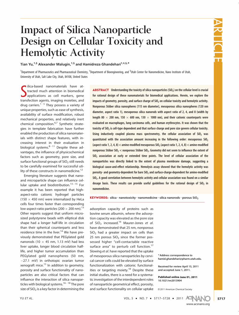
Cellular Association. The amount of cellular-asso-ciated SiO2, which included internalized nanoparticlesor nanoparticles adhering to the extracellular matrix,was quantitated by ICP-MS.19 Results show a similarpattern of cellular association of SiO2 with macro-phages and cancer epithelial cells. However, theamount of silicon associated with macrophages was10�15 times higher than that of the cancer epithelialcells (Figure 6A,B). Nonporous Stöber nanoparticles ledto much higher cellular association than mesoporousnanoparticles both on a particle mass basis and on aparticle number basis (Table 4). The level of cellularassociation was also highest for Stöber nanoparticlesamong all types of SiO2 including the amine-modifiedcounterparts SA. This observation was possibly due tothe highest silanol density on the continuous externalsurface of Stöber nanoparticles, which was reflected bythe highest magnitude of negative charge (�50 mV)for these particles.36 The formation of porous structure
or modification with primary amine groups led toreduced silanol density on the external surface of theparticles (�33 to �39 mV for mesoporous SiO2) orshielding of surface silanol by amine functionalities,which reduced the accessibility of silanol groups to cellsand in turn decreased the level of cellular association.36
Figure 6. Inductively coupled plasmamass spectrometry (ICP-MS) analysis of cellular association of SiO2 in (A) RAW264.7 and(B) A549 cells post-incubation with nanoparticles at 100 μg/mL for 24 h. The graph shows the mass of silicon per 100 μgprotein content versus different nanoparticle treatments. ***The level of cell-associated silicon was significantly higher inStöber or SA-treated cells than in the mesoporous counterpart treated cells (p < 0.001). ###The level of cell-associated siliconwas significantly higher in high aspect ratio, 8A-treated cells than in MA-, 2A-, or 4A-treated cells (p < 0.001). N.D. means “notdetected”. (C) Cellular association of SiO2 after RAW 264.7 cells were incubatedwith 100 μg/mL selected SiO2 at 4 �C (1 h) and37 �C (1 or 24 h). The level of cellular-associated siliconwas significantly higher at 24 h than at 1 h post-incubationwith Stöber(***, p < 0.001), Meso S (###, p < 0.001), or AR8 (##, p < 0.01) at 37 �C. One-hour incubation with Stöber at 37 �C led tosignificantly higher silicon association than incubation at 4 �C (***, p < 0.001). Data are mean ( SD (n = 3).
TABLE 4. Average Cellular Association of Bare SiO2
Detected by ICP-MS on RAW 264.7 Post-incubating with
Nanoparticles at 100 μg/mL for 24 ha
nanoparticle treatment
silicon content
(μg)/100 μg protein
no. nanoparticles/
100 μg protein
Stöber 21.2 2.6� 1011
Meso S 0.7 1.6� 1010
AR2 0.8 2.2 � 1010
AR4 0.5 1.8� 109
AR8 0.5 6.1� 108
a There was no significant difference in the amount of cellular-associated siliconcontent per 100 μg of protein among various types of mesoporous SiO2 (p > 0.05).Stöber nanoparticles were associated with RAW 264.7 at significantly higher levelsthan mesoporous SiO2 of all types either in mass concentration or in numberconcentration (p < 0.001). Data are mean of triplicates.
ARTIC
LE

YU ET AL . VOL. 5 ’ NO. 7 ’ 5717–5728 ’ 2011
www.acsnano.org
5724
On the other hand, amine-modified mesoporousSiO2 (32�40 mV) showed significantly higher cellularassociation than their bare mesoprous counterparts(p < 0.05), which appeared to contradict the aforemen-tioned phenomenon with SA (17 mV) and Stöbernanoparticles. This indicated that there could be asurface charge “threshold” (>30 mV) above which theamine functionalities facilitated nanoparticle�cell in-teraction through electrostatic interaction of positivelycharged amine groups with negatively charged cellmembrane, whereas below the “threshold”, there wereless surface amine groups available and they hadelectrostatic interactions with surface silanols andcovered the sites of silanol,37 which eventually reducedthe level of cellular association.
Bare mesoporous SiO2, irrespective of their shapefeatures, exhibited a similar level but the lowestamount of cellular-associated silicon. For A549 cells,the level of cell-associated silicon from mesoporousSiO2 exposure was even below the detection limit ofICP-MS (<0.1 μg/mL for silicon element). There was nosignificant difference in the level of cellular associationamong all types of mesoporous SiO2 (p > 0.05). Therewas also no significant difference in the cellular asso-ciation among all mesoporous SiO2 post amine mod-ification on both cell lines (p > 0.05), except that 8Ahad a significantly higher cellular association thanother amine-modified mesoporous SiO2 on A549 cells(p < 0.001). This suggests that the curvature of cationicSiO2 could influence the wrapping by the cell mem-brane and affect the cellular association with nonpha-gocytic cells.13
In order to test if porosity, geometry, and surfacemodification can influence the cellular association atearlier time points, selected SiO2 including Stöber,Meso S, AR8, and MA were incubated with RAW 264.7cells for 1 h, and the level of cellular association wasdetected by ICP-MS. The experiment was done at 4 or37 �C to differentiate the amount of membrane-boundSiO2 from that of internalized SiO2, as incubation at lowtemperature (4 �C) drastically reduces the energy-dependent internalization process in cells.38 Consider-ing that the viability of cells could be affected uponincubation at 4 �C, which subsequently could influencethe protein content recovered, the relative viability ofcells post 70min incubation (10min preincubation and60 min incubation with nanoparticles) at 4 �C wasmeasured, and the results show that the percentageof viable cells was 94( 8% comparedwith control cellstreated at 37 �C for the same time duration. As shownin Figure 6C, Stöber nanoparticles led to a significantincrease in cellular association 24 h post-incubation at37 �C comparedwith 1 h incubation at 37 �C (p< 0.001)or at 4 �C (p < 0.001), indicating that there was exten-zsive internalization of nonporous nanoparticles over24 h. There was no significant difference in cellularassociation betweenMeso S andAR8 post 1 h incubation
at 37 �C (p > 0.05). Geometry did not seem to affect thelevel of cellular-associated nanoparticles for mesopor-ous SiO2 at the early time points as well. Most meso-porous SiO2 seemed to bind to the cell membraneinstead of being internalized into the cytoplasmwithinan hour, as the level of silicon association from Meso Sor AR8 exposure was similar for cells incubated at 4 or37 �C for one hour. However, the level of cellularassociation significantly increased post-incubation for24 h comparedwith incubation for 1 h at 37 �C forMesoS (p < 0.001) and AR8 (p < 0.01), which indicated thatinternalization of mesoporous SiO2 had occurred. Onthe contrary, there was no significant difference incellular association between 1 h incubation and 24 hincubation with MA at 37 �C (p > 0.05), which impliedthat the cellular association of MA almost reached aplateau within 1 h post-incubation. The cellular asso-ciation of MA post 1 h incubation at 37 �C was notsignificantly higher than that at 4 �C (p > 0.05). Thecombined results suggest that there was limited inter-nalization over 24 h post-incubation with MA, whichprobably explained why there was a reduction intoxicity of amine-modified SiO2 compared with thatof bare SiO2. It has been suggested that the strongassociation of cationic SiO2 with negatively chargedcell membranes, which made the cationic SiO2 adhereto the cell membrane instead of bringing them into thecytoplasm, led to the reduction in internalization andthe resultant decreased toxicity based on transmissionelectron microscopy analysis39�41 or confocal micro-scopy analysis.42 Our results provide quantitative evi-dence by ICP-MS that there was limited internalizationof amine-modified SiO2.
In summary, it appears that surface charge andporosity mainly influenced the extent of cellular asso-ciation, while geometry did not seem to influencecellular association within the aspect ratio range of1�8 studied. These observations are consistent withthe experiments examining plasma membrane integ-rity post nanoparticle treatment. The level of plasmamembrane damage by nanoparticles was directlyrelated to the extent of nanoparticle cellular associa-tion, which indicated a biological cause-and-effectrelationship between cellular association and cellmembrane damage on both cell lines.
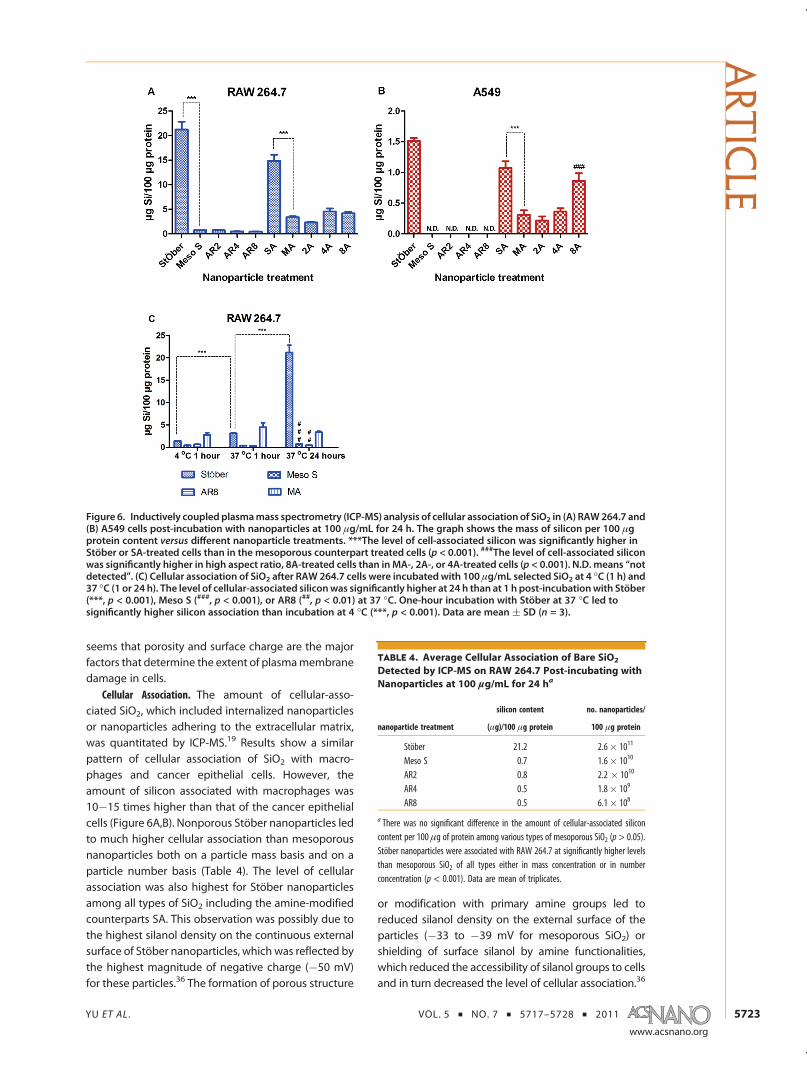
Hemolysis. The impact of nanoparticle porosity, geo-metry, and surface functionality on human red bloodcells (RBCs) was evaluated by a hemolysis assay. Thequantitation of hemoglobin in the supernatant of ananoparticle�RBC mixture was done by recording theabsorbance of hemoglobin at 577 nm with a referencewavelength of 655 nm (Supplemental Figure 5).36,43
Results show that the extent of hemolysis was con-centration-, porosity-, and geometry-dependent forbare SiO2 (Figure 7). Stöber nanoparticles caused animmediate onset of hemolysis that soon reached aplateau of 17% hemolysis at ca. 250 μg/mL probably
ARTIC
LE
YU ET AL . VOL. 5 ’ NO. 7 ’ 5717–5728 ’ 2011
www.acsnano.org
5725
due to its high negative charge, which might preventRBCs (�15 mV)44 from interacting at further increasednanoparticle concentration. For mesoporous SiO2 of allgeometries tested, no hemolytic toxicity was observedbelow 100 μg/mL. The impact of nanoparticle geome-try became pronounced as the concentration furtherincreased. Mesoporous SiO2 with high aspect ratiodemonstrated lower hemolytic activity than sphericalor low aspect ratio mesoporous SiO2. It has beenreported that the external surface area and the curva-ture of SiO2 influence their hemolytic activity by affect-ing the magnitude of binding energy of particles withRBCs or bending energy of the membrane to wraparound the nanoparticles.37 Large external surface areaand small curvature (i.e., 1/r2 for spheres) rendered thehemolysis process thermodynamically favorable.37 Inthis case, the external surface areas of Stöber andMesoS were 24 and 109 m2/g, respectively, which agreedwell with previous similar calculations,37,43 and hadsimilar curvature due to the similar size they possessed.However, Meso S did not lead to a higher hemolyticrate than Stöber until the mass concentration exceededbeyond ca. 190 μg/mL. This indicates that there could
possibly be a threshold in the density of silanol groupson each nanoparticle only above which it could causeimmediate cell membrane damage upon exposure.Hence, the hemolytic activity depends not only onexternal surface area and curvature but also on silanoldensity of each nanoparticle exposed to RBCs.
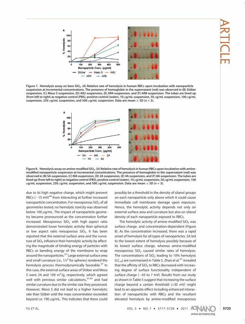
The hemolytic activity of amine-modified SiO2 wassurface charge- and concentration-dependent (Figure8). As the concentration increased, there was a rapidonset of hemolysis for all types of nanoparticles. SA ledto the lowest extent of hemolysis possibly because ofits lowest surface charge, whereas amine-modifiedmesoporous SiO2 caused similar rates of hemolysis.The concentrations of SiO2 leading to 10% hemolysis(LC10) are summarized in Table 5. Zhao et al.37 revealedthat the affinity of SiO2 to RBCs decreased with increas-ing degree of surface functionality independent ofsurface charge (�43 to 7 mV). Results from our studyas shown in Table 5 suggest that increasing the surfacecharge beyond a certain threshold (>30 mV) mightlead to an opposite effect including enhanced interac-tion of nanoparticles with RBCs and the resultantelevated hemolysis by amine-modified mesoporous
Figure 7. Hemolysis assay on bare SiO2. (A) Relative rate of hemolysis in human RBCs upon incubation with nanoparticlesuspension at incremental concentrations. The presence of hemoglobin in the supernatant (red) was observed in (B) Stöbersuspension, (C) Meso S suspension, (D) AR2 suspension, (E) AR4 suspension, and (F) AR8 suspension. The tubes are lined up(from left to right) as negative control (PBS), positive control (water), 10 μg/mL suspension, 50 μg/mL suspension, 100 μg/mLsuspension, 250 μg/mL suspension, and 500 μg/mL suspension. Data are mean ( SD (n = 3).
Figure 8. Hemolysis assay on amine-modified SiO2. (A) Relative rate of hemolysis in humanRBCs upon incubationwith amine-modified nanoparticle suspension at incremental concentrations. The presence of hemoglobin in the supernatant (red) wasobserved in (B) SA suspension, (C) MA suspension, (D) 2A suspension, (E) 4A suspension, and (F) 8A suspension. The tubes arelinedup (from left to right) as negative control (PBS), positive control (water), 10μg/mL suspension, 50μg/mL suspension, 100μg/mL suspension, 250 μg/mL suspension, and 500 μg/mL suspension. Data are mean ( SD (n = 3).
ARTIC
LE
YU ET AL . VOL. 5 ’ NO. 7 ’ 5717–5728 ’ 2011
www.acsnano.org
5726
SiO2, in agreement with the cellular association results(Figure 6A,B). It must be noted that a good correlationwith results of different experiments is based on asimilar dose (ca.e 100 μg/mL SiO2). As the dose changesbeyond a certain range, the pattern from differentexperiments shifts, and the correlation of various experi-ments at that dosage needs to be further validated.
CONCLUSION
In summary, nonporous Stöber silica nanospheres,mesoporous silica nanospheres, mesoporous silicananorods with aspect ratios of 2, 4, and 8, and theircationic charged counterparts were synthesized andcharacterized. The porosity, shape, and surface mod-ification effects on cellular toxicity or hemolytic activitywere evaluated on macrophages, cancer epithelial
cells, and human erythrocytes. The toxicity of SiO2
was found to be cell-type-dependent. Cancer epithelialcells were highly resistant to nanoparticle treatment,while the toxicity on macrophages was predominantlysurface-charge-dependent. The difference in toxicitybetween the two cell types could be due to thedifference in the physiological function of each. Por-osity and surface characteristics of the nanoparticleswere the major factors that influenced the cellularassociation of the nanoparticles. Geometry did notseem to influence the extent of cellular association ofthe nanoparticles either at the early time point or overextended duration. Initial comparison of blood bio-compatibility of nonporous andmesoporous SiO2 withvaried shapes and surface characteristics has beendemonstrated using the hemolysis assay. Bare SiO2
showed a porosity- and geometry-dependent hemo-lytic activity on RBCs with mesoporous SiO2 at highaspect ratio exhibiting a reduced hemolytic activity.The extent of hemolysis was highly zeta-potential-dependent among the amine-modified SiO2, and re-sults indicated that there could be a surface charge“threshold” below which the amine modification onSiO2 could lead to reduced hemolysis compared withtheir bare counterparts. Further studies evaluating thein vivo toxicity of SiO2 in animal models are needed toestablish an in vitro�in vivo correlation for betterprediction of toxicity in biological systems.
METHODSSynthesis of Nonporous and Mesoporous SiO2. Nonporous silica
nanoparticles (Stöber) were produced using the modified Stö-ber method.23 Water (34.82 mL), 3.25 mL of ammonium hydro-xide (29.7%), and 100 mL of ethanol were mixed and stabilizedat 40 �C. Tetraethyl orthosilicate (6.20 mL) was added at aninjection rate of 5mL/min upon stirring at 550 rpm. The reactionwas conducted for 1 h, and the product was washed twice byethanol and stored in ethanol. Mesoporous SiO2 of differentshapes were synthesized through a one-step condensationunder dilute silica source and low surfactant concentrationconditions with ammonium hydroxide as the base catalyst.24�29
Generally, cetyltrimethylammonium bromide was dissolvedin aqueous medium with mild heating (30 �C). After thesolution was cooled to room temperature (22 �C), aqueousammonia was introduced and the mixture was stirred for anhour. TEOS was added at the rate of 5 mL/min while thestirring continued. Themixture was further stirred for 4 h, andthe product was autoclaved at 100 �C for 24 h.30,31 Subse-quently the product was collected by centrifugation at15 000 rpm for 20 min. As-synthesized nanoparticles weresuspended in ethanolic HCl (1.5 mL of HCl in 150 mL ofethanol) and heated at 60 �C for 6 h to remove the surfactant.The complete removal of CTAB was confirmed by Fouriertransform infrared (FT-IR) spectroscopy.
Surface Functionalization. To modify the surface of SiO2 withprimary amine functionalities,33 100 mg of SiO2 was resus-pended in 100 mL of anhydrous ethanol. (3-Aminopropyl-)triethoxysilane (APTES) was introduced dropwise to the SiO2
suspension upon stirring at 500 rpm under nitrogen flow. Themixture was stirred at room temperature for 20 h. Amine-modified SiO2 were collected by centrifugation and washed
extensively with ethanol and water. SiO2 were stored in ethanolat 4 �C and transferred to water immediately before use.
Nanoparticle Characterization. Transmission electron micro-scopy images were taken with a Philips Tecnai microscopeoperating at 120 kV. FT-IR spectra were recorded on a VarianCary FT-IR 1000 spectrometer using KBr pellets. X-ray diffractionpatterns of SiO2 were analyzed on a Philips PANalytical X0PertX-ray diffractometer (Spectris, England) using Cu Ka radiation(λ = 0.1542 nm) at 45 kV and 40 mA. The XRD spectra wererecorded in the 2θ range of 2�10with a step size of 0.02� in a 2θscattering angle and a scanning speed of 0.01 deg/s. The slitsizes and specimen length were also adjusted for divergenceslit, antiscattered slit, and receiving slit to suit the low-angledetection. Nitrogen adsorption�desorption isothermmeasure-mentswere completed on aMicromeritics ASAP 2010 (Norcross,GA) accelerated surface area analyzer at�196 �C. The SiO2 weredried at 100 �C overnight before analysis. The Brunauer�Emmett�Teller specific surface areas were calculated by usingadsorption data at P/P0 = 0.05�0.20.19,21 The external surfaceareas of mesoporous SiO2 were calculated from the t plots oftheir N2 adsorption isotherms.32 Pore volume and pore sizedistributionswere obtained froman adsorption branch by usingthe Barrett, Joyner, and Halenda method.19,21
Acute Cytotoxicity Assay. The acute toxicity effect of SiO2 wasdetermined by the WST-8 assay on A549 cells or RAW 264.7macrophages (ATCC, Manassas, VA). Cells from passages 5through 20 were used with medium changing once every threedays. A549 cells or RAW 264.7 macrophages were seeded at8000 cells/well or 16 000 cells/well in a 96-well plate in F-12kmedium or DMEM supplementedwith 10% FBS andmaintainedin a humidified incubator for 24 h. SiO2 at incremental concen-trationof 100, 250, or 500μg/mLwere added to cells. Supernatants
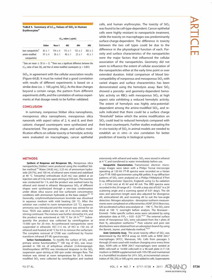
TABLE 5. Summary of LC10 Values of SiO2 in Human
Erythrocytesa
LC10 values (μg/mL)
Stöber Meso S AR2 AR4 AR8
bare nanoparticlesb 36( 1 154( 4 115( 1 152( 2 302( 3amine-modifiednanoparticles
97( 4 30( 1 40( 1 23( 1 43( 1
a Data are mean ( SD (n = 3). b There was a significant difference between theLC10 value of bare SiO2 and that of amine-modified counterparts (p < 0.001).
ARTIC
LE
YU ET AL . VOL. 5 ’ NO. 7 ’ 5717–5728 ’ 2011
www.acsnano.org
5727
from nanoparticle stock solutions and respective growth med-ium served as controls. Post 24 h, old medium was aspirated,and cells werewashed three timeswith PBS. A 100 μL amount ofcomplete medium containing 10% (v/v) Cell Counting Kit-8(Dojindo, Rockville, MD) was added to each well and incubatedwith cells for 2 h. The absorbance of the plate was recorded at450 nm on a UV/vis reader with a reference wavelength of650 nm.
Proliferation Inhibition Assay. The cytotoxicity of SiO2 wasevaluated by the WST-8 viability assay on A549 cells orRAW264.7 macrophages. Initially, A549 or RAW cells wereseeded at 2000 or 4000 cells/well in a 96-well plate and allowedto settle for 24 h. Then 10, 50, 100, 250, 500, or 1000 μg/mL ofbare SiO2 or amine-modified SiO2 was added into the 96-wellplate in triplicates. Supernatants from nanoparticle stock solu-tions and respective growth medium served as controls. Post72 h, old medium was aspirated, and the following steps werethe same as the procedures for the acute cytotoxicity assaydescribed above.
Plasma Membrane Integrity Assay. Determination of propidiumiodide uptake was used to assess the integrity of the plasmamembrane of nanoparticle-dosed cells. A549 cells or RAW cellswere seeded at 8� 104 or 1.6� 105 cells/well on a 12-well platein triplicate. After 24 h, selected nanoparticles were added intoeach well at the concentration of 250 μg/mL. Twenty-four hourslater, cells and medium from each well were collected into a5 mL flow cytometry tube. The cell suspension was centrifugedat 1000 rpm for 5 min, the supernatant was decanted, and thecells were resuspended in 100 μL of PBS. A 5 μL portion ofpropidium iodide solution (50 μg/mL in water) was added toeach tube. The tube was gently vortexed and incubated for15min at room temperature in the dark. Then 400 μL of PBSwasadded into each tube, and the samples were analyzed by flowcytometry (FACScan analyzer, Becton Dickinson, Franklin Lakes,NJ) within an hour.
Quantitation of Cellular Association. Cellular association of SiO2
was evaluated on A549 and RAW264.7 cells. A549 or RAW cellswere seeded at 8� 104 or 1.6� 105 cells/well on a 12-well platein triplicate, 24 h before the addition of particles. Cells wereincubated with 100 μg/mL of SiO2 for 24 h (37 �C, 5% CO2). Aftercell/particle incubation, the old medium was aspirated and thecells were washed three times with PBS. Then the cells weretreated with 0.5 mL of 0.1% (v/v) Triton X-100 solution in waterfor 15 min. After that, the cell lysate was collected into acentrifuge tube, and the wells were further washed with0.5 mL of water. The wash was also collected into the samecentrifuge tube. Aliquots of 100 μL of cell lysate were used forprotein content measurement by BCA assay (Thermo Scientific,Rockford, IL). The concentration of silicon in the cell lysate wasmeasured by direct Si measurement using inductively coupledplasmamass spectrometry (Agilent 7500, Agilent Technologies,Santa Clara, CA). The amount of cellular-associated SiO2 post 1 hincubation at 4 or 37 �C was also measured. RAW cells wereseeded at 3.2� 105 cells/well on a 12-well plate in triplicate andincubated for 24 h. After that, cells were preconditioned to 4 �Cby incubating at 4 �C for a brief period of 10 min. Then silicananoparticles were added to the cells at a concentration of100 μg/mL, and the cells were further incubated at 4 �C foranother hour. The treatment that followed was the same asmentioned above. To make sure that relative cell viability in the4 �C treated plate was not dramatically influenced by exposureto cold temperature for the experimental duration, the relativeviability from the 4 �C treated plate was compared with cellsincubated at 37 �C for 70 min by the WST-8 assay.
Hemolysis. Heparin-stabilized human blood was freshly col-lected according to an approved University of Utah InstitutionalReview Board protocol and used within 3 h of being drawn.36,43
A 4mL sample of whole blood was added to 8mL of Dulbecco'sphosphate-buffered saline (D-PBS), and the RBCs were isolatedfrom serum by centrifugation at 10016g for 5 min. The RBCswere further washed five times with sterile D-PBS solution.Following the last wash, the RBCs were diluted to 40 mL ofD-PBS. Then 0.2 mL of the diluted RBC suspension was added to0.8 mL of the silica nanoparticle suspension in D-PBS at aconcentration of 12.5, 62.5, 125, 312.5, or 625 μg/mL to make
the final nanoparticle concentration 10, 50, 100, 250, or 500μg/mL.All samples were prepared in triplicate, and the suspension wasbriefly vortexed before leaving at static conditions at roomtemperature for 4 h. After that, the mixture was briefly vortexedagain and centrifuged at 10016g for 3 min. A 100 μL amount ofsupernatant from the sample tube was transferred to a 96-wellplate. The absorbance value of hemoglobin at 577 nm wasmeasured with the reference wavelength of 655 nm. A 0.2 mLamount of diluted RBC suspension incubated with 0.8 mL ofD-PBS and 0.8 mL of water was used as the negative or positivecontrol. The percent of hemolysis was calculated as follows:Hemolysis % = [(sample absorbance � negative control)/(positive control � negative control)] � 100%.
Statistical Analysis. The difference between multiple groupswas analyzed by one-way ANOVA. The Tukey post test was usedwhere a difference was detected. For two-group comparison,Student's t-test was used. The difference between two groupswas considered significant when p < 0.05. The LC10 values inthe hemolysis assay were determined by using ED50plus v1.0software.
Acknowledgment. We would like to acknowledge Dr. SajoNaik (Western Research Institute, Cheyenne, WY) for his techni-cal suggestions for mesoporous silica nanoparticle synthesis,and Dr. Khaled Greish for assistance with human blood with-drawal from lab volunteers. Financial support was provided byNIH grant R01 DE19050 and the Utah Science and TechnologyResearch (USTAR) Initiative.
Supporting Information Available: Supplemental Calculation1 and Supplemental Figures 1�5 are available free of charge viathe Internet at http://pubs.acs.org.
REFERENCES AND NOTES1. Barb�e, C.; Bartlett, J.; Kong, L.; Finnie, K.; Lin, H.; Larkin, M.;
Calleja, S.; Bush, A.; Calleja, G. Silica Particles: A Novel Drug-Delivery System. Adv. Mater. 2004, 16, 1–8.
2. Liong, M.; Lu, J.; Kovochich, M.; Xia, T.; Ruehm, S. G.; Nel,A. E.; Tamanoi, F.; Zink, J. I. Multifunctional InorganicNanoparticles for Imaging, Targeting, and Drug Delivery.ACS Nano 2008, 2, 889–896.
3. Vivero-Escoto, J. L.; Slowing, I. I.; Trewyn, B. G.; Lin, V. S.Mesoporous Silica Nanoparticles for Intracellular Con-trolled Drug Delivery. Small 2010, 6, 1952–1967.
4. Lu, J.; Liong, M.; Li, Z.; Zink, J. I.; Tamanoi, F. Biocompat-ibility, Biodistribution, and Drug-Delivery Efficiency ofMesoporous Silica Nanoparticles for Cancer Therapy inAnimals. Small 2010, 6, 1794–1805.
5. Li, L.; Tang, F.; Liu, H.; Liu, T.; Hao, N.; Chen, D.; Teng, X.; He, J.In Vivo Delivery of Silica Nanorattle Encapsulated Doce-taxel for Liver Cancer Therapy with Low Toxicity and HighEfficacy. ACS Nano 2010, 4, 6874–6882.
6. Tsai, C.-P.; Chen, C.-Y.; Hung, Y.; Chang, F.-H.; Mou, C.-Y.Monoclonal Antibody-Functionalized Mesoporous SilicaNanoparticles (MSN) for Selective Targeting Breast CancerCells. J. Mater. Chem. 2009, 19, 5737–5743.
7. Cheng, S.-H.; Lee, C.-H.; Chen, M.-C.; Souris, J. S.; Tseng, F.-G.; Yang, C.-S.; Mou, C.-Y.; Chen, C.-T.; Lo, L.-W. Tri-Functio-nalization of Mesoporous Silica Nanoparticles for Com-prehensive Cancer Theranostics-the Trio of Imaging,Targeting and Therapy. J. Mater. Chem. 2010, 20,6149–6157.
8. Nan, A.; Bai, X.; Son, S. J.; Lee, S. B.; Ghandehari, H. CellularUptake and Cytotoxicity of Silica Nanotubes. Nano Lett.2008, 8, 2150–2154.
9. Son, S. J.; Bai, X.; Nan, A.; Ghandehari, H.; Lee, S. B. TemplateSynthesis of Multifunctional Nanotubes for ControlledRelease. J. Controlled Release 2006, 114, 143–152.
10. Chen, C.-C.; Liu, Y.-C.; Wu, C.-H.; Yeh, C.-C.; Su, M.-T.; Wu,Y.-C. Preparation of Fluorescent Silica Nanotubes andTheir Application in Gene Delivery. Adv. Mater. 2005, 17,404–407.
11. Buyukserin, F.; Medley, C. D.; Mota, M. O.; Kececi, K.; Rogers,R. R.; Tan, W.; Martin, C. R. Antibody-Functionalized Nano
ARTIC
LE

YU ET AL . VOL. 5 ’ NO. 7 ’ 5717–5728 ’ 2011
www.acsnano.org
5728
Test Tubes Target Breast Cancer Cells. Nanomedicine(London) 2008, 3, 283–292.
12. Hudson, S. P.; Padera, R. F.; Langer, R.; Kohane, D. S. TheBiocompatibility of Mesoporous Silicates. Biomaterials2008, 29, 4045–4055.
13. Gratton, S. E.; Ropp, P. A.; Pohlhaus, P. D.; Luft, J. C.;Madden, V. J.; Napier, M. E.; DeSimone, J. M. The Effect ofParticle Design on Cellular Internalization Pathways. Proc.Natl. Acad. Sci. U. S. A. 2008, 105, 11613–11618.
14. Muro, S.; Garnacho, C.; Champion, J. A.; Leferovich, J.;Gajewski, C.; Schuchman, E. H.; Mitragotri, S.; Muzykantov,V. R. Control of Endothelial Targeting and IntracellularDelivery of Therapeutic Enzymes by Modulating the Sizeand Shape of ICAM-1-Targeted Carriers. Mol. Ther. 2008,16, 1450–1458.
15. Arnida; Janat-Amsbury, M. M.; Ray, A.; Peterson, C. M.;Ghandehari, H. Geometry and Surface Characteristics ofGold Nanoparticles Influence Their Biodistribution andUptake by Macrophages. Eur. J. Pharm. Biopharm. 2011,77, 417–423.
16. Nguyen, T. P. B.; Lee, J.-W.; Shim, W. G.; Moon, H. Synthesisof Functionalized SBA-15 with Ordered Large Pore Sizeand Its Adsorption Properties of BSA. Microporous Meso-porous Mater. 2008, 110, 560–569.
17. Maurer-Jones, M. A.; Lin, Y. S.; Haynes, C. L. FunctionalAssessment of Metal Oxide Nanoparticle Toxicity in Im-mune Cells. ACS Nano 2010, 4, 3363–3373.
18. Slowing, I.; Trewyn, B. G.; Lin, V. S. Effect of SurfaceFunctionalization of MCM-41-Type Mesoporous SilicaNanoparticles on the Endocytosis by Human Cancer Cells.J. Am. Chem. Soc. 2006, 128, 14792–14793.
19. Lu, F.; Wu, S. H.; Hung, Y.; Mou, C. Y. Size Effect on CellUptake in Well-Suspended, Uniform Mesoporous SilicaNanoparticles. Small 2009, 5, 1408–1413.
20. He, Q.; Zhang, Z.; Gao, Y.; Shi, J.; Li, Y. Intracellular Localiza-tion and Cytotoxicity of Spherical Mesoporous Silica Nano-and Microparticles. Small 2009, 5, 2722–2729.
21. Tsai, C. P.; Hung, Y.; Chou, Y. H.; Huang, D. M.; Hsiao, J. K.;Chang, C.; Chen, Y. C.; Mou, C. Y. High-Contrast Paramag-netic Fluorescent Mesoporous Silica Nanorods as a Multi-functional Cell-Imaging Probe. Small 2008, 4, 186–191.
22. Dobrovolskaia, M. A.; Aggarwal, P.; Hall, J. B.; McNeil, S. E.Preclinical Studies to Understand Nanoparticle Interactionwith the Immune System and Its Potential Effects onNanoparticle Biodistribution. Mol. Pharm. 2008, 5, 487–495.
23. Chung, Y. S.; Jeon, M. Y.; Kim, C. K. Fabrication of NearlyMonodispersed Silica Nanoparticles by Using Poly(1-Vinyl-2-Pyrrollidinone) and Their Application to the Preparationof Nanocomposites. Macromol. Res. 2009, 17, 37–43.
24. Giri, S.; Trewyn, B. G.; Stellmaker, M. P.; Lin, V. S.-Y. Stimuli-Responsive Controlled-Release Delivery System Based onMesoporous Silica Nanorods CappedwithMagnetic Nano-particles. Angew. Chem., Int. Ed. 2005, 44, 5038–5044.
25. Huh, S.; Wiench, J. W.; Yoo, J.-C.; Pruski, M.; Lin, V. S.-Y.Organic Functionalization and Morphology Control ofMesoporous Silicas Via a Co-Condensation SynthesisMethod. Chem. Mater. 2003, 15, 4247–4256.
26. Naik, S. P.; Elangovan, S. P.; Okubo, T.; Sokolov, I. Morphol-ogy Control of Mesoporous Silica Particles. J. Phys. Chem. C2007, 111, 11168–11173.
27. Yang, S.; Zhao, L.; Yu, C.; Zhou, X.; Tang, J.; Yuan, P.; Chen, D.;Zhao, D. On the Origin of Helical Mesostructures. J. Am.Chem. Soc. 2006, 128, 10460–10466.
28. Lelong, G.; Bhattacharyya, S.; Kline, S.; Cacciaguerra, T.;Gonzalez, M. A.; Saboungi, M.-L. Effect of Surfactant Con-centration on the Morphology and Texture of MCM-41Materials. J. Phys. Chem. C 2008, 112, 10674–10680.
29. Jin, H.; Liu, Z.; Ohsuna, T.; Terasaki, O.; Inoue, Y.; Sakamoto,K.; Nakanishi, T.; Ariga, K.; Che, S. Control of Morphologyand Helicity of Chiral Mesoporous Silica. Adv. Mater. 2006,18, 593–596.
30. Kim, J. M.; Kwak, J. H.; Jun, S.; Ryoo, R. Ion Exchange andThermal Stability of MCM-41. J. Phys. Chem. 1995, 99,16742–16747.
31. Ryoo, R.; Jun, S. Improvement of Hydrothermal Stability ofMCM-41 Using Salt Effects During the CrystallizationProcess. J. Phys. Chem. B 1997, 101, 317–320.
32. Zhu, H. Y.; Zhao, X. S.; Liu, G. Q.; Do, D. D. ImprovedComparison Plot Method for Pore Structrue Characteriza-toin of MCM-41. Langmuir 1996, 12, 6513–6517.
33. Kobler, J.; Moller, K.; Bein, T. Colloidal Suspensions ofFunctionalized Mesoporous Silica Nanoparticles. ACSNano 2008, 2, 791–799.
34. Jones, C. F.; Grainger, D. W. In Vitro Assessments ofNanomaterial Toxicity. Adv. Drug Delivery Rev. 2009, 61,438–456.
35. Riccardi, C.; Nicoletti, I. Analysis of Apoptosis by PropidiumIodide Staining and Flow Cytometry. Nat. Protoc. 2006, 1,1458–1461.
36. Slowing, I. I.; Wu, C. W.; Vivero-Escoto, J. L.; Lin, V. S.Mesoporous Silica Nanoparticles for Reducing HemolyticActivity Towards Mammalian Red Blood Cells. Small 2009,5, 57–62.
37. Zhao, Y.; Sun, X.; Zhang, G.; Trewyn, B. G.; Slowing, I. I.; Lin,V. S. Interaction of Mesoporous Silica Nanoparticles withHuman Red Blood Cell Membranes: Size and SurfaceEffects. ACS Nano 2011, 5, 1366–1375.
38. Xing, X.; He, X.; Peng, J.; Wang, K.; Tan, W. Uptake of Silica-Coated Nanoparticles by Hela Cells. J. Nanosci. Nanotech-nol. 2005, 5, 1688–1693.
39. Tao, Z.; Toms, B. B.; Goodisman, J.; Asefa, T. Mesoporosityand Functional Group Dependent Endocytosis and Cyto-toxicity of Silica Nanomaterials. Chem. Res. Toxicol. 2009,22, 1869–1880.
40. Petushkov, A.; Intra, J.; Graham, J. B.; Larsen, S. C.; Salem,A. K. Effect of Crystal Size and Surface Functionalization onthe Cytotoxicity of Silicalite-1 Nanoparticles. Chem. Res.Toxicol. 2009, 22, 1359–1368.
41. Chung, T. H.; Wu, S. H.; Yao, M.; Lu, C. W.; Lin, Y. S.; Hung, Y.;Mou, C. Y.; Chen, Y. C.; Huang, D. M. The Effect of SurfaceCharge on the Uptake and Biological Function of Meso-porous Silica Nanoparticles in 3t3-L1 Cells and HumanMesenchymal Stem Cells. Biomaterials 2007, 28, 2959–2966.
42. Nabeshi, H.; Yoshikawa, T.; Arimori, A.; Yoshida, T.; Tochigi,S.; Hirai, T.; Akase, T.; Nagano, K.; Abe, Y.; Kamada, H.; et al.Effect of Surface Properties of Silica Nanoparticles on TheirCytotoxicity and Cellular Distribution in Murine Macro-phages. Nanoscale Res. Lett. 2011, 6, 1–6.
43. Lin, Y. S.; Haynes, C. L. Impacts of Mesoporous SilicaNanoparticle Size, Pore Ordering, and Pore Integrity onHemolytic Activity. J. Am. Chem. Soc. 2010, 132, 4834–4842.
44. Jan, K. M.; Chien, S. Role of Surface Electric Charge in RedBlood Cell Interactions. J. Gen. Physiol. 1973, 61, 638–654.
ARTIC
LE