Embed Size (px)
Citation preview

UNLV Theses, Dissertations, Professional Papers, and Capstones
5-1-2015
In Vitro Studies of Gold and Gold Silica Nanoparticle In Vitro Studies of Gold and Gold Silica Nanoparticle
Radiosensitization with Kilovoltage X-Rays Radiosensitization with Kilovoltage X-Rays
Gregory Colarch University of Nevada, Las Vegas
Follow this and additional works at: https://digitalscholarship.unlv.edu/thesesdissertations
Part of the Biophysics Commons, and the Nanoscience and Nanotechnology Commons
Repository Citation Repository Citation Colarch, Gregory, "In Vitro Studies of Gold and Gold Silica Nanoparticle Radiosensitization with Kilovoltage X-Rays" (2015). UNLV Theses, Dissertations, Professional Papers, and Capstones. 2342. http://dx.doi.org/10.34917/7645871
This Thesis is protected by copyright and/or related rights. It has been brought to you by Digital Scholarship@UNLV with permission from the rights-holder(s). You are free to use this Thesis in any way that is permitted by the copyright and related rights legislation that applies to your use. For other uses you need to obtain permission from the rights-holder(s) directly, unless additional rights are indicated by a Creative Commons license in the record and/or on the work itself. This Thesis has been accepted for inclusion in UNLV Theses, Dissertations, Professional Papers, and Capstones by an authorized administrator of Digital Scholarship@UNLV. For more information, please contact [email protected].

IN VITRO STUDIES OF GOLD AND GOLD SILICA NANOPARTICLE
RADIOSENSITIZATION WITH KILOVOLTAGE X-RAYS
by
Gregory Joseph Colarch
Bachelor of Science in Astrophysics University of Calgary
2009
Master of Science in Physics University of Nevada, Las Vegas
2012
A thesis submitted in partial fulfillment of
the requirements for the
Master of Science - Health Physics
Department of Health Physics School of Allied Health Sciences
The Graduate College
University of Nevada, Las Vegas May 2015
ii
We recommend the thesis prepared under our supervision by
G regory Joseph Colarch
entitled
In V itro Studies of Gold and Gold Silica Nanoparticle Radiosensitization with K ilovoltage X-Rays
is approved in partial fulfillment of the requirements for the degree of
Master of Science - Health Physics Department of H ealth Physics and Diagnostic Sciences
Steen Madsen, Ph.D., Committee Chair
Yu Kuang, Ph.D., Committee Member
Bing Ma, Ph.D., Committee Member
Robbin Hickman, Ph.D., Graduate College Representative
Kathryn Hausbeck Korgan, Ph.D., Interim Dean of the Graduate College
May 2015
iii
ABSTRACT
Technological advances in the ability to construct and manipulate nanoscale
particles have opened up the possibility of using solid metallic nanoparticles and mixed
metal nanoshells as a means to increase dose enhancement and treatment efficacy to
tumors. In order for nanoparticles to be an effective form of treatment, they must be
delivered to tumors in sufficient concentrations so that there is a dose enhancement factor
due to ionizing radiation, as well as being essentially non-toxic to healthy cells. Gold
nanoparticles and silica-gold nanoshells fit these requirements. Gold has a high atomic
number (Z=79), which gives a larger cross section for the photoelectric effect vs. tissue
with regards to kilovoltage x-rays. Both gold and silica are also relatively inert and
biocompatible.
The investigation of dose enhancement to cells that have been incubated with
nanoparticles and nanoshells is the focus of this thesis. The effectiveness of the treatment
was determined by measuring the size of multicellular hybrid spheroids consisting of
human glioma cells and murine lymphocytic monocytes. Dose enhancement effects was
also examined in murine lymphocytic monocytes using an MTS assay, which measures
metabolic activity in cells.
A clear dose response was observed for spheroids consisting of human glioma
cells only: increasing doses resulted in decreased spheroid growth. With a few
exceptions, this trend was also observed in hybrid spheroids consisting of glioma cells
and nanoparticle or nanoshell loaded monocytes. Contrary to the premise of utilizing the
photoelectric effect, the most pronounced dose effect was observed in the pure glioma
irradiated spheroids which showed greater growth suppression compared to the
iv
nanoparticle and nanoshell loaded hybrid spheroids at each dose investigated. A similar
trend was found when comparing the viability of bare and nanoparticle/nanoshell loaded
monocytes exposed to kilovoltage x-rays. These results are considered anomalous since
kilovoltage x-rays are expected to be more damaging to cells and spheroids containing
nanoparticles/nanoshells due to enhanced photoelectric absorption. The anomalous
results were attributed to inaccuracies in x-ray tube output.
Optimization of MTS parameters required for accurate determination of monocyte
viability represents the most significant finding of this work. It was found that 50,000
cells per well yielded an accurate MTS signal. Furthermore, the MTS assay should not be
performed less than 96 hours from the time of irradiation. As long as this 96 hour
criterion is satisfied, any of the investigated MTS incubation times (1 – 4 hours) can be
used. Finally, at the concentrations used in these studies, neither nanoparticles nor
nanoshells were toxic to murine lymphocytes.
v
ACKNOWLEDGEMENTS
I would like to thank my advisor Dr. Steen Madsen, for taking me on as a
graduate student and giving me the opportunity to earn this degree. Any knowledge I
now have about biology and cancer is due to this degree. I would also like to thank the
members of my committee: Dr. Robbin Hickman, Dr. Yu Kuang, and Dr. Bing Ma.
Also, I would like to thank Mary Turner for her work in keeping the labs operational. I
respectively acknowledge the financial support given for my graduate studies.
This thesis is dedicated to the memory of my aunts, Francis and Sylvia, and my
uncle John.
vi
TABLE OF CONTENTS
ABSTRACT ....................................................................................................................... iii
ACKNOWLEDGEMENTS ................................................................................................ v
TABLE OF CONTENTS ................................................................................................... vi
LIST OF FIGURES ........................................................................................................... ix
CHAPTER 1: INTRODUCTION ....................................................................................... 1
1.1: NANOPARTICLES ................................................................................................. 1
1.1.1: Gold Nanoparticles ............................................................................................ 2
1.1.2: Gold Nanoshells ................................................................................................ 3
1.1.3: Electromagnetic Interactions: Photoelectric Effect and others ......................... 4
1.1.4: Dose Enhancement ............................................................................................ 8
1.1.5: Effective Z of Silica-Gold Nanoshells ............................................................ 10
1.1.6: Electromagnetic Interactions: Surface Plasmon Resonance ............................ 11
1.1.7: Toxicity ............................................................................................................ 14
1.1.8: Conjugation ..................................................................................................... 14
1.2: MONOCYTES and SPHEROIDS ......................................................................... 15
1.2.1: Enhanced Permeability and Retention Effect .................................................. 15
1.2.2: Tumor Associated Macrophages ..................................................................... 15
1.2.3: Spheroids ......................................................................................................... 16
1.3: SCOPE OF WORK ................................................................................................ 18
CHAPTER 2: MATERIALS AND METHODS .............................................................. 19
2.1: MATERIALS ......................................................................................................... 19
2.1.1: Cell Lines and Culturing ................................................................................. 19

vii
2.1.2: ACBT .............................................................................................................. 20
2.1.3: P388D-1 ........................................................................................................... 20
2.1.4: Silica-Gold Nanoshells and Gold Nanoparticles ............................................. 21
2.1.5: X-Ray Sources ................................................................................................. 22
2.1.6: MTS Assay ...................................................................................................... 24
2.2: METHODS ............................................................................................................ 26
2.2.1: Incubation of Monocytes with Nanoshells and Nanoparticles ........................ 26
2.2.1.1: Incubation of Silica-Gold Nanoshells .......................................................... 26
2.2.1.2: Incubation of Gold Nanoparticles ................................................................ 27
2.2.2: Preparation of Incubated Monocytes for Spheroid Formation or MTS assay . 27
2.2.3: Spheroid Creation ............................................................................................ 28
2.2.4: Spheroid Treatment ......................................................................................... 30
2.2.5: Maintenance and Monitoring .......................................................................... 30
2.2.6: Determination of Monocyte Concentration for MTS Assay ........................... 31
2.2.7: Survivability and MTS Assay IncubationTime ............................................... 32
2.2.8: Toxicity ............................................................................................................ 32
CHAPTER 3: RESULTS .................................................................................................. 34
3.1: SPHEROIDS .......................................................................................................... 34
3.1.1: ACBT Spheroid Growth Kinetics ................................................................... 34
3.1.2: ACBT/P388D-1 Hybrid Spheroid Growth Kinetics ........................................ 34
3.2: Parameters for MTS Assay of P388D-1 Monocytes .............................................. 38
3.2.1: Establishment of Concentration for MTS assay of P388D-1 monocytes. ....... 38
3.2.2: 8 Gy Dose, 1 Hour MTS Incubation at 24, 48 and 72 Hours .......................... 39

viii
3.2.3: 20 Gy Dose, 1,2,3,4 Hr MTS Incubation at 24,48,72,96 and 120 Hours ........ 40
3.3: Treatment and Toxicity of NP and NS incubated P388D-1 Monocytes ................ 46
3.3.1: Toxicity Test of NP and NS on P388D-1 Monocytes ..................................... 46
3.3.2: 20 Gy Dose to NP and NS Incubated P388D-1 Monocytes ............................ 47
CHAPTER 4: DISCUSSION ............................................................................................ 48
CHAPTER 5: CONCLUSION ......................................................................................... 52
REFERENCES ................................................................................................................. 53
VITA ................................................................................................................................. 58
ix
LIST OF FIGURES
Figure 1: Deposition of Gold on Silica Core ...................................................................... 4 Figure 2: Radii of Nanoshell ............................................................................................. 11 Figure 3: LSPR of Gold Nanoshell ................................................................................... 13 Figure 4: Cross Section of Spheroid with Oxygen Gradients ........................................... 17 Figure 5: Schematic of Silica-Gold Nanoshell used in Experiments ................................ 22 Figure 6: Faxitron RX-650 X-Ray Machine ..................................................................... 23 Figure 7: Differing Incubation Times of MTS Assay ....................................................... 25 Figure 8: Tecan Well Plate Reader ................................................................................... 25 Figure 9: ACBT Spheroid Growth Kinetics. .................................................................... 34 Figure 10: Gold Nanoparticle Incubated 5:1 ACBT/P388D-1 Spheroids. ....................... 35 Figure 11: Silica-Gold Nanoshell Incubated 5:1 ACBT/P388D-1 Spheroids. ................. 36 Figure 12: Silica-Gold Incubated 2:1 ACBT/P388D-1 Spheroids. .................................. 37 Figure 13: Relative Spheroid Size at 21 Days Post Treatment ......................................... 38 Figure 14: Linear Trend and Saturation Concentration of P388D-1 Monocytes. ............. 39 Figure 15: 8 Gy Dose, 1 Hr MTS incubation at 24, 48 and 72 Hours. ............................. 40 Figure 16: 20 Gy Dose 24 Hours after Treatment ............................................................ 41 Figure 17: 20 Gy Dose 48 Hours after Treatment ............................................................ 42 Figure 18: 20 Gy Dose 72 Hours after Treatment ............................................................ 42 Figure 19: 20 Gy Dose 96 Hours after Treatment ............................................................ 43 Figure 20: 20 Gy Dose 120 Hours after Treatment .......................................................... 43 Figure 21: 1 Hour MTS Incubation .................................................................................. 44 Figure 22: 2 Hour MTS Incubation .................................................................................. 44 Figure 23: 3 Hour MTS Incubation .................................................................................. 45 Figure 24: 4 Hour MTS Incubation .................................................................................. 45 Figure 25: Toxicity Test of Incubated Monocytes ............................................................ 46 Figure 26: Survival Curve after Irradiation ...................................................................... 47
1
CHAPTER 1: INTRODUCTION
1.1: NANOPARTICLES
Technological advances in the ability to construct and manipulate nanoscale
particles has opened up the possibility of using solid metallic nanoparticles and mixed
metal nanoshells as a means to enhance treatments to cancerous tissue (malignant
tumors). Different modalities of treatment utilizing nanoparticles exist such as
radiosensitization, photothermal therapy, photodynamic therapy, and as a delivery
mechanism for site-specific compounds. The purpose of radiosensitization is to increase
dose deposition in tumors through the use of high atomic number nanoparticles which
have an increased probability of interacting with ionizing radiation via the photoelectric
effect (Hainfeld et al 2004, Hainfeld et al 2008). Photothermal therapy (PTT) utilizes
electromagnetic properties of nanoparticles that can cause them to heat up when exposed
to certain frequencies of electromagnetic radiation, causing thermal damage to tumors
(Baek et al 2011). The surfaces of nanoparticles can have various compounds attached to
them and then be delivered to the tumor. This enables the compounds to infiltrate the
tumor where they may interact directly as in the case of chemotherapeutic agents, or be
used as a photosensitizer or photosensitizing agent that responds to certain frequencies of
electromagnetic radiation depending on the nature of the compound and the type of
interaction desired (Brown et al 2004, Trinidad et al 2014). These latter cases are
examples of photodynamic therapy (PDT).
The nanoparticles used in the chemical and biological fields are typically
concerned with structures that are on the order of less than 500 nm in size. There are
solid metallic nanoparticles and mixed metal nanoshells. Metallic nanoparticles can have
2
differing shapes such as spheres, rods, or cages. The nanosphere is spheroidal in shape,
the nanorod is cylindrical in shape with an axis that is longer than the radius, and the
nanocage is a lattice type framework. Mixed metal nanoshells are composed of a central
spheroidal core surrounded by a layer of material with a different dielectric constant than
the core. The central core is typically a dielectric such as silica, e.g. silicon dioxide,
(SiO2) surrounded by a metallic shell, e.g. gold (Au) or silver (Ag) (Hirsch et al 2005).
Metallic nanoparticles are typically made of solid gold (Au), silver (Ag), or iron
(III) oxide (Fe2O3). The latter is a paramagnetic material and has uses as an advanced
contrast agent for MRI among other things and won’t be discussed further here (Babes et
al 1999, Morales et al 2005).
The nanoparticles that are of particular interest in this paper are silica-gold
nanoshells (NS) and solid gold nanoparticles (NP). When either of these particles is
mentioned in a non-specific way they will be called nanoparticles.
1.1.1: Gold Nanoparticles
Gold nanoparticles are nanometer-sized particles that can come in a variety of
shapes: spherical, rod, and polygonal among others. They are essentially solid particles
of gold, which can be synthesized through a variety of chemical methods. This typically
involves the reduction of a gold salt or the seeding of gold ions. The first monodisperse
gold nanoparticles were synthesized in 1951 and enhancements and improvements on
their manufacture improved over the years (Turkevich et al 1951, Frens et al 1972,
Brown et al 1998). The nanoparticles typically exist as a colloid, a suspension of
nanoparticles in a medium such as de-ionized water or citrate buffer. In order to reduce
aggregation of the nanoparticles in the colloid, they typically have a stabilizing agent that
is attached to the surface of the nanoparticle. In some cases the citrate buffer acts as the
3
stabilizer, in others polyethylene glycol (PEG) is coated on the surface of the nanoparticle
to minimize aggregation.
1.1.2: Gold Nanoshells
The construction of mixed metal nanoshells is technologically more challenging
than solid nanoparticles, and as such they were not synthesized until the 1990’s, although
they were conceived of in 1951 (Aden et al 1951, Welch et al 1995, Averitt et al 1997,
Oldenburg et al 1998). Having a gold outer shell gives the nanoshells the same chemical
properties as gold nanoparticles, which implies that they should have similar toxicity and
conjugation profiles as solid gold nanoparticles.
The first gold nanoshells were made of an Au2S dielectric core surrounded by a
shell of Au and were synthesized in 1994 by mixing two chemical compounds (Zhou,
Welch et al 1995). This method of synthesis limits the control over the core to shell ratio
as well as the size of the nanoshell. Advancement was made in 1998 through the
construction of silica-gold core-shell nanoshells (Oldenburg et al 1998). Silica is the
name for the molecule silicon dioxide, SiO2. To create these nanoshells, a silicate
compound is reduced which creates a spherical silica particle. Functionalization of these
cores with amine groups allows gold colloid to be adsorbed onto their surface. The
thickness of gold deposited on the nanoshell can then be controlled by reducing Au out of
chloroauric acid.
4
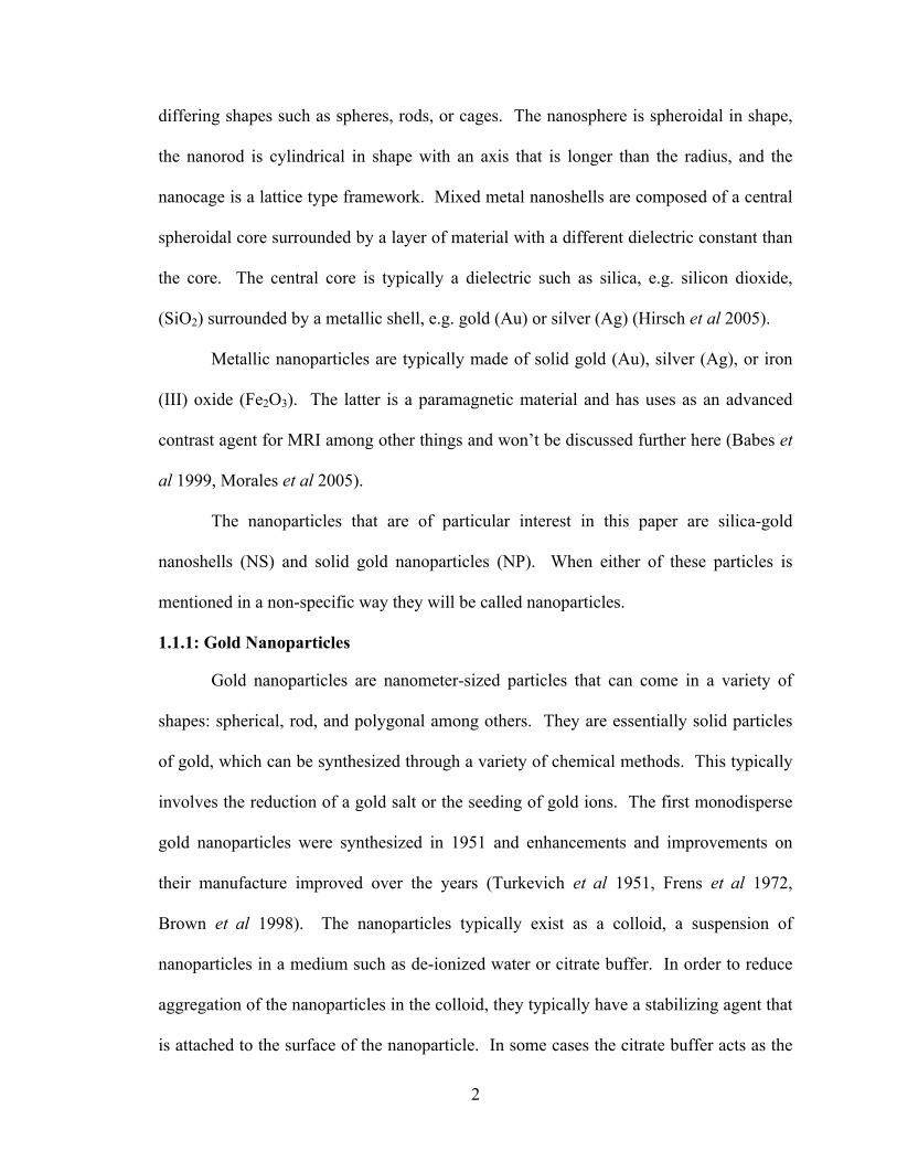
Figure 1: Deposition of Gold on Silica Core (Erickson Tunnell 2010)
The advantage to this method is that the core-shell ratios can be manipulated so
that surface plasmon resonance effects can be maximized depending on the desired use.
Surface plasmon resonance effects will be discussed in section 1.1.4.
1.1.3: Electromagnetic Interactions: Photoelectric Effect and others
The research that is being investigated in this thesis is to compare the effects of x-
ray dose enhancement on the viability of cells that have been incubated with gold
nanoparticles vs. silica-gold nanoparticles. In order to optimize photoelectric interactions
and minimize Compton scattering, it is desirable that photons have energies less than 500
kVp (Mesbahi 2010). The details of the photoelectric effect and Compton scattering will
be discussed below.
The proof of concept for using gold nanoparticles as a means to enhance dose to a
tumor was done by injecting mice, which had subcutaneous EMT-6 mammary
carcinomas, with 1.9 nm gold particles directly into the tumor site and then treating them
with 250 kVp x-rays (Hainfeld et al 2004).
Dose enhancement effects due to materials with a high atomic number (Z) relative
to the surrounding tissue, was first discussed concerning the interface between bone and
5
soft tissue (Spiers 1949). The phenomenon was also observed in patients receiving
radiation treatments that had metal wires in their jaws following reconstructive surgery
(Castillo et al 1988). Further historical investigation into the dose enhancing effects of
high Z materials and ionizing radiation can be found in the literature (Matsudaira et al
1980, Santos Mello et al 1983, Nath et al 1990, Regulla et al 1998, Herold et al 2000).
There are five interactions that can occur which will attenuate the x-rays as they
interact with the nanoparticles. The attenuation can be due to either scattering or
absorption and the type of interaction that occurs is primarily dependent on the energy of
the x-ray, and secondarily on characteristics of the nanoparticle, e.g. it’s atomic number,
Z, or the electron density, σe. The probability of a particular interaction occurring is
given by the linear attenuation coefficient or the mass attenuation coefficient. The mass
attenuation coefficient is the linear attenuation coefficient divided by the density of the
material. The five interactions that can occur, in order of increasing probability with
increasing energy are: Rayleigh (coherent) scattering, the photoelectric effect
(absorption), Compton (incoherent) scattering, pair production (absorption) and
photodisintegration (absorption).
Rayleigh scattering, also known as coherent scattering, is an interaction with an
electromagnetic wave (photon) and an outer shell electron of the atom making up the
material, which in this case would be gold, silicon, or oxygen, as these are the constituent
elements of the nanoparticles or nanoshells used. This is an elastic event resulting in a
change of direction of the photon while its energy remains unchanged. The scattered
photon may go on to interact further in the region of interest or may escape. This effect
6
can be significant when the energy is low (<10 keV) and the atomic number is high, as
the mass attenuation coefficient scales as approximately Z2.
The interaction of electromagnetic waves and metals has been studied extensively
for many years and details of the physics behind this can be found in books on
electromagnetic theory and scattering (Jackson 1998, Newton 2002, van de Hulst 1981).
The scattering of electromagnetic waves from small spherical particles, ie. where the
wavelength is much greater than the particle diameter is known as Rayleigh scattering.
This is treated as an elastic scattering event and no energy from the photon is transferred
to the material. The scattering of electromagnetic waves from particles that are a similar
size to the electromagnetic wave is described by Mie scattering theory (Aden et al 1951,
van de Hulst 1981). When the object has a size that is significantly greater than the
wavelength, then geometrical scattering is used. However, when the wavelength of the
incident radiation is small enough that its energy exceeds the binding energy of electrons
in the atoms, then classical electromagnetic theory must be abandoned and quantum
theory needs to be used.
The photoelectric effect is the type of interaction with which this thesis is
primarily interested. In this interaction, a photon is absorbed by an inner shell electron
which is then ejected from its orbital and is known as a photoelectron. The vacancy left
by the photoelectron is then filled by an electron in a higher shell, releasing its potential
energy in the form of a characteristic x-ray, which in turn may escape the medium or
interact with a higher shell electron. If a higher shell electron absorbs the characteristic
x-ray, that electron is also ejected from the atom and is known as an Auger electron.
7
The photoelectric effect is highly dependent upon the energy of the x-ray and the
atomic number of the material. The mass attenuation coefficient for this interaction is
proportional to (Z/E)3 (Khan 2010). It is because of this relationship that research is
being conducted into the use of metallic nanoparticles and nanoshells as a means to
enhance dose to tumors with kVp x-rays. The increase in dose to the tumor is known as
the dose enhancement factor (DEF) (Cai et al 2013). This factor is the ratio of dose
required for a certain effect in the presence of nanoparticles vs. the dose required to
achieve an identical effect in the absence of nanoparticles.
There are increases in the mass attenuation coefficients, which occur at the
binding energies of the electron shell in question. For the K-edge in gold, which
corresponds to the increase in the mass attenuation coefficient of the inner most shell, this
occurs at an energy of 80.7 keV. The L- and M-edge correspond to the next two highest
shells at energies of ~ 13 keV and ~ 3 keV respectively (Hainfeld et al 2008). Although
the absorption cross-section occurs at the stated energies, higher energies are required in
order to impart enough kinetic energy to the electron so that it can cross the length of
several cells. One effect of this is that, in order for the electron to be ejected with enough
kinetic energy to traverse a few cells, the photon must be of a higher energy than the
optimal absorption edge thus reducing the absorption cross-section (Hainfeld et al 2008).
When the energy of the photon becomes significantly greater than the binding
energy of the electrons, the photoelectric effect gives way to Compton scattering. In this
inelastic event, a photon transfers some of its energy to an outer shell electron, typically
ejecting the electron from the atom. The loss in energy of the photon may be enough that
it is able to go on to interact via the photoelectric effect. The photon may only excite the
8
electron, which can then spontaneously decay as fluorescent photons, or Auger electrons.
The first ionization energy of Au is 9.2 eV (Haynes et al 2014). Compton scattering is
essentially independent of the atomic number and is dependent on the electron density of
the material, as the interaction involves “free” electrons. For almost all materials, this is
on the order of 1023 electrons per gram of material (Khan 2010). This effect also
diminishes as photon energy increases.
Pair production is an effect that occurs in the presence of matter, whereby a high-
energy photon (>1.022 MeV) is transformed into an electron and a positron. The
positron will then interact with an electron, annihilating both in the process and
producing two 511 keV photons.
The final effect, photodisintegration, occurs at even higher photon energies than
pair production. This involves a photon interacting with a nucleus directly, ejecting a
proton, neutron, or alpha particle by a de-excitation of the nucleus.
Since this research involves X-rays with energies of 110-130 kVp, the role of pair
production and photodisintegration will not be covered further as they involve
interactions requiring megavoltage and higher energies.
1.1.4: Dose Enhancement
The main advantage to using kilovoltage x-rays and gold nanoparticles/nanoshells
as a potential treatment of malignancies is the high cross section for photoelectric events
compared to higher energy beams and/or lower Z materials. The increase in dose to
tissue due to these events is called radiosensitization and the increase in dose to tissue can
be quantified by the dose enhancement factor (DEF) or dose enhancement ratio (DER).
The dose deposited to tissue surrounding the gold nanoparticle from low-energy
x-rays is due to photoelectrons, characteristic x-rays and Auger electrons. The
9
characteristic x-rays can cause secondary photoelectric events, and the effect of all of the
electrons is to deposit their kinetic energy in the tissue. Dose is defined as the energy
transferred by ionizing radiation per unit mass of material, with units of gray (Gy) where
1 Gy = 1 J/kg (Khan 2010).
As discussed earlier, the effectiveness due to increase in absorption cross-section
at the energy of an electron shell edge is offset by the lack of kinetic energy of the
photoelectron at these same energies. In order to increase the kinetic energy of the
photoelectron, so that it can impart significant relative dose to tissue, requires choosing a
higher incident photon energy that unfortunately has the disadvantage of decreasing the
probability of a photoelectric event occurring. There is a significant increase in the
relative absorbance of gold vs. tissue at 20 keV, on the order of 95 times (Hainfeld et al
2008). This is below the 80.7 keV K edge of gold but about 7 keV above the L edge of
gold. At an energy of ~ 35 keV, there is a factor of ~ 50 increase in the ratio of gold
attenuation relative to soft tissue (Hainfeld et al 2008).
Monte Carlo simulations have shown that for a Pd-103 source (20.48 keV γ-ray)
the probability of a photoelectric event from the L shell is 76.13 % relative to all
photoelectric events. By comparison, an I-125 source (26.07 keV γ-ray) and a Yb-169
source (62.11 keV γ-ray) yield a 76.35 % and 66.61 % probability of an L shell
photoelectric event relative to all photoelectric events, respectively (Lechtman et al
2011). There have been many investigations into modeling radiosensitivity
enhancements with gold nanoparticles (Rose et al 1999, Lechtman et al 2011, Lechtman
et al 2013, Douglass et al 2013, Cai et al 2013).
10
1.1.5: Effective Z of Silica-Gold Nanoshells
Composite materials will obviously have a certain probability for the
photoelectric effect to occur. An element is determined by the number of protons in its
nucleus (the atomic number, Z), and for a neutral element this also determines the
number of electrons in it. A composite material has an effective atomic number, Zeff, that
is determined by
!!"" = !!!!!.!"!.!"
!
!!!
where fi is the fractional weighting of the number of electrons of the ith element and Zi is
the atomic number of the ith element (Mayneord 1937, Khan 2010).
A silica-gold nanoshell has a core of silicon dioxide surrounded by a layer of
elemental gold. Assuming the compounds and elements are pure and the densities are
standard, then MSiO2 = 60.08 g/mole, MAu = 196.97 g/mole, ρSiO2 = 2.648 g/cm3, ρAu =
19.3 g/cm3, #eSiO2/molecule = 30, #eAu/atom = 79 where M is the molecular/atomic mass
and ρ is the molecular/atomic density (Haynes WM (Ed.) 2014).
The volume of a sphere is given by V1 = (4/3) π r13 and that of a shell surrounding
the sphere is given by V2 = (4/3) π (r23-r1
3) where r1 is the radius of the inner core and r2
is the radius of the entire nanoshell. For the nanoshells used in this experiment r1 = 60
nm and r2 = 75 nm.
11
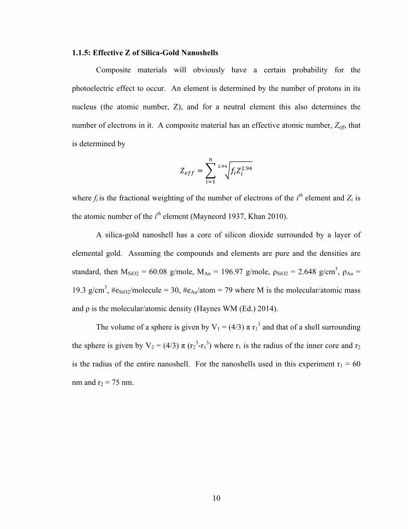
Figure 2: Radii of Nanoshell (ε represent dielectric constants). (Erickson Tunnell 2010)
Using the values given above, it can be shown that V1 = 9.048 x 10⁻¹⁶ cm3, V2 = 8.624 x
10-16 cm3, #eSiO2 = 7.202 x 10⁸ and #eAu = 4.019 x 10⁹ so that f1 = 0.152 and f2 = 0.848.
Inserting these values into the formula given above for the effective atomic number gives
Zeff =74.96. The calculated effective atomic number of the silica gold nanoshell is close
to Z = 75, the element rhenium, and is ~ 95 % of the value of a solid gold nanoparticle.
Thus silica-gold nanoshells should not show a significant decrease in dose due to the
photoelectric effect as compared to gold nanoparticles.
1.1.6: Electromagnetic Interactions: Surface Plasmon Resonance
There is a unique effect that can be exploited due to the interaction of optical
wavelengths of electromagnetic radiation (approximately 400 – 1100 nm) and gold
nanoparticles, specifically with silica-gold nanoshells. When an electromagnetic wave of
12
a certain frequency interacts with a medium of differing dielectric constants, of which the
surface one is a thin layer, a resonance effect occurs due to the collective oscillation of
the conduction electrons on the surface layer. This couples the incident electromagnetic
field to the conduction electrons and then propagates in a direction parallel to the
interface between differing dielectrics. This effect is known as surface plasmon
resonance (SPR) and is sensitive to changes in the boundary layer, i.e. its thickness or the
chemical structure of the layer. When this effect occurs where there is a nanoscale size
spherical object with differing dielectric constants, e.g. a silica-gold nanoshell, and the
wavelength of the incident electromagnetic field is on the size order of the nanoshell, the
plasmon is constrained to the surface of the nanoshell. In this case the effect is called
localized surface plasmon resonance (LSPR).
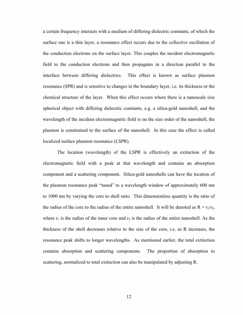
The location (wavelength) of the LSPR is effectively an extinction of the
electromagnetic field with a peak at that wavelength and contains an absorption
component and a scattering component. Silica-gold nanoshells can have the location of
the plasmon resonance peak “tuned” to a wavelength window of approximately 600 nm
to 1000 nm by varying the core to shell ratio. This dimensionless quantity is the ratio of
the radius of the core to the radius of the entire nanoshell. It will be denoted as R = r1/r2,
where r1 is the radius of the inner core and r2 is the radius of the entire nanoshell. As the
thickness of the shell decreases relative to the size of the core, i.e. as R increases, the
resonance peak shifts to longer wavelengths. As mentioned earlier, the total extinction
contains absorption and scattering components. The proportion of absorption to
scattering, normalized to total extinction can also be manipulated by adjusting R.
13
Figure 3: LSPR of Gold Nanoshell (Erickson Tunnell 2010)
The ability to specify the location of the resonance peak and the proportion of
absorbance to scattering, gives silica-gold nanoshells a distinct advantage over gold
nanoparticles. The plasmon resonance peak window of nanoshells contains the near infra
red (NIR) tissue window, which ranges from 700 nm to 900 nm. Tissue is quite
transparent in this range and this opens up the potential of using gold nanoshells for PTT
and PDT (Baek et al 2011, Trinidad et al 2014). By comparison, the resonance peak of
gold nanoparticles is located at ~ 520 nm (Hirsch et al 2006). This wavelength is not
suited for most light-based therapies due to its strong absorption and hence limited
penetration in biological tissues.
14
1.1.7: Toxicity
There are several reasons that gold is a desirable element to construct
nanoparticles out of. One is that it is relatively inert and biocompatible. This is an
important feature for use in biological applications. The low toxicity of gold is required
if these structures are to be left in the target for an indefinite amount of time and not
cause additional damage. Gold in its metallic form (non-ionized) is inert to chemical
processes that occur in the body and so is not susceptible to being transformed into toxic
gold compounds that could potentially be toxic (Merchant 1998). The incubation of
macrophages in vitro with silica-gold nanoshells has been found to be non-toxic to the
macrophages (Shukla et al 2005). Various in vivo studies have been performed to verify
the non-toxicity of silica-gold nanoshells (Hirsch et al 2003, O’Neal et al 2004, Stern et
al 2008, Gad et al 2012).
1.1.8: Conjugation
The gold surface of nanoparticles is capable of having a variety of different
compounds conjugated to its surface. In one method there is a passive attachment of
molecules or proteins to the gold surface which is known as adsorption. Adsorption may
not lead to a permanent attachment of the compound to the surface and that could limit its
usefulness. This method may also cause an active area of the attached compound to be
on the gold face, which renders it inert for whatever biological effect it is intended.
Another method of attachment is using a chemical linker, such as PEG to
permanently attach molecules to the surface of the nanoparticle. In this manner, the
required compound can be attached to the nanoparticle so that the active area of interest
is not in contact with the surface of the nanoparticle. The conjugation of certain
15
compounds to the gold surface has been shown to limit systemic clearance or to have
increased affinity to malignant cells (Kong et al 2008, Mody et al 2009).
1.2: MONOCYTES and SPHEROIDS
1.2.1: Enhanced Permeability and Retention Effect
In the absence of a tumor, bare nanoparticles that are circulating in a host’s
vascular system will continue to circulate until they are engulfed by macrophages
(Madsen et al 2011, Maeda et al 2000.). This is due to normal blood vessels being
essentially impermeable to nanoscale particles (Dvorak et al 1988, Jain 1999). Tumors
have the ability to emit a vascular epithelial growth factor (VEGF), which enables the
tumor to create new blood vessels (angiogenesis) (Goto et al 1993). This vasculature is
“leaky” and it lacks the relative impenetrability of normal blood vessels. As such,
nanoparticles with sizes on the order of hundreds of nanometers are able to extravasate
into the tumor through the leaky vasculature (Yuan 1995). The enhanced permeability of
the tumor vasculature, along with decreased lymphatic drainage associated with most
tumors results in passive accumulation of nanoscale structures in tumors. This is known
as the enhanced permeability and retention effect (EPR) (Maeda et al 1999).
1.2.2: Tumor Associated Macrophages
Solid tumors typically have a necrotic center consisting of dead or dying cells.
Surrounding this is a zone of cells that are in stasis due being in a zone that is low in
oxygen (hypoxic). Outside of this is the region of proliferating cells that have a well
developed capillary network and high oxygen levels. The necrotic core occurs due to the
rapid growth of malignant cells, which increasingly push out the proliferating cells which
have the ability to create the vasculature required to nourish the tumor (Hall et al 2012).
16
The lack of vasculature in the hypoxic region essentially nullifies the effectiveness of
exploiting the EPR effect for nanoparticle delivery to the tumor (Choi et al 2007).
Monocytes are the precursors to macrophages, the white blood cells that are
derived from the myeloid progenitor cells in the bone marrow (Madsen et al 2011). Once
monocytes leave the circulatory system and cross the endothelial basement membrane,
they differentiate into macrophages (Owen et al 2004, Choi et al 2007).
Tumors are able to attract monocytes via a chemo-attractive gradient. Once
differentiated into macrophages inside the tumor, there is evidence that the tumor is able
to manipulate them into promoting tumor growth (Lewis et al 2006). Macrophages that
have been recruited by the tumor are called tumor associated macrophages (TAMs).
TAMs can exist in significant numbers in tumors and have been shown to make up ~
65% - 70 % of the mass of a tumor (Kelly et al 1988, Fleige et al 2001). Exploiting the
ability of monocytes/macrophages to phagocytize nanoparticles, has led to the idea of
using them as a delivery vector to transport the nanoparticles to the tumor. This has been
called a cellular “Trojan Horse” (Choi et al 2007
1.2.3: Spheroids
Spheroids are an in vitro agglomeration of cells that have grouped together into a
spheroidal shaped mass that roughly mimics a tumor. This model was first developed and
used as a model to study in vitro responses to tumors in the early 1970’s (Inch et al 1970,
Sutherland et al 1971). One of the main advantages of spheroids over monolayer cell
cultures is the appearance of an oxygen gradient that decreases radially towards the
center of the spheroid (Santini et al 1999). The spheroid contains a necrotic core that is
located at 50 to 300 µm from the outer rim of the spheroid (Sutherland 1988). It is
believed that hypoxia is a key factor in creating the necrotic core of the spheroid
17
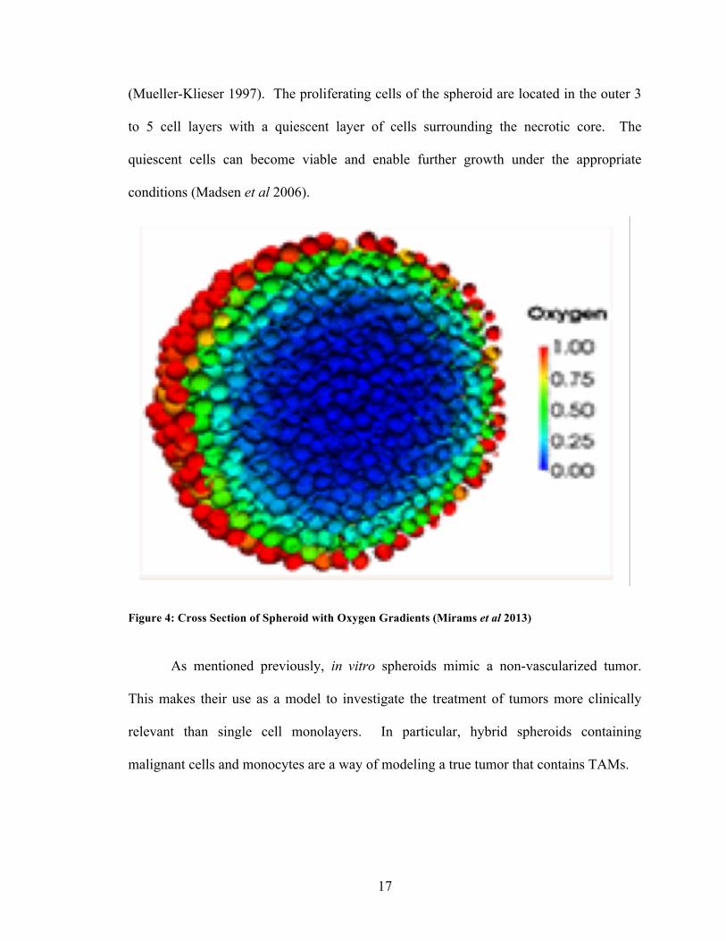
(Mueller-Klieser 1997). The proliferating cells of the spheroid are located in the outer 3
to 5 cell layers with a quiescent layer of cells surrounding the necrotic core. The
quiescent cells can become viable and enable further growth under the appropriate
conditions (Madsen et al 2006).
Figure 4: Cross Section of Spheroid with Oxygen Gradients (Mirams et al 2013)
As mentioned previously, in vitro spheroids mimic a non-vascularized tumor.
This makes their use as a model to investigate the treatment of tumors more clinically
relevant than single cell monolayers. In particular, hybrid spheroids containing
malignant cells and monocytes are a way of modeling a true tumor that contains TAMs.
18
1.3: SCOPE OF WORK
There are two main areas of research in this thesis. The first is to investigate the
efficacy of using 130 kVp x-rays on multicellular hybrid spheroids using silica-gold
nanoshell incubated monocytes. The multicellular hybrid spheroids consist of human
ACBT glioblastoma multiforme as the malignant cell line and P388D-1 murine
lymphocytic monocytes as the nanoparticle/nanoshell delivery vector. It has been shown
that cells containing gold nanoparticles are susceptible to dose enhancement effects from
kilovoltage x-rays due to the photoelectric effect. It is hypothesized that: (1) x-ray
irradiated hybrid spheroids containing nanoparticle loaded monocytes will show
decreased survival compared to irradiated hybrid spheroids with empty monocytes, and
(2) no statistically significant difference in survival between hybrid spheroids containing
gold nanoparticles and silica-gold nanoshells will be observed.
The second area of research is to establish parameters for the MTS assay which
will be used to measure the effects of ionizing radiation on P388D-1 monocytes. This
includes determining the number of cells to be plated in each well of a flat bottom 96
well plate, determining the number of days after radiation treatments for measurable cell
death to occur, and determining the number of hours the cells are incubated with the MTS
reagent. Two different radiation doses (8 and 20 Gy) will be tested on silica-gold
nanoshell, and gold nanoparticle incubated monocytes as well as controls (empty
monocytes). It is hypothesized that: (1) the 20 Gy dose will yield greater cell death than
the 8 Gy dose, (2) a significant decrease in survival will be observed following irradiation
of the nanoparticle loaded monocytes compared to empty monocytes at both doses, and
(3) no statistically significant difference will be observed when comparing survival of
irradiated monocytes containing either gold nanoparticles or silica gold nanoshells.
19
CHAPTER 2: MATERIALS AND METHODS
2.1: MATERIALS
2.1.1: Cell Lines and Culturing
All experiments were carried out in the Bigelow Health Sciences building (BHS)
and the Chemistry building (CHE) at the University of Nevada, Las Vegas (UNLV). The
malignant cell line used in the experiments, both as the basis for spheroid formation and
to measure the efficacy of the treatment techniques was a human grade IV glioblastoma
multiforme (GBM) (ACBT - G. Granger, University of California, Irvine, CA). The cell
line used to represent the cellular vector for the delivery of gold nanoparticles/nanoshells
to the tumor spheroids was the murine lymphocytic monocyte cell line P388D-1 (ATCC
CCL-46, American Type Culture Collection, Manassas, VA). This cell line was also
used as the basis for experiments measuring treatment efficacy and the establishment of
measurement parameters for the MTS assay.
All cultured cells and spheroids were kept in a CO2 Water Jacketed Incubator
(Sheldon Manufacturing Co., Cornelius, OR) which maintained a temperature of 37 °C,
80 % humidity and a CO2 level of 5%. Culturing was performed in a Labconco™ Purifier
Class A2 Biological Safety Cabinet (Labconco, Kansas City, MO). The ACBT cultures
were contained in T-25 BD Falcon tissue culture flasks and the P388D-1 cultures were
contained in T-75 BD Falcon tissue culture flasks (BD Biosciences, Franklin Lakes, NJ).
Culturing was done once per week in gibco™ Dulbecco’s Modified Eagle Medium
(1X DMEM) with 4.5 g/L D-Glucose, L-Glutamine, 110 mg/L Sodium Pyruvate , 25 mM
HEPES, and no phenol red (Thermo Fisher Scientific - Life Technologies, Carlsbad, CA).
The media was supplemented with 50 mL of 10% fetal bovine serum (FBS) and 5 mL
Pen-Strep (10,000 U/mL Penicillin and 10,000 μg/mL Streptomycin).
20
2.1.2: ACBT
ACBT exists as an adherent monolayer in a flask with media. The culturing of
ACBT cells was done by aseptically pipetting the old media out of the flask, which leaves
the adherent monolayer on the bottom of the flask. The flask was then lightly rinsed with
5 mL of gibco phosphate buffered saline (PBS), pH 7.4. To detach the adherent cells, 1
mL of 0.25 % Trypsin-EDTA (1X) with phenol red (Thermo Fisher Scientific - Life
Technologies, Carlsbad, CA) was added to the flask. In order to ensure proteolytic
cleavage, it was left in the flask for 5 minutes and sporadically rocked back and forth.
Once detached, 4 mL of PBS was added to the flask, aspirated by pipetting and then
transferred to a 15 mL centrifuge tube. The tube was then centrifuged for 5 minutes at
200 g in a Heraeus™ Megafuge™ 16 (Thermo Scientific, Waltham, MA). The
supernatant was pipetted off and 5 mL of media was added to the remaining cellular
pellet. The purpose of this step was to remove any dead cells, trypsin and old media.
The cells were then re-suspended using a VWR Digital Vortex Mixer (VWR
International, Radnor, PA) at 3000 rpm and a 1 mL aliquot was transferred to a T-25
flask containing 4 mL media.
2.1.3: P388D-1
Murine monocytes are essentially non-adherent and exist as a suspension in
media. The culturing of the P388D-1 murine monocytes was done by aseptically
pipetting a flask containing the monocytes into a centrifuge tube, stirred using a VWR
Digital Vortex Mixer (VWR International, Radnor, PA) at 3000 rpm, and then
centrifuging for 5 minutes at 200g using a Heraeus™ Megafuge™ 16 (Thermo Scientific,
Waltham, MA). The supernatant was then removed via pipetting and 10 mL of fresh
media was added to the tube and the cells re-suspended using the mixer. The purpose of
21
this step was to remove dead cells and old media. A 3 mL aliquot of the suspension was
transferred to a T-75 flask containing 15 mL of media.
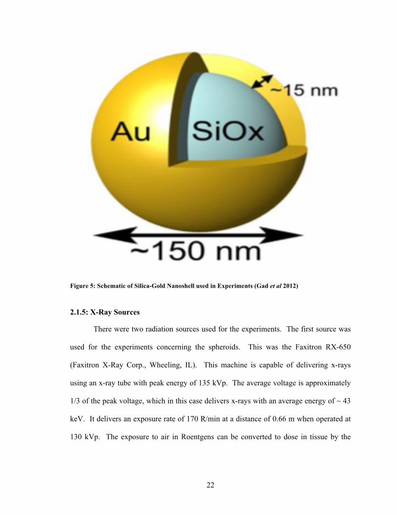
2.1.4: Silica-Gold Nanoshells and Gold Nanoparticles
The experiments were carried out using two different types of nanoparticles:
1. Silica-‐Gold Nanoshells (NS): The AuroShell™ nanoparticle is a silica-‐gold
core-‐shell coated with a functionalized poly(ethylene glycol) (PEG)
hydrophilic outer layer (Nanospectra Biosciences Inc., Houston, TX). It is
kept as a suspension in deionized water. The purpose of the PEGylated
coating is to reduce aggregation of the nanoparticles. The silica core has a
diameter of 150 nm and the gold coating is 15 nm thick, which gives the
overall diameter of the nanoparticle, sans PEGylated coating, as 150 nm. The
stock concentration is 2.82 x 10¹¹ particles/mL.
2. Gold Nanoparticles (NP): Cytodiagnostics™ 150 nm Stabilized Gold
Nanoparticle, supplied in a 0.1 mg/mL citrate buffer with a proprietary
stabilizing surfactant (Cytodiagnostics, Burlington, ON). The purpose of the
citrate buffer and stabilizing surfactant was to reduce aggregation of the
nanoparticles. The stock concentration was 3.9 x 10⁹ particles/mL.
22
Figure 5: Schematic of Silica-Gold Nanoshell used in Experiments (Gad et al 2012)
2.1.5: X-Ray Sources
There were two radiation sources used for the experiments. The first source was
used for the experiments concerning the spheroids. This was the Faxitron RX-650
(Faxitron X-Ray Corp., Wheeling, IL). This machine is capable of delivering x-rays
using an x-ray tube with peak energy of 135 kVp. The average voltage is approximately
1/3 of the peak voltage, which in this case delivers x-rays with an average energy of ~ 43
keV. It delivers an exposure rate of 170 R/min at a distance of 0.66 m when operated at
130 kVp. The exposure to air in Roentgens can be converted to dose in tissue by the
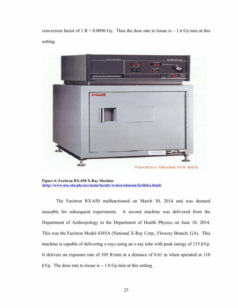
23
conversion factor of 1 R = 0.0096 Gy. Thus the dose rate to tissue is ~ 1.6 Gy/min at this
setting.
Figure 6: Faxitron RX-650 X-Ray Machine (http://www.uta.edu/physics/main/faculty/wchen/nbmain/facilities.html)
The Faxitron RX-650 malfunctioned on March 30, 2014 and was deemed
unusable for subsequent experiments. A second machine was delivered from the
Department of Anthropology to the Department of Health Physics on June 10, 2014.
This was the Faxitron Model 4385A (National X-Ray Corp., Flowery Branch, GA). This
machine is capable of delivering x-rays using an x-ray tube with peak energy of 115 kVp.
It delivers an exposure rate of 105 R/min at a distance of 0.61 m when operated at 110
kVp. The dose rate to tissue is ~ 1.0 Gy/min at this setting.
24
Both machines undergo a warm-up procedure where they are operated for 5
minutes at 30, 60, and 90 kVp respectively. After this is done they are ready for
operation at the desired voltage.
2.1.6: MTS Assay
The MTS assay (Promega Corp., Madison, WI) is a colorimetric method to
determine cell viability. It does this by counting the number of viable cells with a well
plate reader. Viable cells are metabolically active and they contain dehydrogenase
enzymes which, in the presence of phenazine methosulfate (PMS; Sigma, St. Louis, MO),
can convert MTS [3-(4,5-dimethylthiazol-2-yl)- 5-(3-carboxy-methoxyphenyl)-2-(4-
sulfophenyl)-2H-tetrazolium] into a purple formazan compound. This compound is
soluble in media and has an absorption peak at a wavelength of 490 nm (Santos et al
2014).
To determine the net number of viable cells, a negative control was subtracted
from the wells containing the cells of interest. For any particular experiment, 100 µL of
media containing a concentration of cells that is required, was pipetted into a minimum of
2 wells in a Costar™ flat bottom 96-Well Cell Culture Cluster Plate (Corning Inc.,
Corning, NY). Most experiments performed in this research used 16 wells of interest and
16 wells of negative control for each trial. In the negative control group, 80 µL of the
clear media was removed and replaced with 80 µL of 95 % ethanol to kill the cells. The
96 well plate was placed in the incubator for 10 min and then 80 µL of the media-ethanol
mixture was removed and replaced with 80 µL of clear media. To this, 20 µL of the MTS
assay was added to all wells containing cells and then allowed to incubate for 1 to 4
hours. Thus there was a 5:1 ratio of media to MTS assay with a total volume of 120
µL/well. Longer incubation times resulted in a darker color but cytotoxic effects due to
25
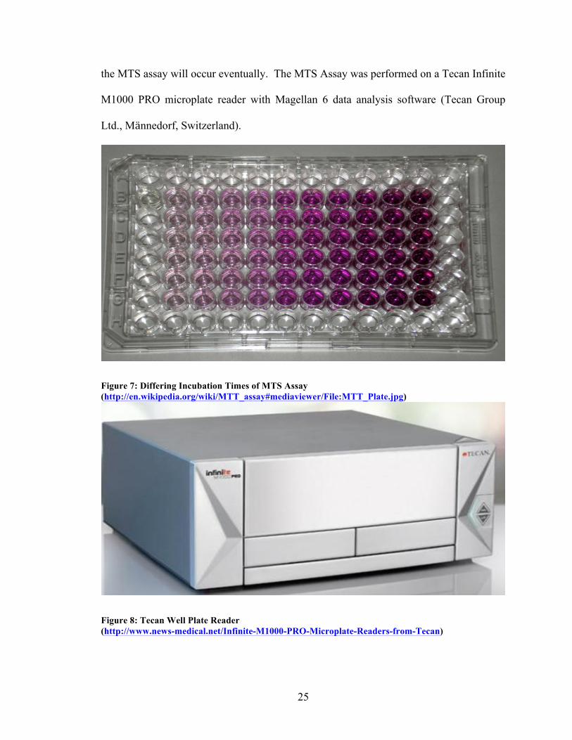
the MTS assay will occur eventually. The MTS Assay was performed on a Tecan Infinite
M1000 PRO microplate reader with Magellan 6 data analysis software (Tecan Group
Ltd., Männedorf, Switzerland).
Figure 7: Differing Incubation Times of MTS Assay (http://en.wikipedia.org/wiki/MTT_assay#mediaviewer/File:MTT_Plate.jpg)
Figure 8: Tecan Well Plate Reader (http://www.news-medical.net/Infinite-M1000-PRO-Microplate-Readers-from-Tecan)
26
2.2: METHODS
2.2.1: Incubation of Monocytes with Nanoshells and Nanoparticles
The monocytes need to be loaded (incubated) with the nanoparticles in order for
the experiment comparing the efficacy of radiation on the incubated vs. bare cells. It has
been shown that the optimum time for the uptake of 150 nm PEG silica-gold nanoshells
is approximately 24 hours (Chhetri 2013). Previous experiments performed at UNLV
have used a concentration of 4.285 x 10⁹ NS/mL and 5 x 10⁶ cells/mL for the PEG silica-
gold nanoshells (Makkouk 2010, Chhetri 2013).
The cell concentration was determined using an iNCYTO™ C-Chip disposable
hemocytometer (Neubauer Improved) (Incyto, Korea). Counting was performed under a
VWR inverted microscope (VWR International, Radnor, PA). The monocyte culture (10
µL was pipetted into the C-Chip and the number of monocytes in the central large square
was determined. This number was then multiplied by 104 to give the concentration of
monocytes/mL. The counting was done under a VWR microscope using a hand counter.
To obtain the required concentration of monocytes, an existing flask of culture was
centrifuged, the supernatant removed followed by the addition of the requisite amount of
media and re-suspension of the pellet. This step was done whether loaded or empty
monocytes were used.
2.2.1.1: Incubation of Silica-Gold Nanoshells
As mentioned above, the stock concentration of NS was 2.82 x 10¹¹ NS/mL in
deionized water. To get the required concentration of 4.285 x 10⁹ NS/mL the following
formula was used: V1 C1= V2 C2, where V is the volume and C is the concentration. This
calculation yielded a required volume of 15 μL NS per mL of media-nanoshell mixture.
27
This was then incubated for 24 hours and centrifuged for 5 minutes at 200 g to
remove un-endocytosed nanoshells. This step was repeated twice more using 10 mL of
media and the supernatant was removed each time. Resuspension was accomplished by
the addition of 10 mL of fresh media to the pellet. A cell count was performed and the
suspension diluted to achieve 5 x 10⁵ / mL of nanoshell incubated monocytes. It has been
shown that there is an uptake of 3.9 % of the silica-gold nanoshells by the P388D-1
monocytes over a 24 hour incubation period (Chhetri 2013).
2.2.1.2: Incubation of Gold Nanoparticles
The stock concentration of the NP was 3.6 x 10⁹ / mL in a citrate buffer solution.
This concentration was lower than the concentration used for the nanoshell experiments.
The experiment involving the nanoshells had 1.5 % of the total volume being attributable
to the nanoshells. Since the stock concentration of NP was lower than needed, it was not
possible to have the same ratio of nanoparticle to media volume. The experiment
performed used a ratio of 1 mL of nanoparticles to 1 mL of monocytes at a concentration
of 5 x 10⁵ monocytes / mL. This resulted in a final concentration of 1.8 x 10⁹ monocytes
/ mL of nanoparticles and a concentration of 2.5 x 10⁵ monocytes / mL.
The uptake of the gold nanoparticles by the P388D-1 monocytes was not known
as a blank citrate buffer could not be obtained for a background measurement.
2.2.2: Preparation of Incubated Monocytes for Spheroid Formation or MTS assay
As stated above, 24 hours prior to the creation of the spheroids, 4 mL of the
monocytes at 5 x 10⁵ / mL were placed into a 10 mL petri dish, and to this was added
either 15 µL of nanoshells or 4 mL of nanoparticles. This mixture was gently aspirated
with the pipette to mix the contents. The dish was then placed in the incubator until the
next day. One hour prior to the formation of the spheroids, mitomycin-C was added to
28
the dish in order to halt the division of the monocytes in a concentration of 20 μL/mL of
culture, i.e., 80 μL of mitomycin-C for the nanoshells and 160 μL of mitomycin C for the
nanoparticles. In cases where the MTS assay was used, mitomycin C was NOT added
and, as such, monocyte division was not halted.
2.2.3: Spheroid Creation
The spheroids were kept in well plates that had an agar mixture added to each
well. This agar is a nutrient base that the spheroids rest on while they are being
monitored for growth. The same day that incubation of the monocytes with nanoparticles
was started, the well plates with the agar mixture were prepared. These are Costar™ Flat
Bottom 48-Well Cell Culture Cluster Plates (Corning Inc., Corning, NY). First a 2%
agarose gel was heated in boiling water on a hot plate. Once the agarose was liquefied, it
was mixed in a 1:1:0.005 ratio of agarose: 2x DMEM : 1 M NaOH. 0.25 mL of this
mixture was pipetted into each well of the plate. This was then placed in the incubator to
cool. Once the agar had solidified in the well plate, 0.75 mL of media was added to each
well.
Spheroid creation was based on the rapid generation technique using a centrifuge
(Ivascu and Kubbies 2006). Two ratios of ACBT-to-P388D-1 cells were used (2:1 and
5:1). For the 5:1 ratio experiments, hybrid spheroids were created that initially contained
5000 ACBT cells and 1000 monocytes. For the 2:1 ratio experiment, 5000 ACBT cells
and 2500 monocytes were used. The same amount of ACBT per spheroid was used since
monocyte division was inhibited with the addition of mitomycin-C. Once the mitomycin-
C was added to the monocytes, the ACBT cells were prepared for the experiment. This
was done first by detaching them as described above in the culturing section. The
29
spheroids were formed in Costar™ Ultra Low Cluster 96-Well Round Bottom Plates via
centrifugation. 200 μL of combined ACBT/macrophages were pipetted into each well.
For the 5:1 ratio experiments a concentration of 50,000 ACBT cells/mL was used and for
the 2:1 ratio experiment, a concentration of 40,000 ACBT cells/mL was used.
Monocytes were harvested one hour after the addition of mitomycin-C was added.
They were then centrifuged to remove nanoparticles that had not undergone
phagocytosis. This was done at 200 rpm for 5 min followed by the removal of the
supernatant. Monocytes were then re-suspended in media and the process repeated twice.
Media was added to the final pellet to get a concentration of 10,000 monocytes/mL for
the 5:1 experiments and a concentration of 25,000 monocytes/mL for the 2:1 experiment.
The ACBT cells that were previously prepared were mixed with the respective
nanoparticle/nanoshell loaded monocytes for the requisite concentration in a 50 mL
centrifuge tube. The tube was then placed on a hand centrifuge to ensure the ACBT and
monocytes were uniformly distributed. The contents were then put in a multi-channel
reagent container and a multi-pipetter was used to transfer 200 μL of the mixture to each
of the round-bottomed well plates.
In cases where control groups of spheroids were required, the exact same
procedure as described above was performed, sans incubation of the monocytes. For the
ACBT only spheroids, 5,000 cells per spheroid were used.
To create the spheroids, they were centrifuged in their well plates at 800 g for 10
minutes. The plate was then transferred to the incubator to give the cells time to form the
spheroid structure. It has been observed that spheroid creation has not always been
consistent, and can range from 1-2 days and may require additional centrifugation.
30
2.2.4: Spheroid Treatment
The spheroids were irradiated with the Faxitron RX-650 at an energy of 130 kVp.
Experiments were performed using doses of 8, 10, 12, 14, and 20 Gy at a dose rate of 1.6
Gy/min.
Prior to irradiation, spheroids were transferred individually from each well of the
round bottom plate into a 10 mL petri dish containing 4 mL of medium. There were 16
spheroids per petri dish per experiment. This gave six-petri dishes per well plate so that
the spheroids could be treated at the five doses as well as having a control The petri
dishes were kept in the incubator until they were used in a treatment. Over the course of
an experiment, a few spheroids were typically lost or damaged, and as such they were no
longer tracked for the remainder of the experiment.
On the day of an experiment, the Faxitron was run through its warm-up
procedure, which required approximately 15 minutes. One petri dish at a time was then
moved from the room containing the incubator to the treatment room. In order to deliver
the doses of 8, 10, 12, 14, and 20 Gy, the Faxitron was operated at 130 kVp for 4:54,
6:08, 7:21, 8:35, and 12:15 minutes respectively. The treatments were done with the lid
of the petri dish removed to reduce x-ray attenuation and electron contamination. After
each treatment was performed, the spheroids were transferred by micro-pipetting each
spheroid into the flat bottomed well plates containing the agar and media.
2.2.5: Maintenance and Monitoring
The success of the treatments was based on monitoring the relative size of the
spheroids over a period of three weeks. Two orthogonal measurements were made of the
diameter of a spheroid using a calibrated micrometer on the VWR microscope. Each
measurement was rounded to the nearest 25 μm and then the two measurements
31
averaged. The volume of each spheroid was then calculated by the formula V=(1/6π)d3
where d is the average diameter of the spheroid. The day of treatment was counted as
day zero for measurements, and then two measurements were taken twice per week for
the following three weeks. This gave 7 measurements per spheroid from day zero to day
21. On the day of every measurement, the media was refreshed by removing 400 μL of
old media and replacing it with 425 μL of new media. The increase of new media was to
account for evaporation.
2.2.6: Determination of Monocyte Concentration for MTS Assay
In order to perform the experiments using the MTS assay to check for cell
viability, the optimum number of cells per well in a Costar™ 96-Well Flat Bottom Well
Plate had to be determined. The signal strength of the absorbance at 490 nm should
increase linearly as the number of cells increases per well. At a certain concentration, the
signal will reach saturation and the absorbance reading will plateau. The first experiment
was performed to determine the optimal number of monocytes per well. Once the linear
trend had been established, a concentration corresponding to the midpoint of the trend
line was used in subsequent experiments.
This experiment was performed by pipetting 100 μL of varying concentrations of
monocytes into the well plates. For each trial, 32 wells were used, 16 of which were used
for the negative control. The concentrations used were: 500, 1000, 2000, 5000, 10000,
20000, 50000, 100000, 150000, 200000, and 250000 monocytes per well. An MTS assay
incubation time of 1 hour was used for this initial experiment. It was found that 50,000
monocytes per well for 100 μL of media was on the midpoint of the linear trend. These
results are discussed further in Chapter 3.
32
2.2.7: Survivability and MTS Assay IncubationTime
The second set of experiments was performed to determine the optimal time to
apply the MTS assay following irradiation. In the first part of this experiment, a dose of
8 Gy was delivered to a 10 mL petri dish containing 4 mL of 5 x 10⁵ monocytes/mL. A
control group consisting of an identical preparation of monocytes was brought into the
treatment room, but was not irradiated. After each treatment, 50,000 monocytes were
pipetted into each well plate and left in the incubator for 24, 48 or 72 hours. After each
of the time periods cells were incubated with the MTS assay for one hour. The results of
this experiment were not conclusive so a second experiment with a higher dose and
longer post treatment times was done as described below.
In the second part of this experiment, a dose of 20 Gy was delivered to a 10 mL
petri dish containing 4 mL of 5 x 10⁵ monocytes/mL. As before, an unirradiated control
group consisting of an identical preparation of monocytes was included. After each
treatment, 50,000 monocytes were pipetted into each well plate and left in the incubator
for 24, 48, 72, 96 or 120 hours. In this experiment, not only were there five days of
experiments performed, but the MTS assay duration was also varied: 1, 2, 3 or 4 hour
incubation times were examined.
2.2.8: Toxicity
The third part of the experiments was performed to determine whether there was
any toxicity to the monocytes due to their incubation with gold nanoparticles or
nanoshells. In the experiments with the spheroids, this was not relevant as the monocytes
were given mitomycin-C and therefore they did not contribute to spheroid growth.
Monocytes were incubated with nanoparticles and nanoshells as described in Ch. 2.2.1.1
and Ch. 2.2.1.2 and were prepared as in Ch. 2.2.2. The MTS assay was performed at 72
33
hours after irradiation using an MTS incubation time of one hour. As will be shown in
Ch. 3, 1 hour of MTS incubation yielded an optimum result.
34
CHAPTER 3: RESULTS
3.1: SPHEROIDS
3.1.1: ACBT Spheroid Growth Kinetics
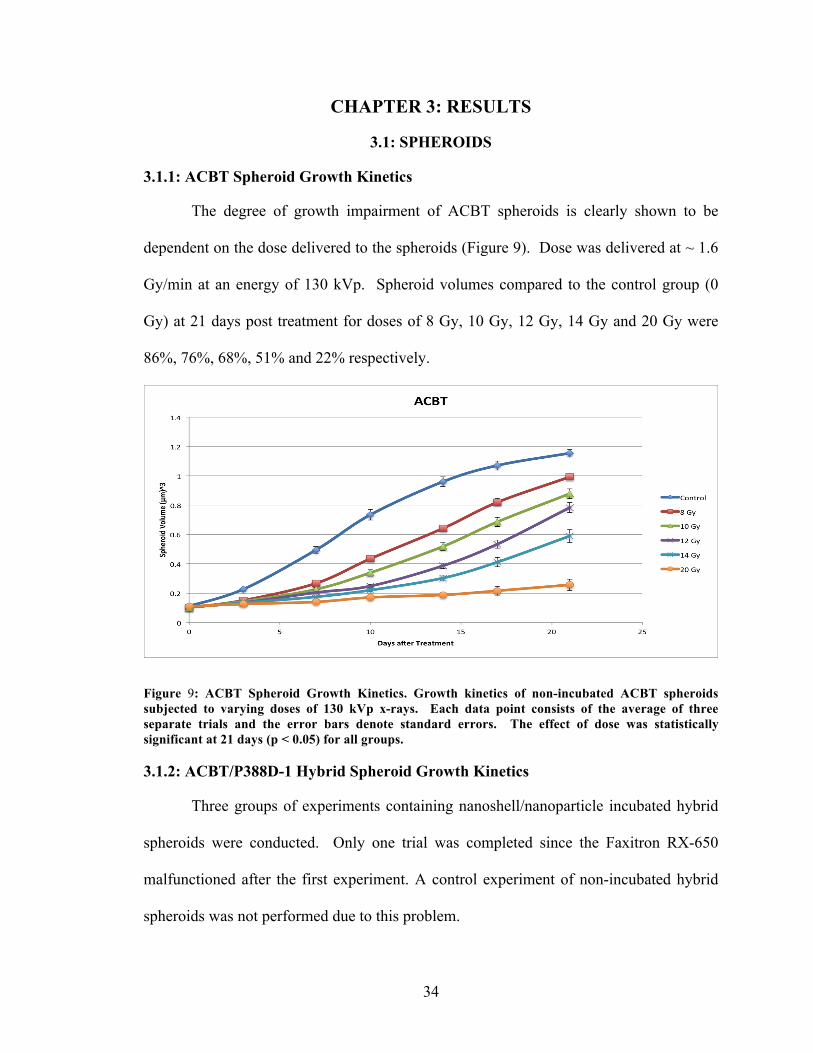
The degree of growth impairment of ACBT spheroids is clearly shown to be
dependent on the dose delivered to the spheroids (Figure 9). Dose was delivered at ~ 1.6
Gy/min at an energy of 130 kVp. Spheroid volumes compared to the control group (0
Gy) at 21 days post treatment for doses of 8 Gy, 10 Gy, 12 Gy, 14 Gy and 20 Gy were
86%, 76%, 68%, 51% and 22% respectively.
Figure 9: ACBT Spheroid Growth Kinetics. Growth kinetics of non-incubated ACBT spheroids subjected to varying doses of 130 kVp x-rays. Each data point consists of the average of three separate trials and the error bars denote standard errors. The effect of dose was statistically significant at 21 days (p < 0.05) for all groups. 3.1.2: ACBT/P388D-1 Hybrid Spheroid Growth Kinetics
Three groups of experiments containing nanoshell/nanoparticle incubated hybrid
spheroids were conducted. Only one trial was completed since the Faxitron RX-650
malfunctioned after the first experiment. A control experiment of non-incubated hybrid
spheroids was not performed due to this problem.
35
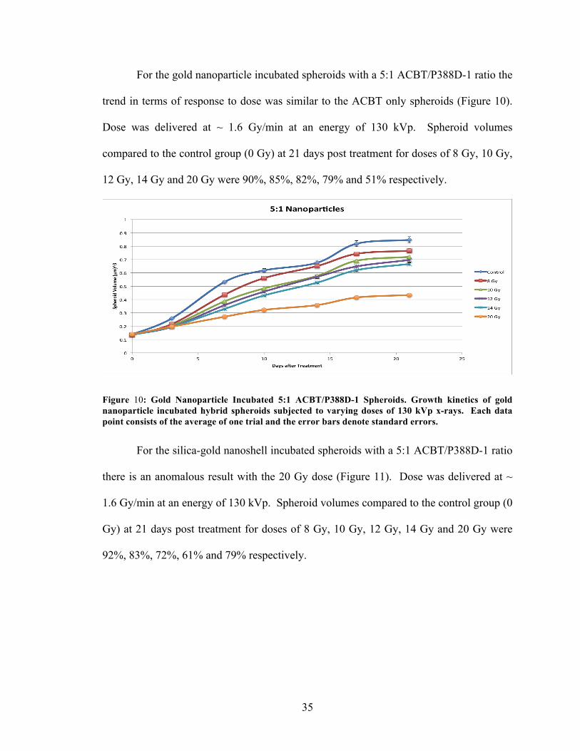
For the gold nanoparticle incubated spheroids with a 5:1 ACBT/P388D-1 ratio the
trend in terms of response to dose was similar to the ACBT only spheroids (Figure 10).
Dose was delivered at ~ 1.6 Gy/min at an energy of 130 kVp. Spheroid volumes
compared to the control group (0 Gy) at 21 days post treatment for doses of 8 Gy, 10 Gy,
12 Gy, 14 Gy and 20 Gy were 90%, 85%, 82%, 79% and 51% respectively.
Figure 10: Gold Nanoparticle Incubated 5:1 ACBT/P388D-1 Spheroids. Growth kinetics of gold nanoparticle incubated hybrid spheroids subjected to varying doses of 130 kVp x-rays. Each data point consists of the average of one trial and the error bars denote standard errors.
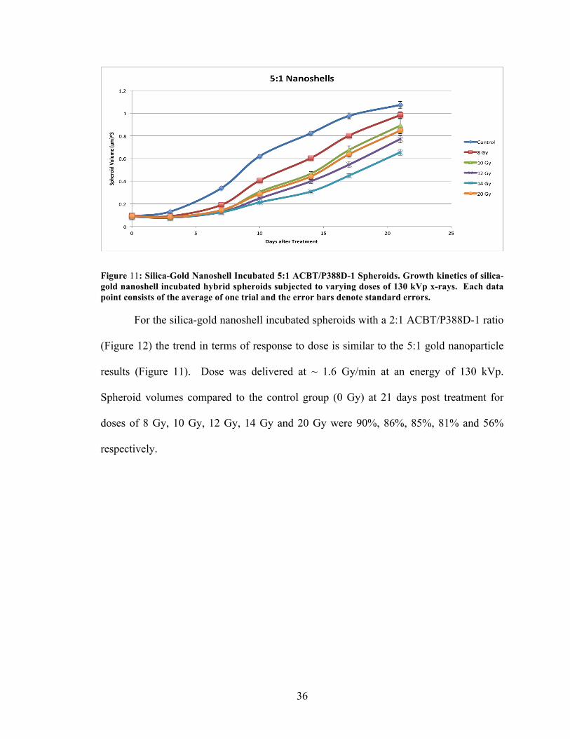
For the silica-gold nanoshell incubated spheroids with a 5:1 ACBT/P388D-1 ratio
there is an anomalous result with the 20 Gy dose (Figure 11). Dose was delivered at ~
1.6 Gy/min at an energy of 130 kVp. Spheroid volumes compared to the control group (0
Gy) at 21 days post treatment for doses of 8 Gy, 10 Gy, 12 Gy, 14 Gy and 20 Gy were
92%, 83%, 72%, 61% and 79% respectively.
36
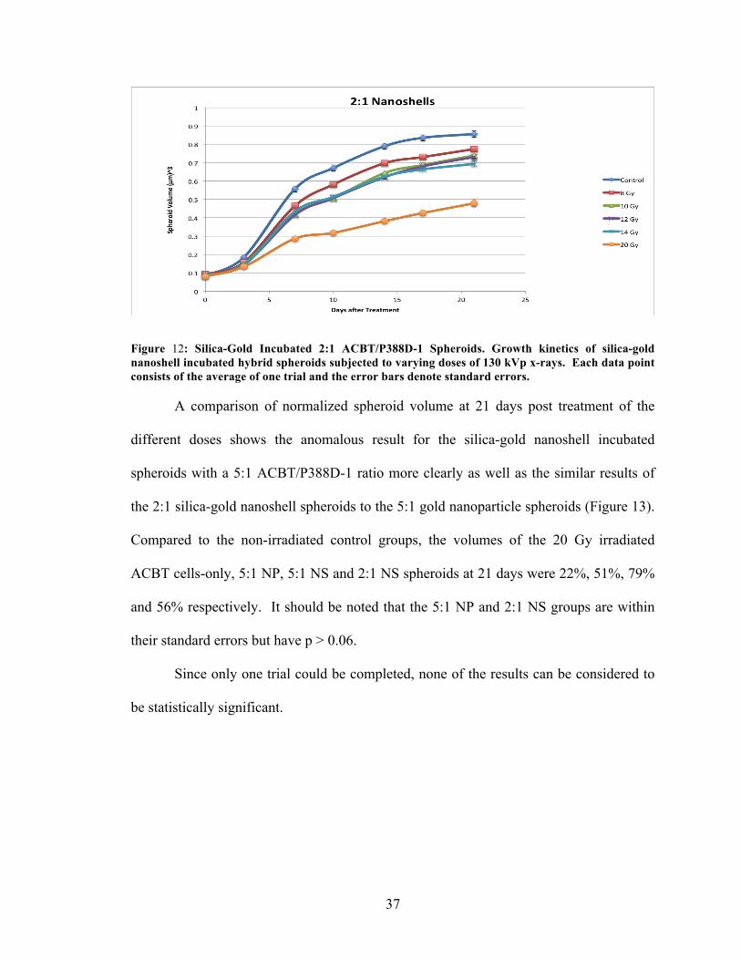
Figure 11: Silica-Gold Nanoshell Incubated 5:1 ACBT/P388D-1 Spheroids. Growth kinetics of silica-gold nanoshell incubated hybrid spheroids subjected to varying doses of 130 kVp x-rays. Each data point consists of the average of one trial and the error bars denote standard errors. For the silica-gold nanoshell incubated spheroids with a 2:1 ACBT/P388D-1 ratio
(Figure 12) the trend in terms of response to dose is similar to the 5:1 gold nanoparticle
results (Figure 11). Dose was delivered at ~ 1.6 Gy/min at an energy of 130 kVp.
Spheroid volumes compared to the control group (0 Gy) at 21 days post treatment for
doses of 8 Gy, 10 Gy, 12 Gy, 14 Gy and 20 Gy were 90%, 86%, 85%, 81% and 56%
respectively.
37
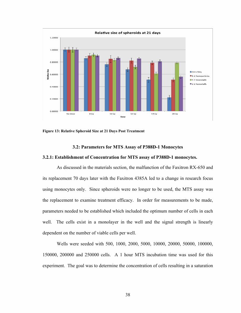
Figure 12: Silica-Gold Incubated 2:1 ACBT/P388D-1 Spheroids. Growth kinetics of silica-gold nanoshell incubated hybrid spheroids subjected to varying doses of 130 kVp x-rays. Each data point consists of the average of one trial and the error bars denote standard errors. A comparison of normalized spheroid volume at 21 days post treatment of the
different doses shows the anomalous result for the silica-gold nanoshell incubated
spheroids with a 5:1 ACBT/P388D-1 ratio more clearly as well as the similar results of
the 2:1 silica-gold nanoshell spheroids to the 5:1 gold nanoparticle spheroids (Figure 13).
Compared to the non-irradiated control groups, the volumes of the 20 Gy irradiated
ACBT cells-only, 5:1 NP, 5:1 NS and 2:1 NS spheroids at 21 days were 22%, 51%, 79%
and 56% respectively. It should be noted that the 5:1 NP and 2:1 NS groups are within
their standard errors but have p > 0.06.
Since only one trial could be completed, none of the results can be considered to
be statistically significant.
38
Figure 13: Relative Spheroid Size at 21 Days Post Treatment
3.2: Parameters for MTS Assay of P388D-1 Monocytes
3.2.1: Establishment of Concentration for MTS assay of P388D-1 monocytes.
As discussed in the materials section, the malfunction of the Faxitron RX-650 and
its replacement 70 days later with the Faxitron 4385A led to a change in research focus
using monocytes only. Since spheroids were no longer to be used, the MTS assay was
the replacement to examine treatment efficacy. In order for measurements to be made,
parameters needed to be established which included the optimum number of cells in each
well. The cells exist in a monolayer in the well and the signal strength is linearly
dependent on the number of viable cells per well.
Wells were seeded with 500, 1000, 2000, 5000, 10000, 20000, 50000, 100000,
150000, 200000 and 250000 cells. A 1 hour MTS incubation time was used for this
experiment. The goal was to determine the concentration of cells resulting in a saturation
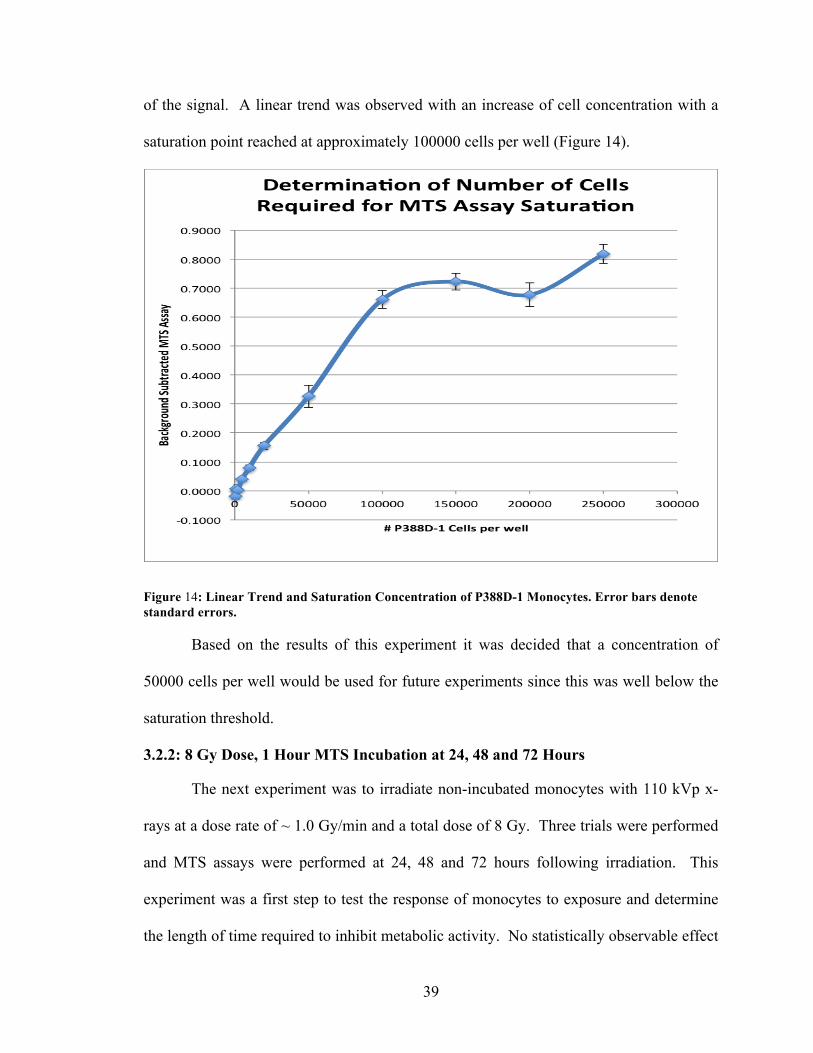
39
of the signal. A linear trend was observed with an increase of cell concentration with a
saturation point reached at approximately 100000 cells per well (Figure 14).
Figure 14: Linear Trend and Saturation Concentration of P388D-1 Monocytes. Error bars denote standard errors. Based on the results of this experiment it was decided that a concentration of
50000 cells per well would be used for future experiments since this was well below the
saturation threshold.
3.2.2: 8 Gy Dose, 1 Hour MTS Incubation at 24, 48 and 72 Hours
The next experiment was to irradiate non-incubated monocytes with 110 kVp x-
rays at a dose rate of ~ 1.0 Gy/min and a total dose of 8 Gy. Three trials were performed
and MTS assays were performed at 24, 48 and 72 hours following irradiation. This
experiment was a first step to test the response of monocytes to exposure and determine
the length of time required to inhibit metabolic activity. No statistically observable effect
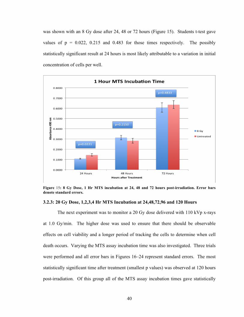
40
was shown with an 8 Gy dose after 24, 48 or 72 hours (Figure 15). Students t-test gave
values of p = 0.022, 0.215 and 0.483 for those times respectively. The possibly
statistically significant result at 24 hours is most likely attributable to a variation in initial
concentration of cells per well.
Figure 15: 8 Gy Dose, 1 Hr MTS incubation at 24, 48 and 72 hours post-irradiation. Error bars denote standard errors. 3.2.3: 20 Gy Dose, 1,2,3,4 Hr MTS Incubation at 24,48,72,96 and 120 Hours
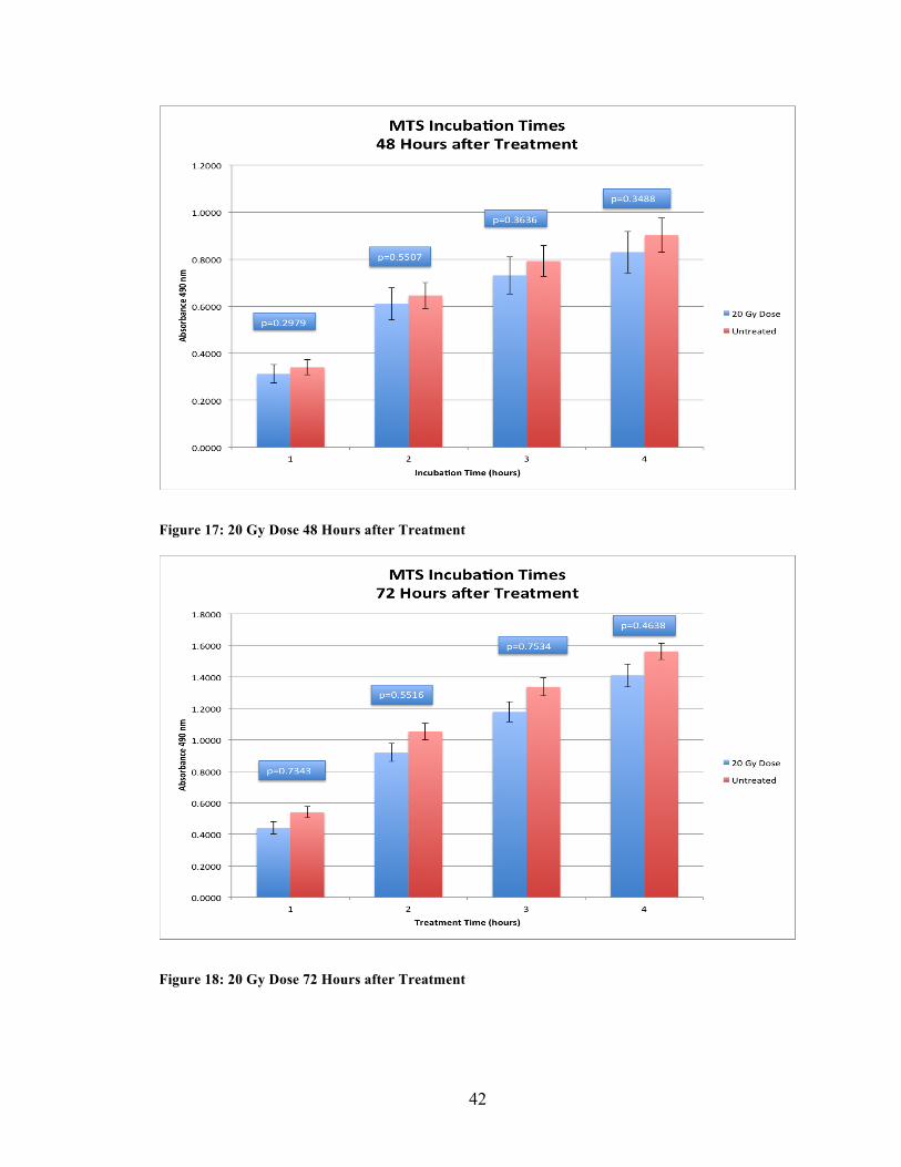
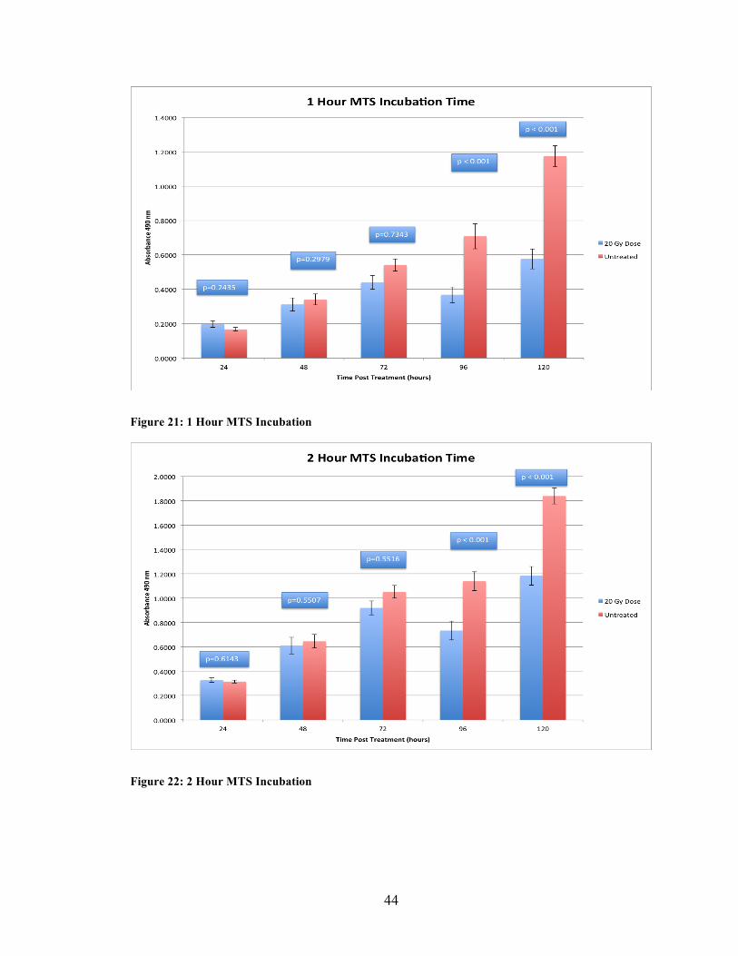
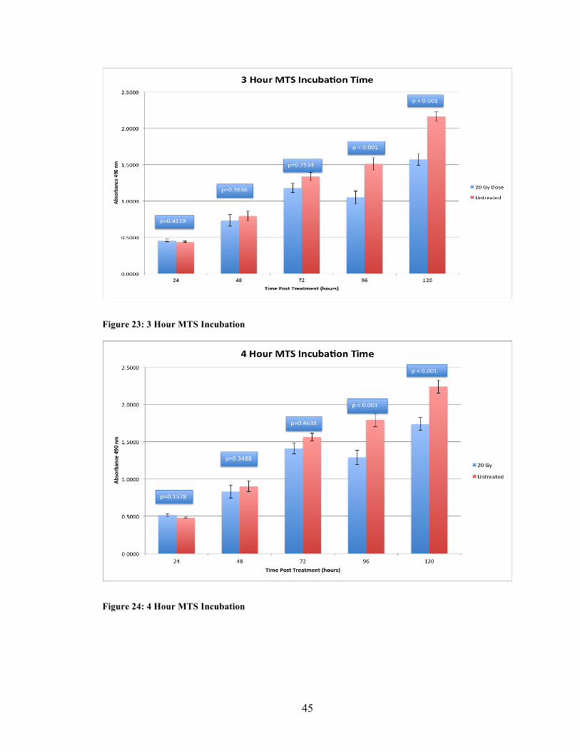
The next experiment was to monitor a 20 Gy dose delivered with 110 kVp x-rays
at 1.0 Gy/min. The higher dose was used to ensure that there should be observable
effects on cell viability and a longer period of tracking the cells to determine when cell
death occurs. Varying the MTS assay incubation time was also investigated. Three trials
were performed and all error bars in Figures 16–24 represent standard errors. The most
statistically significant time after treatment (smallest p values) was observed at 120 hours
post-irradiation. Of this group all of the MTS assay incubation times gave statistically
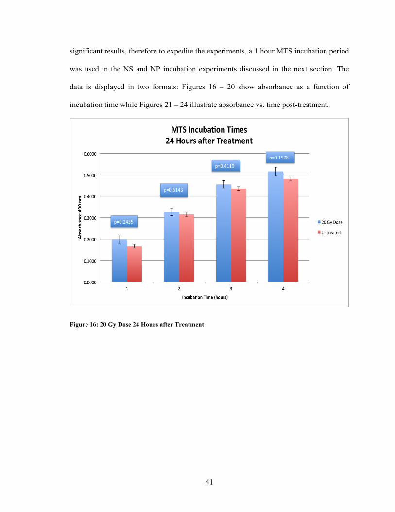
41
significant results, therefore to expedite the experiments, a 1 hour MTS incubation period
was used in the NS and NP incubation experiments discussed in the next section. The
data is displayed in two formats: Figures 16 – 20 show absorbance as a function of
incubation time while Figures 21 – 24 illustrate absorbance vs. time post-treatment.
Figure 16: 20 Gy Dose 24 Hours after Treatment
42
Figure 17: 20 Gy Dose 48 Hours after Treatment
Figure 18: 20 Gy Dose 72 Hours after Treatment

43
Figure 19: 20 Gy Dose 96 Hours after Treatment
Figure 20: 20 Gy Dose 120 Hours after Treatment
44
Figure 21: 1 Hour MTS Incubation
Figure 22: 2 Hour MTS Incubation
45
Figure 23: 3 Hour MTS Incubation
Figure 24: 4 Hour MTS Incubation
46
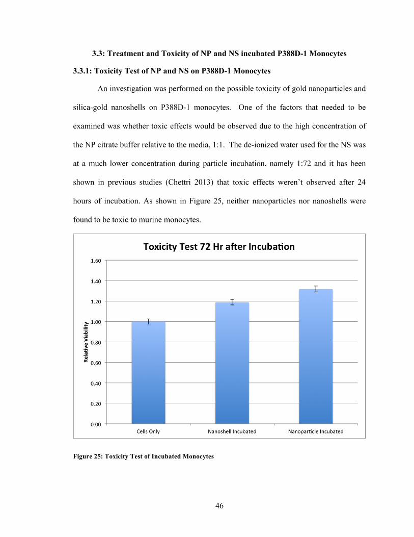
3.3: Treatment and Toxicity of NP and NS incubated P388D-1 Monocytes
3.3.1: Toxicity Test of NP and NS on P388D-1 Monocytes
An investigation was performed on the possible toxicity of gold nanoparticles and
silica-gold nanoshells on P388D-1 monocytes. One of the factors that needed to be
examined was whether toxic effects would be observed due to the high concentration of
the NP citrate buffer relative to the media, 1:1. The de-ionized water used for the NS was
at a much lower concentration during particle incubation, namely 1:72 and it has been
shown in previous studies (Chettri 2013) that toxic effects weren’t observed after 24
hours of incubation. As shown in Figure 25, neither nanoparticles nor nanoshells were
found to be toxic to murine monocytes.
Figure 25: Toxicity Test of Incubated Monocytes
47
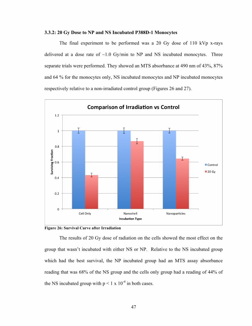
3.3.2: 20 Gy Dose to NP and NS Incubated P388D-1 Monocytes
The final experiment to be performed was a 20 Gy dose of 110 kVp x-rays
delivered at a dose rate of ~1.0 Gy/min to NP and NS incubated monocytes. Three
separate trials were performed. They showed an MTS absorbance at 490 nm of 43%, 87%
and 64 % for the monocytes only, NS incubated monocytes and NP incubated monocytes
respectively relative to a non-irradiated control group (Figures 26 and 27).
Figure 26: Survival Curve after Irradiation The results of 20 Gy dose of radiation on the cells showed the most effect on the
group that wasn’t incubated with either NS or NP. Relative to the NS incubated group
which had the best survival, the NP incubated group had an MTS assay absorbance
reading that was 68% of the NS group and the cells only group had a reading of 44% of
the NS incubated group with p < 1 x 10⁻⁴ in both cases.
48
CHAPTER 4: DISCUSSION
The use of nanoparticles in cancer treatments, particularly gold nanoparticles and
silica-gold nanoshells, is a novel and emergent technology that has been shown to
enhance dose effects in vitro, in silico and in vivo. Additionally, nanoparticles show
promise in other treatment modalities such as photothermal therapy, and photodynamic
therapy. Silica-gold nanoshells have the advantage over gold nanoparticles of having
tunable localized surface plasmon resonances (LSPR), which can take advantage of NIR
wavelength light for photothermal or photodynamic therapy, while maintaining an
effective atomic number that is within 10% of that of gold nanoparticles (Z = 79)
allowing for their use in combined radiation and NIR treatments.
The proof of concept of using gold nanoparticles was established when mice with
subcutaneous EMT-6 mammary carcinomas were injected with 1.9 nm gold nanoparticles
directly into the tumor (Hainfeld et al 2004). Treatment proceeded with 250 kVp x-rays
and a dose of 30 Gy. Tumor volume was significantly smaller 30 days post-treatment for
these mice vs. irradiation only controls. The 1.9 nm NP were chosen to avoid high liver
intake and to take advantage of the permeable vasculature of tumors (Dvorak et al 1988).
An additional study has shown that 68 keV (42 Gy) x-rays are more effective than 157
keV (44 Gy) x-rays in terms of tumor growth suppression (Hainfeld et al 2010). Energies
greater than 80.75 keV are required to take advantage of K-shell interactions in gold
while energies greater than ~ 13 keV are required for L-shell interactions.
The concept of dose enhancement from x-ray radiation due to the presence of
gold nanoparticles is based on the photoelectric effect. Not only have experiments
proven the effectiveness of this method, but Monte Carlo simulations have been carried
49
out that show the validity of enhancing dose to cells via photoelectric interactions due to
the presence of gold nanoparticles (Cho et al 2005, Cai et al 2013, Lechtman et al 2013).
It was shown that a dose enhancement factor of at least 2 was realized for 140 kVp
photon beams (Cho et al 2005). Furthermore, a peak in the DEF was shown at 30/40 keV
in a separate simulation (Cai et al 2013). These studies show that the experiments carried
out with the equipment used in this thesis project should be capable of producing
observable dose enhancement effects (kVp 110-130).
Spheroids are a proven in vitro method to model tumors in laboratory
experiments. Aside from lacking the vascularization of a true in situ tumor, spheroids
mimic the oxygen gradients that are observed in tumors. Multi-cellular hybrid spheroids
constitute a more accurate representation of tumors as they model the effects of tumor
associated monocytes/macrophages. The recognition that monocytes/macrophages can
compose a significant portion of a tumor (~ 60 % – ~ 70 %), has provided the rationale
for using them as a transport mechanism of nanoparticles in a technique called the
“Cellular Trojan Horse” (Choi et al 2007). The monitoring and measurement of spheroid
growth post-treatment is a standard technique for determining treatment efficacy.
The results in Figure 9 demonstrate a clear dose response for pure ACBT
spheroids. As expected, spheroid survival decreases with increasing x-ray dose. These
results are in qualitative agreement with those of Madsen et al. (2002) who used a Cs-137
gamma source (0.66 MeV) to irradiate ACBT spheroids to doses up to 16 Gy at a dose
rate of ~ 1.6 Gy/min.
The hybrid spheroid results presented in Figures 10-13 are somewhat difficult to
interpret. With the exception of a few anomalies, the data do show a dose response,
50
although not as pronounced as the corresponding data for the pure ACBT spheroids.
Unfortunately, as illustrated in Figure 13, the most pronounced dose effect was observed
in the pure ACBT irradiated spheroids which showed greater volume reduction compared
to the nanoparticle and nanoshell loaded hybrid spheroids at each dose investigated. This
is the exact opposite result one would expect based on increased photoelectric
interactions in the nanoparticle and nanoshell spheroids. These anomalous results are
likely due to the fact that the experiment was performed only once (due to equipment
malfunction) and, as such, the data cannot be considered statistically significant.
Furthermore, as the experiment was conducted at the very end of the useful lifetime of
the x-ray tube, one cannot rule out the possibility of significant output fluctuations
resulting in substantial dose variations and, hence significant dosimetry errors.
In the interest of expediency, it was decided to forego further investigations with
hybrid spheroids in favor of murine monocytes in which survival was monitored using a
colorimetric (MTS) assay. MTS assays allows for rapid determination of cell viability (a
few days) compared to the time consuming spheroid growth assay which requires at least
3 weeks.
The data presented in Figures 14-24 provide valuable information concerning the
optimal parameters for the MTS assay, and collectively, they represent the most
significant finding of this thesis. As illustrated in Figure 14, there is a relatively broad
range over which the MTS signal is linear with cell concentration (approximately 100 –
100,000) and this provided the rationale for choosing 50,000 cells per well (the
approximate midpoint). The data shows that the MTS signal saturates at a concentration
of approximately 100 000 cells above which the assay is no longer accurate.
51
The results illustrated in Figures 16-24 show that the duration between monocyte
irradiation and expression of damage was at least 96 hours (Figure 19) since this
represents the first time point at which there was a statistically significant survival
difference between irradiated and non-irradiated bare monocytes. Furthermore, the data
show that, as long as this 96 hour time interval was satisfied, any of the investigated MTS
incubation times (1 – 4 hours) could be used. For expediency, the shortest (1 hour) time
was chosen.
At the concentrations used in these experiments, neither the nanoshells nor the
nanoparticles were toxic to murine lymphocytic monocytes. This is in good agreement
with the findings of other investigators (Chhetri 2013; Makkouk 2010).
As shown in Figure 26, radiation was most effective in bare monocytes. This is
similar to the findings for the hybrid spheroids (Figure 13) and is clearly anomalous
based on expectations of increased photoelectric absorption in nanoshell and nanoparticle
loaded monocytes. The reasons for these anomalous results are not entirely clear, but are
likely due to inaccuracies in dosimetry. The replacement x-ray source used in these
experiments was very old and the reliability of the tube output was uncertain since
detailed dose measurements were not performed.
52
CHAPTER 5: CONCLUSION
The overall aim of this work was to investigate the dose enhancing effects of
ionizing radiation on multi-cellular hybrid tumor spheroids consisting of human glioma
cells and nanoparticle-loaded murine monocytes. The failure of a key piece of equipment
(Faxitron RX-650) after only one trial halted further experiments and, as a consequence,
the results obtained were inconclusive.
A second set of experiments was conducted with a different x-ray source
(Faxitron 4385 A) and utilizing the MTS assay to determine cell viability. It was
determined that a concentration of 50,000 monocytes per well in a 96 well plate yielded a
sufficiently accurate MTS signal. Furthermore, it was shown that the time interval from
irradiation to application of the MTS assay must be at least 96 hours in this in vitro
system. As long as the 96 hour interval is satisfied, any of the investigated MTS
incubation times (1 – 4 hours) may be used. As was the case with the hybrid spheroid
results, the monocyte studies were inconclusive. This was most likely due to fluctuations
in the x-ray tube output.
Future studies should be focused on using more reliable radiation sources
including commonly used x-ray irradiators based on radioisotopes such as 137Cs and 60Co.
With the opening of UNLV’s Accelerator Center, access to high energy (megavoltage)
sources will become available. This will facilitate more clinically relevant studies since
the energies are identical to those used in radiation therapy of cancer patients and, as
such, the true clinical potential of gold-based nanoparticles can be evaluated.
53
REFERENCES
Aden AL, Kerker M. Scattering efficiency for a layered sphere. J. Appl. Phys. 22:1242-1246, 1951. Averitt RD, Sarkar D, Halas NJ. Plasmon resonance shifts of Au-coated Au2S nanoshells: insight into multicomponent nanoparticles growth. Phys. Rev. Lett. 78:4217-4220, 1997. Babes L, Denizot B, Tanguy G, Le Jeune JJ, Jallet P. Synthesis of iron oxide nanoparticles used as MRI contrast agents: A Parametric Study. J. Colloid Interface Sci. 212: 474-482, 1999.
Baek S-K, Makkouk AR, Krasieva T, Sun C-H, Madsen SJ, Hirschberg H. Photothermal treatment of glioma; an in vitro study of macrophage-mediated delivery of gold nanoshells. J. Neurooncol. 104:439-448, 2011. Brown KR, Natan MJ. Hydroxylamine seeding of colloidal Au nanoparticles in solution and on surfaces. Langmuir. 14:726-728, 1998. Brown SB, Brown EA, Walker I. The present and future role of photodynamic therapy in cancer treatment. Lancet Oncol. 5:497-508, 2004. Cai Z, Pignol J-P, Chattopadhyay N, Kwon YL, Lectman E, Reilly RM. Investigation of the effects of cell model and subcellular location of gold nanoparticles on nuclear dose enhancement factors using Monte Carlo simulation. Med. Phys. 40:1141011-1141018, 2013.
Castillo MH, Button TM, Doerr R, Homs MI, Pruett CW, Pearce JI. Effects of radiotherapy on mandibular reconstruction plates. Am. J. Surg. 156:261-263, 1988.
Chhetri, S. Efficacy of gold silica nanoshells and gold nanorods for photothermal therapy of human glioma spheroids. University of Nevada, Las Vegas, ProQuest, UMI Dissertations Publishing, 2013. Cho SH. Estimation of tumour dose enhancement due to gold nanoparticles during typical radiation treatments: a preliminary Monte Carlo study. Phys. Med. Biol. 50:N163-N173, 2005.
Choi M-R, Stanton-Maxey KJ, Stanley JK, Levin CS, Bardhan R, Akin D, Badve S, Sturgis J, Robinson JP, Bashir R, Halas NJ, Clare SE. A cellular Trojan horse for delivery of therapeutic nanoparticles into tumors. Nano Lett. 7:3759-3765, 2007. Douglass M, Bezak E, Penfold S. Monte Carlo investigation of the increased radiation deposition due to gold nanoparticles using kilovoltage and megavoltage photons in a 3D randomized cell model. Med. Phys. 40(7):1-9, 2013.
Dvorak HF, Nagy JA, Dvorak JT, Dvorak AM. Identification and characterization of the blood vessels of solid tumors that are leaky to circulating macromolecules. Amer. Jour. Path. 133: 95-109, 1988. Fleige G, Nolte C, Synowitz M, Seeberger F, Kettenmann H, Zimmer C. Magnetic labeling of activated microglia in experimental gliomas. Neoplasia. 3:489-499, 2001.
54
Frens G. Particle size and sol stability in metal colloids. Colloid Polym Sci. 250:736-741, 1971.
Gad SC, Sharp KL, Montgomery C, Payne JD, Goodrich GP. Evaluation of the toxicity of intravenous delivery of auroshell particles (gold-silica nanoshells). Int. Jour. Toxi. 0:1-11, 2012. Goto F, Goto K, Weindel K, Folkman J. Synergistic effects of vascular endothelial growth factor and basic fibroblast growth factor on the proliferation and cord formation of bovine capillary endothelial cells within collagen gels. Laboratory Investigation. 69(5): 508–17, 1993. Hainfeld JF, Dilmanian FA, Slatkin DN, Smilowitz HM. Radiotherapy enhancement with gold nanoparticles. J. Pharm. Pharmacol. 60:977-985, 2008. Hainfeld JF, Dilmanian FA, Zhong Z, Slatkin DN, Kalef-Ezra JA, Smilowitz HM. Gold nanoparticles enhance the radiation therapy of a murine squamous cell carcinoma. Phys. Med. Biol. 55:3045-3059, 2010.
Hainfeld JF, Slatkin DN, Smilowitz HM. The use of gold nanoparticles to enhance radiotherapy in mice. Phys. Med. Biol. 49:N309-N315, 2004.
Hall EJ, Giaccia AJ. Radiobiology for the radiologist, seventh edition. Wolters Kluwer Health | Lippincott Williams & Wilkins, 2012.
Haynes WM (Eds.). Handbook of chemistry and physics, 95th edition. CRC Press, Taylor & Francis Group, London, 2014.
Herold DM, Das IJ, Stobbe CC, Iyer RV, Chapman JD. Gold microspheres: a selective technique for producing biologically effective dose enhancement. Int. J. Radiat. Biol. 76: 1357-1364, 2000. Hickey VF. Leukocyte traffic in the central nervous system: the participants and their roles. Semin. Immunol. 11:125-137, 1999. Hirsch LR, Gobin AM, Lowery AR, Tam F, Drezek RA, Halas NJ, West JL. Metal nanoshells. Ann Biomed Eng. 34:15-22, 2006. Hirsch LR, Stafford RD, Bankson JA, Serchen SR, Rivera B, Price RE, Hazle JD, Halas NJ, West JL. Nanoshell-mediated near-infrared thermal therapy of tumors under magnetic resonance guidance. Proc. Nat. Acad. Sci. USA. 100: 13549-13554, 2003.
Inch WR, McCredie JA, Sutherland RM. Growth of nodular carcinoma in rodents compared with multi-cell spheroids in tissue culture. Growth 34:271-282, 1970.
Ivascu A, Kubbies M. Rapid generation of single-tumor spheroids for high-throughput cell function and toxicity analysis. J. Biomol. Screen 11:922:932, 2006.
Jackson, JD. Classical electrodynamics, third edition. Wiley. New York, 1998. Jain PK, Lee KS, El-Sayed IH, El-Sayed MA. Calculated absorption and scattering properties of gold nanoparticles of different size, shape, and composition: applications in biological imaging and biomedicine. J Phys. Chem. B. 110:7238-7248, 2006.
Jain RK. Transport of molecules, particles, and cells in solid tumors. Ann. Rev. Biomed. Eng. 1:241-263, 1999.
55
Kelly KL, Coronado E, Zhao LL, Schatz GC. The optical properties of metal nanoparticles: the influence of size, shape, and dielectric environment. J. Phys Chem. B. 107:668-677, 2002. Kelly PM, Davison RS, Bliss E, McGee JO. Macrophage infiltration of breast tumors: a prospective study. Br. J. Cancer. 57:174-177, 1988. Khan FM. The physics of radiation therapy, fourth edition. Wolters Kluwer Health | Lippincott Williams & Wilkins, 2010. Kong T, Zeng J, Wang X, Yang X, Yang J, McQuarrie S, McEwan A, Roa W, Chen J, Xing JZ. Enhancement of radiation cytotoxicity in breast-cancer cells by localized attachment of gold nanoparticles. Small. 4:1537-1543, 2008.
Lechtman E, Chattopadhyay N, Cai Z, Mashouf S, Reilly R, Pignol J-P. Implications on clinical scenario of gold nanoparticle radiosensitization in regards to photon energy, nanoparticle size, concentration and location. Phys. Med. Biol. 56:4631-4647, 2011. Lechtman E, Mashouf S, Chattopadhyay N, Keller B M, Lai P, Cai Z, Reilly R M, Pignol J-P. A Monte Carlo-based model of gold nanoparticle radiosensitization accounting for increased radiobiological effectiveness, Phys. Med. Biol. 58:3075-3087, 2013.
Lewis C E, Pollard J W. Distinct role of macrophages in different tumor microenvironments. Cancer Res. 59:605-612, 2006.
Madsen SJ, Baek S-K, Makkouk AR, Krasieva T, Hirschberg H. Macrophages as cell-based delivery systems for nanoshells in photothermal therapy. Ann. Biom. Eng. 40:507-515, 2011. Madsen SJ, Sun C-H, Tromberg BJ, Yeh AT, Sanchez R, Hirschberg H. Effects of combined photodynamic therapy and ionizing radiation on human glioma spheroids. Photochem. Photobiol. 76:411-416, 2002.
Maeda H, Wu J, Sawa T, Matsumura Y, Hori K. Tumor vascular permeability and the EPR effect in macromolecular therapeutics: a review. Controlled Release. 65:271-284, 2000. Makkouk, AR. Macrophages loaded with gold nanoshells for photothermal ablation of glioma: An in vitro model. University of Nevada, Las Vegas, ProQuest, UMI Dissertations Publishing, 2010.
Matsudaira H, Ueno AM, Furuno I. Iodine contrast medium sensitizes cultured mammalian cells to x-rays but not to γ rays. Radiat. Res. 84:144-148, 1980.
Mayneord WV. The significance of the röntgen. Acta Int. Union Against Cancer. 2:271, 1937.
Merchant B. Gold, the noble metal and the paradoxes of its toxicology. Biologicals 26:49-59, 1998.
Mesbahi A. A review on gold nanoparticles radiosensitization effect in radiation therapy of cancer. Rep. Pract. Oncol. Radiother. 15(6): 176-180, 2010.
Mody VV, Nounou MI, Bikram M. Novel nanomedicine-based MRI contrast agents for gynecological malignancies. Adv. Drug Deliv. Formul. 61:795-807, 2009.
56
Morales MP, Bomati-Miguel O, de Alejo RP, Ruiz-Cabello J, Veintemillas-Verdaguer S, O’Grady K. Contrast agents for MRI based on iron oxide nanoparticles prepared by laser pyrolysis. J. Magn. Magn. Mater. 266:102-109, 2003. Mueller-Klieser W. three-dimensional cell cultures: From molecular mechanisms to clinical applications. Am. J. Physiol. 273:C1109-C1123, 1997. Nath R, Bongiorni P, Rockwell S. Iododeoxyurdine radiosensitization by low- and high-energy photons for brachytherapy dose rates. Radiat. Res. 124:249-258, 1990. Newton, RG. Scattering theory of waves and particles, second edition. Dover Publications, Inc. Mineola, 2002. O’Neal DP, Hirsch IR, Halas NJ, Payne JD, West JL. Photo-thermal tumor ablation in mice using near infrared-absorbing nanoparticles. Cancer Letters. 209:171-176, 2004. Oldenburg SJ, Averitt RD, Westcott SL, Halas NJ. Nanoengineering of optical resonances. Chem. Phys. Lett. 28:243-247, 1998. Owen MR, Byrne HM, Lewis CE. Mathematical modeling of the use of macrophages as vehicles for drug delivery to hypoxic tumour sites. J. Theor. Biol. 226: 377-391, 2004. Regulla DF, Hieber LB, Seidenbusch M. Physical and biological interface dose effects in tissue due to x-ray-induced release of secondary radiation from metallic gold surfaces. Radiat. Res. 150:92-100, 1998.
Rose JH, Norman A, Ingram M, Aoki C, Solberg T, Mesa A. First radiotherapy of human metastatic brain tumors delivered by a computerized tomography scanner (CTRx). Int. J. Radiat. Oncol. Biol. Phys. 45:1127-1132, 1999. Santini MT, Rainaldi G, Indovina PL. Multicellular tumor spheroids in radiation biology. Int. J. Radiat. Biol. 75:787-799, 1999. Santos GS, Tsutsumi S, Vieira DP, Bartolini P, Okazaki K. Effect of Brazilian propolis (AF-08) on genotoxicity, cytotoxicity and clonogenic death of Chinese hamster ovary (CHO-K1) cells irradiated with 60Co gamma-radiation. Mutation Research. 762:17-23, 2014. Santos Mello R, Callisen H, Winter J, Kagan AR, Norman A. Radiation dose enhancement in tumors with iodine. Med. Phys. 10: 75-78, 1983. Shukla R, Bansal V, Chaudhary M, Basu A, Bhonde RR, Sastry M. Biocompatibility of gold nanoparticles and their endocytotic fate inside the cellular compartment: a microscopic overview. Langmuir, 21:10644-10654, 2005.
Spiers FW. The influence of energy absorption and electron range on dosage in irradiated bone. Br. J. Radiol. 22:521-533, 1949.
Stern JM, Stanfield J, Kabbani W, Hsieh JT, Cadeddu JA. Selective prostate cancer thermal ablation with laser activated gold nanoshells. Jour. Urology. 179:748-753, 2008.
Sutherland RM, McCredie JA, Inch WR. Growth of multi-cellular spheroids in tissue culture as a model of nodular carcinomas. J. Natl. Cancer Inst. 46:113-120, 1971.
Sutherland RM. Cell and environment interactions in tumor microregions: The multicell spheroid model. Science. 240:177-184, 1988.
57
Trinidad AJ, Hong SJ, Peng Q, Madsen SJ, Hirschberg H. Combined concurrent photodynamic and gold nanoshell loaded macrophage-mediated photothermal therapies: an in vitro study on squamous cell head and neck carcinoma. Lasers Surg. Med. 46:310-318, 2014.
Turkevich J, Stevenson PC, Hillier J. A study of the nucleation and growth process in the synthesis of colloidal gold. Discussions Faraday Soc. 11:55-75, 1951.
van de Hulst, HC. Light scattering by small particles. Dover Publications, Inc. Mineola 1981
Welch A, van Gemert M (Eds.). Optical-Thermal Response of Laser-Irradiated Tissue, Plenum Press, New York, 1995.
Yuan F. Vascular permeability in a human tumor xenograft: molecular size dependence and cutoff size. Cancer Research. 55: 3752-3756, 1995.

58
VITA
Graduate College University of Nevada, Las Vegas
Gregory Joseph Colarch
Degrees: Bachelor of Science in Astrophysics, May 2009 University of Calgary Master of Science in Physics, December 2012 University of Nevada, Las Vegas Thesis Title: In Vitro Studies of Gold and Gold Silica Nanoparticle Radiosensitization with Kilovoltage X-Rays Thesis Examination Committee: Chairperson, Steen Madsen, Ph.D. Committee Member, Yu Kuang, Ph.D. Committee Member, Bing Ma, Ph.D. Graduate Faculty Representative, Robin Hickman, Ph.D.