Embed Size (px)
Citation preview
1
SYNTHESIS AND CHARACTERISATION
OF
SILICA COATED MAGNETITE NANOPARTICLE
A Dissertation Submitted in partial fulfillment
FOR THE DEGREE OF MASTER OF SCIENCE IN CHEMISTRY
Under The Academic Autonomy NATIONAL INSTITUTE OF TECHNOLOGY, ROURKELA
By
Ranjana Panigrahi
Under the Supervision of Dr. Sasmita Mohapatra
DEPARTMENT OF CHEMISTRY
NATIONAL INSTITUTE OF TECHNOLOGY
ROURKELA – 769008, ORISSA
2
CERTIFICATE
This is to certify that the dissertation entitled “Synthesis and characterization of silica coated
magnetite nanoparticle” being submitted by Miss Ranjana Panigrahi to the Department of
Chemistry, National Institute of Technology, Rourkela, Orissa, for the award of partial
fulfillment of the degree of Master of Science is a record of the research training carried out by
her under my supervision and guidance. This work is a scale-up synthesis process of silica coated
magnetite following the procedure earlier reported. The matter embodied in the dissertation has
not been submitted to any other University / Institute for the award of any Degree or Diploma.
Rourkela Date: 05-05-2011 Dr. Sasmita Mohapatra Dept. of Chemistry National Institute of Technology Rourkela, Orissa
3
ACKNOWLEDGEMENT With deep regards and profound respect, I avail the opportunity to express my deep sense of
Gratitude and indebtedness to Dr. Sasmita Mohapatra, Department of Chemistry, National
Institute of Technology, Rourkela for introducing the present project topic and for her inspiring
guidance, constructive criticism and valuable suggestion throughout the project work. I most
gratefully acknowledge her constant encouragement and help in different ways to complete this
project successfully.
I acknowledge my sincere regards to Dr. B.G. Mishra (HOD) and all the faculty
members,Department of Chemistry, NIT Rourkela for their enthusiasm in promoting the research
inchemistry and for their kindness and dedication to students. I specially record my deep
appreciation and thanks to Dr. B. G. Mishra, Dr. N. Panda, Dr. S.Patel, Dr. S. Chatterjee, Dr.
A. Mondal and their Ph.D scholars for giving me the necessary permission to use their
laboratory facilities whenever I needed.
I would like to add a special note of thanks to Mr. Smruti, and Ms. Swagatika Ph.D.
Scholars, Dept. of Chemistry for her kind help and guidance whenever required.
I acknowledge the support of my classmates throughout this course.Last but not the least,
I also take the privilege to express my deep sense of gratitude to my parents, for selflessly
extending their ceaseless help and moral support at all times.
Thanking you
Ms. Ranjana Panigrahi Roll No.:-409cy2018 M.Sc. Chemistry N.I.T. Rourkela

4
Table of contents
Contents Subject Page no
Chapter-1 Introduction 6-7
Chapter-2 Experiment 8-9
Chapter-3 Result and Discussion 9-15
Chapter-4 Conclusion 16
Chapter-5 References 17
5
Abstract
Magnetite nanoparticle was prepared by co-precipitation of Fe2+ and Fe3+, and then it was coated
with silica. The silica coated magnetite nanoparticles were characterized in terms of their
structure, morphology, hydrodynamic size and presence of surface functional groups by X-ray
diffraction analysis, Scanning electron microscopy, Dynamic light scattering and Infrared
spectroscopy respectively. It was found that the silica coating prevents magnetic particles from
aggregation and imparts excellent stability in aqueous medium.
6
Chapter-1
INTRODUCTION
Nanoscience is one of the most important research & development frontiers in modern
science. The use of nanoparticles offers many advantages due to their unique size & physical
properties. Key importance of these magnetic nanoparticles is their unique properties [1], their
controlled dimension maintaining their physical properties. Characteristics of magnetic
nanoparticles such as high magnetization, superparamagnetism, high field irreversibility , high
saturation field[2], smaller size than 100 nm and its narrow particle size distribution found to be
advantageous for many technological applications such as for bio separation in life science[3]
biomedicine and bioengineering such as magnetically assisted drug delivery [4], cell isolation
[5], MRI contrast agents [6], immunoassay [7] and bio macromolecule purification [8]
Magnetic nanoparticle is an interesting system in nanotechnology applications due to
the magnetic switching behavior and because of the transition between SPM and FM behaviors,
which can be adjusted by modifying the distance between the particles. Nanoparticles
possessing magnetic properties offer great advantages in that they can provide selective
attachment to a functional molecule, confer magnetic properties to the target, and allow
manipulation and transportation to a desired location through the control of a magnetic field
produced by an electromagnet or permanent magnet. The important physical property that
provides its wide application is its chemical inertness and resistance to surface oxidation.
The materials used in bio separation processes are superparamagnetic, meaning that they
respond strongly to magnetic fields, but retain no residual magnetism after the field is removed.
The morphology, surface area and the magnetic susceptibility of the support contribute in a
major fashion to the efficiency of the separation processes. Additionally, the chemical nature of
the support surface can be used to specify the separation process: the adsorption of the molecular
species can be driven by the interactions at the molecular level between the surface groups of the
paramagnetic particles and those of the target molecules.[9]
The surface of magnetite nanoparticle can be modified with several polymers such as
polyethylene glycol [10], Polystyrene [11], starch [12], chitosan [13], inorganic chemicals such
as SiO2,ZrO2,TiO2. Coating of magnetic nanoparticles with silica is becoming a promising and
7
important approach in the development of magnetic nanoparticles for both fundamental study
and technology application. First, silica formed on the surface of magnetic nanoparticles could
screen the magnetic dipolar attraction between magnetic nanoparticles, which favors the
dispersion of magnetic nanoparticles in liquid media and protects them from leaching in an
acidic environment. Second, due to the existence of abundant silanol groups on the silica layer,
silica-coated magnetic nanoparticles could be easily activated to provide the surface of silica-
coated magnetic nanoparticles with various functional groups� Finally, the most important is that
the silica layer provides a chemically inert surface for magnetic nanoparticles in biological
systems [14].
Recently much attention has been focused on the development of suitable method or
improvement of existing methods for large scale production of silica coated magnetic
nanoparticles with reliable size, colloidal stability and good magnetic property. In the present
dissert, we have synthesized magnetite nanoparticles by coprecipitation and then it was coated
with silica following a sol-gel route. The material was characterized XRD analysis, SEM. The
aqueous dispersion stability was investigated using dynamic light scattering.
8
Chapter-2
EXPERIMENTAL
2.1. Chemicals
The chemicals used for the synthesis of silica coated Fe3O4 magnetic nanoparticle were:
1. Anhydrous FeCl3 and FeSO4
2. Trisodium citrate and NH4OH
3. tetra ethyl orthosilicate (TEOS)
2.2. Synthesis of Fe3O4 magnetic Nanoparticles:
0.648g of FeCl3 and 0.548g of FeSO4.6H2O were taken to which 40-45 ml of Millipore water
was added under N2 environment.5-10 ml of 25% NH3 was added to the reaction mixture with
continuous stirring and under nitrogen environment. And was kept for further stirring for1hr.
100 ml of 0.3 molar trisodium citrate was added to the reaction mixture and again was allowed
for stirring for another 30 min at 90 0C. The synthesized particles were collected using magnetic
separator and was washed with (3×20 ml) Millipore water. Fe3O4 were again dispersed in
Millipore water [15].
2.3. Synthesis of silica coated Fe3O4 nanoparticles:
4g of the Fe3O4 despersed in Millipore water was taken in a solution of CH3OH (160 ml)
and 40 ml H2O. To this reaction mixture 3ml of NH3 solution and 2ml of tetraethyl
orthosilicate(TEOS) were added one after another and was kept for stirring for further 24hr
followed by washing with (2×30)ml Millipore water. Then dried at 80 0C and further heated at
400 0C [15].
2.4. Characterization
The identification of crystalline phase of the synthesized ferrite particles was performed
by an Expert Pro Phillips X-ray diffractometer. The morphology and microstructure were
analysed using scanning electron microscope (HITACHI COM-S-4200). The hydrodynamic size
and dispersion stability of particle aggregates were investigated through dynamic light scattering
using a Malvern ZS90 zetasizer. Hydrodynamic size was measured by dispersing 0.1 mg of
sample in 1 ml phosphate buffered saline. The surface charge was determined through zeta
potential measurements. The surface chemistry of the nanoparticles was studied using FTIR
spectroscopy (Perkin Elmer (BX 12).
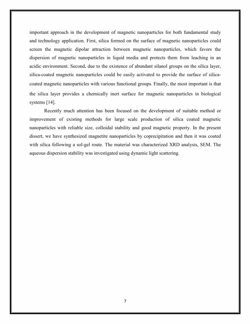
Figure1 Schematic presentation of the synthesis of silica coated magnetite nanoparticle
9
potential measurements. The surface chemistry of the nanoparticles was studied using FTIR
(BX 12).
Schematic presentation of the synthesis of silica coated magnetite nanoparticle
potential measurements. The surface chemistry of the nanoparticles was studied using FTIR
Schematic presentation of the synthesis of silica coated magnetite nanoparticle
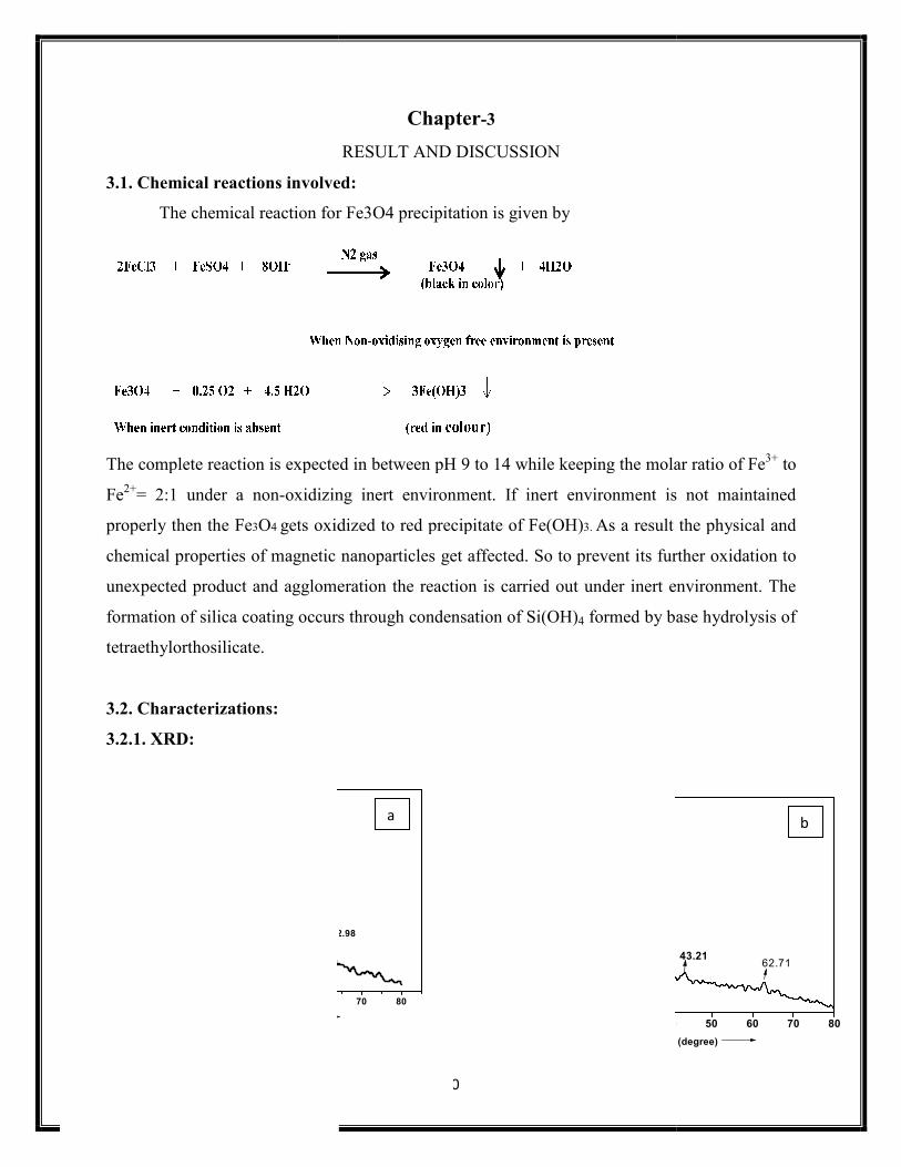
3.1. Chemical reactions involved:
The chemical reaction for Fe3O4 precipitation is given by
The complete reaction is expected in between pH 9 to 14 while keeping the molar ratio o
Fe2+= 2:1 under a non-oxidizing
properly then the Fe3O4 gets oxidized to red precipitate
chemical properties of magnetic nanoparticles get
unexpected product and agglomeration the reaction is carried out under inert environment.
formation of silica coating occurs through condensation of Si(OH)
tetraethylorthosilicate.
3.2. Characterizations:
3.2.1. XRD:
20 30 40 50 60150
200
250
300
350
400
450
57.3753.98
43.56
62.98
30.33
35.72
Intensity
2θ in degree
10
Chapter-3
RESULT AND DISCUSSION
Chemical reactions involved:
chemical reaction for Fe3O4 precipitation is given by
The complete reaction is expected in between pH 9 to 14 while keeping the molar ratio o
oxidizing inert environment. If inert environment is not maintained
ts oxidized to red precipitate of Fe(OH)3. As a result the physi
chemical properties of magnetic nanoparticles get affected. So to prevent its further oxi
unexpected product and agglomeration the reaction is carried out under inert environment.
formation of silica coating occurs through condensation of Si(OH)4 formed by base hydrolysis of
70 80
62.98
10 20 30 4050
100
150
200
250
Intensity
2θθθθ (degree)
35.74
a
The complete reaction is expected in between pH 9 to 14 while keeping the molar ratio of Fe3+ to
environment is not maintained
As a result the physical and
ffected. So to prevent its further oxidation to
unexpected product and agglomeration the reaction is carried out under inert environment. The
formed by base hydrolysis of
40 50 60 70 80
(degree)
62.7143.21
b
11
Table 1. 2Ѳ, d and corresponding [h k l] values of synthesized Fe3O4 nanoparticles
2Ѳ 30.3 35.7 43.5 53.7 57.5 62.9
D 2.96 2.53 2.09 1.71 1.61 1.48
[h k l] [220] [331] [400] [422] [511] [440]
Figure 1 illustrates the XRD patterns of black precipitations of Fe3O4 of magnetic particles.
The position and relative intensity of all diffraction peaks match well with those of the magnetite
(JCPDS 19-629) and broad peaks indicates nano-crystalline nature of the particles. Figure 2
shows XRD pattern of silica coated Fe3O4 magnetic nano-particle, the broad peak is due to
amorphous silica. Thus XRD pattern indicates the presence of amorphous silica coating on
magnetite nanoparticles surface. The broadened peak at 10-20 degree corresponds to SiO2 and
the rest of peaks are as those in XRD patterns of Fe3O4.
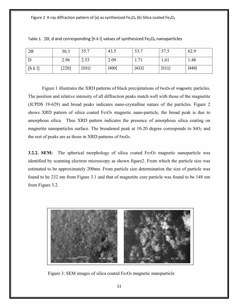
3.2.2. SEM: The spherical morphology of silica coated Fe3O4 magnetic nanoparticle was
identified by scanning electron microscopy as shown figure2. From which the particle size was
estimated to be approximately 200nm. From particle size determination the size of particle was
found to be 232 nm from Figure 3.1 and that of magnetite core particle was found to be 148 nm
from Figure 3.2.
Figure 3: SEM images of silica coated Fe3O4 magnetic nanoparticle
Figure 2 X-ray diffraction pattern of (a) as synthesized Fe3O4 (b) Silica coated Fe3O4
12
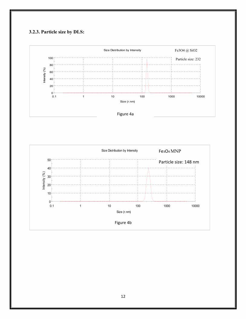
3.2.3. Particle size by DLS:
0
20
40
60
80
100
0.1 1 10 100 1000 10000
Intensity (%)
Size (r.nm)
Size Distribution by Intensity
Record 3: Ranjana 1
0
10
20
30
40
50
0.1 1 10 100 1000 10000
Intens
ity (%)
Size (r.nm)
Size Distribution by Intensity
Record 4: Ranjana 1
Fe3O4 MNP
Particle size: 148 nm
Fe3O4 @ SiO2
Particle size: 232
Figure 4a
Figure 4b
13
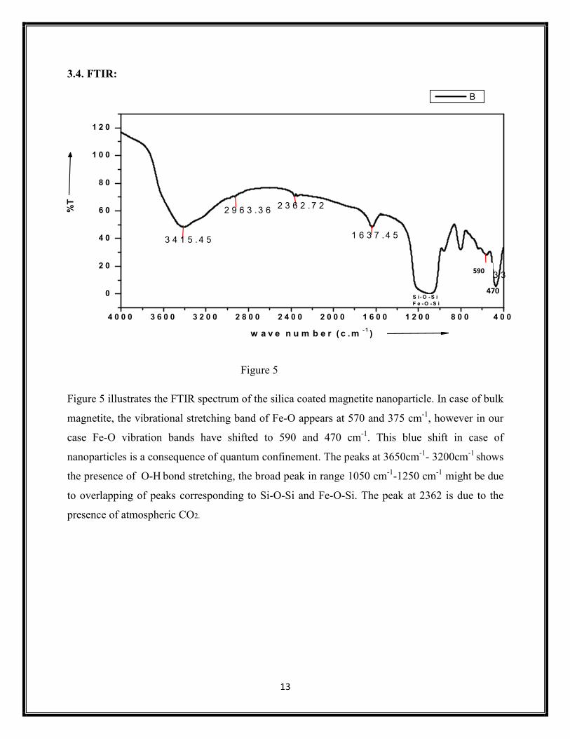
3.4. FTIR:
Figure 5
Figure 5 illustrates the FTIR spectrum of the silica coated magnetite nanoparticle. In case of bulk
magnetite, the vibrational stretching band of Fe-O appears at 570 and 375 cm-1, however in our
case Fe-O vibration bands have shifted to 590 and 470 cm-1. This blue shift in case of
nanoparticles is a consequence of quantum confinement. The peaks at 3650cm-1- 3200cm-1 shows
the presence of O-H bond stretching, the broad peak in range 1050 cm-1-1250 cm-1 might be due
to overlapping of peaks corresponding to Si-O-Si and Fe-O-Si. The peak at 2362 is due to the
presence of atmospheric CO2.
4 0 0 0 3 6 0 0 3 2 0 0 2 8 0 0 2 4 0 0 2 0 0 0 1 6 0 0 1 2 0 0 8 0 0 4 0 0
0
2 0
4 0
6 0
8 0
1 0 0
1 2 0
%T
w a v e n u m b e r ( c .m - 1 )
B
3 4 1 5 . 4 5 1 6 3 7 .4 5
2 3 6 2 .7 2
5 5 6 .3 3
S i -O -S iF e -O -S i
2 9 6 3 .3 6
590
470
14
Chapter-4
Conclusion
A systematic study of the formation of silica coated magnetite nanoparticle via co-precipitation method was successfully made. The result shows that the reaction parameters such as the volume ratio of alcohol/water, the amount of aqueous ammonia and TEOS influence the formation of silica coating on magnetic nanoparticles. Silica-coated magnetic nanoparticle of size 232 nm with spherical morphology could be conveniently prepared in large scale using the developed process. The amount of catalyst (ammonia aqueous) plays an important role on the formation of silica coated magnetic nanoparticles. By increase in amount of precursor (TEOS) larger silica-coated magnetic particles with more regular shape and monodispersed could be produced.
15
REFFERENCE:
[1] I. Prigogine, A. Stuart, in: J.L. Darmann, D. Fiorani, E. Ronc (Eds.), Advance in
Chemical Physics, Wiley, New York, (1977).
[2] Y.H. Deng, C.C. Wang, J.H. Hu, Wu-Li Yang, S.K. Fu, Colloids and Surfaces A:
Physicochemical. Eng. Aspects 262 (2005) 87–93
[3] Aranzazu del Campo,Tapas sen,Jean-Paul lellouche,Ian J.Bruce,journal of
magnetismand magneti materials 293 (2005) 33-40
[4] A.S. Lubbe, C.C. Alexiou, et al., J. Surg. Res. 95 (2001) 200.
[5] A.L. Paul, G.R. Chandra, et al., J. Magn. Magn. Mater. 225 (2001) 301.
[6] J. Halavaara, P. Tervahartiala, et al., Acad. Radiol. 43 (2002) 180.
[7] M. Mary, in: U. Hafeli, W. Schutt, M. Zborowski (Eds.), Scientific and Clinical
Applications of Magnetic Carriers, Plenum Press, NewYork, 1997
[8] A. Elaissari, M. Rodrigue, et al., J. Magn. Magn. Mater. 225 (2001)127.
[9] A.n.d. Campoa,b,1,Tapas Sena,Jean-Paul Lellouchec,Ian J. Bruce Journal of
Magnetism and Magnetic Materials 293 (2005) 33–40
[10] X.G. Li, S. Takahashi, K. Watanabe, Y. Kikuchi, M. Koishi, Fabrication and
characteristististics of Fe3O4-polymer composite particles by hybridization, Powder
Technol. 133 (2003) 156–163.
[11] H.X. Guo, X.P. Zhao, The synthesis of composite particles responsive to electric and
magnetic fields, Opt. Mater. 22 (2003) 39.
[12] G.L. Qiu, Y.L. Li, G.L. Siri, S.Y. Li, Sci. Technol. Chem. Ind. 9 (2001) 15.
[13] E.M. Denkbas, E. Kilicay, C. Birlikseven, E. Ozturk, Magnetic chitosan microspheres:
preparation and characterization, React. Funct. Polym. 50 (2002) 225
[14] Y. H. Deng, C.C. Wang, J.H. Hu, W.L. Yang, S.K. Fu .Colloids and Surfaces A:
Physicochemical. Eng. Aspects 262 (2005) 87–93
16
[15] S.Mohapatra, D.Pal ,S.K. Ghosh 2,and P.Pramanik,design of superparamagnetic iron
oxide nanoparticle for purification of recombinant proteins,journal of nanoscience and
nanotechnology,vol.7,(2007)1-7
![SYNTHESIS AND CHARACTERIZATION OF CORE/SHELL ......Functionalized nanoparticles such as core/shell silica coated gold [2], alumina coated Titania [3], silver coated magnetite [4],](https://img.pdfslide.us/doc/110x75/60bd533bb67f6c68462c9209/synthesis-and-characterization-of-coreshell-functionalized-nanoparticles.jpg)