Embed Size (px)
Citation preview
Proc. Nati. Acad. Sci. USAVol. 84, pp. 969-972, February 1987Biophysics
Images of a lipid bilayer at molecular resolution by scanningtunneling microscopy
(biological membranes/cadmium isocanoate/graphite)
D. P. E. SMITH*, A. BRYANT*, C. F. QUATE*t, J. P. RABEt§, CH. GERBERt¶, AND J. D. SWALENt*Department of Applied Physics, Stanford University, Stanford, CA 94305; *IBM Almaden Research Center, San Jose, CA 95120; and TIBM ResearchLaboratory, Zurich, Switzerland
Contributed by C. F. Quate, September 23, 1986
ABSTRACT The molecular structure of a fatty acid bilay-er has been recorded with a scanning tunneling microscopeoperating in air. The molecular film, a bilayer of cadmiumicosanoate (arachidate), was deposited onto a graphite sub-strate by the Langmuir-Blodgett technique. The packing of thelipid film was found to be partially ordered. Along one axis ofthe triclinic unit cell the intermolecular distance varied ran-domly around a mean of 5.84 A with a SD of 0.24 A. Along theother axis the mean distance was 4.1 A and appeared to varymonotonically over several intermolecular distances, indicat-ing that a superstructure of longer range may exist. Themolecular density was one molecule per 19.4 A2. The surprisingability of the scanning tunneling microscope to image theindividual molecular chains demonstrates that electrons fromthe graphite can be transferred along the molecular chains fora distance of 50 A.
z
PIEZOELECTRIC FSCANNER
TUNGSTEN -A.xTIP
ARACHIDATE ABILAYER
GRAPHITE SUBSTRATE
Molecular monolayers, bilayers, and multilayers are impor-tant in the fields of biology and technology. In biology wherethey occur as lipid membranes the details of the two-dimensional packing are important for our understanding oftheir biological function (1). The proposed technological usesof Langmuir-Blodgett films as thin film dielectrics make itnecessary to characterize them if we are to optimize theinteractions that occur within the film and at the interfaceswith the environment (2). In recent years a number oftechniques have been applied toward the problem of deter-mining the structure, orientation, and packing of molecularmonolayers and bilayers. The methods that have been usedto characterize these films include infrared spectroscopy (3),Raman spectroscopy (4), electron diffraction (5), x-ray dif-fraction (6), and fluorescence microscopy (7). However, evenfor the simple cases of pure fatty acids, it is difficult tounambiguously determine the packing structure. The mostdirect way to study molecular packing is with a microscopethat is capable of resolving the intermolecular distances,which in the case of the fatty acids is about 5 A. In this reportwe will demonstrate that the scanning tunneling microscope(STM) is useful for this purpose. A preliminary report on thiswork has been presented elsewhere (25).The technique of scanning tunneling microscopy has been
used to image the distribution of electronic charge density fora variety of conducting and semiconducting solid surfaces(8-10). Viruses (11) and DNA (G. Binnig, H. Rohrer, E.Courtens, H. Gross, and J. Sogo, personal communication)deposited on conducting substrates have also been imagedwith the STM, but the resolution in these experiments wasonly about 50 A. In this report we present STM studies of afatty acid bilayer deposited on a graphite substrate; we areable to reproducibly image the individual molecules in thefilm with a resolution of about 2 A. By using the STM in a
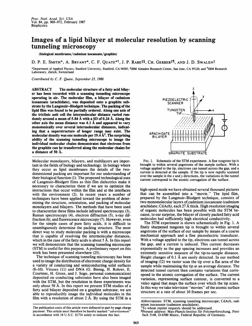
FIG. 1. Schematic of the STM experiment. A fine tungsten tip isbrought to within several angstroms of the sample surface. With avoltage applied to the tip, electrons can tunnel across the gap, and acurrent is detected at the sample. If the tip is now rapidly scannedover the sample in the x and y directions, the variations in the tunnelcurrent correspond to the atomic corrugation of the surface.
high-speed mode we have obtained several thousand picturesthat can be assembled into a "movie." The lipid film,prepared by the Langmuir-Blodgett technique, consists oftwo monomolecular layers of cadmium isocanoate (cadmiumarachidate; CdAch), each 27 A thick. High resolution imagingof organic molecules has been possible with the STM be-cause, to our surprise, the bilayer of closely packed fatty acidmolecules had sufficiently high electrical conductivity.The STM experiment is shown schematically in Fig. 1. A
finely sharpened tungsten tip is brought to within severalangstroms of the surface of our sample by means of a coarsemechanical approach and a fine piezoelectric transducer.With a voltage applied to the tip, electrons can tunnel acrossthe gap, and a current is induced. This current decreasesexponentially as the gap spacing increases and provides anextremely sensitive measure of the tip-to-sample distance.Height changes of 0.1 A are easily detected. In our methodof imaging (12) we raster scan the tip over a flat area of thesample while maintaining the tip at an average distance. Thedetected tunnel current then contains variations that corre-spond to the atomic corrugation of the surface. The currentvariation, representing surface contour, is converted to avideo signal that maps the surface over which the tip scans.In this way we take television "movies" ofthe atomic surfacestructure at a rate of about 10 frames per sec.
Abbreviations: STM, scanning tunneling microscope; CdAch, cad-mium isocanoate (cadmium arachidate).tTo whom reprint requests should be addressed.§Present address: Max-Planck-Institut fur Polymerforschung, Post-fach 3148, D-6500 Mainz, Federal Republic of Germany.
969
The publication costs of this article were defrayed in part by page chargepayment. This article must therefore be hereby marked "advertisement"in accordance with 18 U.S.C. §1734 solely to indicate this fact.
Proc. Natl. Acad. Sci. USA 84 (1987)
STAI NLESS
APPROACHMICROMETER
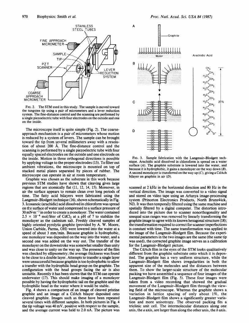
FIG. 2. The STM used in this study. The sample is moved towardthe tungsten tip using a pair of micrometers and a lever reductionsystem. The fine-distapce control and the scanning are performed bya single piezoelectric tube with four electrodes on the outside and oneon the inside.
The microscope itself is quite simple (Fig. 2). The coarse-approach mechanism is a pair of micrometers whose motionis reduced by a system of levers. The sample can be broughttoward the tip from several millimeters away with a resolu-tion of about 200 A. The fine-distance control and thescanning is performed by a single piezoelectric tube with fourequally spaced electrodes on the outside and one electrode onthe inside. Motion in three orthogonal directions is possibleby applying voltage to the proper electrodes (13). To filter outambient vibrations, the microscope is mounted on top ofstacked metal plates separated by pieces of rubber. Themicroscope can operate in air at room temperature.
Graphite was chosen as the substrate in this work becauseprevious STM studies have shown that cleaving gives largeregions that are atomically flat (11, 12, 14, 15). Moreover, inair the surface appears to remain clean over long periods oftime. The fatty acid bilayers were fabricated using theLangmuir-Blodgett technique (16), shown schematically in Fig.3. Icosanoic (arachidic) acid dissolved in chloroform was spreadon the surface ofwater and compressed to a surface pressure of30mN m- in order to create a monolayer. The water contained2.5 x 10-4 mol/liter of CdCl2 at a pH of 7 to stabilize themonolayer as the cadmium salt. Freshly cleaved samples ofhighly oriented pyrolytic graphite (provided by A. W. Moore,Union Carbide, Parma, OH) were lowered into the water at aspeed of about 3 mm/min. Because graphite is hydrophobic,one monolayer was deposited on the way into the water, and asecond one was added on the way out. The transfer of themonolayer on the downstroke was somewhat smaller than unityand was close to unity on the way up. The presence of the filmwas checked by ellipsometry; the average thickness was foundto be close to a double layer. Attempts to transfer a single layerwere unsuccessful because graphite is too hydrophobic to allowa transfer with the hydrophilic head group on the graphite. Theconfiguration with the head groups facing the air is alsounstable. Recently it has been shown that the STM can operateunderwater (17). This should make imaging of a monolayerpossible by having the hydrophobic tail on the graphite and thehydrophilic head in the water where it would be stable.
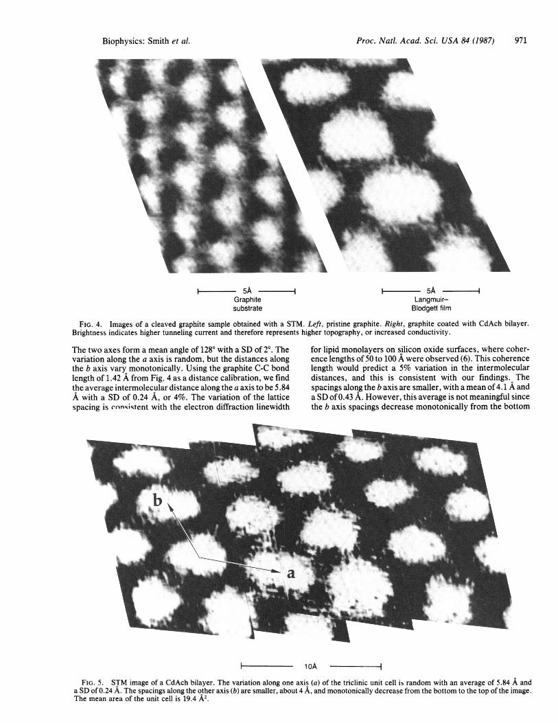
Fig. 4 shows a comparison of an image of cleaved pristinegraphite and an image of a CdAch bilayer deposited ontocleaved graphite. Images such as these have been repeatedseveral times with different samples. In both pictures in Fig. 4the tip voltage was 40 mV, positive with respect to the sample,and the average current was held to 2.0 nA. The picture was
A_
__Graphite
-_--Water
-------Cu&-D (
Arachidic Acid
CH3
/CH2H2C\
/CH2H2C\
/CH2H2C\
/CH2H2C\
/CH2H2C\
/CH2H2C\
/CH2H2C\
/CH2H2C\
/CH2H2C\c
0\D
2 A
FIG. 3. Sample fabrication with the Langmuir-Blodgett tech-nique. Arachidic acid dissolved in chloroform is spread on a watersurface (A). The graphite substrate is lowered into the water, andbecause it is hydrophobic, it gains a monolayer on the way down (B).A second monolayer is transferred on the way up (C), giving a CdAchbilayer on graphite in air (D).
scanned at 2 kHz in the horizontal direction and 80 Hz in thevertical direction. The image was converted to a video signaland stored on video tape using an Arlunya image-processingsystem (Princeton Electronics Products, North Brunswick,NJ). It was then temporally filtered using the same machine andspatially filtered by a digital computer. The distortion intro-duced into the picture due to scanner nonorthogonality andunequal scan ranges was removed by linearly transforming thegraphite image to agree with its known hexagonal structure (18);the transformation required to correct for scanner imperfectionsis constant with time. The same transformation was applied tothe image of the Langmuir-Blodgett film. Because the experi-mental parameters in the two images are the same (the same tipwas used), the corrected graphite image serves as a calibrationfor the Langmuir-Blodgett picture.The CdAch film in the eyes of the STM looks qualitatively
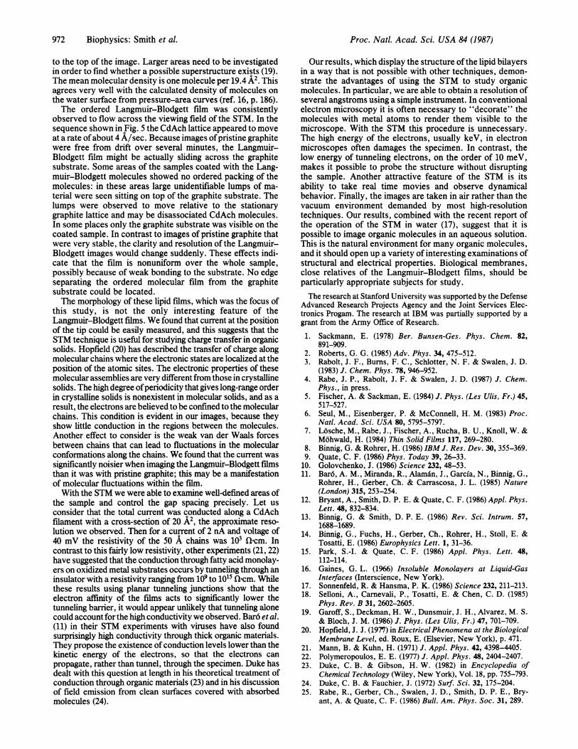
different from the graphite substrate onto which it is depos-ited. The graphite has a very uniform structure, while theLangmuir-Blodgett film shows irregularities in both theapparent size of the molecules and the distances betweenthem. To show the larger-scale structure of the molecularpacking we have assembled a sequence of four images of theLangmnuir-Blodgett film (Fig. 5). These four images weretaken from a video recording that showed the gradualmovement of the Langmuir-Blodgett film through the view-ing field of the microscope. Whereas the graphite shows avariation in lattice spacing of only about 1%, theLangmuir-Blodgett film shows a significantly greater varia-tion and more anisotropy. The observed packing fits atriclinic unit cell. The intermolecular distances along oneaxis, the a axis, are larger than along the other axis, the b axis.
970 Biophysics: Smith et al.
---
Proc. Natl. Acad. Sci. USA 84 (1987)
- 5A - I i - 5A -Graphite Langmuir-substrate Blodgett film
FIG. 4. Images of a cleaved graphite sample obtained with a STM. Left, pristine graphite. Right, graphite coated with CdAch bilayer.Brightness indicates higher tunneling current and therefore represents higher topography, or increased conductivity.
The two axes form a mean angle of 1280 with a SD of 20. Thevariation along the a axis is random, but the distances alongthe b axis vary monotonically. Using the graphite C-C bondlength of 1.42 A from Fig. 4 as a distance calibration, we findthe average intermolecular distance along the a axis to be 5.84A with a SD of 0.24 A, or 4%. The variation of the latticespacing is conniqtent with the electron diffraction linewidth
for lipid monolayers on silicon oxide surfaces, where coher-ence lengths of 50 to 100 A were observed (6). This coherencelength would predict a 5% variation in the intermoleculardistances, and this is consistent with our findings. Thespacings along the b axis are smaller, with a mean of4.1 A anda SD of 0.43 A. However, this average is not meaningful sincethe b axis spacings decrease monotonically from the bottom
- 1oA -
FIG. 5. STM image of a CdAch bilayer. The variation along one axis (a) of the triclinic unit cell is random with an average of 5.84 A anda SD of 0.24 A. The spacings along the other axis (b) are smaller, about 4 A, and monotonically decrease from the bottom to the top of the image.The mean area of the unit cell is 19.4 A2.
Biophysics: Smith et al. 971
Proc. Natl. Acad. Sci. USA 84 (1987)
to the top of the image. Larger areas need to be investigatedin order to find whether a possible superstructure exists (19).The mean molecular density is one molecule per 19.4 A2. Thisagrees very well with the calculated density of molecules onthe water surface from pressure-area curves (ref. 16, p. 186).The ordered Langmuir-Blodgett film was consistently
observed to flow across the viewing field of the STM. In thesequence shown in Fig. 5 the CdAch lattice appeared to moveat a rate of about 4 A/sec. Because images ofpristine graphitewere free from drift over several minutes, the Langmuir-Blodgett film might be actually sliding across the graphitesubstrate. Some areas of the samples coated with the Lang-muir-Blodgett molecules showed no ordered packing of themolecules: in these areas large unidentifiable lumps of ma-terial were seen sitting on top of the graphite substrate. Thelumps were observed to move relative to the stationarygraphite lattice and may be disassociated CdAch molecules.In some places only the graphite substrate was visible on thecoated sample. In contrast to images of pristine graphite thatwere very stable, the clarity and resolution of the Langmuir-Blodgett images would change suddenly. These effects indi-cate that the film is nonuniform over the whole sample,possibly because of weak bonding to the substrate. No edgeseparating the ordered molecular film from the graphitesubstrate could be located.The morphology of these lipid films, which was the focus of
this study, is not the only interesting feature of theLangmuir-Blodgett films. We found that current at the positionof the tip could be easily measured, and this suggests that theSTM technique is useful for studying charge transfer in organicsolids. Hopfield (20) has described the transfer of charge alongmolecular chains where the electronic states are localized at theposition of the atomic sites. The electronic properties of thesemolecular assemblies are very different from those in crystallinesolids. The high degree ofperiodicity that gives long-range orderin crystalline solids is nonexistent in molecular solids, and as aresult, the electrons are believed to be confined to the molecularchains. This condition is evident in our images, because theyshow little conduction in the regions between the molecules.Another effect to consider is the weak van der Waals forcesbetween chains that can lead to fluctuations in the molecularconformations along the chains. We found that the current wassignificantly noisier when imaging the Langmuir-Blodgett filmsthan it was with pristine graphite; this may be a manifestationof molecular fluctuations within the film.With the STM we were able to examine well-defined areas of
the sample and control the gap spacing precisely. Let usconsider that the total current was conducted along a CdAchfilament with a cross-section of 20 A2, the approximate reso-lution we observed. Then for a current of 2 nA and voltage of40 mV the resistivity of the 50 A chains was 103 flQcm. Incontrast to this fairly low resistivity, other experiments (21, 22)have suggested that the conduction through fatty acid monolay-ers on oxidized metal substrates occurs by tunneling through aninsulator with a resistivity ranging from 109 to 1015 &cm. Whilethese results using planar tunneling junctions show that theelectron affinity of the films acts to significantly lower thetunneling barrier, it would appear unlikely that tunneling alonecould account for the high conductivity we observed. Bar6 et al.(11) in their STM experiments with viruses have also foundsurprisingly high conductivity through thick organic materials.They propose the existence of conduction levels lower than thekinetic energy of the electrons, so that the electrons canpropagate, rather than tunnel, through the specimen. Duke hasdealt with this question at length in his theoretical treatment ofconduction through organic materials (23) and in his discussionof field emission from clean surfaces covered with absorbedmolecules (24).
Our results, which display the structure ofthe lipid bilayersin a way that is not possible with other techniques, demon-strate the advantages of using the STM to study organicmolecules. In particular, we are able to obtain a resolution ofseveral angstroms using a simple instrument. In conventionalelectron microscopy it is often necessary to "decorate" themolecules with metal atoms to render them visible to themicroscope. With the STM this procedure is unnecessary.The high energy of the electrons, usually keV, in electronmicroscopes often damages the specimen. In contrast, thelow energy of tunneling electrons, on the order of 10 meV,makes it possible to probe the structure without disruptingthe sample. Another attractive feature of the STM is itsability to take real time movies and observe dynamicalbehavior. Finally, the images are taken in air rather than thevacuum environment demanded by most high-resolutiontechniques. Our results, combined with the recent report ofthe operation of the STM in water (17), suggest that it ispossible to image organic molecules in an aqueous solution.This is the natural environment for many organic molecules,and it should open up a variety of interesting examinations ofstructural and electrical properties. Biological membranes,close relatives of the Langmuir-Blodgett films, should beparticularly appropriate subjects for study.The research at Stanford University was supported by the Defense
Advanced Research Projects Agency and the Joint Services Elec-tronics Progam. The research at IBM was partially supported by agrant from the Army Office of Research.
1. Sackmann, E. (1978) Ber. Bunsen-Ges. Phys. Chem. 82,891-909.
2. Roberts, G. G. (1985) Adv. Phys. 34, 475-512.3. Rabolt, J. F., Burns, F. C., Schlotter, N. F. & Swalen, J. D.
(1983) J. Chem. Phys. 78, 946-952.4. Rabe, J. P., Rabolt, J. F. & Swalen, J. D. (1987) J. Chem.
Phys., in press.5. Fischer, A. & Sackman, E. (1984) J. Phys. (Les Ulis, Fr.) 45,
517-527.6. Seul, M., Eisenberger, P. & McConnell, H. M. (1983) Proc.
Natl. Acad. Sci. USA 80, 5795-5797.7. Losche, M., Rabe, J., Fischer, A., Rucha, B. U., Knoll, W. &
Mohwald, H. (1984) Thin Solid Films 117, 269-280.8. Binnig, G. & Rohrer, H. (1986) IBM J. Res. Dev. 30, 355-369.9. Quate, C. F. (1986) Phys. Today 39, 26-33.
10. Golovchenko, J. (1986) Science 232, 48-53.11. Bar6, A. M., Miranda, R., Alamdn, J., Garcia, N., Binnig, G.,
Rohrer, H., Gerber, Ch. & Carrascosa, J. L. (1985) Nature(London) 315, 253-254.
12. Bryant, A., Smith, D. P. E. & Quate, C. F. (1986) Appl. Phys.Lett. 48, 832-834.
13. Binnig, G. & Smith, D. P. E. (1986) Rev. Sci. Intrum. 57,1688-1689.
14. Binnig, G., Fuchs, H., Gerber, Ch., Rohrer, H., Stoll, E. &Tosatti, E. (1986) Europhysics Lett. 1, 31-36.
15. Park, S.-I. & Quate, C. F. (1986) Appl. Phys. Lett. 48,112-114.
16. Gaines, G. L. (1966) Insoluble Monolayers at Liquid-GasInterfaces (Interscience, New York).
17. Sonnenfeld, R. & Hansma, P. K. (1986) Science 232, 211-213.18. Selloni, A., Carnevali, P., Tosatti, E. & Chen, C. D. (1985)
Phys. Rev. B 31, 2602-2605.19. Garoff, S., Deckman, H. W., Dunsmuir, J. H., Alvarez, M. S.
& Bloch, J. M. (1986) J. Phys. (Les Ulis, Fr.) 47, 701-709.20. Hopfield, J. J. (1977) in Electrical Phenomena at the Biological
Membrane Level, ed. Roux, E. (Elsevier, New York), p. 471.21. Mann, B. & Kuhn, H. (1971) J. Appl. Phys. 42, 4398-4405.22. Polymeropoulos, E. E. (1977) J. Appl. Phys. 48, 2404-2407.23. Duke, C. B. & Gibson, H. W. (1982) in Encyclopedia of
Chemical Technology (Wiley, New York), Vol. 18, pp. 755-793.24. Duke, C. B. & Fauchier, J. (1972) Surf. Sci. 32, 175-204.25. Rabe, R., Gerber, Ch., Swalen, J. D., Smith, D. P. E., Bry-
ant, A. & Quate, C. F. (1986) Bull. Am. Phys. Soc. 31, 289.
972 Biophysics: Smith et al.